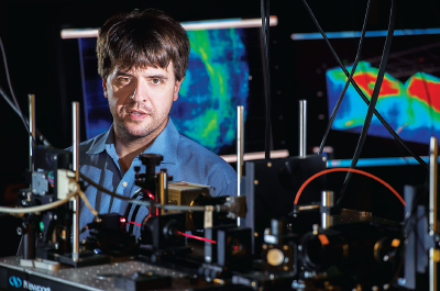
Using an innovative brain imaging technique, a research team at Stanford University has for the first time been able to track brain activity in real time in specific kinds of neurons. Their efforts have helped to uncover the brain circuits that regulate vigilance, or alertness.
“Vigilance gone awry marks states such as mania and those seen in posttraumatic stress disorder [PTSD] and depression,” explained National Institute of Mental Health (NIMH) Director Joshua Gordon, M.D., Ph.D., in a press release highlighting the research. “Gaining familiarity with the molecular players in a behavior—as this new tool promises—may some day lead to clinical interventions targeting dysfunctional brain states.”
The locus coeruleus has long been recognized as a key regulator of vigilance. In the current study, the Stanford team led by Karl Deisseroth, M.D., Ph.D., showed in animal models that two other regions play important roles: the tegmentum (an area in the brainstem that controls reflexive behaviors) and the hypothalamus (which regulates many important metabolic processes such as body temperature and hunger).
“These results may explain why it is hard to get a disorder like PTSD to respond to pharmacological treatment,” Deisseroth told Psychiatric News. “A drug typically works on one system, and if there are multiple circuits working independently, it won’t have much effect.”
The study was published November 2, 2017, in the journal Cell.
Diesseroth and his team used zebrafish to test out its Multi-MAP technique (which stands for multiplexed alignment of molecular and activity phenotypes). Zebrafish are excellent models for neuroscience since these animals, when young, are completely translucent—allowing researchers to easily observe the brains of the fish.
The fish in this study were bioengineered such that any neurons that became active were fluorescent. For the alertness task, the fish were tethered in their aquariums so that their heads were constrained but their tails free. The researchers then displayed a visual stimulus that startled the fish and caused them to swish their tails rapidly in response. The team recorded which neurons became fluorescent between the stimulus and the response. The researchers then superimposed images of zebrafish brains in which all the neurons were labeled based on their primary neurotransmitter (for example, dopamine neurons, serotonin neurons, and so on) onto these recordings. This way, they could identify which types of neurons were becoming active during the behavioral task.
The Multi-MAP approach confirmed that norepinephrine-secreting neurons in the locus coeruleus contributed to the alertness. But, many other neurons also turned on in response to the stimulus; these included neurons in the hypothalamus that release serotonin and dopamine, as well as cholinergic neurons in the tegmentum.
“This is where neuroscience is going,” Deisseroth said. “It’s not enough to just see flashing lights in the brain; you need to know what these neurons do, and where they are wired.”
Diesseroth and his team examined these same neuronal pathways in mice during a reaction-time task and observed almost precisely the same activity patterns as they had seen in the zebrafish. In addition, by using another innovative technique Deisseroth pioneered called optogenetics—which enables individual neurons to be activated by a pulse of light—the researchers concluded that only a subset of these neurons was directly involved in triggering the vigilance response.
The other neurons, Deisseroth said, likely play an indirect role, perhaps relaying information to other parts of the brain that something important is about to happen. But these indirect signals should not be discounted, he added, as they may contribute to alertness-related disorders such as PTSD, anxiety, or sleep problems.
This study was funded by grants from the National Institutes of Health, with additional support in part from the Howard Hughes Medical Institute and the Brain and Behavior Research Foundation. ■
An abstract of “Ancestral Circuits for the Coordinated Modulation of Brain State” can be accessed
here.