Although general paresis is quite rare nowadays, we still cannot clinically ignore this disease, as reports from the United States point out that the number of patients with neurosyphilis as well as acquired immunodeficiency syndrome (AIDS) is now increasing.
1,2 As neurosyphilis advances, serious dementia or a personality change remains, but it is considered to be “treatable” dementia
3,4 because it can be treated with antibiotics, mainly penicillin.
2,5,6 Early diagnosis and treatment are therefore most important.
We scarcely find any descriptions of neuroimages of neurosyphilis, including general paresis, in recent textbooks.
2,7 This may be because neuroimages of neurosyphilis generally are not very specific and are of little value in making a diagnosis. We performed magnetic resonance imaging (MRI) to understand the relationship between MRI findings and prognosis for patients with general paresis.
DISCUSSION
The most important MRI finding in this study is atrophy of the medial temporal lobe including the hippocampus in 3 of the 7 cases. MRI findings in patients with neurosyphilis have been reported. Zifko et al.
8 performed MRI in 4 HIV-negative patients with general paresis. The outcome for social functioning of 3 of these patients was quite poor, and the personality changes and dementia remained. They had subcortical gliosis as well as cerebral atrophy mainly of the frontal lobes, and 1 of the 3 also had hippocampal atrophy. Furthermore, 1 year later they found in 1 of the 3 patients signs suggesting ferritin deposition in the basal ganglia. In this patient, psychiatric and neurological symptoms were worsening even after the treatment had been finished, and MRI examinations showed that atrophy was also advancing. On the other hand, the outcome for the remaining 1 patient was quite good, and he was able to get a job although he had manic symptoms. He had only slight frontal atrophy, and even at the time of reexamination 1 year later there were no changes on MRI. Cerebral atrophy and gliosis, dominantly in the frontal lobes, were equivalent to well-known neuropathological findings of general paresis;
2,9 they were not very unusual findings and had little diagnostic value. There have been no studies of ferritin deposition in general paresis.
Russouw et al.
10 performed MRI in 20 patients with neurosyphilis, and results in 13 of them proved to be abnormal. T
2-weighted images showed high-intensity areas in 11 of the 20 patients and atrophy in the remaining 9. Furthermore, lesions in the frontal lobes proved to have a significant correlation with score on the Brief Psychiatric Rating Scale, and lesions in the temporoparietal lobes had a significant correlation with score on the Mini-Mental State Examination.
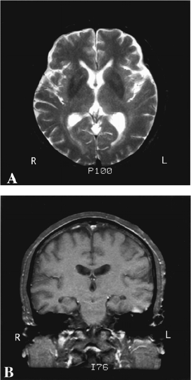
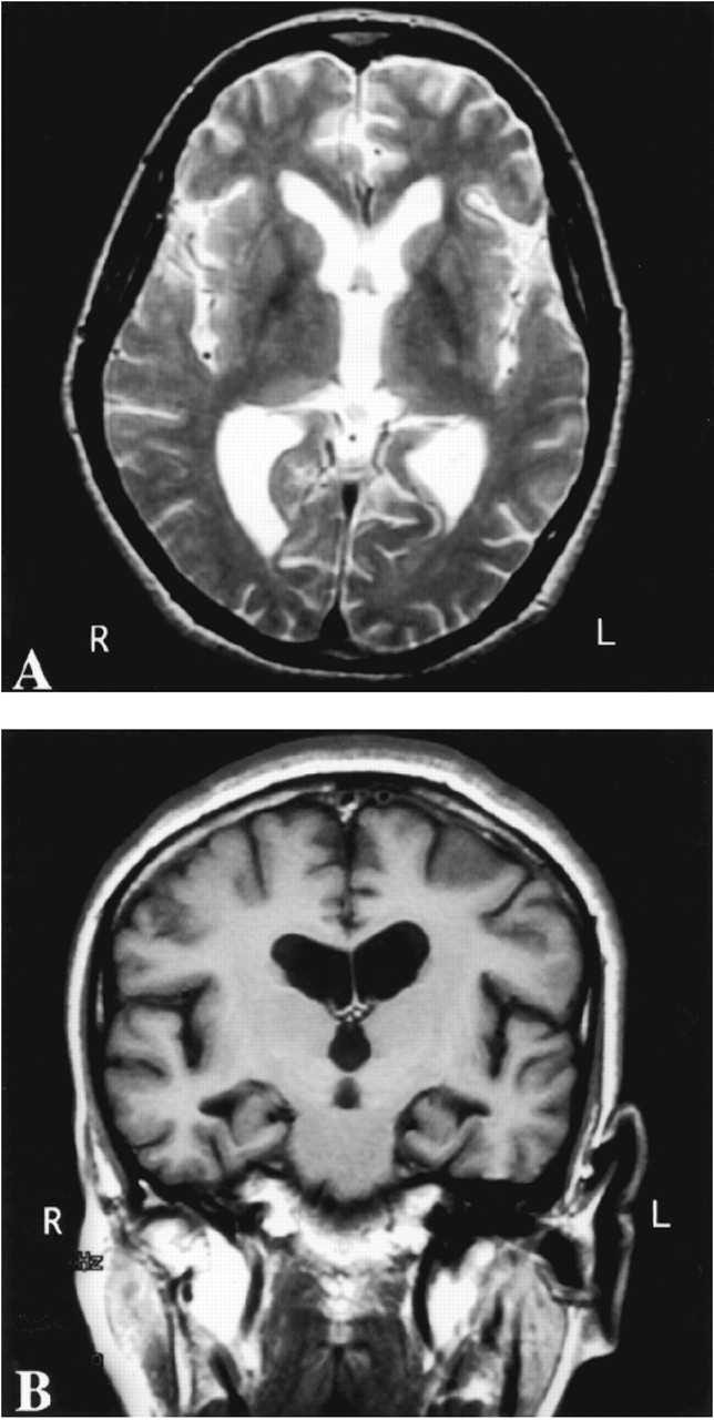
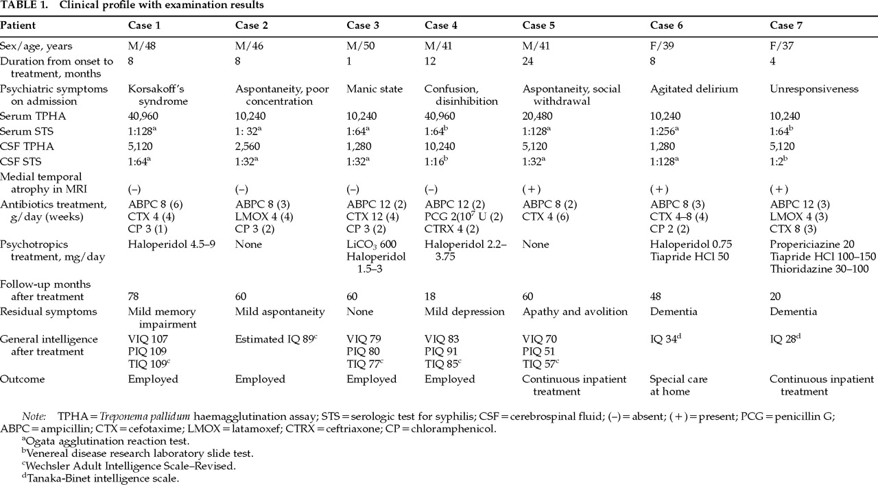
Longevity did not appear to be decreased in any of our patients, but only 4 were able to return to their jobs and regain almost the same social functioning level that they had before becoming ill. When we performed MRI before treatment, these 4 did not have any abnormal MRI findings except slight cerebral atrophy. On the other hand, in the remaining 3 cases, serious dementia or a personality change remained and a long stay in the hospital or special care at home was necessary. These 3 patients already had cerebral atrophy or dilation of the ventricles; moreover, medial temporal lobe atrophy had been recognized as a special feature before treatment. We looked for an etiology that might explain medial temporal lobe atrophy and residual impairment. There were no abnormal findings except syphilis-related abnormality in the patients' sera and CSF.
We consider that a neuroimaging finding of cerebral atrophy of various degrees mainly in the frontal lobes is equivalent to the pathological findings of general paresis reported so far,
2,9 but we can scarcely find any pathological reports on atrophy of the medial temporal lobe including the hippocampus. Why atrophy occurs in the medial temporal lobe is unclear, but it may occur secondarily to dysfunction in the diffuse cerebral cortex, as the medial temporal regions have connections with the cerebral association areas, including the frontal, temporal, and parietal lobes. In our experience, when atrophy is observed in the medial temporal lobe including the hippocampus, the prognosis for the patient's social functioning may be poor in spite of proper treatment.
Early treatment for general paresis is considered to be effective in deciding the prognosis. But delay in starting treatment is not always the reason for a poor prognosis; some of our medial temporal atrophy patients (with a bad prognosis) had treatment started relatively soon after onset. We still do not know whether there is a type of general paresis in which cerebral atrophy would begin to worsen at a very early stage of the disease once its symptoms appeared. But even in these serious cases the patients benefited from treatment, their prognoses across the lifespan were good, and their activities in daily living were greatly improved.
On the other hand, in the case of a slight atrophy seen in neuroimaging, a high dose of antibiotics should be given from the beginning, irrespective of the patient's clinical condition. With this method we can expect the prognosis to be greatly improved. If the patient's clinical condition remained poor, it would be advisable to continue the large dose of antibiotics. Case 1 had shown cognitive disorders, mainly Korsakoff's syndrome, but after long-term treatment of 75 days with a high dose of antibiotics he returned to society with little memory disturbance. There are some reports
11,12 from other groups concerning cases in which cerebral atrophy on CT worsened even after starting treatment. These patients had been given a small amount of penicillin (600,000 to 3 million units per day). When the patients showed prolongation and/or recurrence of psychiatric symptoms, the method of treatment was changed to a high dose of penicillin, but memory disturbance with personality change remained.
Among our cases, there has not been one in which cerebral atrophy worsened after starting treatment. In 4 cases we performed MRI again 3 to 6 years later; in 3 cases there was no change and in the remaining case only a slightly increased atrophy. Judging from the neuroimaging results, we believe that treatment with a high dose of antibiotics was very effective.
In summary, when patients with general paresis are treated, prognosis can be predicted on the basis of the existence of cerebral atrophy, especially medial temporal lobe atrophy that is detected by MRI. Patients with slight cerebral atrophy will have good social functioning after an effective high-dose antibiotics treatment. Even in patients with diffuse cerebral atrophy including the medial temporal lobe, a good prognosis across the lifespan can be predicted and more serious cerebral deterioration can be prevented when an effective high-dose antibiotics treatment starts during an early stage of the disease.