The prefrontal cortex (PFC) is known to be involved in basal ganglia–thalamocortical circuits mediating mood and emotions in humans.
1–3 A large body of evidence derived from neuroimaging and neuropsychological studies suggests that abnormalities of PFC function play a role in the pathophysiology of affective disorders, such as major depression.
4–6 Therefore, it might be clinically relevant to understand how the PFC modulates emotions in healthy volunteers and patients with affective disorders. Several models have been proposed to explain how the brain mediates emotions. One model (the “valence model”) states that emotions are mediated differently depending on their valence, with positive emotions being mediated by the left and negative emotions by the right hemisphere.
1 Although originally based on lesion studies, this notion was recently supported by studies applying repetitive transcranial magnetic stimulation (rTMS) as a noninvasive tool to focally stimulate cortical areas.
7In healthy volunteers, fast rTMS of the DLPFC has also been demonstrated to modulate mood differently depending on which side was stimulated.
11–13 Stimulation of the left DLPFC produced a mild increase in self-rated sadness
11–13 and rTMS of the right DLPFC produced a mild increase in self-rated happiness.
12,13 Therefore, the mood effects of rTMS in healthy volunteers showed the opposite laterality to those seen in patients with major depression. Although previous studies reported mood effects, they did not address the question of whether the expression of emotions, which is altered in affective disorders,
14 is modulated differently by the left and right PFC. To clarify the contrasting results in healthy subjects and depressed patients, it seems worthwhile to study rTMS effects on emotions of healthy volunteers at both sides by subjective mood rating as well as an objective behavior measure.
The aim of the present study was to explore rTMS-induced changes of both self-rated mood and facial expressions, which—in contrast to self-rated mood changes—can be objectively measured by applying a computerized three-dimensional movement analysis system with high spatial and temporal resolution. The measurement of facial expressions is based on the idea that emotional states are expressed by motor activity such as locomotor activity, gestures, or facial expressions in humans.
15 Facial expressions can finely and specifically indicate inner emotional states under normal conditions and in psychiatric disorders.
16,14 Using facial expression analysis in conjunction with subjective mood rating, we assessed laughing reactions in healthy volunteers during an emotion-induction paradigm, to investigate 1) whether previous findings of TMS-induced mood changes can be replicated and 2) whether emotionally induced facial expressions indicate such lateralized changes of emotional valence.
DISCUSSION
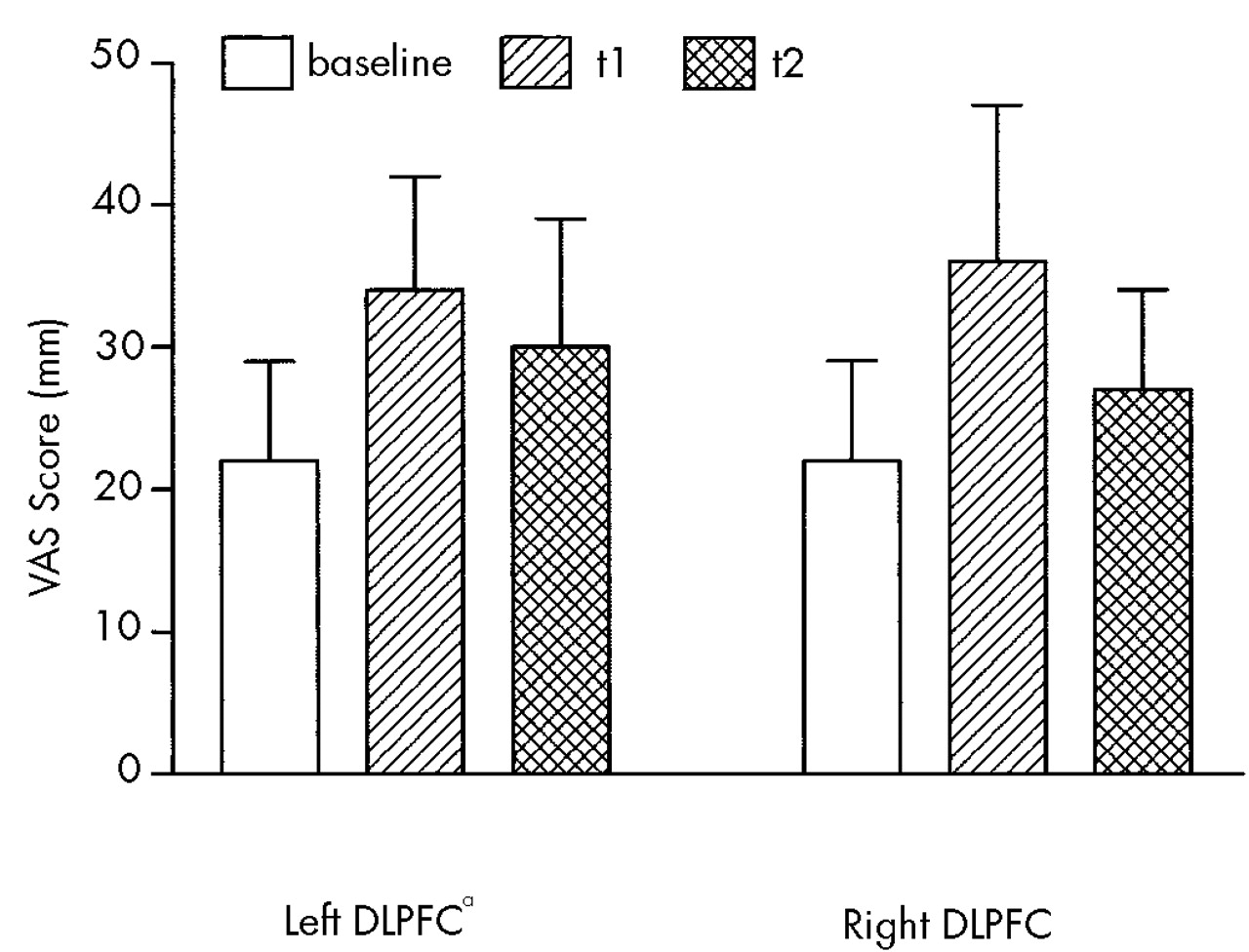
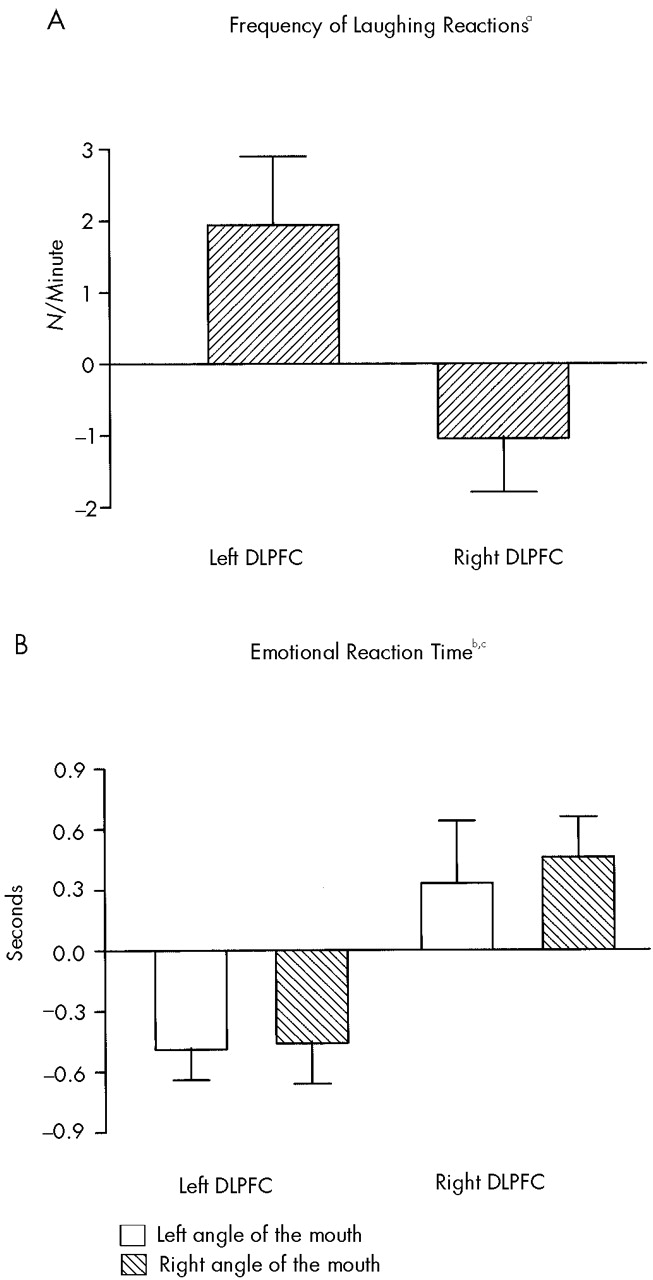
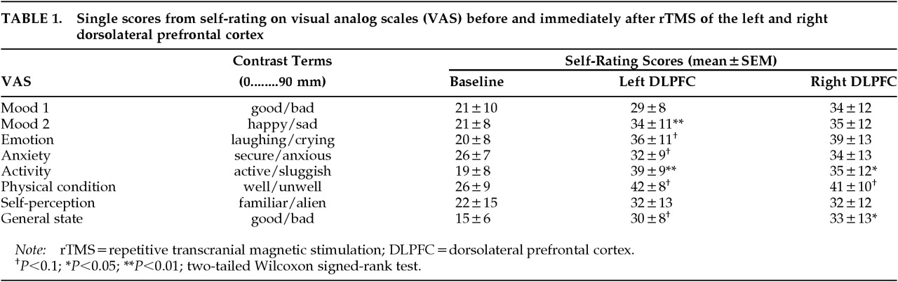
Left and right prefrontal rTMS altered facial expression parameters in a selective manner. Significant differences between rTMS of the left and the right DLPFC were found for frequency of laughing reactions (increased after left rTMS) and emotional reaction time (shortened after left rTMS). In contrast, VAS scores worsened for both sites, and no statistically significant differences were detected between left and right prefrontal rTMS.
Lateralized changes of mood with a mild increase in self-rated sadness after rTMS of the left DLPFC and a mild increase in self-rated happiness after rTMS of right DLPFC have been reported previously.
11–13 Pascual-Leone et al.
11 studied 10 healthy volunteers and found a significant increase in self-ratings of sadness and a significant decrease in self-ratings of happiness immediately after rTMS of the left DLPFC, as compared with right prefrontal and midfrontal stimulation. In another study on 10 healthy subjects, five stimulation sites were compared: left DLPFC, right DLPFC, midfrontal cortex, occipital cortex and cerebellum.
12 The comparison of left and right DLPFC revealed significant differences between the hemispheres, with decreased happiness after left and decreased sadness after right prefrontal rTMS. In a third study, a significant decrease in happiness and a nonsignificant increase in sadness were found after left compared with right prefrontal stimulation in 9 healthy subjects.
13In the present study, we only partially replicated prior findings and did not observe a clear lateralization of rTMS-induced mood changes. Consistent with previous studies, we found subjective impairment in mood and activity after left prefrontal rTMS. However, we did not detect any statistically significant differences between rTMS of the left DLPFC and that of the right DLPFC. Likewise, two more recent studies, one investigating changes of rapid eye movement sleep after rTMS
20 and the other comparing mood changes after verum and sham rTMS,
21 did not demonstrate a significant difference between active and sham rTMS regarding effects on self-rated mood.
Methodological differences may account for the discrepancy between our findings and previous reports. First, VAS with two contrasting polarities were used in our study, in contrast to scales with one polarity used in previous studies
11–13 or explicit questions about sadness and happiness.
12 Second, we did not follow the subjects over several hours as George et al.
12 did; they observed maximal effects on self-rated mood 3 to 8 hours after rTMS. However, Pascual-Leone et al.
11 reported changes in mood immediately after rTMS. Third, rTMS conditions were investigated consecutively in a single session, as in one previous study,
11 but not on separate days.
12,13 Therefore we cannot exclude the possibility of carryover effects, although the sequence of rTMS conditions was randomized and counterbalanced between subjects. Fourth, our stimulation parameters were selected according to one prior study,
11 but differed from parameters used by other investigators.
12,13,20,21 Variation in these parameters, i.e., frequency and intensity, may well contribute to discrepancies between studies.
Physical discomfort caused by rTMS may have additionally influenced self-ratings of mood. Subjects spontaneously reported discomfort or pain during rTMS. This was also observed in VAS scores, with parallel increases in both physical discomfort and sad mood. Similarly, Pascual-Leone et al.
11 found the largest increase in sadness and decrease in happiness after rTMS of the left DLPFC, which subjects spontaneously reported to be most distressing. As overall differences were only minimally significant by rTMS position, however, Pascual-Leone et al.
11 concluded that discomfort did not account for observed mood changes. In contrast, our impression was that rTMS-induced physical discomfort has to be further considered as a biasing factor in studies using rTMS to probe the functional anatomy of mood.
Studies using rTMS to investigate the role of the PFC in emotions have so far not addressed the question of PFC modulation of emotion perception or expression
11–13,20,21 Because facial expressions can finely and specifically indicate inner emotional states,
16,14 we investigated whether fine analysis of facial expressions shows lateralized effects of prefrontal rTMS, as suggested by rTMS-induced changes in self-rated mood.
11,12,13 We found significant differences between left and right prefrontal rTMS: after rTMS of the left DLPFC, the frequency of laughing reactions was increased and the emotional reaction time decreased as compared with rTMS of the right DLPFC. Higher frequency of laughing reactions and shorter reaction time to funny stimuli could be related to an increase in positive emotions. As reported, however, there was no obvious correspondence between facial expression parameter changes and changes of self-rated mood. This incongruity may be explained by the fact that facial expressions and self-rated mood are influenced by several internal and external covariables, such as pain or self-beliefs, which are difficult to control experimentally.
18 Another possibility is that TMS-induced effects on emotionally induced facial expressions are purely motor in nature. However, no changes were observed for voluntary facial expressions after prefrontal rTMS, as one would expect in case of primary motor effects. It is more likely that both the analysis of facial expressions and self-rating of mood evaluate two different aspects of emotions. Facial expressions may be more related to short-term kinetics of emotions, while subjective mood rating reflects a lasting emotional state of an individual. The two do not necessarily correspond; discrepancies between mood and facial expressions have been observed in healthy subjects
22 as well as in neurological or psychiatric patients.
23 The present study is hypothesis-generating in this respect, and future studies will have to investigate the relation between the two phenomena.
It is intriguing that the finding of more and faster laughing reactions after left than right prefrontal rTMS fits with the major body of literature on hemispheric lateralization of emotional behavior in humans.
1,3 Mirth and laughter can be evoked by electrical stimulation of the left frontal cortex and other left hemispheric sites.
24,25 Furthermore, epileptic foci localized in the left hemisphere were found in patients suffering from gelastic seizures, whereas dacrystic seizures were associated with right hemispheric foci.
1,26,27 Pathological laughing and euphoria were associated with right hemispheric lesions; pathological crying and depressed mood with left hemispheric lesions, particularly those affecting the left PFC.
1,28,29 In depressive disorders, both decreased rates of glucose metabolism and reduced cerebral blood flow were found in the left PFC.
4,5 Finally, antidepressant effects have been reported after fast rTMS of the left PFC and antimanic effects after fast rTMS of the right PFC.
8 However, the putative laterality of rTMS-induced effects in affective disorders is not sufficiently established, since left prefrontal rTMS can also exert antidepressant effects at a low, possibly “inhibitory” frequency.
9,10 The effects of rTMS in affective disorders appear to be opposite to the changes of self-rated mood previously reported in healthy volunteers.
11–13 In the present study, however, changes of emotional facial expression parameters point in a similar direction to the effects of rTMS in depression and mania.
We are aware that the interpretation of lateralized changes of emotions found in rTMS studies is limited, since one cannot know whether pathways that regulate mood, emotions, and emotional facial expressions have been activated or inhibited by rTMS. Moreover, rTMS appears also to exert effects on the contralateral hemisphere, as has been demonstrated by functional neuroimaging studies. Neurophysiological investigations and imaging studies have not so far reached conclusive results as to whether rTMS has positive or negative net effects on neural activity.
10,30 The presented findings, however, support the idea that combining rTMS and facial expression analysis may provide a promising tool to further investigate which regions of the brain are involved in the expression of emotions. Most of this system is usually thought to be located in subcortical structures such as the hippocampus, amygdala, or nucleus accumbens.
31 It will be worthwhile to focus on the role of the PFC, or specific parts of it such as the DLPFC, in the generation and processing of emotions and their facial expressions.