Rats that drink like college students at a frat party are teaching researchers about how alcohol affects the growth and death of brain cells and providing insight into how alcohol produces addictive behavior. They may even point to ways to help alcoholics reach sobriety.
Alcohol's effects on the brain occur through a mixture of genetics and inflammation, Said Fulton Crews, Ph.D., a professor of pharmacology and psychiatry and director of the Bowles Center for Alcohol Studies at the University of North Carolina at Chapel Hill.
Crews delivered the annual Mark Keller Honorary Lecture at the National Institute on Alcohol Abuse and Alcoholism in November.
His alcoholism research for several decades has been based on the fact that neural stem cells continue dividing in adulthood and give rise to new neurons in the dentate gyrus of the hippocampus and the subventricular zone of the anterior lateral ventricles.
However, ethanol blocks development of new adult neurons, decreasing neural stem cell proliferation, reducing cell survival, and altering maturation of new neurons. Part of that effect is due to genetics, since rats bred to prefer alcohol exhibit greater brain damage after drinking than do controls.
Age is another factor. Adolescent rats are less sensitive to the intoxicating effects of alcohol, but are more likely to sustain brain damage.
A family history of alcoholism and lower age at drinking onset are known to increase the prevalence of alcohol dependence, said Crews. Adolescent rats show more damage in the forebrain from alcohol, and other studies have shown that damage in the orbitofrontal cortex leads to maladaptive decision making.
Crews' recent research has concentrated on how immune regulatory and transcription factors affect neurogenesis. For instance, CREB (cAMP response element-binding) proteins are transcription factors that increase or decrease the transcription of certain genes. Given the equivalent of binge-drinking doses of alcohol, rats show a dose-dependent decrease in CREB binding, which protects neurons, and an increase in necrosis factor-kB (NF-kB) DNA binding activity, a proinflammatory agent.
“Ethanol intoxication decreases neurogenesis, and cell death occurs during intoxication, not withdrawal,” said Crews.
Crews and his colleagues also found that butylated hydroxytoluene (BHT)—an antioxidant used as a food preservative— blocks tumor necrosis factor-a (TNF-a), an inflammatory cytokine, in brain slice cultures and in vivo by blocking NF-k B activation. That observation led them to look more closely at the effects of inflammation. Giving BHT with ethanol produced some reversal of neuronal death by suppressing the inflammatory reaction, leading Crews to speculate about whether diet plays a role in this process. Perhaps the BHT—much reviled by health-food advocates—might protect neurogenesis.
“Natural foods may not be as good for you in this case,” he joked.
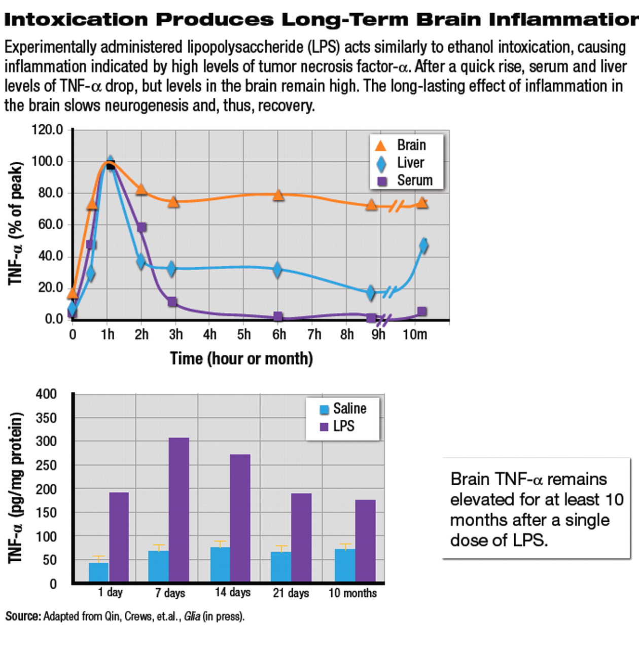
To test the role of TNF-a, he gave rats a dose of lipopolysaccheride, a bacterial toxin that produces inflammation (see
graphs). in an hour, the toxin produced a 1,000-fold increase in TNF-a in liver, serum, and brain. Levels in liver and serum dropped within hours, but brain levels stayed up, said Crews.
“Cytokines stay in the brain a long time,” said Crews.“ Ten months after a single dose of the toxin, brain levels remained high.” Further study found that alcohol potentiated the toxin's effect in producing long-lasting effects of TNF-a, indicating a similar pathway. At the same time, alcohol also reduces brain levels of IL-10, an anti-inflammatory cytokine.
In another experiment, rats given high doses of alcohol for four days exhibited cognitive deficits associated with neurotoxicity in the dentate gyrus, which is involved with memory and learning. In a water-maze test, both control and alcohol-drinking rats learned equally well, but even three weeks into abstinence the alcoholic rats were much worse at relearning the task. They kept making the same mistake over and over again.
“Such perseveration in rats only brings to light how difficult therapy is for the human alcoholic,” said crews. “They have to regenerate to relearn.”
The implication, said Crews, is that neurons must be regenerated to provide a substrate for recovery. Yet that does seem possible once the extended, harmful inflammatory effects of alcohol fade.
“Within 20 weeks of stopping alcohol, the damage can be reversed,” he said. “So if brain regrowth is useful for the return of executive function, how do we make the brain grow?”
Crews has an answer for the rats and maybe for humans, one that doesn't even require approval by the Food and Drug Administration. Heavy physical exercise is known to increase neurogenesis, he said, so he and his colleagues tried an experiment with three groups of rats. One group drank alcohol, but got no exercise. A second drank water and exercised.
“The third drank huge amounts of alcohol and also ran huge amounts,” said Crews. The sedentary, alcoholic rats lost neurons, but running increased neurogenesis equally in both the water-drinking rats and the exercising, alcoholic animals, he said. Perhaps an exercise regimen could reverse neurodegeneration, improve executive function, and help alcoholics along the path to recovery.
“Therapists should challenge their patients to engage in vigorous physical exercise and see if it helps recovery,” he said.
An abstract of “Cytokines and Alcohol” is posted at<www.blackwell-synergy.com/doi/abs/10.1111/j.1530-0277.2006.00084.x>.▪

