Full access
Article
Published Online: 1 February 2001

Three-Dimensional Mapping of Gyral Shape and Cortical Surface Asymmetries in Schizophrenia: Gender Effects

Publication: American Journal of Psychiatry

Abstract

OBJECTIVE: People with schizophrenia exhibit abnormalities in brain structure, often in the left hemisphere. Disturbed structural lateralization is controversial, however, and effects appear mediated by gender. The authors mapped differences between schizophrenic and normal subjects in gyral asymmetries, complexity, and variability across the entire cortex. METHOD: Asymmetry and shape profiles for 25 schizophrenic patients (15 men) and 28 demographically similar normal subjects (15 men) were obtained for 38 gyral regions, including the sylvian fissure and temporal and postcentral gyri, by using magnetic resonance data and a novel surface-based mesh-modeling approach. Cortical complexity was examined for sex and diagnosis effects in lobar regions. Intragroup variability was quantified and visualized to assess regional group abnormalities at the cortical surface. RESULTS: The patients showed greater variability in frontal areas than the comparison subjects. They also had significant deviations in gyral complexity asymmetry in the superior frontal cortex. In temporoparietal regions, significant gyral asymmetries were present in both groups. Sex differences were apparent in superior temporal gyral measures, and cortical complexity in inferior frontal regions was significantly greater in men. CONCLUSIONS: Cortical variability and complexity show regional abnormalities in the frontal cortex potentially specific to schizophrenia. The results indicate highly significant temporoparietal gyral asymmetries in both diagnostic groups, contrary to reports of less lateralization in schizophrenia. Substantially larger study groups are necessary to isolate smaller deviations in surface asymmetries, if present in schizophrenia, suggesting their diagnostic value is minimal.
Gender and disease may influence structural brain asymmetries and their functional homologues (14). There is evidence, for example, that the pathophysiology of schizophrenia is lateralized such that patients show preponderantly left hemisphere deficits (5). This hypothesis is supported by observations that 1) lesions of the left hemisphere are associated with schizophrenia-like psychosis (5, 6), 2) patients with schizophrenia exhibit more language-related deficits (left hemisphere dysfunction) (7), and 3) deviations in structural asymmetries are reported from postmortem and imaging studies (5).
The reported abnormalities of hemispheric asymmetries in schizophrenia include 1) the reversal of petalias (3, 8, 9), 2) a lack of asymmetry in frontal volumes (10), 3) disproportionately low volumes in the left hemisphere temporal lobe (11, 12), and 4) less than normal asymmetry of the superior temporal cortex, the planum temporale proper, and the sylvian fissure (4, 1315). Furthermore, hemispheric effects on indices of gyrification complexity in schizophrenia have been reported (16, 17). Findings of normal structural lateralization in schizophrenia, however, are present in spite of publication biases toward positive findings (2, 3, 18). Notwithstanding, neurodevelopmental disturbances influenced by aberrant genetic or environmental factors are thought to contribute to the reported predominantly left hemisphere pathology in schizophrenia (19, 20). Furthermore, these disturbances are hypothesized to possess a genetic and evolutionary relationship to language specialization in the left hemisphere (1).
Cerebral asymmetries and their relationships to gender have also been a focus of research. Sex differences exist in structural lateralization: males exhibit somewhat greater hemispheric asymmetries that may be influenced by sex steroid hormones in development (e.g., references 21 and 21). Furthermore, gender differences occur in the phenomenology and neurobiology of schizophrenia (7, 23, 24); cortical and subcortical brain abnormalities are more frequently reported in male patients (25). By studying abnormalities in cerebral asymmetries, which become manifest early in development (2), as well as patterns of gyrification, which occur after neuronal migration is complete, the temporal patterns and hemispheric contributions of neuroanatomic variation underlying the etiology of schizophrenia may be elucidated (3, 4). Furthermore, gender differences may provide insight into potentially disturbed sexually dimorphic developmental processes in schizophrenia.
To investigate the hypothesized less than normal cerebral asymmetry and abnormalities in cortical complexity in schizophrenia, we used a probabilistic brain atlasing approach and methods that generate average surface models of anatomy (2630). These methods retain information on morphometric measures from individual subjects and facilitate detection of fine abnormalities in brain morphology, including local shape differences and asymmetries that may segregate groups according to diagnostic and/or demographic features. Specifically, we applied these techniques to examine sulcal asymmetries in temporoparietal regions, shape and variability profiles for 38 distinct cortical regions (13, 31), and gyral complexity in lobar regions. Given that gender differences in normal cerebral structure (32) and in schizophrenia have been described, anatomical mapping and statistical techniques were designed to discriminate sex effects and interactions with diagnostic groups.

Method

Subjects

The subjects were 25 patients with chronic schizophrenia (15 men and 10 women; mean age=31.1 years, SD=5.6) and 28 normal subjects (15 men and 13 women; mean age=30.5 years, SD=8.7). Scanning took place at the Institute of Psychiatry, London. The groups did not differ significantly in age, years of education, height, or parental socioeconomic class. Socioeconomic status was derived from the Standard Occupational Classification of the Office of Population Censuses and Surveys (33) by using the “best-ever” parental occupation. There were two left-handed subjects in each group, according to the Annett Handedness scale (34). These subjects were included in analyses as they were not determined to have outlying values on the dependent measures.
All of the patients with schizophrenia met the DSM-III-R criteria and were receiving regular antipsychotic medication. The comparison subjects were screened for any personal or family history of psychiatric illness. The exclusion criteria for both patients and comparison subjects included head trauma, drug abuse, and hereditary neurological disorders. All of the subjects gave informed written consent with ethical permission from the Bethlem and Maudsley Ethical Committee (Research).

Image Analysis

High-resolution three-dimensional spoiled gradient recall acquisition magnetic resonance (MR) images (256×256 matrix; 20-cm field of view) were acquired for each subject on a GE Signa 1.5-T scanner as a series of 124 contiguous 1.5-mm coronal slices. The image data (16-bit) passed through several steps that included removal of the data for the cerebellum and extracerebral tissues. The images were digitally transformed into stereotaxic coordinates; the anterior commissure (AC) at midline was placed at the origin (0, 0, 0 in x, y, and z coordinates) and the posterior commissure (PC) at –23.5, 0, 0 such that in each three-dimensional brain volume the AC-PC line was aligned and scaled to the same dimensions (35). This corrects for the relative position and orientation of each brain and places data in a common coordinate space for group comparisons. Cortical surfaces representing boundaries between gray matter and external CSF were extracted from each MR volume by using a three-dimensional active surface algorithm (36). Briefly, a spherical mesh surface is continuously deformed to fit tissue threshold intensity values from “scalp-edited” brain volumes. The resulting cortical surfaces consist of 100,000–150,000 polygons forming high-resolution meshes of discrete triangular elements (37).

Delineation of Cortical Surface Sulci

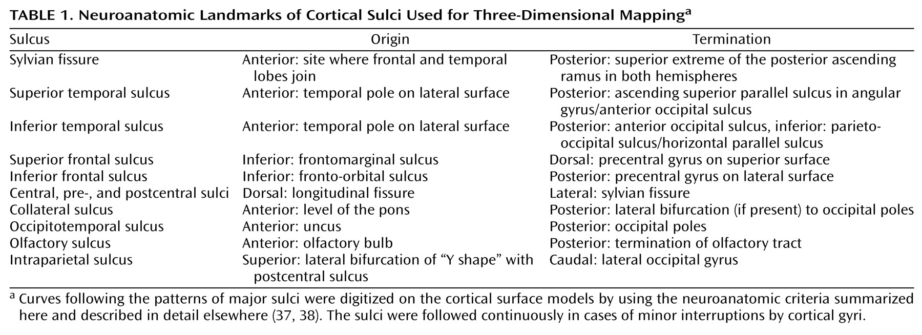
Curves following the patterns of 12 major sulci were outlined manually on magnified cortical surface models. The sulci included were the 1) central sulcus, 2) precentral sulcus, 3) postcentral sulcus, 4) inferior frontal sulcus, 5) superior frontal sulcus, 6) inferior temporal sulcus, 7) superior temporal sulcus, 8) sylvian fissure, 9) intraparietal sulcus, 10) olfactory sulcus, 11) collateral sulcus, and 12) occipitotemporal sulcus. The termination points of the cortical sulci followed the rules summarized in Table 1 (37, 38) and were applied by viewing all three planes simultaneously. Seven pairs of midline landmark curves bordering the longitudinal fissure were delimited to establish hemispheric gyral limits. Surface contours were outlined in all brains by a single investigator (K.L.N.) blind to group status. Intrarater reliability was evaluated by repeatedly outlining the same structures (six times) in a randomly chosen brain. The contouring error was less than 1 mm (three-dimensional root mean square distance) in all cases. Interrater reliability for this delineation protocol was established previously for cross-study comparisons with an interrater error between 0 and 2 mm for all sulci (three-dimensional root mean square distance) (37 and unpublished articles by E.R. Sowell et al. and by R.E. Blanton et al., 2000).

Calculation of Cortical Complexity

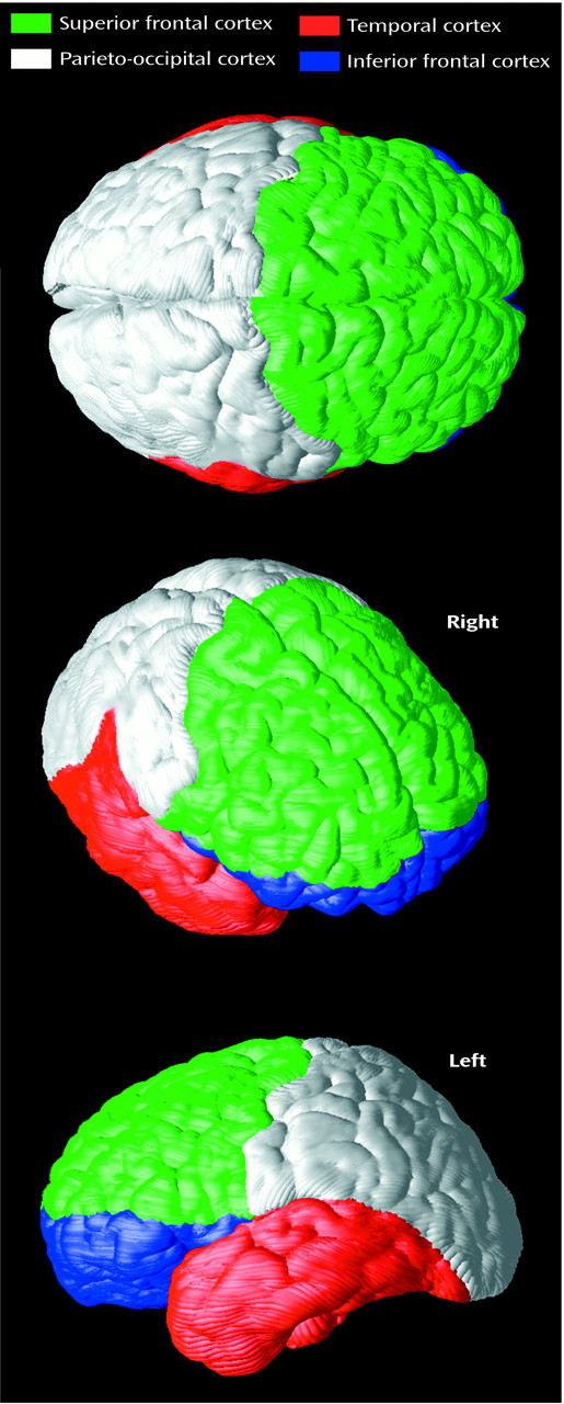
Cortical complexity was calculated in distinct neuroanatomic regions by using the sulcal outlines as landmarks for delineation. The cortical surface was separated into 1) superior frontal (dorsolateral), 2) parieto-occipital, 3) temporal, and 4) inferior frontal (orbitofrontal) regions in each subject (Figure 1). The superior frontal region was bordered by the central sulcus posteriorly, and the sylvian fissure and the inferior points of the inferior frontal and frontomarginal sulci were used as ventral landmarks. The inferior frontal region included the orbital cortex and was defined by superior frontal region boundaries dorsally. The temporal region included the cortex inferior to the sylvian fissure, and the posterior boundaries followed a line from the posterior and superior extremes of the sylvian fissure, inferior temporal, and collateral sulci inferiorly. The central sulcus formed the anterior boundary of the parieto-occipital region, and the temporal region boundaries served to delimit the lateral and inferior extents (unpublished article by Blanton et al.). Given that the same sulcal anatomies were used for delineation, the reliability for lobar parcellation was as already described.
Cortical complexity was defined by the frequency of sulcal/gyral convolutions in all three planes in each lobar subregion of the cortical surface model (39). The fractal dimension (surface complexity) is computed by obtaining the logarithmic least squares regression of the surface area against the spatial frequency of the surface mesh representing each region. A complexity value of 2.00 describes a surface region that is planar (flat), while increasing values between 2.00 and 3.00 indicate more cortical convolutions.

Averaging of the Cortical Surface

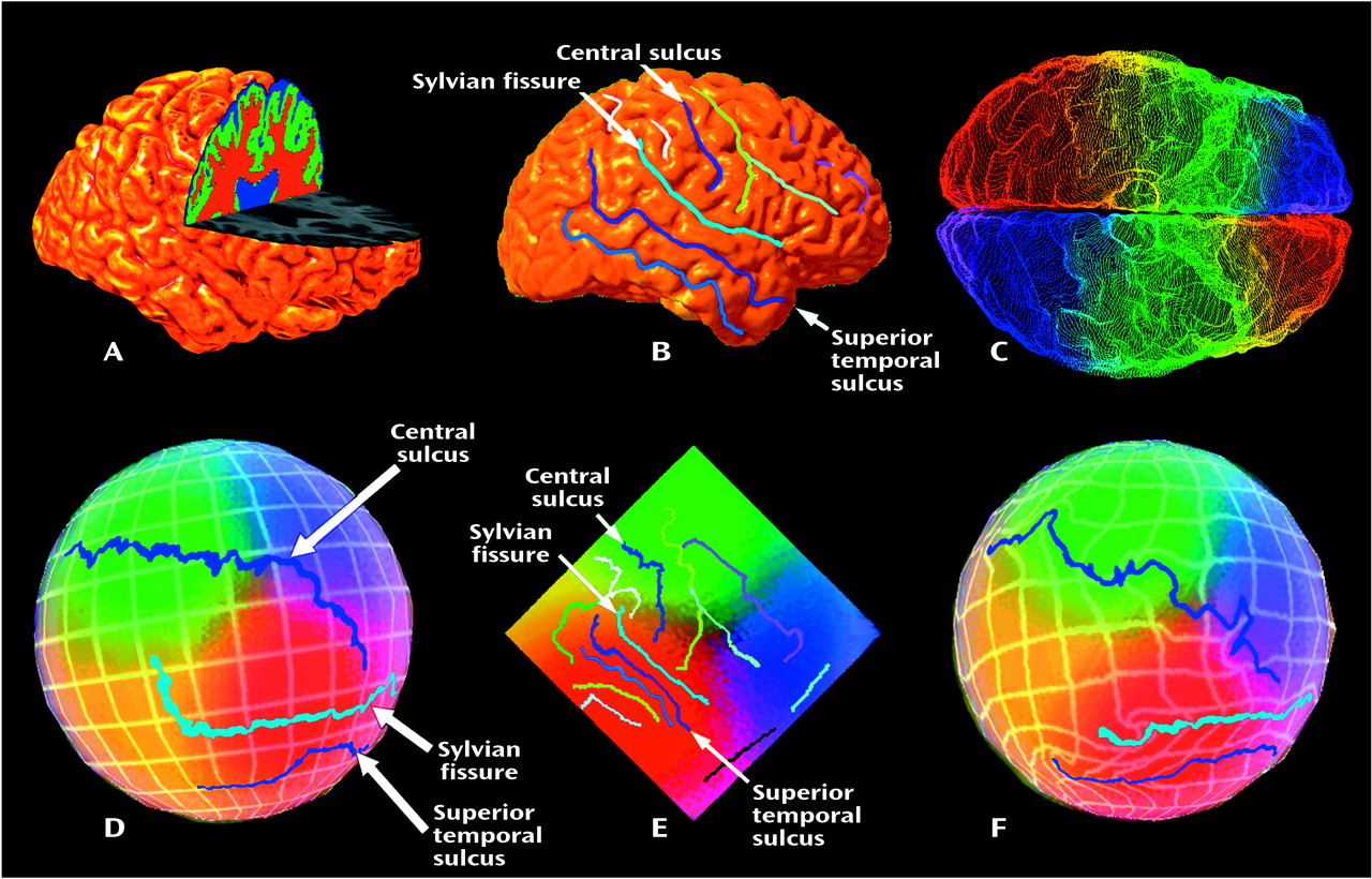
The manually outlined sulcal curves were reparameterized to render digitized points uniformly spaced. Average models of each curve were obtained by matching equally spaced points from corresponding sulci in each subject. Individual point-wise sulcal deviations were retained, providing information on intragroup sulcal variability (Figure 2) (27, 28, 37, 39, 40). The variability of the entire cortical surface was determined by using the averaged sulcal and landmark curves as anchors to drive the three-dimensional cortical surface models from each subject into correspondence, as shown in Figure 2 (28, 29, 36, 37). Corresponding sulcal curve points for the two hemispheres were subtracted to obtain asymmetry maps that showed averages and magnitudes of asymmetry within each group (mirrored in each hemisphere) in color.

Measurement of Brain Volume

Total brain volumes for all tissue types were obtained, after correction for radio-frequency inhomogeneities (41), by classifying brain matter as white matter, gray matter, CSF, and background (42). Reliability in selecting representative intensity values for these tissue types was evaluated by classifying 10 different test brains (r>0.94 for all tissue types [43]).

Statistical Analyses

Measures of sulcal dimensions at the cortex were obtained in terms of three-dimensional Talairach stereotaxic coordinates. To compensate for any confounding effects of AC-PC scaling, the original AC-PC distances were cubed and assessed, along with brain volumes, as possible covariates by examining statistical linkages with the dependent variables. Source variables hypothesized to characterize gyral asymmetries and shape profiles of temporoparietal sulcal anatomy were chosen as dependent variables. The dependent measures included the superior extreme (y-maximum coordinate), the posterior extreme (x-minimum), and slope of the sylvian fissure and temporal sulci. The anterior extreme (x-maximum coordinate) and curvature were included as postcentral sulcal measures. The measures of cortical complexity have already been described. To examine gyral geometry and cortical complexity, we used right and left hemispheric measures as repeated measures in analyses of variance (ANOVAs) or analyses of covariance (ANCOVAs) as determined by the significance of the covariates in a two (sex) by two (diagnosis) by two (hemisphere) design. Type I error was controlled by adopting Bonferroni corrections for each set of three sulcal measures (p<0.016) and the four complexity measures (p<0.0125).

Results

Covariates

To examine whether the AC-PC distance and raw brain volume differed between groups, these measures were used as dependent variables in separate two (sex) by two (diagnosis) ANOVAs. A significant sex effect was found for AC-PC distance (F=8.42, df=1, 49, p<0.006), with greater distances in the men (mean=26.84 mm, SD=1.33) than in the women (mean=25.72 mm, SD=1.43), and for brain volume (F=13.58, df=1, 49, p<0.0005; men: mean=1270.9 cm3, SD=117.8; women: mean=1161.5 cm3, SD=86.8). Finally, a similar analysis assessing the effect of AC-PC scaling on brain volume revealed that although a significant sex effect on brain volumes remained after AC-PC scaling (F=4.54, df=1, 48, p<0.04; men: mean=945.8 cm3, SD=228.2; women: mean=949.0 cm3, SD=147.7), the highly significant effect of the covariate (F=31.29, df=1, 48, p<0.00001) largely controlled for differences in head size between the sexes, although here the goal was to allow comparison of anatomical maps in stereotaxic space.
Finally, the original AC-PC distances and brain volumes were evaluated as covariates by examining correlations with the dependent variables to remove potential differences in AC-PC scaling and head size from the data. None of the sulcal measures revealing significant statistical effects showed a significant association with AC-PC distance. Higher brain volumes, however, may exhibit greater cortical folding that may segregate the sexes. Brain volume was assessed as a potential covariate in the complexity analyses given that although surface area is controlled for in this measure, the brain volumes were scaled according to the AC-PC line. Raw brain volume was significantly associated with the complexity measures (median r=0.38) and was included as a covariate in analyses.

Asymmetry

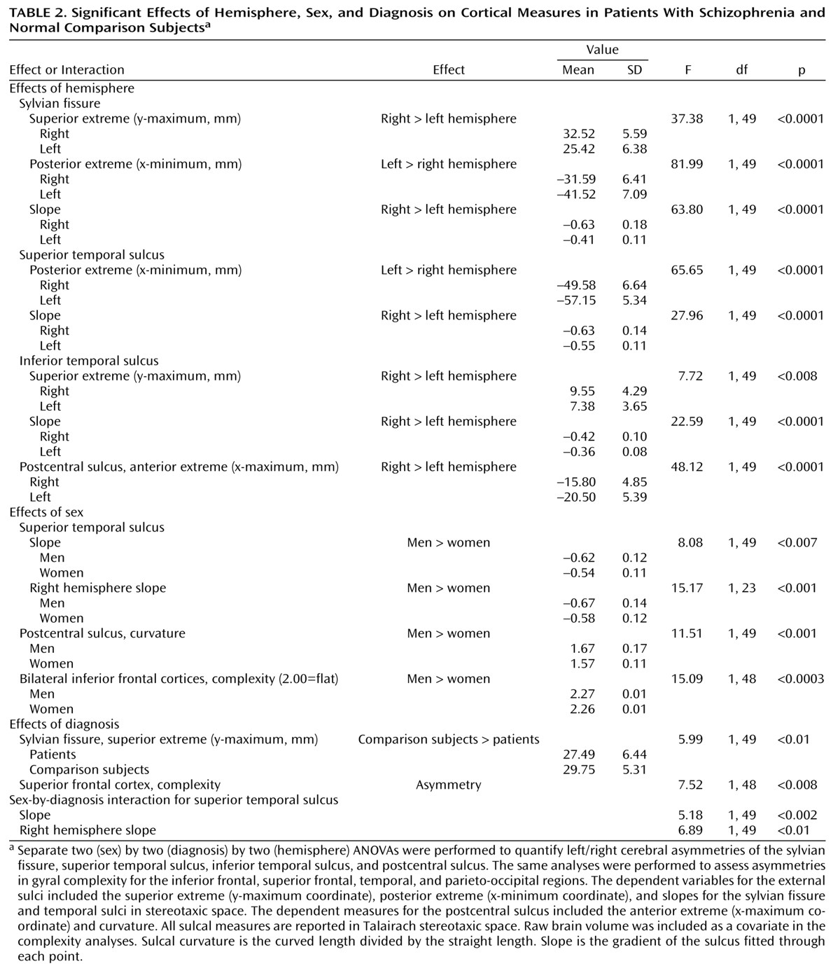
Table 2 lists the sulcal measures showing significant asymmetries, effects of sex and diagnosis, and interactions with hemisphere from the ANOVAs described in the Method section. The significant sulcal asymmetries included the following: 1) greater right hemisphere sulcal slopes, 2) posterior extension of the sylvian fissure and superior temporal sulcus in the left hemisphere, 3) greater superior extremes of the sylvian fissure and inferior temporal sulcus in the right hemisphere, and 4) more rostral anterior extreme of the postcentral sulcus in the right hemisphere.

Variability Maps

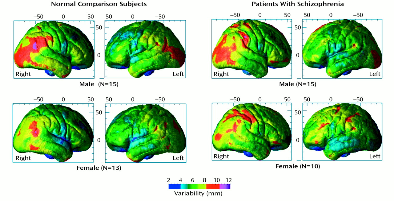
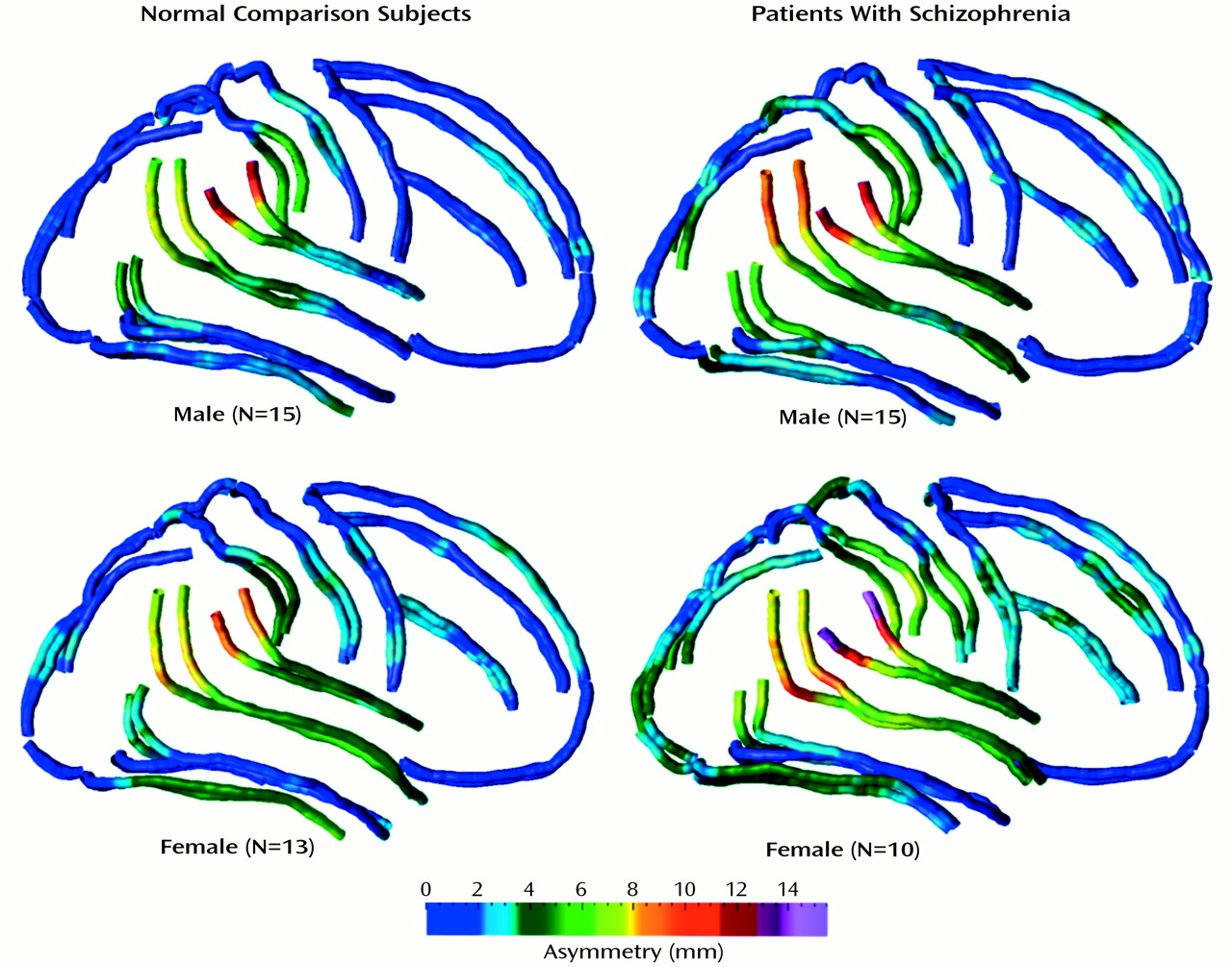
Average surface maps for the four groups defined by diagnosis and sex show localized asymmetries in the inferior and superior temporal sulci, sylvian fissure, and postcentral sulcus (Figure 3). Variability on the lateral surfaces appears greatest at the superior extreme of the superior temporal sulcus and is greater in the right hemisphere in all groups (Figure 3). Overall, the variability maps suggest greater individual differences in neopallial association areas in respect to phylogenetically and ontogenetically older areas of the cortex (Figure 3, Figure 4, Figure 5). Asymmetries in the temporoparietal regions mapped in three dimensions were confirmed in statistical analyses of the extents of the temporoparietal sulci (Table 2).

Diagnostic Effects

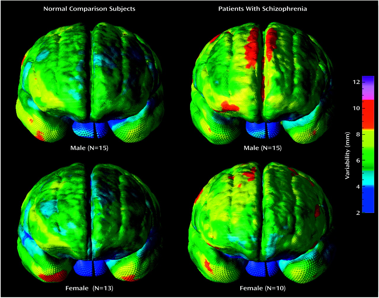
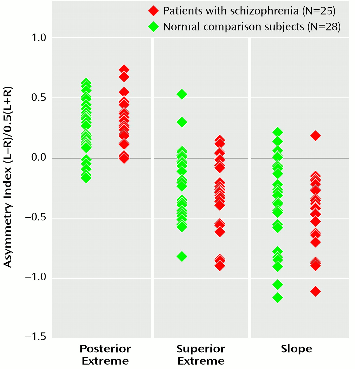
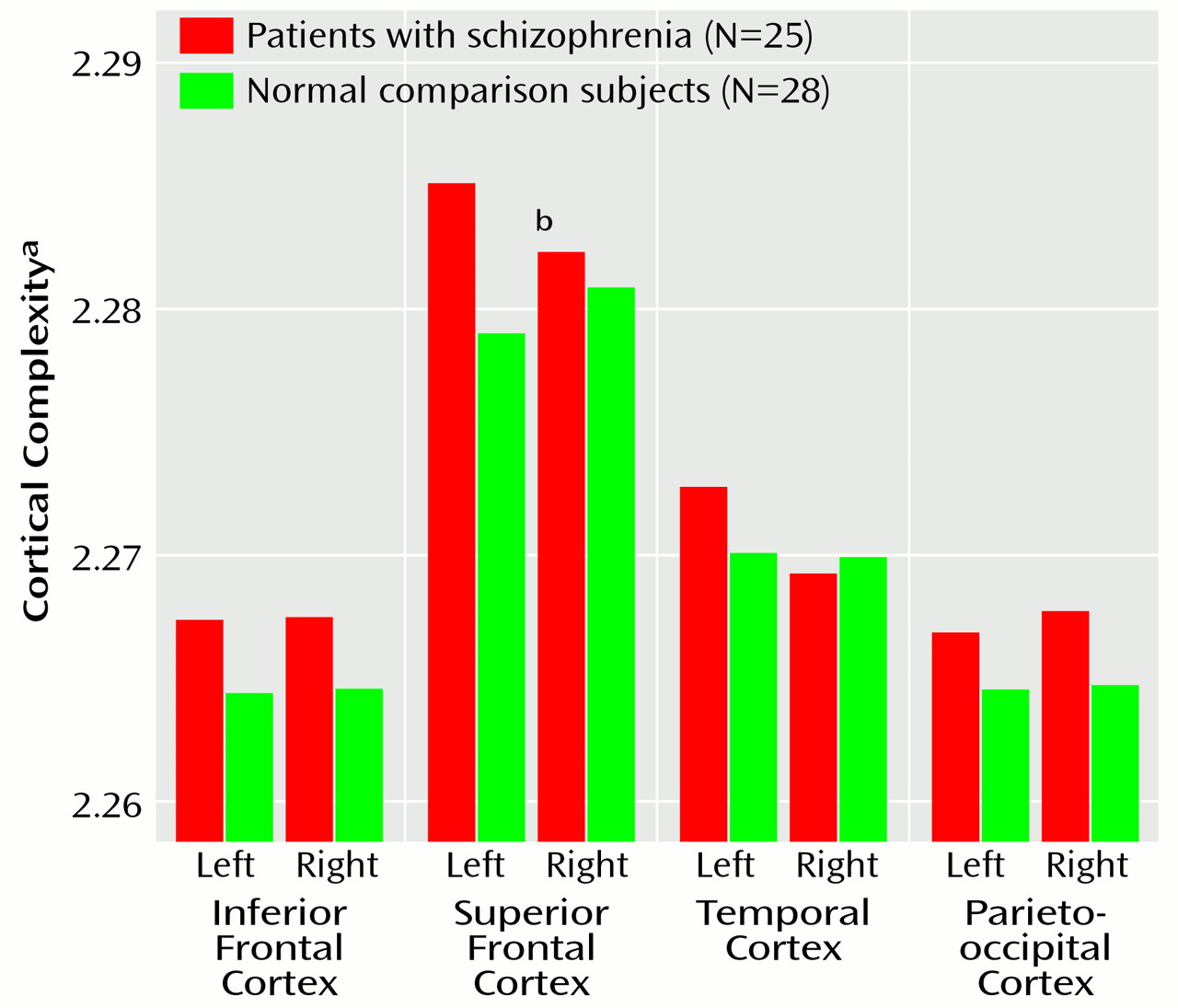
In the variability maps the patients showed greater variability in the frontal cortex than did the comparison subjects (Figure 4). The patients also showed greater variability in frontal midline curves, suggesting a larger interhemispheric fissure and localized vulnerability in the frontal cortices. The perisylvian asymmetries, however, appeared similar in the two diagnostic groups (Figure 5). Asymmetry coefficients of sylvian fissure measures, calculated with the formula (L–R)/0.5(L+R), clearly showed that there were minimal laterality differences between diagnostic groups (Figure 6). ANOVAs however, revealed significantly lower superior extremes of the sylvian fissure in the patients with schizophrenia, but no interaction with hemisphere (Figure 6). Finally, an ANCOVA showed a significant diagnosis-by-hemisphere interaction for gyral complexity in the superior frontal cortices (Figure 7).

Sex Effects

The maps showed greater variability in the ascending ramus of the sylvian fissure of the male groups and in the ascending superior ramus of the inferior temporal sulcus in the female groups (Figure 3). Statistical tests revealed significantly greater curvature of the postcentral sulcus in the men than in the women. In the comparison group, the slope of the superior temporal sulcus in the right hemisphere was also significantly greater for the men than for the women. Finally, cortical complexity was significantly greater for male than female subjects in inferior frontal regions.

Discussion

Disturbances in structural brain asymmetries in schizophrenia have been reported, although some empirical data contradict this general hypothesis (2, 3, 18, 44, 45). The majority of evidence suggests, however, that the pathophysiological mechanisms are diffuse, involving structural and functional systems in both hemispheres, although the left hemisphere is more widely implicated (5). At the cortex, gyral asymmetries have been shown as undisturbed, less lateralized compared to normal, and even reversed in schizophrenia (e.g., references 2, 3, 8, 9, 15, 18, 46).

Cortical Asymmetries

We mapped sulcal asymmetries across the entire cortical surface in schizophrenic and comparison subjects. Statistical analyses confirmed the presence of significant sulcal asymmetries in temporoparietal regions in both diagnostic groups (Figure 5). Several of these asymmetries are well established in normal populations, but to our knowledge, they have not been previously mapped in three dimensions. For example, it is known that the sylvian fissure extends further posteriorly in the left hemisphere (corresponding to greater planum temporale asymmetries in these regions) and rises more steeply in the right hemisphere (e.g., references 27, 47, 48). Our results are similar, showing greater than normal left hemisphere sylvian fissure length and right hemisphere slope and height (Figure 6). These asymmetries were largely mirrored in the superior and inferior temporal sulci.
Postcentral gyrus hemispheric asymmetries have been reported in normal adults (49) and in Alzheimer’s disease patients (right-handed men) (29). Our results corroborate these findings, revealing significant asymmetry of the postcentral sulcal anterior extreme (right hemisphere greater than left hemisphere) in both diagnostic groups. Such asymmetry may be associated with asymmetries of the slope and horizontal length of the sylvian fissure and temporal sulci and may reflect asymmetries in the parietal operculum that complement planum temporale asymmetries (not necessarily related) in right-handed subjects (50).
Many earlier studies assessing perisylvian asymmetries in patients with schizophrenia focused on the planum temporale, a triangular region on the superior temporal gyral surface that is involved in language functions. The findings indicate abnormally low planum temporale volume and area in the left hemisphere of patients with schizophrenia (e.g., references 15 and 51). Negative findings exist, however, and discrepancies in results are difficult to interpret given the differences in the measurement techniques used (2, 18). Although sylvian fissure measurements and planum temporale area/volume asymmetries overlap, they should be considered different measures (5254). That is, typically the planum is found to be symmetrical when the boundaries include the posterior ascending ramus of the sylvian fissure (55). This appears to indicate, as others have suggested, that the horizontal segment is a better indicator of planum temporale asymmetry (52) and that the right hemisphere ascending segment compensates for the shorter horizontal segment. Cytoarchitectonically, however, the auditory association cortices extend into the ascending part of the sulcus (55).
In this study, the sylvian fissure was not divided at the cortex to specifically isolate planum temporale regions, and no conclusions regarding abnormalities of laterality of the planum in schizophrenia are assumed. In previous studies, both the sylvian fissure and the anatomically overlapping planum temporale showed large hemispheric asymmetry effects in normal subjects. According to Cohen (56, 57), an effect size, f, is considered small if f is 0.10, medium if f is 0.25, and large if f is beyond 0.40. As in the earlier studies, we found hemispheric asymmetry effects with f values greater than 0.40. Since studies with relatively small study groups have shown hemispheric asymmetries in schizophrenia that are less than in healthy subjects, and since some studies have even shown complete reversals of hemispheric asymmetries (15), effect sizes for diagnosis-by-hemisphere interactions for sylvian fissure measurements might similarly be expected to be large or at least medium in magnitude.
Post hoc power analyses revealed that empirically measured effect sizes for asymmetries were extremely large: f=0.90, f=0.60, and f=0.80 for the sylvian fissure posterior extent, superior extreme, and slope, respectively, with power between 0.90 and 0.99. Hemisphere-by-diagnosis interactions for these measures, however, showed extremely small empirically measured effect sizes (f<0.10). The probability of type II error for such small effects was therefore greater than 0.50. If these effect sizes are truly representative, our analyses indicate that only unreasonably large study groups would likely reveal effects showing small differences in sulcal asymmetries, even with alpha at 0.05. Notwithstanding, if the diagnosis-by-hemisphere effects were large, as implied by previous study results, our study would have had reasonable power to detect group differences in asymmetry.
These findings suggest that 1) sylvian fissure asymmetries at the cortex that include the planum parietale, which follows the terminal ascending ramus, do not reflect less than normal planum temporale asymmetry in schizophrenia (54) and 2) group differences in perisylvian sulcal asymmetries, if present at the cortex, are likely so small that they are of minimal diagnostic importance (Figure5 and Figure 6). Supporting our findings, Bartley et al. (58) used similar sylvian fissure measurements and found normal hemispheric asymmetries in monozygotic twins discordant for schizophrenia. Furthermore, DeLisi et al. (3) reported somewhat less laterality in the horizontal component of the sylvian fissure in schizophrenia (although the difference was not statistically significant) but no difference for the vertical segment. Finally, we found the sylvian fissure superior extreme to be more inferior in the patients than in the comparison subjects but not to exhibit diagnosis-by-hemisphere effects.

Sulcal Asymmetries and Sex

Structural asymmetries are influenced by gender (e.g., references 2, 48, 59), and gender interactions in schizophrenia have been reported (24) but not always supported (60). Significant sex effects in this study indicated greater asymmetries of the superior temporal sulcal slope and postcentral sulcal curvature in men. Gender differences in sylvian fissure asymmetries were absent, however, but as mentioned, discrete partitions were not assessed, and this may have obscured sex differences relating to planum asymmetries (9, 48). Finally, the greater right hemisphere superior temporal sulcal slope in men than in women in the comparison group was not present in the patients, suggesting that sexually dimorphic developmental processes that may result in greater structural asymmetries in males may be disturbed in schizophrenia.

Cortical Complexity

Cortical complexity reflects the number of primary, secondary, and tertiary sulcal bifurcations in a region and may reveal potentially different patterns of gyral geometry that become manifest during development or disease (27, 40). Procedures for cortical surface averaging may underestimate interindividual differences in sulcal continuity (38) that could discriminate diagnostic groups. Kikinis and colleagues (61), for example, reported greater than normal vertical orientation and interruptions in the patterns of left hemisphere temporal sulci in schizophrenia. Furthermore, studies have shown hemispheric differences in frontal complexity in schizophrenia, which appear to interact with gender (16, 17). For instance, significantly greater right hemisphere prefrontal complexity was found in male patients than in comparison subjects (17) when a gyrification index (62, 63) representing the ratio of the outer (superficial) to the inner (deep) perimeter of a cortical slice was determined. In contrast, we report abnormal asymmetry in superior frontal regions in patients (left hemisphere greater than right). Cortical complexity indices, however, showed average differences between patients and comparison subjects (except right hemisphere temporal lobe) (Figure 7) that were at least in the same direction as those indicating greater complexity in right frontal and left temporal regions in schizophrenia (17, 61). Notwithstanding, different methods may make cross-study comparisons of complexity measures unsuitable. For example, here fractal complexity was measured in three dimensions such that group differences in gyral geometry in the vertical, horizontal, and lateral planes could be detected. That is, complexity was not computed on the basis of a single two-dimensional plane of reference defined by a particular, noncontiguous brain slice. Moreover, gyral complexity was measured across the cortical surface in anatomically homologous regions in each subject.
Greater cortical complexity in male than female subjects was found in inferior frontal regions. Other investigators (62, 64) have failed to find differences in cortical complexity across gender in normal populations, but again, methodological differences between studies exist. For example, gyral complexity has been computed by using the a ratio of total cortical surface area to brain volume (64), which may not be sensitive to the same features as the three-dimensional approach used here. Furthermore, in an earlier postmortem study (62), only temporal regions were assessed in addition to the gyral complexity from coronal slices over the entire cortex. It is therefore possible that gender effects may have been overlooked in the inferior frontal cortices. Clearly, new studies may establish whether sexually dimorphic development processes, in specific cortical regions, influence gyral complexity.
Conclusion

In this study, we detected highly significant asymmetries at the cortex in patients with schizophrenia, contrary to prior findings of less than normal lateralization. Subtle differences in sulcal surface asymmetries, if present in temporoparietal regions in schizophrenia, however, may require substantially larger study groups for detection, suggesting their diagnostic value is minimal. Our results, however, suggest the absence of reversed asymmetry in schizophrenia. The discrepancy in results may be related to the criteria for sylvian fissure measurement and whether the horizontal or posterior regions are included. In contrast, cortical complexity indices indicated a disturbance in the patterns of gyral asymmetries in frontal regions of patients with schizophrenia. Many studies have implicated lateralized structural deviations in frontal regions in schizophrenia, showing abnormalities in petalias, lobar asymmetries, and lateralized low gray matter volume, which lend some support for these results (3, 10, 25, 6567). Finally, there is substantial individual variation in the patterns and degree of hemispheric differences as seen in the cortical variability maps (9, 29, 48). Subject variables such as hand preference and sex as well as clinical features may all influence patterns of cortical variation (51). Examination of these factors may help clarify some differences across studies concerning the abnormalities of asymmetries in perisylvian and frontal regions in schizophrenia.
TABLE 1
TABLE 2
Figure 1. Regional Parcellation of Cortex for Measurement of Cortical Complexity a
aThe measure of sulcal/gyral complexity is based on the rate at which the computed surface area of the region increases as the scale of the measurement is reduced.
Figure 2. Illustration of Cortical Surface Matching Between Subjectsa
aPart A: A parametric cortical model is extracted from each image set by using intensity information after removal of extracortical tissue. The cortical surface is arrived at by the deformation of a spherical mesh, so that any point on the cortical surface corresponds to exactly one point on the sphere and planar map. Part B: Surface curves—for example, the sylvian fissure, superior temporal sulcus, and central sulcus—are mapped onto each surface model. Part C: Each point on the cortical surface is color coded in red/green/blue format such that every color value is accurately represented at point locations in three-dimensional stereotaxic space on a colorized cortical map. The cortical surface model and the corresponding colorized spherical map (part D) are flattened while all three-dimensional stereotaxic point locations in color are retained (right hemisphere shown). Part E: Individual sulcal anatomies are mapped back onto the flattened hemispheric surface. Sulcal curves within each group are reparameterized to create an average template of sulcal anatomy while variability information is retained. Part F: Finally, a complex vector-valued flow field defined on the spherical and planar maps drives the cortical surface anatomy into correspondence with the average sulcal anatomical template and retains variability information about the extent of the deformation. The average sulcal curves and surface anatomy are now aligned and translated back into three dimensions for visualization of variability maps.
Figure 3. Three-Dimensional Maps of Variability in Cortical Surfaces in Both Hemispheres of Male and Female Patients With Schizophrenia and Normal Comparison Subjectsa
aThe color bar indicates variability in each group as the root mean square magnitude of displacement vectors required to map each individual onto the group average mesh. Variability was greater at the superior limits of the superior temporal sulcus in all groups, particularly in the right hemisphere. Greater variability was most apparent in the ascending ramus of the sylvian fissure in the male groups and in the ascending superior ramus of the inferior temporal sulcus in the female groups. Overall, posterior association areas showed greater variability in all groups.
Figure 4. Three-Dimensional Maps of Variability in Frontal Cortical Surfaces in Male and Female Patients With Schizophrenia and Normal Comparison Subjectsa
aThe color bar indicates variability in each group as the root mean square magnitude of displacement vectors required to map each individual onto the group average mesh. Greater variability can be see in the frontal regions in the patients, especially the men, who have greater variability bordering the longitudinal fissure, perhaps reflecting larger sulci and larger CSF space. Greater variability in the orbitofrontal region appears unilaterally (right hemisphere) in the comparison subjects and bilaterally in the orbitofrontal region in the male patients.
Figure 5. Maps of Sulcal Asymmetry in Male and Female Patients With Schizophrenia and Normal Comparison Subjectsa
aAsymmetry maps were created by subtracting the sulcal mesh averages of one hemisphere from the mirror of the other hemisphere to create displacement vectors representing asymmetry in color. These maps not only represent the magnitude of average sulcal asymmetry in color but also display differences in sulcal shape profiles between hemispheres since the right hemisphere is mapped onto the left hemisphere and vice versa. The color bar reemphasizes the shape difference between the hemispheres.
Figure 6. Asymmetry Indices of Sylvian Fissure Measures for Patients With Schizophrenia and Normal Comparison Subjectsa
aL, left hemisphere measure; R, right hemisphere measure. Positive values indicate leftward asymmetries, and negative values indicate rightward asymmetries.
Figure 7. Regional Cortical Complexity in Left and Right Hemispheres of Patients With Schizophrenia and Normal Comparison Subjectsa
aLogarithmic least squares regression of lobar surface area against the spatial frequency of the surface mesh in three dimensions. A value of 2.00 indicates a surface region that is flat; values between 2.00 and 3.00 indicate more cortical convolutions.
bSignificant diagnosis-by-hemisphere interaction (F=7.52, df=1, 48, p<0.008).

Footnote

Received Nov. 2, 1999; revisions received April 7 and Aug. 24, 2000; accepted Sept. 14, 2000. From the Laboratory of Neuro Imaging, Department of Neurology, Division of Brain Mapping, UCLA School of Medicine; and the Section of Cognitive Psychopharmacology, Institute of Psychiatry, London. Address reprint requests to Dr. Toga, Laboratory of Neuro Imaging, Department of Neurology, Division of Brain Mapping, UCLA School of Medicine, 710 Westwood Plaza, Los Angeles, CA 90095-1769; [email protected] (e-mail). Supported by grant BIR 93-22434 from the National Science Foundation, by grant LM/MH-05639 from the National Library of Medicine and NIMH, by grant RR-05056 and P41 Resource Grant RR-13642 from the National Center for Research Resources, by grant NS-38253 from the National Institute of Neurological Disorders and Stroke, and by Human Brain Project grant P20 MH/DA-52176 to the International Consortium for Brain Mapping (funded jointly by NIMH and the National Institute on Drug Abuse). The authors thank Dr. Sharma’s colleagues at the Institute of Psychiatry for data collection and Andrew Lee for graphics input.

References

1.
Crow TJ: Schizophrenia as failure of hemispheric dominance for language. Trends Neurosci 1997; 20:339–343
2.
Beaton AA: The relation of planum temporale asymmetry and morphology of the corpus callosum to handedness, gender, and dyslexia: a review of the evidence. Brain Lang 1997; 60:255–322
3.
DeLisi LE, Sakuma M, Kushner M, Finer DL, Hoff AL, Crow TJ: Anomalous cerebral asymmetry and language processing in schizophrenia. Schizophr Bull 1997; 23:255–271
4.
Falkai P, Bogerts B, Schneider T, Greve B: Disturbed planum temporale asymmetry in schizophrenia: a quantitative post-mortem study. Schizophr Res 1995; 14:161–176
5.
Petty RG: Structural asymmetries of the human brain and their disturbance in schizophrenia. Schizophr Bull 1999; 25:121–139
6.
Flor-Henry P: Lateralized temporal-limbic dysfunction and psychopathology. Ann NY Acad Sci 1976; 280:777–797
7.
Ragland JD, Gur RE, Klimas BC, McGrady N, Gur RC: Neuropsychological laterality indices of schizophrenia: interactions with gender. Schizophr Bull 1999; 25:79–89
8.
Nasrallah HA: A controlled magnetic resonance imaging study of corpus callosum thickness in schizophrenia. Biol Psychiatry 1986; 21:274–282
9.
Witelson SF, Kigar DL: Sylvian fissure morphology and asymmetry in men and women: bilateral differences in relation to handedness in men. J Comp Neurol 1992; 323:326–340
10.
Sharma T, Lancaster E, Sigmundsson T, Lewis S, Takei N, Gurling H, Barta P, Pearlson G, Murray R: Lack of normal pattern of cerebral asymmetry in familial schizophrenic patients and their relatives—the Maudsley Family Study. Schizophr Res 1999; 40:111–140
11.
Bogerts B, Ashtari M, Degreef G, Alvir JM: Reduced temporal limbic structure volumes on magnetic resonance images in first episode schizophrenia. Psychiatry Res Neuroimaging 1990; 35:1–13
12.
Dauphinais ID, DeLisi LE, Crow TJ, Alexandropoulos K, Colter N, Tuma I, Gershon ES: Reduction in temporal lobe size in siblings with schizophrenia: a magnetic resonance imaging study. Psychiatry Res 1990; 35:137–147
13.
Shenton ME, Kikinis R, Jolesz FA, Pollak SD, LeMay M, Wible CG, Hokama H, Martin J, Metcalf D, Coleman M, McCarley RW: Abnormalities of the left temporal lobe and thought disorder in schizophrenia. N Engl J Med 1992; 327:604–612
14.
Hoff AL, Riordan H, O’Donnell DW, Stritzke P: Anomalous lateral sulcus asymmetry and cognitive function in first-episode schizophrenia. Schizophr Bull 1992; 18:257–272
15.
Gur RE, Chin S: Laterality in functional brain imaging studies of schizophrenia. Schizophr Bull 1999; 25:141–156
16.
Kulynych JJ, Luevano LF, Jones DW, Weinberger DR: Cortical abnormality in schizophrenia: an in vivo application of the gyrification index. Biol Psychiatry 1997; 41:995–999
17.
Vogeley K, Schneider-Axmann T, Pfeiffer U, Tepest R, Bayer TA, Bogerts B, Honer WG, Falkai P: Disturbed gyrification of the prefrontal region in male schizophrenic patients: a morphometric postmortem study. Am J Psychiatry 2000; 157:34–39
18.
Cutting J: The role of right hemisphere dysfunction in psychiatric disorders. Br J Psychiatry 1992; 160:583–588
19.
Gur RE: Is schizophrenia a lateralized brain disorder? editor’s introduction. Schizophr Bull 1999; 25:7–9
20.
Nopoulos P, Torres I, Flaum M, Andreasen NC, Earhardt JC, Yuh WTC: Brain morphology in first-episode schizophrenia. Am J Psychiatry 1995; 152:1721–1723
21.
Geschwind N, Galaburda AM: Cerebral lateralization: biological mechanisms, associations, and pathology, II: a hypothesis and a program for research. Arch Neurol 1985; 42:521–552
22.
Wisniewski AB: Sexually-dimorphic patterns of cortical asymmetry, and the role for sex steroid hormones in determining cortical patterns of lateralization. Psychoneuroendocrinology 1998; 23:519–547
23.
DeLisi LE, Dauphinais ID, Hauser P: Gender differences in the brain: are they relevant to the pathogenesis of schizophrenia? Compr Psychiatry 1989; 30:197–208
24.
Cowell PE, Kostianovsky DJ, Gur RC, Turetsky BI, Gur RE: Sex differences in neuroanatomical and clinical correlations in schizophrenia. Am J Psychiatry 1996; 153:799–805
25.
Lawrie SM, Abukmeil SS: Brain abnormality in schizophrenia: a systematic and quantitative review of volumetric magnetic resonance imaging studies. Br J Psychiatry 1998; 172:110–120
26.
Mazziotta JC, Toga AW, Evans A, Fox P, Lancaster J: A probabilistic atlas of the human brain: theory and rationale for its development. Neuroimage 1995; 2:89–101
27.
Thompson PM, Moussai J, Zohoori S, Goldkorn A, Khan AA, Mega MS, Small GW, Cummings JL, Toga AW: Cortical variability and asymmetry in normal aging and Alzheimer’s disease. Cereb Cortex 1998; 8:492–509
28.
Thompson PM, Toga AW: Anatomically-driven strategies for high-dimensional brain image warping and pathology detection, in Brain Warping. Edited by Toga AW. New York, Academic Press, 1998, pp 311–336
29.
Thompson PM, Woods RP, Mega MS, Toga AW: Mathematical/computational challenges in creating deformable and probabilistic brain atlases. Hum Brain Mapp 2000; 9:81–92
30.
Toga AW: An introduction to maps and atlases of the brain, in Brain Mapping: The Systems. Edited by Mazziotta JC, Toga AW, Frackowiak RSJ. New York, Academic Press, 1998, pp 3–28
31.
Aso M, Kurachi M, Suzuki M, Yuasa S: Asymmetry of the ventricle and age at the onset of schizophrenia. Eur Arch Psychiatry Clin Neurosci 1995; 245:142–144
32.
DeCourten-Myers: The human cerebral cortex: gender differences in structure and function. Exp Neurol 1999; 58:217–226
33.
Office of Population Censuses and Surveys: Standard Occupational Classification, vol 3. London, Her Majesty’s Stationery Office, 1991
34.
Annett M: A classification of hand preference by association analysis. Br J Psychol 1970; 61:303–321
35.
Talairach J, Tournoux P: Co-Planar Stereotaxic Atlas of the Human Brain. New York, Thieme Medical, 1988
36.
MacDonald D, Avis D, Evans AC: Multiple surface identification and matching in magnetic resonance imaging. Proceedings of the International Society for Optical Engineering 1994; 2359:160–169
37.
Thompson PM, MacDonald D, Mega MS, Holmes CJ, Evans AC, Toga AW: Detection and mapping of abnormal brain structure with a probabilistic atlas of cortical surfaces. J Comput Assist Tomogr 1997; 21:567–581
38.
Ono M, Kubik S, Abernathey CD: Atlas of the Cerebral Sulci. New York, Thieme Verlag, 1990
39.
Thompson PM, Schwartz C, Lin RT, Khan AA, Toga AW: Three-dimensional statistical analysis of sulcal variability in the human brain. J Neurosci 1996; 16:4261–4274
40.
Thompson PM, Schwartz C, Toga AW: High-resolution random mesh algorithms for creating a probabilistic 3D surface atlas of the human brain. Neuroimage 1996; 3:19–34
41.
Sled JG, Pike GB: Standing-wave and RF penetration artifacts caused by elliptic geometry: an electrodynamic analysis of MRI. IEEE Trans Med Imaging 1998; 17:653–662
42.
Zijdenbos AP, Dawant BM: Brain segmentation and white matter lesion detection in MR images. Crit Rev Biomed Eng 1994; 22:401–465
43.
Sowell ER, Thompson PM, Holmes CJ, Batth R, Jernigan TL, Toga AW: Localizing age-related changes in brain structure between childhood and adolescence using statistical parametric mapping. Neuroimage 1999; 9:587–597
44.
Frangou S, Sharma T, Sigmudsson T, Barta P, Pearlson G, Murray RM: The Maudsley Family Study, 4: normal planum temporale asymmetry in familial schizophrenia: a volumetric MRI study. Br J Psychiatry 1997; 170:328–333
45.
Satz P, Green MF: Atypical handedness in schizophrenia: some methodological and theoretical issues. Schizophr Bull 1999; 25:63–78
46.
Harris LJ: Early theory and research on hemispheric specialization. Schizophr Bull 1999; 25:11–39
47.
Galaburda AM, Sanides F, Geschwind N: Human brain: cytoarchitectonic left-right asymmetries in the temporal speech region. Arch Neurol 1978; 35:812–817
48.
Ide A, Rodríguez E, Zaidel E, Aboitiz F: Bifurcation patterns in the human Sylvian fissure: hemispheric and sex differences. Cereb Cortex 1996; 6:717–725
49.
Hustler JJ, Loftus WC, Gazzaniga MS: Individual variation of cortical surface area asymmetries. Cereb Cortex 1998; 8:11–17
50.
Habib M, Robichon F, Lévrier O, Khalil R, Salamon G: Diverging asymmetries of temporo-parietal cortical areas: a reappraisal of Geschwind/Galaburda theory. Brain Lang 1995; 48:238–258
51.
Kwon JS, McCarley RW, Hirayasu Y, Anderson JE, Fischer IA, Kikinis R, Jolesz FA, Shenton ME: Left planum temporale volume reduction in schizophrenia. Arch Gen Psychiatry 1999; 56:142–148
52.
Barta PE, Petty RG, McGilchrist I, Lewis RW, Jerram M, Casanova MF, Powers RE, Brill LB II, Pearlson GD: Asymmetry of the planum temporale: methodological considerations and clinical associations. Psychiatry Res 1995; 61:137–150
53.
Steinmetz H, Rademacher J, Huang YX, Hefter H, Zilles K, Thron A, Freund HJ: Cerebral asymmetry: MR planimetry of the human planum temporale. J Comput Assist Tomogr 1989; 13:996–1005
54.
Steinmetz H: Structure, functional and cerebral asymmetry: in vivo morphometry of the planum temporale. Neurosci Biobehav Rev 1996; 20:587–591
55.
Shapleske J, Rossell SL, Woodruff PW, David AS: The planum temporale: a systematic, quantitative review of its structural, functional and clinical significance. Brain Res 1999; 29:26–49
56.
Cohen J: Statistical Power Analysis for the Behavioral Sciences. New York, Academic Press, 1977
57.
Cohen J: Statistical Power Analysis for the Behavioral Sciences, 2nd ed. Hillsdale, NJ, Lawrence Erlbaum Associates, 1988
58.
Bartley AJ, Jones DW, Torrey EF, Zigun JR, Weinberger DR: Sylvian fissure asymmetries in monozygotic twins: a test of laterality in schizophrenia. Biol Psychiatry 1993; 34:853–863
59.
Paus T, Tomaiuolo F, Otaky N, MacDonald D, Petrides M, Atlas J, Morris R, Evans AC: Human cingulate and paracingulate sulci: pattern, variability, asymmetry, and probabilistic map. Cereb Cortex 1996; 6:207–214
60.
Flaum M, Swayze VW II, O’Leary DS, Yuh WTC, Ehrhardt JC, Arndt SV, Andreasen NC: Effects of diagnosis, laterality, and gender on brain morphology in schizophrenia. Am J Psychiatry 1995; 152:704–714
61.
Kikinis R, Shenton ME, Gerig G, Hokama H, Haimson J, O’Donnell BF, Wible CG, McCarley RW, Jolesz FA: Temporal lobe sulco-gyral pattern anomalies in schizophrenia: an in vivo MR three-dimensional surface rendering study. Neurosci Lett 1994; 182:7–12
62.
Zilles K, Armstrong E, Schleicher A, Kretschmann HJ: The human pattern of gyrification in the cerebral cortex. Anat Embryol (Berl) 1988; 179:173–179
63.
Armstrong E, Schleicher A, Omran H, Curtis M, Zilles K: The ontogeny of human gyrification. Cereb Cortex 1995; 1:56–63
64.
Nopoulos P, Flaum M, O’Leary D, Andreasen NC: Sexual dimorphism in the human brain: evaluation of tissue volume, tissue composition and surface anatomy using magnetic resonance imaging. Psychiatry Res 2000; 98:1–13
65.
Goldman-Rakic PS, Selemon LD: Functional and anatomical aspects of prefrontal pathology in schizophrenia. Schizophr Bull 1997; 23:437–458
66.
Crow TJ, Colter N, Frith CD, Johnstone EC: Developmental arrest of cerebral asymmetries in early onset schizophrenia. Psychiatry Res 1989; 29:247–253
67.
Bilder RM, Wu H, Chakos MH, Bogerts B: Cerebral morphometry and clozapine treatment in schizophrenia. J Clin Psychiatry 1994; 55:53–56

Information & Authors

Information

Published In

Go to American Journal of Psychiatry
American Journal of Psychiatry
Pages: 244 - 255
PubMed: 11156807

History

Published online: 1 February 2001
Published in print: February 2001

Authors

Details

Katherine L. Narr, B.A.
Paul M. Thompson, Ph.D.

Metrics & Citations

Metrics

Citations

Export Citations

If you have the appropriate software installed, you can download article citation data to the citation manager of your choice. Simply select your manager software from the list below and click Download.

For more information or tips please see 'Downloading to a citation manager' in the Help menu.

Format
Citation style
Style
Copy to clipboard

View Options

View options

PDF/EPUB

View PDF/EPUB

Login options

Already a subscriber? Access your subscription through your login credentials or your institution for full access to this article.

Personal login Institutional Login Open Athens login
Purchase Options

Purchase this article to access the full text.

PPV Articles - American Journal of Psychiatry

PPV Articles - American Journal of Psychiatry

Not a subscriber?

Subscribe Now / Learn More

PsychiatryOnline subscription options offer access to the DSM-5-TR® library, books, journals, CME, and patient resources. This all-in-one virtual library provides psychiatrists and mental health professionals with key resources for diagnosis, treatment, research, and professional development.

Need more help? PsychiatryOnline Customer Service may be reached by emailing [email protected] or by calling 800-368-5777 (in the U.S.) or 703-907-7322 (outside the U.S.).

Media

Figures

Other

Tables

Share

Share

Share article link

Share