Posttraumatic stress disorder (PTSD) is an incapacitating clinical syndrome characterized by intrusive recollections, emotional numbing and withdrawal, cue-related responses, and psychological and physiological hyperarousal (DSM-IV).
There is no definitive pharmacotherapy for PTSD. Agents studied that have resulted in various degrees of improvement in core PTSD symptoms and/or the associated anxiety and depressive symptoms
(1–
3) have included antidepressants (monoamine oxidase inhibitors, tricyclic antidepressants, and selective serotonin reuptake inhibitors [SSRIs])
(4–
6), adrenergic agonists and antagonists (clonidine, propranolol)
(7,
8), mood-stabilizing drugs (carbamazepine, lithium, valproic acid)
(9–
11), and benzodiazepines (alprazolam)
(12). The Food and Drug Administration has to date approved the use of sertraline and paroxetine (which are SSRIs) for the indication of PTSD.
Transcranial magnetic stimulation (TMS) is a noninvasive technique for directly stimulating cortical neurons. Electrical energy crosses the brain almost painlessly, without causing convulsions or cognitive impairment but causing a depolarization of neurons
(13) with cortical changes in monoamines
(14). TMS has been shown to have some antidepressant-like effects
(15–
18).
In a prior open pilot study
(19), one session of slow TMS with 30-m/sec pulses, 15 stimuli to each side of the vertex, was found to be effective in lowering the core PTSD symptom of avoidance, as well as anxiety and somatization, and in improving ratings on the Clinical Global Impression scale. These preliminary results were taken to show that TMS is a safe and tolerable intervention with possible therapeutic efficacy for PTSD patients
(19).
Treatment with 1-Hz repetitive transcranial magnetic stimulation (rTMS) over the right frontal cortex has been reported
(20) to demonstrate normalization of positron emission tomography (PET) findings, decreasing regional cerebral blood flow (rCBF) as well as improving the clinical condition of two patients with PTSD.
The prefrontal cortex has been implicated in the organization and control of behavior
(21) by way of extensive dorsolateral prefrontal subcortical connections to limbic structures. The prefrontal cortex is involved in many complex cognitive and behavioral functions that are potentially relevant to PTSD, such as working memory
(22–
24), supervisory attentional control
(25), reasoning and decision making
(26), temporal organization of behavior
(27), and inhibition of cognition.
Structural and functional neuroimaging studies have demonstrated abnormalities in the prefrontal cortex in PTSD patients. Low metabolism was found at baseline in temporal and prefrontal cortical areas
(28) and in the parietal cortex of substance-dependent PTSD patients
(29,
30), according to PET scans. PTSD patients showed increased rCBF in the amygdala and decreased blood flow in the medial prefrontal cortex in response to provocation of symptoms by script-driven imagery
(31–
34). These findings have been found in both subjects with combat-related PTSD and subjects whose PTSD was related to childhood abuse. Using a pharmacological challenge in Vietnam combat veterans with PTSD and healthy age-matched comparison subjects, Bremner et al.
(28) found that the responses of brain metabolism to yohimbine were significantly smaller in the prefrontal, temporal, parietal, and orbitofrontal cortices of PTSD patients than those of healthy subjects.
Proton magnetic resonance spectroscopy (MRS) measurement of medial temporal lobe neural density in Vietnam combat veterans with PTSD and in healthy veterans
(35) showed that the ratio of
N-acetyl-
l-aspartic acid to creatinine in the right hemisphere of the PTSD patients was significantly lower than that on the left and was also significantly lower than that of the comparison subjects. De Bellis et al.
(36) reported lower levels of
N-acetylaspartate in the anterior cingulate region in maltreated children with PTSD than in healthy matched subjects. The lower
N-acetylaspartate/creatine ratio suggests neuronal loss in the anterior cingulate
(36). Functional neuroimaging studies are beginning to reveal fairly consistent data indicating a hyperresponsive amygdala accompanied by hypoactivation of the prefrontal cortex in PTSD patients.
A specific pattern of prefrontal and limbic abnormalities is suggested by neuropsychological tests sensitive to frontal lobe damage, which demonstrate impaired performance on tests reflecting abnormalities of the dorsolateral prefrontal cortex, the orbitofrontal cortex (part of the ventral prefrontal system intimately connected with the limbic system), and the limbic system in general
(37–
39).
Taken together, these findings suggest that right limbic and paralimbic structures are intimately involved with PTSD abnormalities and could potentially be the target of neurobiological treatment strategies. Therefore, the stimulation in this study was focused on the right dorsolateral prefrontal cortex.
It is well documented that rTMS effects on cortical excitability may depend on the frequency of stimulation
(15). rTMS to the motor cortex has been reported to increase the excitability of some cortical neurons when delivered at high frequencies (5–20 Hz) or to depress excitability at low frequencies (1–5 Hz)
(40,
41). Therefore, we compared low-frequency (1 Hz), high-frequency (10 Hz), and sham stimulation.
The aim of this study was to evaluate the therapeutic effects of two different frequencies of active rTMS, as compared to sham stimulation, administered to the right dorsolateral prefrontal cortex of PTSD patients.
Method
Subjects
Twenty-nine consecutive patients fulfilling the DSM-IV diagnostic criteria for PTSD (as assessed by the Structured Clinical Interview) were recruited from the inpatient and outpatient treatment programs at the Beer Sheva Mental Health Center. Twenty-four patients completed a course of 10 daily rTMS sessions.
The exclusion criteria included substance use disorder, cardiac pacemaker implant, or a history of epilepsy, neurosurgery, or brain trauma. Patients suffering from chronic medical conditions of any sort were excluded from the study.
After receiving a full explanation of the procedures, all subjects signed a written informed consent statement approved by the Helsinki Ethics Committee of Ben-Gurion University. Subjects were randomly assigned (by N.G.) to either sham treatment or active treatment, with two different active treatment frequencies, faster (10 Hz) or slower (1 Hz).
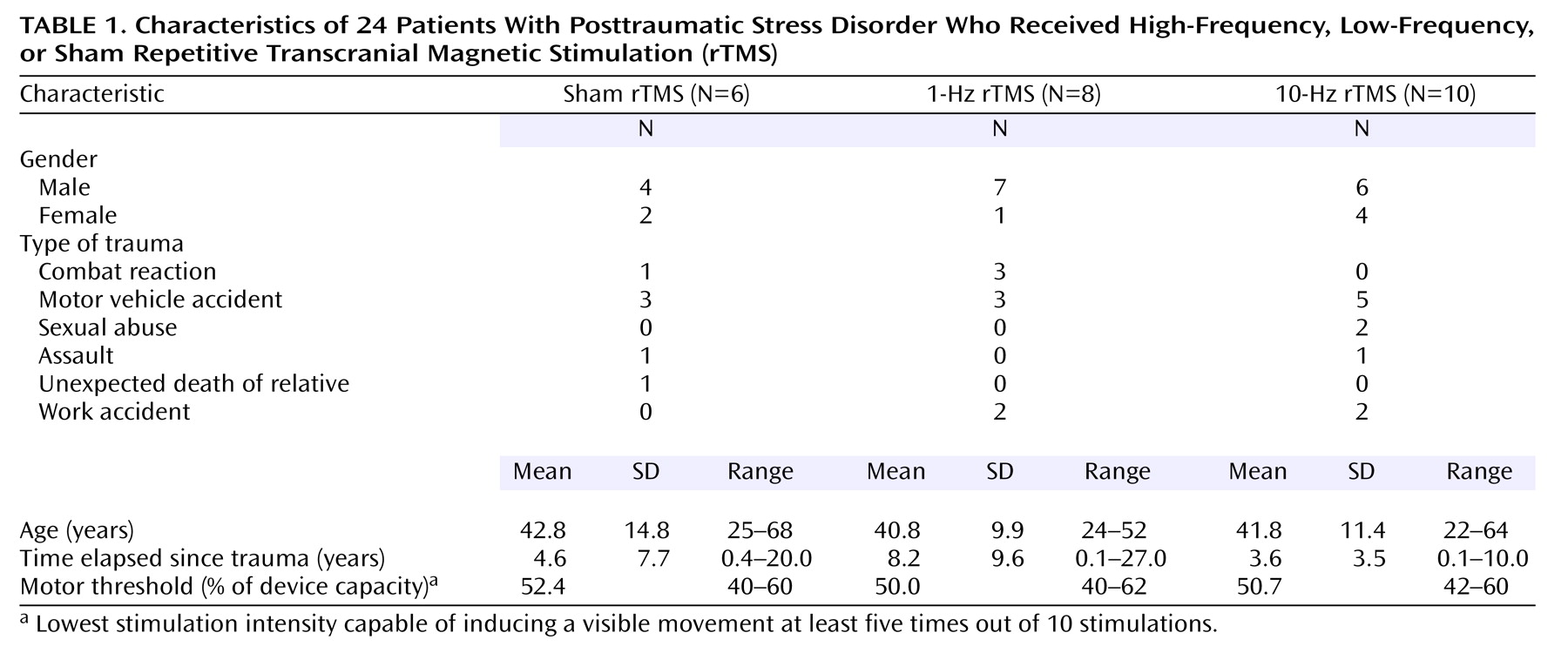
The subject group consisted of 17 men and seven women, with a mean age of 41.7 years (SD=11.4, range=22–68). The types of trauma were combat reaction (N=4), motor vehicle accident (N=11), sexual abuse (N=2), assault (N=2), work accident (N=4), and unexpected death of a relative (N=1). The mean time elapsed since the trauma was 5.4 years (SD=7.0, range=0.1–27.0) (
Table 1). None of the patients suffered from any chronic medical disease.
The participants were asked to answer questions regarding their personal background, demographic characteristics, marital status, place of birth, education, and place of residence and the type of trauma that led them to seek help.
Four subjects were drug free. Polypharmacy was the rule for the majority. Two were receiving clomipramine, 15 were taking SSRIs or a serotonin and norepinephrine reuptake inhibitor, 19 were taking benzodiazepines (for five, the benzodiazepine was the only therapy), and four were receiving mood stabilizers. Three subjects with severe PTSD who were receiving long-term antipsychotic treatment enrolled in the study. Two of the three were taking olanzapine (10 mg/day), and one was taking clothiapine (40 mg/day); in addition, they were receiving mood stabilizers, SSRIs, and benzodiazepines. No significant differences in pharmacotherapy were found between groups. Drug treatment was neither stopped nor changed in the 3 weeks before the study or during the study. The patients continued to receive the same individual and group supportive psychotherapy as before the intervention.
Treatment Characteristics
In this double blind, placebo-controlled study, the patients were randomly assigned to one of three stimulation groups. None of the patients had any experience with rTMS before the study.
rTMS was performed with a Magstim stimulator (Magstim Company, Whitland, U.K.) having a circular coil with a 9-cm diameter.
The motor threshold was determined in each subject once, before treatment. This was defined as the lowest stimulation intensity capable of inducing a visible movement at least five times out of 10 stimulations
(42).
The position of the right dorsolateral prefrontal cortex was defined as 5 cm anterior (in a parasagittal line) to the motor cortex. The stimulus intensity was 80% of the patient’s motor threshold intensity. The mean motor threshold for the group receiving sham treatment was 52.4%, with a range of 40%–60%; for the group receiving slow-frequency rTMS the mean was 50.0%, with a range of 40%–62%; and for the group receiving high-frequency treatment the mean was 50.7%, with a range of 42%–60%.
Treatments were given for 20 minutes per day over 10 working days. The patients were randomly assigned to the three treatment groups after assessment of their motor thresholds. Group 1 (sham rTMS) was treated in the same way as the group receiving high-frequency rTMS, but the coil was held at 90° vertical over the stimulated head area (no significant magnetic field was evoked, just the auditory artifact). Group 2 (slow-frequency rTMS) received 1 Hz for 5 seconds per train; the intertrain interval was 55 seconds. Group 3 (high-frequency rTMS) received 10 Hz for 2 seconds per train; the intertrain interval was 58 seconds.
For each participant the stimulus was administered over the right dorsolateral prefrontal cortex.
Rating Scales
The ratings of PTSD symptoms, anxiety, and depression were carried out by an expert investigator (R.M.) who was blind to the stimulation condition. The patients were assessed at four time points—before TMS (baseline), at day 5, at day 10, and at day 24 (14 days after the intervention). The instruments used were as follows.
The PTSD Checklist is a 17-item self-report checklist of PTSD symptoms based closely on the DSM-IV criteria
(43). The respondents rate each item from 1 (“not at all”) to 5 (“extremely”) to indicate the degree to which they have been bothered by that particular symptom over the past month. Thus, the total scores can range from 17 to 85.
The Treatment Outcome PTSD Scale
(44) is a clinician-rated instrument that measures the presence and severity of PTSD. This eight-item instrument measures symptoms that occur frequently within the PTSD population and is sensitive to the three major PTSD symptom dimensions (intrusive thoughts, avoidance behavior, and hyperarousal symptoms). Each symptom is rated on a defined step scale (0 to 4). Higher scores reflect greater severity on each measure.
The Hamilton Anxiety Rating Scale
(45) is a clinician-rated instrument that measures the presence and severity of anxiety. This instrument covers 14 symptoms. Each symptom is rated on a defined scale (0 to 4). Here, too, a higher numeric rating reflects greater symptom severity.
The Hamilton Rating Scale for Depression
(46) is a 23-item instrument that measures the presence and severity of depression. Each symptom is rated on a defined scale (0 to 4), whereby a higher numeric rating reflects greater symptom severity.
PTSD symptoms were assessed by using the Hebrew version of the Clinician-Administered PTSD Scale
(47). This is a structured interview for assessing PTSD according to DSM-IV criteria. It quantifies symptom frequency and intensity for each of the criteria, yielding both a continuous measure of symptom severity and a dichotomous classification of PTSD status. A severity score for each symptom is calculated by summing the frequency and intensity scores. Thus, the total range of the instrument is 0–136. If a particular symptom was not present, the individual item was automatically scored as zero, as a default option. The Hebrew version of the scale has been extensively used and validated
(49).
The questionnaires were filled out in the presence of an interviewer, and the subjects were assisted in answering the questions if necessary. The interviewer made sure that all subjects clearly understood the content of each item and the different aspects of the various component questions.
Statistical Methods
The ratings of psychopathology (PTSD Checklist, Treatment Outcome PTSD Scale, Hamilton anxiety scale, Hamilton depression scale) were entered into two-way repeated-measures analyses of variance with covariance for baseline scores (ANCOVAs), with treatment as the between-group factor (fast rTMS, slow rTMS, sham rTMS) and time as the within-subjects factor (day 5, day 10, and day 24). Repeated-measures analysis of variance (ANOVA) was used in order to compare the scores on the Clinician-Administered PTSD Scale of the three treatment groups (fast rTMS, slow rTMS, sham rTMS) and to estimate the effect of time (baseline and day 24, i.e., 14 days after the last treatment).
All scale scores were analyzed as change from baseline to the end of the treatment (day 10) or to follow-up 14 days later, by using ANOVA.
Dropouts
Five of the original 29 patients did not complete a course of rTMS. Two patients (receiving sham treatment) asked that the procedure stop immediately after motor threshold location, before stimulation began. One patient (receiving 1-Hz rTMS) refused to continue after one session, complaining that dizziness that existed before treatment had worsened. Another patient (1-Hz rTMS) asked that treatment stop after two sessions because of reasons unrelated to any side effect of the treatment. One patient (receiving 10-Hz rTMS) was excluded at the end of the treatment because of problems with device calibration.
Results
The demographic data are summarized in
Table 1. The three groups did not differ significantly on age, gender, or duration of illness.
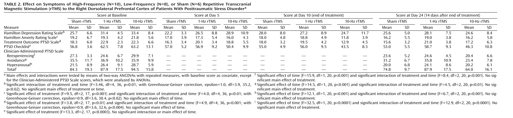
Scores on the PTSD Checklist, Treatment Outcome PTSD Scale, Hamilton anxiety scale, Hamilton depression scale, and Clinician-Administered PTSD Scale of the patients treated with active rTMS and sham rTMS are shown in
Table 2.
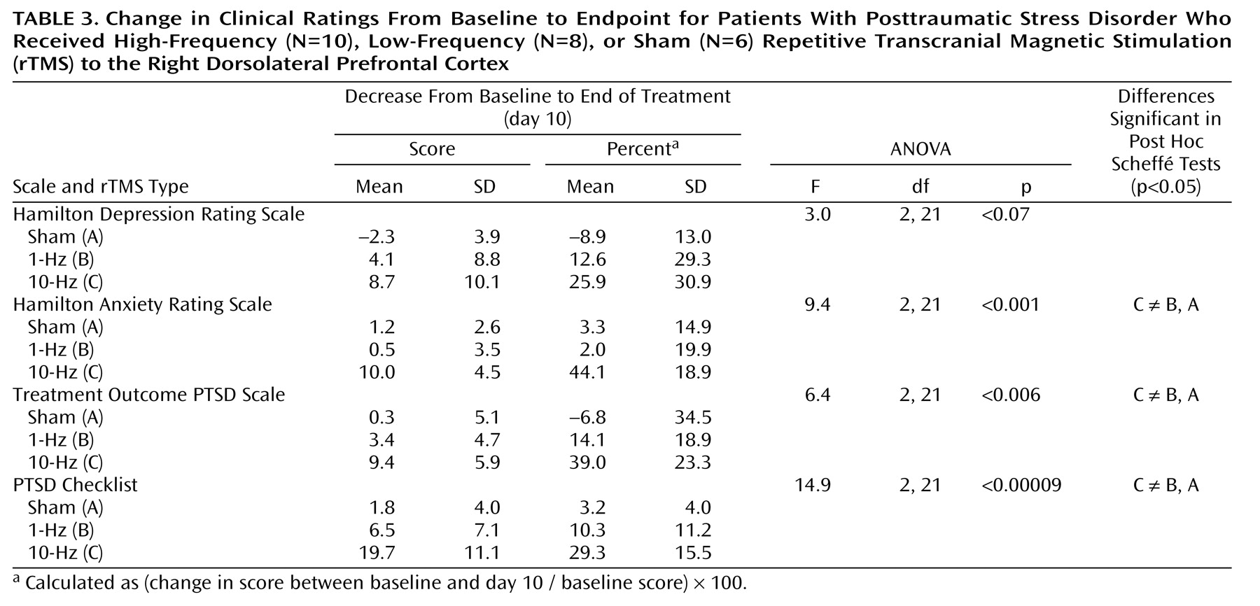
Two-way repeated-measures ANCOVA for the PTSD Checklist showed a significant effect of rTMS treatment group (
Table 2). Post hoc Sheffé tests showed a significant effect of rTMS treatment group; active 10-Hz rTMS was significantly different from the sham (p<0.01) and 1-Hz (p<0.002) treatments. As shown in
Table 3, during high-frequency active rTMS treatment, the mean PTSD Checklist scores decreased by 29.3% from baseline to the end of treatment (day 10).
Two-way repeated-measures ANCOVA for the Treatment Outcome PTSD Scale showed a significant effect of rTMS treatment type and a significant interaction of time and treatment (
Table 2). Post hoc Sheffé tests showed a significant effect of rTMS treatment; active 10-Hz rTMS was significantly different from the sham (p<0.05) and 1-Hz (p<0.02) treatments. As shown in
Table 3, during the active high-frequency rTMS treatment, the mean scores decreased by 39.0% from baseline to the end of treatment.
Two-way repeated-measures ANCOVA for the total Hamilton anxiety scale showed a significant effect of rTMS treatment and a significant interaction of time and rTMS treatment (
Table 2). Post hoc Sheffé tests showed that active 10-Hz rTMS was significantly different from the sham (p<0.04) and 1-Hz (p<0.01) treatments. As shown in
Table 3, during the active high-frequency rTMS treatment, the mean Hamilton anxiety scale scores decreased by 44.1% from baseline to the end of treatment.
Two-way repeated-measures ANCOVA for the Hamilton depression scale showed a significant interaction of time and rTMS treatment (
Table 2). Post hoc tests revealed no significant difference between treatments or times.
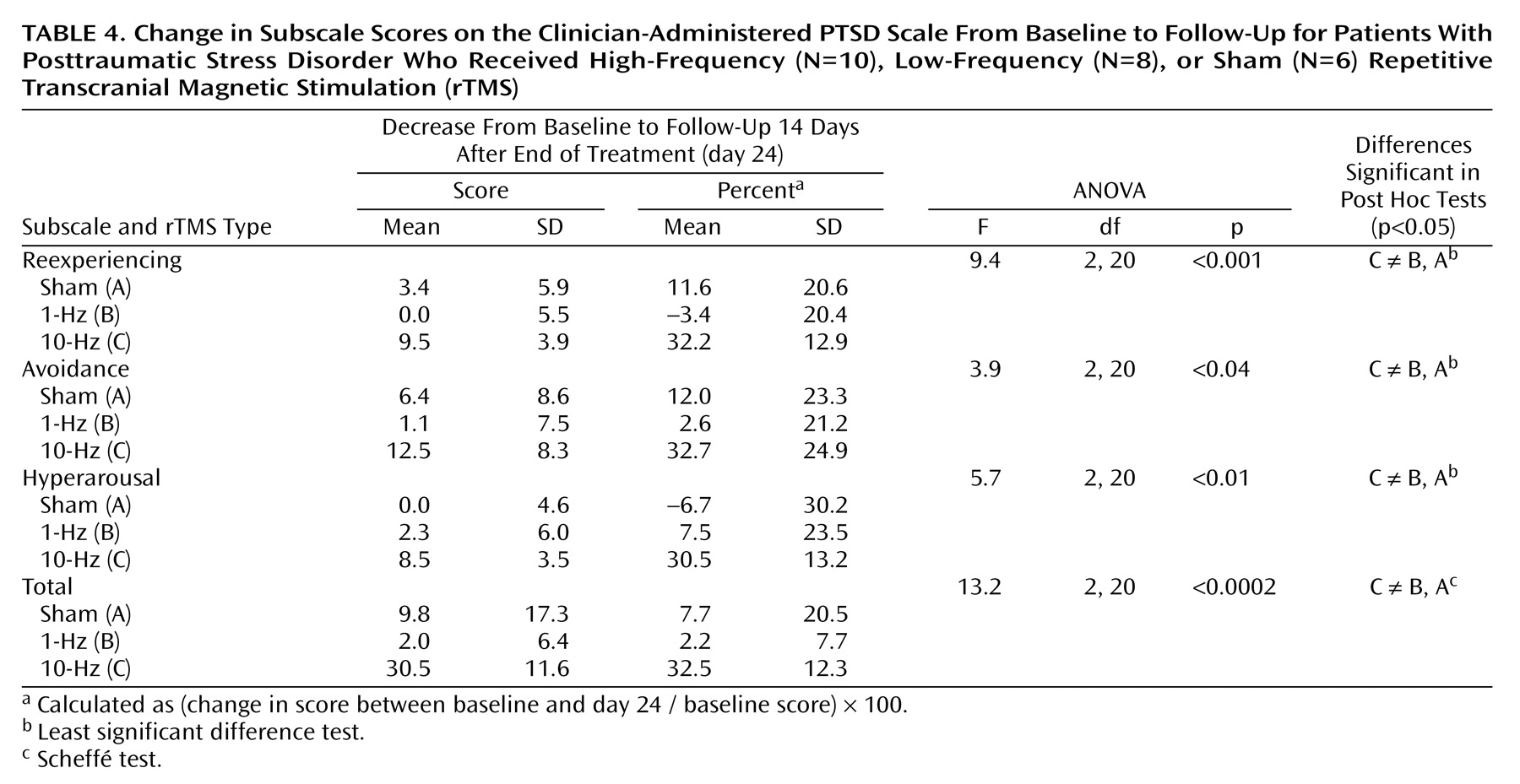
Two-way repeated-measures ANOVA for the reexperiencing subscale of the Clinician-Administered PTSD Scale showed a significant effect of time and a significant interaction of time and rTMS treatment groups (
Table 2). Post hoc Sheffé tests showed a significant effect of time (p=0.0005). As shown in
Table 4, for patients receiving the active high-frequency rTMS treatment, the mean score for reexperiencing decreased by 32.2% from baseline to follow-up 14 days after the end of treatment (day 24).
Two-way repeated-measures ANOVA for the avoidance subscale of the Clinician-Administered PTSD Scale showed a significant effect of time and a significant interaction of time and rTMS treatment group (
Table 2). Post hoc Sheffé tests showed a significant effect of time (p=0.0007). As shown in
Table 4, for patients receiving the active high-frequency rTMS treatment, the mean avoidance score decreased by 32.7% from baseline to follow-up.
Two-way repeated-measures ANOVA for the hyperarousal subscale of the Clinician-Administered PTSD Scale showed a significant effect of time and a significant interaction of time and rTMS treatment group (
Table 2). Post hoc Sheffé tests showed a significant effect of time (p=0.002).
Two-way repeated-measures ANOVA for the total score on the Clinician-Administered PTSD Scale showed a significant effect of time and a significant interaction of time and rTMS treatment (
Table 2). Post hoc Sheffé tests showed a significant effect of time (p=0.00001). As shown in
Table 4, for patients receiving the active high-frequency rTMS treatment, the mean score decreased by 32.5% from baseline to follow-up.
Side Effects
Generally, the treatment was well tolerated, and no serious adverse effects were reported by any of the patients. Headache was the main side effect reported, regardless of stimulation group. It was reported by 14 patients: eight patients reported headache after one rTMS treatment, five patients reported it after two sessions, and one (receiving sham treatment) reported it after three sessions. In most cases, this side effect was reported several hours after the stimulation or on the following morning. Only four patients reported headache immediately after the stimulation. In three cases, headache was a symptom before the study. However, the total number of headaches after treatment was 21, out of approximately 250 treatment sessions, for an incidence of 8%.
Two patients receiving high-frequency rTMS reported neck pain and muscular contraction in the area. Another patient receiving high-frequency treatment reported an exacerbation of previously existing dizziness.
One patient in the group receiving slow-frequency rTMS and one patient from the high-frequency group developed a manic episode; in both cases this occurred after the third session of rTMS. One patient reported a mild rage attack, probably related to the stimulation. Although we did not use earplugs, only two patients reported ear discomfort, which lasted less than 1 minute.
In general, TMS was well tolerated and cooperation was excellent among all patients who completed the course of treatment. rTMS had no effect on any patient’s blood pressure or heart rate during the treatments. No serious side effects, such as seizures, neurological complications, or cognitive difficulties, occurred.
Additional Benefits
Eleven participants reported sleep improvement: six receiving 10-Hz rTMS, four receiving 1-Hz rTMS, and one receiving sham treatment. Six patients reported a sense of calmness or a deep sensation of comfort. One patient (receiving 1-Hz stimulation) reported a marked improvement and sharpening of taste and smell.
Discussion
In this double-blind study, high-frequency rTMS (10 Hz for 2 seconds) and slow-frequency rTMS (1 Hz for 5 seconds) were compared to sham rTMS. Our findings demonstrate that 10 daily sessions of 10-Hz rTMS at 80% motor threshold over the right dorsolateral prefrontal cortex has therapeutic effects on PTSD patients.
The PTSD symptoms markedly improved after 10 daily treatment sessions with 10-Hz rTMS, as reflected by the patients’ scores on the Treatment Outcome PTSD Scale and other PTSD scales. At follow-up 14 days after the end of the treatments, the total score on the Clinician-Administered PTSD Scale and the scores on the subscales reflecting the PTSD core symptoms of reexperiencing and avoidance were significantly reduced. These findings therefore widen the scope and possibilities of treatment applications of TMS.
Active 10-Hz rTMS, relative to 1-Hz treatment and sham, significantly reduced Hamilton anxiety scale scores but not Hamilton depression scale scores. Improvement in anxiety symptoms during high-frequency rTMS treatment has been reported previously. Greenberg et al.
(49) administered single sessions of high-frequency rTMS stimulation to the left and right dorsolateral prefrontal cortex and to a parieto-occipital control site, in a randomized design, to patients with obsessive-compulsive disorder (OCD). They reported that rTMS treatment to the right dorsolateral prefrontal cortex, but not to the left or to the parieto-occipital area, significantly reduced the compulsive urges. The patients also reported significant mood elevation after right prefrontal stimulation. Conversely, Alonso et al.
(50) reported that low-frequency rTMS of the right prefrontal cortex failed to produce significant improvement of OCD and was not significantly different from sham treatment. These results suggest that right prefrontal high-frequency rTMS might affect mechanisms involved in anxiety disorders.
The effect of 10-Hz rTMS was significant and stable for at least 14 days after the last treatment. It is suggested that in further studies the stimulation could be repeated as maintenance therapy, as in ECT procedures.
In an attempt to explain our results, several variables such as frequency and location of stimulation must be discussed. First is the potential lateralization of effects. Several findings suggest that the right hemisphere, especially the right paralimbic and limbic structures, is involved in the emotional and cognitive symptoms associated with traumatic memories and PTSD symptoms
(51–
53). Therefore, we stimulated the right dorsolateral prefrontal cortex, as did McCann et al.
(20), who reported normalization of metabolic activity, as shown by PET imaging, and symptom improvement after 17 treatments with low-frequency rTMS. Second, our results, comparing high- and slow-frequency rTMS over the dorsolateral prefrontal cortex, show that only high-frequency rTMS (10 Hz) achieved clinically significant efficiency, as opposed to the findings of the study by McCann et al. The open design in that study did not exclude placebo effects or changes in severity due to the natural course of the illness as explanations for the observed changes in clinical state. The differences in design and the small patient group may explain that study’s conflicting findings.
The medial prefrontal cortex has an important role in mediating responses to stressful situations. It does so by modulating the hypothalamic-pituitary-adrenal (HPA) axis
(54–
56), acting as a site for glucocorticoids to exert negative-feedback modulation of HPA activity
(57) and regulating a variety of autonomic functions
(58,
59).
In a series of studies conducted in rats, Sullivan and Gratton
(60) showed that unilateral lesions of the right medial prefrontal cortex abolished stress-induced secretion of glucocorticoids, whereas lesions of the left medial prefrontal cortex were without effect. Animals with lesions failed to interpret sensory input related to stress and failed to integrate this with neuroendocrine responses. The laterality of control of HPA axis activity by the prefrontal cortex is highly relevant to PTSD patients, because these patients suffer from symptoms associated with both dysfunction in HPA axis regulation
(61) and prefrontal cortical functional abnormalities
(28–
34,
36,
62–65). Although still controversial, there is a large body of evidence pointing to low baseline cortisol levels and greater sensitivity of negative-feedback inhibition in the HPA axis as characteristics of PTSD patients
(66). Moreover, two studies have shown a deficiency of HPA activity in the aftermath of a traumatic event among individuals who developed PTSD
(67,
68). It is not clear whether hypoactivity of the HPA axis in PTSD patients is a consequence of down-regulation of the endocrine stress system, resulting from chronic anxiety/stress, or is a preexisting condition, which may contribute to vulnerability to PTSD.
On the basis of the preceding animal experiments, the findings of low basal cortisol together with several findings revealing low cerebral blood flow in the prefrontal cortex in PTSD patients suggest a predictive association between hypoactivity of the HPA axis and right hypofrontality. High-frequency rTMS to the right dorsolateral prefrontal cortex of PTSD patients may cause activation in this area, resulting in improvement in PTSD symptoms through activation of the HPA axis. This hypothesis may be applicable to the findings of several studies of depressive patients. An antidepressant effect of TMS was found in depressive patients when high-frequency impulses were applied over the left prefrontal cortex or when low-frequency stimuli were applied to the right prefrontal cortex
(69–
71). Both of these treatments lead to inhibition of HPA axis activity, if the preceding hypothesis is correct.
There is a growing body of evidence implicating the medial prefrontal cortex as a modulator not only of the HPA axis but also of the autonomic nervous system
(72). This area contains neurons (principally in the prelimbic cortex and infralimbic cortex) that, when stimulated, result in an inhibitory influence on sympathetic function. Although the precise mechanisms of how the medial prefrontal cortex affects autonomic regulation are not completely understood, these findings suggest the hypothesis that hypoactivation of the medial prefrontal cortex (as reported in PTSD patients) elicits a sympathoexcitatory response, resulting in an excessive autonomic function.
Lesions of the medial prefrontal cortex have been shown to alter behavioral measures of anxiety in rats (ultrasonic vocalization, immobility duration, and respiratory rate)
(73,
74). Morgan and LeDoux
(73) examined the emotional reactivity of rats with lesions of the dorsal portion of the medial prefrontal cortex, using a classical fear-conditioning paradigm. Conditioned fear behavior (freezing responses) was measured during both the acquisition and extinction phases of the task. Lesions of the dorsal portion of the medial prefrontal cortex were associated with enhanced fear reactivity to both the conditioned stimulus and contextual stimuli during both phases.
Another area involved in regulating the cardiovascular component of the conditioned fear response is the amygdala
(75). Neurons in layers II, III, and V of the prelimbic and infralimbic cortex project to the amygdaloid complex, and a proportion of these projections issue collaterals to the contralateral medial prefrontal cortex. The medial prefrontal cortex modulates emotional responses through inhibition of amygdalar responsiveness to fearful cues. Thus, hypofrontality may abolish this suppression response, resulting in an elevated fearful response as shown in PTSD patients.
Overall, one may speculate that the improvement seen in PTSD symptoms is the result of activation of the right dorsolateral prefrontal cortex by the high-frequency rTMS. This improvement may also be associated with activation of the HPA axis, enhanced activity of the depressor area of autonomic responsiveness, or increased suppression of the amygdala. Although these hypotheses are supported by empirical evidence at several points, more studies are needed to directly assess our hypotheses.
Grosso modo, the brain appears to adhere to two fundamental principles: functional organization/specialization and functional integration (where the integration within and among specialized areas is mediated by effective connectivity). Therefore, in order to achieve complete characterization of brain responses in imaging experiments, it is necessary to assess both specific regional changes and regional interaction (connectivity). Shaw and colleagues
(76) found direct evidence of differences in functional connectivity between PTSD patients and comparison subjects. They reported that the functional connectivity pattern of the PTSD patients was characterized by more activation in the bilateral inferior parietal lobes and the left precentral gyrus than in the comparison group and less activation in the inferior medial frontal lobe, bilateral middle frontal gyri, and right inferior temporal gyrus. Thus, we may speculate that high-frequency rTMS over the dorsolateral prefrontal cortex improves functional connectivity in these PTSD patients.
In conclusion, this double-blind, controlled trial suggests that in PTSD patients, 10 days of daily right prefrontal high-frequency rTMS has therapeutic effects greater than those of low-frequency or sham stimulation. However, given the small number of patients, our study must be considered preliminary. Our findings suggest that trials using high frequencies to the right prefrontal cortex may be a promising avenue for future research with PTSD patients.