Abstract

Objective:

The purpose of this study was to determine the neural correlates of excessive habit formation in obsessive-compulsive disorder (OCD). The authors aimed to test for neurobiological convergence with the known pathophysiology of OCD and to infer, based on abnormalities in brain activation, whether these habits arise from dysfunction in the goal-directed or habit system.

Method:

Thirty-seven OCD patients and 33 healthy comparison subjects learned to avoid shocks while undergoing a functional MRI scan. Following four blocks of training, the authors tested whether the avoidance response had become a habit by removing the threat of shock and measuring continued avoidance. Task-related differences in brain activity in three regions of interest (the caudate, the putamen, and the medial orbitofrontal cortex) were tested at a statistical threshold set at <0.05 (family-wise-error corrected).

Results:

Excessive habit formation in OCD patients, which was associated with hyperactivation in the caudate, was observed. Activation in this region was also associated with subjective ratings of increased urge to perform habits. The OCD group, as a whole, showed hyperactivation in the medial orbitofrontal cortex during the acquisition of avoidance; however, this did not relate directly to habit formation.

Conclusions:

OCD patients exhibited excessive habits that were associated with hyperactivation in a key region implicated in the pathophysiology of OCD, the caudate nucleus. Previous studies indicate that this region is important for goal-directed behavior, suggesting that habit-forming biases in OCD may be a result of impairments in this system, rather than differences in the buildup of stimulus-response habits themselves.
The habit hypothesis of obsessive-compulsive disorder (OCD) suggests that the disorder reflects dysfunction in the brain systems that support automatic habits and more purposeful, goal-directed control over action (1). Habits are automatic stimulus-driven behaviors that can arise under many conditions, the most commonly accepted of which is the overtraining of simple responses (2). However, habits can also arise from failures in goal-directed control, which can render behavior habitual even very early on in training (3, 4). Therefore, these two systems, habit and goal-directed, each contribute to the likelihood that a habit will be performed in a given situation. In OCD, it is currently unclear which of these putative systems drives the exaggerated tendency to display habits, which has been observed regardless of whether they work toward gaining reward (5) or toward avoiding punishment (6). However, two recent studies found deficits in goal-directed behavior during trial-by-trial learning in OCD, using paradigms that did not involve repeating simple responses (7, 8). This suggests that excessive habits in OCD could arise as a result of disturbances in the goal-directed system, rather than the habit system. The present study aimed to test for neurobiological convergence in support of this possibility, drawing on a rich cross-species neuroscience literature, which has identified dissociable neural substrates of these two systems (9).
The medial orbitofrontal cortex and the caudate nucleus each contribute to goal-directed control over our behavior. Specifically, the caudate and medial orbitofrontal cortex both have been shown to subserve learning involving action-outcome contingencies (4, 10, 11). Additionally, the medial orbitofrontal cortex plays a pivotal role in tracking the current value of outcomes (1214). Another region in the basal ganglia, the putamen, is necessary for the formation of stimulus-response habits with practice (11, 15, 16). We tested whether functional activation in these three regions was associated with habit-forming biases in OCD, and in doing so we aimed to reveal whether dysfunction in the goal-directed or habit-learning system accounted for excessive habits in OCD.
Generally, the habit hypothesis of OCD exhibits good face validity in that both habits and compulsions continue in spite of awareness that these actions are not useful/wanted (i.e., ego-dystonic) and are associated with the experience of an urge to perform them (6). A secondary goal of this study was to test the neurobiological validity of the OCD habit hypothesis by assessing whether activation associated with habit forming in OCD overlaps with activation implicated in the disorder's symptoms. The literature has broadly converged on a model of OCD that involves hyperactivity within fronto-striatal circuits (17), with effects in the orbital gyri and caudate nucleus head being among the most reliable (1820). Evidence for this model comes primarily from functional brain imaging studies examining brain activity at rest (2123), during symptom provocation (2426), and pre- and posttreatment with psychotherapy or pharmacotherapy (22, 2729). A more widely distributed network of regions, including the nucleus accumbens, amygdala, and other parts of the prefrontal cortex (3032), has also been implicated in OCD in studies employing task-related functional MRI (fMRI) analysis. However, given that fMRI activation patterns are entirely dependent on the task employed, results have been unsurprisingly heterogeneous and have not confirmed activation seen during earlier studies examining task independent activity patterns that are characteristic of OCD. We hypothesized that if excessive habits are an appropriate model of OCD, then brain activation associated with habit formation in OCD patients should overlap with activation associated with the symptoms, specifically in the medial orbitofrontal cortex and caudate. Although less consistently implicated, there is some suggestion that the putamen may be enlarged in OCD, an effect related to age and plausibly the chronic performance of compulsive behavior (33). Therefore, we also tested the possibility that aberrant activation in the putamen, perhaps reflecting overactive habit learning, would be associated with habits in OCD.
To investigate the neural basis of habit-forming biases in OCD, we used fMRI to examine changes in brain activation while patients acquired and later performed habits. To do this, we used an avoidance task that has previously been shown to be sensitive to differences in habit formation between OCD patients and comparison subjects (6). Only individuals who had not previously participated in the previously published behavioral study using this task were eligible to enroll. This task was selected because avoidance rather than appetitive compulsions are characteristic of OCD. Therefore, this approach allowed us to model the disorder more closely and, secondarily, to investigate how habit learning might relate to anxiety and explicit fears in OCD. To test for habits, we used the “outcome devaluation” technique, in which behavior is defined as a habit if it persists despite changes in the value the action produces (34); in other words, habits are behaviors that are driven by stimuli, not by motivation or goals.

Method

Participants

Participants were 37 OCD patients and 33 healthy comparison subjects matched at a mean level for age, handedness, smoking behavior, education, and gender. Lower premorbid verbal IQ (using the National Adult Reading Test [35]) in the OCD group nearly reached statistical significance (p=0.09); however, analyses of covariance confirmed that this did not drive any of the results. Individuals were excluded if they had participated in a previous study examining avoidance habits in OCD in our laboratory (6) (i.e., all participants were task-naive). OCD patients were free of comorbid psychiatric diagnoses. Fourteen OCD patients were not taking psychotropic medication. The remaining 23 patients had been stabilized on medication for a minimum of 6 weeks prior to taking part in the study, and the majority of these were receiving selective serotonin reuptake inhibitors (SSRIs) (see the data supplement accompanying the online version of this article). For all of the results presented, there were no significant differences between medicated and nonmedicated OCD patients, unless otherwise stated. For further details regarding participant characteristics and recruitment, see the online data supplement.

Procedure

Avoidance training and habit test.

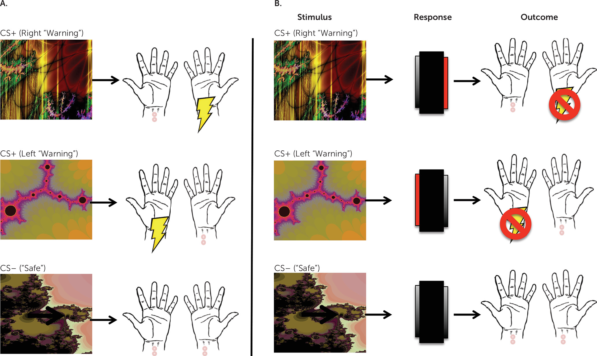
Participants completed a shock avoidance paradigm similar to that described in detail elsewhere (6), as well as in the data supplement. Participants were instructed that their goal was to avoid receiving shocks, which would be delivered to their right and left wrists following the presentation of a conditioned stimulus (warning stimulus) (Figure 1). During fMRI scanning, we overtrained the avoidance response across four blocks, each containing 30 trials (10 per conditioned stimulus), prior to testing for habits using outcome devaluation. Before the final block, the left shock outcome was “devalued” by disconnecting the electrodes from the participants' left wrists. The shock to the right wrist remained threatening or “valued.” Importantly, up until this point, participants had identical training with both the left and right conditioned stimuli. In the final block, we defined these as devalued (left) and valued (right) based on the connection status of the electrodes. Participants were informed onscreen that they could no longer receive a shock to the left wrist and that their only goal was to avoid the remaining shock (which was on the right). Following this, participants completed one more block of the task, constituting the habit test. To prevent new learning during the test, shocks were no longer delivered to any conditioned stimulus.
FIGURE 1. Task Schematica
a Panel A depicts the Pavlovian (stimulus-outcome) contingencies, which were 100% deterministic. There were two warning conditioned stimuli (CS+); one predicted a shock to the left wrist, and another predicted a shock to the right wrist. A safe conditioned stimulus (CS–) never predicted shock. Panel B depicts the avoidance contingencies, including stimuli, responses, and outcomes. When the top (right warning) predictive CS+ appears onscreen, it indicates that a shock to the right wrist is imminent. If participants press on the right side of the foot-box (highlighted in red) while this CS+ is onscreen, they will avoid this shock. Likewise, pressing on the left side of the foot-box when the middle (left warning) CS+ appears cancels an otherwise imminent shock to the left wrist. The safe stimulus always remained safe, regardless of responding.
To better clarify how habits in OCD relate to implicit and explicit fear and belief, we collected supporting data, including skin conductance responses, explicit contingency knowledge and subjective ratings of shock expectancy, shock unpleasantness, urge to perform habits, and attempts to suppress habits (also see Table S2 in the online data supplement). Participants completed an additional and unrelated experiment in the same session (after this task was completed), the results of which will be published elsewhere.

Data Analysis

Behavior.

Behavioral data were analyzed using analysis of variance for parametric data and the Mann-Whitney U test, chi-square test, and Spearman’s rho correlations. In the habit test, which followed outcome devaluation, we compared the number of avoidance responses to the devalued and valued conditioned stimuli. We also measured false alarms in response to the safe conditioned stimulus. OCD patients were divided into two habit groups: “habit” and “no habit,” which was determined by whether or not they made any response to the devalued conditioned stimulus during the habit test. In subsequent analyses, we compared the two habit groups defined by this distinction. During training, since the left and right conditioned stimuli each predicted an avoidable shock, we collapsed these into one factor: warning stimulus. We analyzed accuracy during training in terms of percentage correct avoidance responses over each of the four experimental blocks. Results are presented as significant p values <0.05, and values that fell short of statistical significance were defined as 0.1>p>0.05.

fMRI.

Based on previous literature on habit learning in healthy humans and the known neurobiological profile of OCD, we examined anatomically defined bilateral a priori regions of interest: the medial orbitofrontal cortex (1113), the caudate (10, 11), and the putamen (11, 16). We used the PickAtlas software toolbox in SPM8 (36) to define regions of interest according to the Anatomical Automatic Labeling atlas. Activation within these regions of interest was deemed significant at a p value <0.05, corrected for family-wise error at the voxel level and for testing across multiple regions of interest (p<0.05/3). Results from whole-brain exploratory analyses are presented at a p value <0.001 (uncorrected), with a minimum cluster size of 10 voxels. Although we discuss these results to some extent, we caution that replication is needed.
First-level analyses of the habit test data from the final block modeled three conditioned stimuli (valued, devalued, and safe), along with the six movement parameters produced during realignment (further details are presented in the online data supplement). The habit group was defined as patients who had formed habits, and the no habit group was defined as those who had not, based on their responding to the devalued conditioned stimulus. While behavioral responses to the devalued conditioned stimulus allow us to discern whether a habit has formed, neural responses to this conditioned stimulus are confounded by the experience of the devaluation procedure, as well as the associated differences in behavioral responding, the urge to respond, and attempts to suppress responding. Therefore, to capture the neural signature of habits, we examined the contrast of “valued-safe,” which captures the neural activation associated with unperturbed habitual responding. This analysis therefore relies on the reasonable assumption that if avoidance responding to one warning conditioned stimulus (i.e., devalued) has become habitual, then so has responding to the other warning conditioned stimulus (i.e., valued), given the equivalence of contingencies and training duration.
We analyzed training data using two different first-level contrasts. First, we analyzed activity associated with the early acquisition of avoidance (i.e., in block 1), in a first-level contrast of warning stimulus (collapsed left + right) – safe stimulus. Secondly, we examined brain regions involved in the development of habits over time using a mixed-factor general linear model across the four training blocks. Here, we tested for an interaction between condition stimuli (warning [collapsed left + right] – safe stimulus) and block (14). This allowed us to examine changes in activation that progressed with overtraining or the putative “stamping in” of habits. For both analyses, we tested for group differences at the second level.

Results

Habit Test

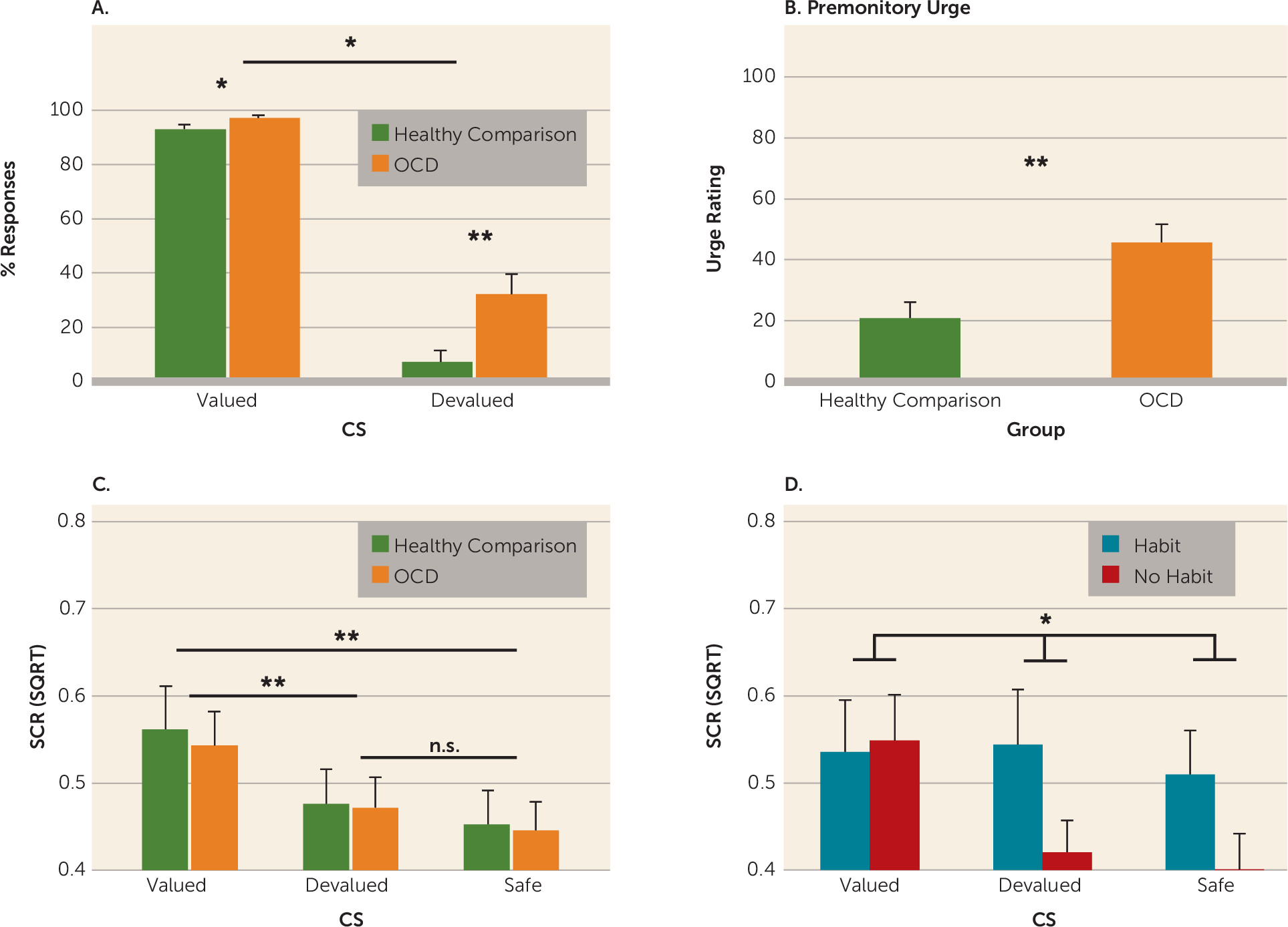
OCD patients showed increased habits compared with healthy comparison subjects, replicating previous findings using both avoidance (6) and appetitive (5, 7) paradigms. There was a significant main effect of group (OCD, healthy comparison) on the number of responses overall (F=10.691, df=1, 68, p=0.002) and interaction between group and conditioned stimuli (valued, devalued) during the devaluation test (F=5.408, df=1, 68, p=0.02) (Figure 2A). Simple-effects analyses revealed that OCD patients, compared with comparison subjects, responded at a significantly higher rate to the devalued conditioned stimulus (F=8.139, df=1, 69, p=0.006) and to the valued conditioned stimulus (F=4.896, 1, 69, p=0.03). However, the significant interaction indicates that the group difference was greater for the devalued compared with the valued conditioned stimulus, and responding to the valued and devalued conditioned stimuli were not significantly correlated (Spearman’s r=−0.276, p=0.099). The trend was in the opposite direction to what would be predicted by a disinhibition account, such that the greater the habits, the fewer the responses to the valued conditioned stimulus.
FIGURE 2. Habit Test: Behavioral Dataa
a Panel A depicts the percentage of accurate avoidance responses made to the devalued and valued stimuli. In line with previous research, obsessive-compulsive disorder (OCD) patients developed more habits than healthy comparison subjects, evidenced by greater responding to the devalued conditioned stimulus (CS) (p<0.006). Overall, OCD patients (N=37) responded more to both the devalued and valued CSs compared with healthy comparison subjects (N=33). However, a significant interaction between group (OCD, healthy comparison) and CS (valued, devalued) (F=5.335, df=1, 69, p=0.02) indicated that the difference was greater for the devalued compared with the valued CS (p<0.04). Panel B depicts the urge to respond ratings. OCD patients reported a greater urge to respond compared with healthy subjects (U=345, Z=–3.191, p=0.001). Panel C depicts skin conductance response (SCR) data. There were no differences between OCD patients (N=36) and comparison subjects (N=31) in SCR (two comparison subjects and one OCD patient were excluded from this analysis due to insufficient SCR data). There was a main effect of CS (F=16.163, df=2, 130, p<0.001). Conditioned fear responses extinguished to the devalued CS to a level equivalent to the safe CS (p=0.18). Responses to the valued CS remained elevated relative to the devalued (p<0.002) and safe (p<0.001) CSs. Panel D depicts SCR differences between the habit group (N=15) and no habit group (N=21; one patient was excluded due to insufficient SCR data) from within the OCD group. There was a significant interaction between CS and habit group (F=4.818, df=2, 68, p=0.01). While the no habit group had a significant main effect of CS (F=9.934, df=2, 40, p=0.001), the habit group did not (F<1). Error bars denote standard error of the mean. n.s.=not significant; SQRT=square root.
** p<0.01; *p<0.05.

Supporting Data

Explicit contingency knowledge was equivalent across groups (F<1) (6). OCD patients reported a greater urge to respond to the devalued conditioned stimulus compared with healthy comparison subjects (U=345, Z=–3.191, p=0.001) (Figure 2B), and this urge correlated with the number of responses made to the devalued conditioned stimulus by OCD patients (Spearman’s r=0.668, N=37, p<0.001). There were no differences in skin conductance responses between OCD patients and comparison subjects (F<1); however, those OCD patients who formed habits during the habit test showed inferior discrimination between the three conditioned stimuli (devalued, valued, and safe) during the habit test compared with those who did not form habits (conditioned stimulus-by-group interaction: p=0.01). There was no difference between the habit and no habit groups in their skin conductance during training (F<1.8). Detailed analyses, along with expectancy and suppression data, are presented in the online data supplement.

fMRI

Habit test.

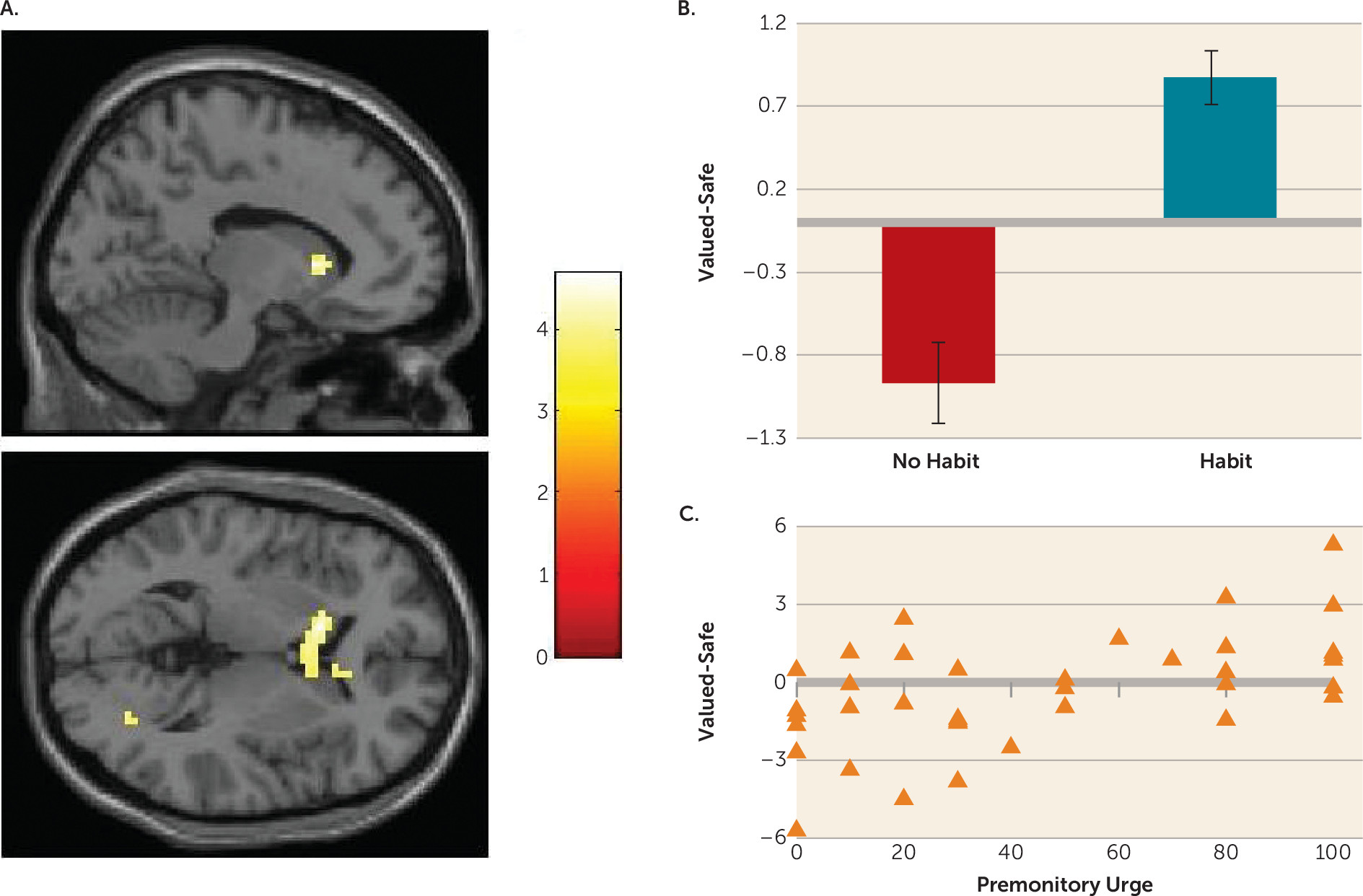
Activation associated with habitual responding in OCD patients was captured using the contrast of the valued-safe conditioned stimuli, comparing the habit groups (habit, N=15; no habit, N=22). OCD patients exhibiting habits during the test showed hyperactivation in the left caudate nucleus (Montreal Neurological Institute coordinates x, y, z: –12, 17, 4) (extending to the right) compared with those who did not (t=4.68, df=35, p<0.05, family-wise-error-corrected region of interest) (Figure 3). Using the urge to respond as a regressor in an independent analysis at the second level, replacing the binary habit group factor, we found that within the OCD group, there was a positive relationship between activation in the right caudate (coordinates x, y, z: 6, 8, 1) for this same contrast (t=3.81, df=35, p<0.001, uncorrected). This pattern was also observed in the left caudate at a more liberal threshold (p<0.005). There was no such relationship in the healthy comparison group. While there were no significant differences between the two study groups (OCD, healthy comparison) with regard to activation in response to the valued-safe contrast, the OCD habit group showed significantly greater activation in the right caudate (coordinates x, y, z: 15, 26, 1) compared with healthy comparison subjects (p<0.001, uncorrected). Conversely, when comparing OCD patients in the no habit group with healthy comparison subjects, we found hypoactivation in the right caudate (p<0.05, family-wise-error-corrected region of interest).
FIGURE 3. Comparison of Obsessive-Compulsive Disorder (OCD) Patients Who Did and Did Not Develop Habitsa
a Panel A depicts the interaction between group (habit, N=15; no habit, N=22) and conditioned stimulus ([CS]; valued, safe) during the devaluation test in the left caudate (t=4.68, df=35, p<0.05, family-wise-error-corrected level) using a bilateral caudate region of interest. Panel B is a plot of the first eigenvariate of the valued-safe contrast extracted from the left caudate cluster (Montreal Neurological Institute coordinates x, y, z: –12, 17, 4). Patients with habits show significant hyperactivation of the caudate compared with those who did not exhibit habits. Error bars denote standard error of the mean. Panel C is a plot showing the parametric association between activity in the right caudate (coordinates x, y, z: 6, 8, 1) and the self-reported urge to respond in OCD patients for the valued-safe contrast (t=3.81, df=35, p<0.001). This pattern was also observed in the left caudate at a more liberal threshold of p<0.005.

Acquisition of avoidance.

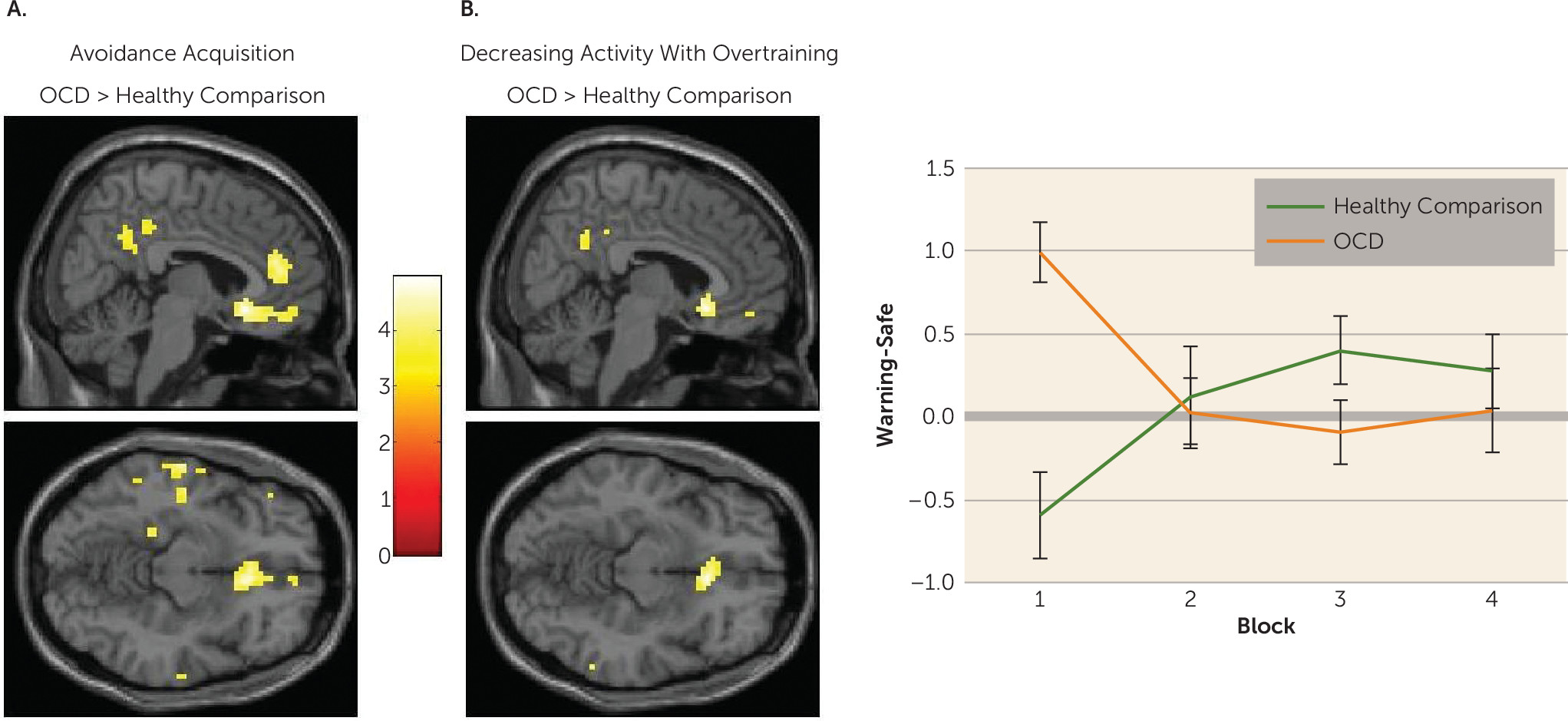
We tested for differences between OCD patients and healthy comparison subjects in activity associated with the acquisition of avoidance (i.e., block 1) using a contrast of conditioned stimuli (warning, safe). We found significant hyperactivation in the medial orbitofrontal cortex in OCD patients compared with comparison subjects (t=4.96, df=68, p<0.05, family-wise-error-corrected region of interest [coordinates x, y, z: 6, 23, –11]) (Figure 4A). A more extensive set of regions were hyperactive in OCD during this initial learning at a p value <0.001 (uncorrected [see Table S3 in the data supplement]). Comparison subjects did not show greater activation than OCD patients in any region. There were no significant differences between OCD patients in the habit and no habit groups.
FIGURE 4. Group Differences During Avoidance Acquisition and Overtraininga
a Panel A shows areas of hyperactivation in obsessive-compulsive disorder (OCD) patients relative to healthy comparison subjects during the initial acquisition of avoidance (i.e., block 1). There was a significant difference in the medial orbitofrontal cortex (t=4.96, df=68, p,0.05, family-wise-error-corrected medial orbitofrontal cortex region of interest; Montreal Neurological Institute coordinates x, y, z: 6, 23, –11). Panel B depicts a significant interaction between group (OCD, healthy comparison) and stimulus (warning, safe) and block (1–4) in the medial orbitofrontal cortex (t=4.82, df=68, p<0.05, family-wise-error corrected medial orbitofrontal cortex region of interest). The peak voxel was as follows: coordinates x, y, z: 6, 23, –11. Panel C is a plot of the mean eigenvariate for the contrast of warning-safe plotted across time for each of the groups, using the medial orbitofrontal cortex cluster displayed at an uncorrected p value <0.001. This graph reveals a pattern of hyperactivation in OCD patients during initial acquisition that decreases over time. Comparison subjects show initial hypoactivation, which increases with extended training. Results are displayed at an uncorrected p value <0.001. Error bars denote standard error of the mean.

Overtraining of avoidance.

To test whether overtraining was associated with changes in brain activation, we compared changes in blood-oxygen-level-dependent (BOLD) activation between OCD patients and comparison subjects across blocks. There was a significant interaction with group in the medial orbitofrontal cortex (t=4.82, df=68, p<0.05 family-wise-error-corrected region of interest) (Figure 4B), such that OCD patients showed a decrease in activation over successive blocks, whereas comparison subjects showed an increasing pattern in this region (Figures 4C). The peak of this interaction was the same as that showing initial hyperactivation in OCD patients during block 1 (coordinates x, y, z: 6, 23, –11). Other regions showing a similar pattern at a p value <0.001 (uncorrected) are presented in Table S3 in the online data supplement.
We compared the habit groups on this contrast to test whether differences in activation during training foreshadowed the expression of habits. There were no effects in our a priori regions of interest. However, at an uncorrected p value <0.001, we found a significant interaction in the right precuneus (t=4.11, df=1, 35, Z=3.68 [coordinates x, y, z: 18, –49, 34]; cluster extent=14) and the right superior occipital gyrus (t=4.42, df=1, 35, Z=3.91 [coordinates x, y, z: 27, –94, 16], cluster extent=22). In both of these regions, OCD patients in the no habit group showed a decreasing pattern of activation, while those who later formed habits did not (see Figure S2 in the data supplement).

Psychophysiological interaction analysis.

We conducted post hoc psychophysiological interaction analyses to interrogate whether the caudate, which was hyperactive in OCD patients who formed habits and correlated with the self-reported urge to respond, showed abnormal neuronal connectivity during the acquisition of avoidance. To do this, we tested for functional connectivity between activation in the bilateral caudate (region of interest) and the whole brain during block 1 (warning-safe condition). There was a significant difference in neural coupling between the habit and no habit groups, such that in the no habit group, there was positive coupling between the caudate and the right inferior frontal gyrus (cluster corrected at family-wise error, p<0.05) and the left pallidum (p<0.001, uncorrected) (Figure 5) during the early acquisition of avoidance and negative coupling with activation in the subgenual anterior cingulate cortex/olfactory cortex (Brodmann’s area 25) (p<0.001, uncorrected). This cluster was rostral to the medial orbitofrontal cortex cluster observed to be hyperactive in OCD patients (relative to healthy comparison subjects) during this stage but overlapped in two voxels (p<0.05, family-wise-error-corrected region of interest of the medial orbitofrontal cortex cluster where OCD patients showed hyperactivity during avoidance acquisition). This pattern was reversed for patients in the habit group (Figure 5; also see Table S5 in the data supplement), such that they exhibited negative coupling between the caudate and the right inferior frontal gyrus and pallidum and positive coupling with the subgenual anterior cingulate cortex/olfactory cortex. There were no differences between patients and comparison subjects overall.
FIGURE 5. Regions Where Neural Coupling Between the Caudate Differed Between the Habit and No Habit Groupsa
a Psychophysiological interaction analysis results at an uncorrected p value <0.001 are shown. The psychological variable was the conditioned stimulus (warning-safe) during acquisition of avoidance (i.e., block 1), and the physiological variable (psychophysiological interaction seed) was the activity for this contrast in an anatomical caudate region of interest. Eigenvariates from clusters showing interaction effects (between psychophysiological interaction and habit group) are plotted. Error bars denote standard error of the mean. The three lower images show significant psychophysiological interactions in the olfactory/subgenual anterior cingulate cortex (ACC) (t=4.4, df=35), the pallidum (t=3.91, df=35), and the right inferior frontal gyrus (t=5.11, df=35) at an uncorrected p value <0.001.

Discussion

Habits in OCD were associated with hyperactivation in the caudate nucleus. Specifically, greater caudate activity was observed in patients whose actions had become habitual, compared with healthy comparison subjects and OCD patients who did not form habits. Independent analysis revealed that, across the entire OCD group, greater activation in this region was correlated with the self-reported urge to perform these habits; there was no such relationship in healthy comparison subjects.
Translational work in rodents and humans has previously revealed that the caudate is necessary for goal-directed control over action. Lesions to this region in rodents render behavior habitual after only moderate training (4). Cocaine-induced habitual responding is associated with increased excitability in the rodent homolog of the caudate (37), and in humans, white matter connectivity between the caudate and the medial orbitofrontal cortex is predictive of improved goal-directed control over action (11) on a task that reveals habit biases in OCD (5). Findings from studies examining dynamic mechanisms of associative learning (rather than devaluation) suggest an important role for the caudate in linking outcomes to actions (i.e., contingency learning [10, 38, 39]). However, in the present study, since explicit contingency knowledge was matched across groups, we would not expect the direction of activity in our study to mirror these results. Rather, our results may reflect difficulties in translating explicit contingency knowledge into action preferences, similar to a recent study by Corbit et al. (37). Hyperactivation in the caudate is one of the most consistent neurobiological markers of OCD symptoms (with medial orbitofrontal cortex hyperactivation being the other) (18, 19), and our data therefore lend strong support to a model of OCD centered on deficits in goal-directed control over actions, resulting in compulsive habits.
OCD patients showed initial hyperactivation in the medial orbitofrontal cortex during avoidance learning, which reduced with extended training; whereas healthy comparison subjects showed the opposite pattern. A post hoc psychophysiological interaction analysis revealed that during the acquisition of avoidance, positive coupling between the caudate and the subgenual anterior cingulate cortex (partially overlapping with the medial orbitofrontal cortex cluster) was observed in OCD patients who later demonstrated habits, but a negative coupling was observed in those who did not. The role of the subgenual anterior cingulate cortex in habit forming has been sparsely studied, but two studies have shown that its likely homolog, the infralimbic cortex, must be intact for habits to persist in rodents (40, 41), suggesting that excessive connectivity between this region and the caudate may be one possible way that goal-directed control is compromised in OCD. Other differences in BOLD activity associated with habit formation within our OCD group were observed in the precuneus and superior occipital gyrus, which were sensitive to extended training, as well as functional connectivity between the caudate and the right inferior frontal gyrus and pallidum during early learning. These results require replication but suggest the possibility that a more distributed network may be involved in habit-forming biases in OCD.
Excessive habit formation in OCD was not related to differences in activation in the putamen. This region is critical for habit formation in rodents, such that lesions to the homologous dorsolateral striatum allow animals to remain goal-directed despite overtraining (15). Moreover, a similar dependency has been observed in healthy humans, such that the formation of habits is associated with white matter connectivity strength between the putamen and premotor cortex (11), as well as changes in putamen activity over time (16) (although directionality of the latter association has been inconsistent and may not relate to cue-evoked responses but rather responses in general [4244]). Although applying the usual caveat when interpreting null effects, the present data suggest that acquisition of automatic action tendencies may not be affected in OCD. Rather, habit biases in OCD appear to emerge as a result of deficits in goal-directed control associated with caudate (and possibly medial orbitofrontal cortex) hyperactivity. This conclusion dovetails with recent data showing that model-based instrumental learning, which is a constituent of goal-directed control, is impaired in OCD and reliant on the structural integrity of the medial orbitofrontal cortex and caudate but not the putamen (8).
The present study investigated avoidance, rather than appetitive, habits in order to determine how conditioned and explicit fear relate to habit formation in OCD. Since this is the first study, to our knowledge, to examine the neural correlates of avoidance habits, whether these results can be generalized to appetitive habit forming, which is similarly overactive in OCD (5), is an open question. The study of avoidance is critical in OCD; however, previous studies have shown that aberrant fear-conditioning processes are characteristic of OCD. For example, patients exhibit a pattern of hypoactivation in the ventromedial prefrontal cortex (partially subsuming the medial orbitofrontal cortex) and caudate during fear conditioning (45). The results of the present study converge with these previous findings in terms of localization but diverge with respect to directionality, a difference presumably associated with passive fear learning versus avoidance (46).
While we observed no differences in skin conductance response between OCD patients and comparison subjects, OCD patients who formed habits did not show differential skin conductance responses to the stimuli (valued, devalued, and safe) during the habit test (for results, see the data supplement). This could reflect overgeneralization of fear, which has been shown to relate to maladaptive instrumental avoidance (47). However, because these patients were able to discriminate during learning, this effect is likely a consequence rather than a cause of habitual responding. Taken together with the findings of Milad et al. (45), as well as with findings from studies suggesting that stress and anxiety contribute to habit biases in healthy people (48, 49), it is likely that a complex interaction between fear learning and habits may be critical to understanding the pathogenesis of OCD.
The findings in the caudate pertain primarily to patients who have formed habits compared with those who have not, rather than representing a difference between OCD patients and healthy comparison subjects. As such, it is possible that with further training, for example, a similar pattern might be observed in control subjects who form habits. This is a question for future research. If this is the case, our results suggest that habit forming and associated caudate hyperactivity is hastened in OCD, rather than this process being qualitatively different.
The majority of our medicated patients were taking serotonergic medication (mainly SSRIs), which in previous work has been shown to affect avoidance responding and inhibition in healthy humans (50). However, we found no evidence for differences between medicated and nonmedicated patients (matched for symptom severity) in terms of behavior and in almost all brain activation contrasts, indicating that medication effects did not drive our results.
Conclusions

These data implicate dysfunction in regions that support goal-directed control over action in excessive habit formation in OCD. These data also add convergent support to the habit hypothesis of OCD, such that it exhibits excellent neurobiological convergence with the known pathophysiology of OCD.

Supplementary Material

File (appi.ajp.2014.14040525.ds001.pdf)

References

1.
Gillan CM, Robbins TW: Goal-directed learning in obsessive-compulsive disorder. Philos Trans R Soc Lond B Biol Sci 2014; 369(1655) pii: 20130475
2.
Dickinson A: Actions and habits: the development of behavioural autonomy. Philos Trans R Soc Lond B Biol Sci 1985; 308:67–78
3.
de Wit S, Ridderinkhof KR, Fletcher PC, et al: Resolution of outcome-induced response conflict by humans after extended training. Psychol Res 2013; 77:780–793
4.
Yin HH, Ostlund SB, Knowlton BJ, et al: The role of the dorsomedial striatum in instrumental conditioning. Eur J Neurosci 2005; 22:513–523
5.
Gillan CM, Papmeyer M, Morein-Zamir S, et al: Disruption in the balance between goal-directed behavior and habit learning in obsessive-compulsive disorder. Am J Psychiatry 2011; 168:718–726
6.
Gillan CM, Morein-Zamir S, Urcelay GP, et al: Enhanced avoidance habits in obsessive-compulsive disorder. Biol Psychiatry 2014; 75:631–638
7.
Gillan CM, Morein-Zamir S, Kaser M, et al: Counterfactual processing of economic action-outcome alternatives in obsessive-compulsive disorder: further evidence of impaired goal-directed behavior. Biol Psychiatry 2014; 75:639–646
8.
Voon V, Derbyshire K, Rück C, et al: Disorders of compulsivity: a common bias towards learning habits. Mol Psychiatry (Epub ahead of print, May 20, 2014)
9.
Balleine BW, O’Doherty JP: Human and rodent homologies in action control: corticostriatal determinants of goal-directed and habitual action. Neuropsychopharmacology 2010; 35:48–69
10.
Tanaka SC, Balleine BW, O’Doherty JP: Calculating consequences: brain systems that encode the causal effects of actions. J Neurosci 2008; 28:6750–6755
11.
de Wit S, Watson P, Harsay HA, et al: Corticostriatal connectivity underlies individual differences in the balance between habitual and goal-directed action control. J Neurosci 2012; 32:12066–12075
12.
de Wit S, Corlett PR, Aitken MR, et al: Differential engagement of the ventromedial prefrontal cortex by goal-directed and habitual behavior toward food pictures in humans. J Neurosci 2009; 29:11330–11338
13.
Valentin VV, Dickinson A, O’Doherty JP: Determining the neural substrates of goal-directed learning in the human brain. J Neurosci 2007; 27:4019–4026
14.
Gremel CM, Costa RM: Orbitofrontal and striatal circuits dynamically encode the shift between goal-directed and habitual actions. Nat Commun 2013; 4:2264–2264
15.
Yin HH, Knowlton BJ, Balleine BW: Lesions of dorsolateral striatum preserve outcome expectancy but disrupt habit formation in instrumental learning. Eur J Neurosci 2004; 19:181–189
16.
Tricomi E, Balleine BW, O’Doherty JP: A specific role for posterior dorsolateral striatum in human habit learning. Eur J Neurosci 2009; 29:2225–2232
17.
Graybiel AM, Rauch SL: Toward a neurobiology of obsessive-compulsive disorder. Neuron 2000; 28:343–347
18.
Whiteside SP, Port JD, Abramowitz JS: A meta-analysis of functional neuroimaging in obsessive-compulsive disorder. Psychiatry Res 2004; 132:69–79
19.
Saxena S, Rauch SL: Functional neuroimaging and the neuroanatomy of obsessive-compulsive disorder. Psychiatr Clin North Am 2000; 23:563–586
20.
Rotge JY, Guehl D, Dilharreguy B, et al: Provocation of obsessive-compulsive symptoms: a quantitative voxel-based meta-analysis of functional neuroimaging studies. J Psychiatry Neurosci 2008; 33:405–412
21.
Swedo SE, Schapiro MB, Grady CL, et al: Cerebral glucose metabolism in childhood-onset obsessive-compulsive disorder. Arch Gen Psychiatry 1989; 46:518–523
22.
Baxter LR Jr, Phelps ME, Mazziotta JC, et al: Local cerebral glucose metabolic rates in obsessive-compulsive disorder. A comparison with rates in unipolar depression and in normal controls. Arch Gen Psychiatry 1987; 44:211–218
23.
Baxter LR Jr, Schwartz JM, Mazziotta JC, et al: Cerebral glucose metabolic rates in nondepressed patients with obsessive-compulsive disorder. Am J Psychiatry 1988; 145:1560–1563
24.
Rauch SL, Jenike MA, Alpert NM, et al: Regional cerebral blood flow measured during symptom provocation in obsessive-compulsive disorder using oxygen 15-labeled carbon dioxide and positron emission tomography. Arch Gen Psychiatry 1994; 51:62–70
25.
Schienle A, Schäfer A, Stark R, et al: Neural responses of OCD patients towards disorder-relevant, generally disgust-inducing and fear-inducing pictures. Int J Psychophysiol 2005; 57:69–77
26.
Cottraux J, Gérard D, Cinotti L, et al: A controlled positron emission tomography study of obsessive and neutral auditory stimulation in obsessive-compulsive disorder with checking rituals. Psychiatry Res 1996; 60:101–112
27.
Baxter LR Jr, Schwartz JM, Bergman KS, et al: Caudate glucose metabolic rate changes with both drug and behavior therapy for obsessive-compulsive disorder. Arch Gen Psychiatry 1992; 49:681–689
28.
Swedo SE, Pietrini P, Leonard HL, et al: Cerebral glucose metabolism in childhood-onset obsessive-compulsive disorder. Revisualization during pharmacotherapy. Arch Gen Psychiatry 1992; 49:690–694
29.
Schwartz JM, Stoessel PW, Baxter LR Jr, et al: Systematic changes in cerebral glucose metabolic rate after successful behavior modification treatment of obsessive-compulsive disorder. Arch Gen Psychiatry 1996; 53:109–113
30.
Figee M, Vink M, de Geus F, et al: Dysfunctional reward circuitry in obsessive-compulsive disorder. Biol Psychiatry 2011; 69:867–874
31.
Cannistraro PA, Wright CI, Wedig MM, et al: Amygdala responses to human faces in obsessive-compulsive disorder. Biol Psychiatry 2004; 56:916–920
32.
Remijnse PL, Nielen MMA, van Balkom AJLM, et al: Differential frontal-striatal and paralimbic activity during reversal learning in major depressive disorder and obsessive-compulsive disorder. Psychol Med 2009; 39:1503–1518
33.
de Wit SJ, Alonso P, Schweren L, et al: Multicenter voxel-based morphometry mega-analysis of structural brain scans in obsessive-compulsive disorder. Am J Psychiatry 2013; 171:340–349
34.
Adams C: Post-conditioning devaluation of an instrumental reinforcer has no effect on extinction performance. Q J Exp Psychol 1980; 32:447–458
35.
Nelson HE: National Adult Reading Test (NART): Test Manual. Windsor, United Kingdom, NFER-Nelson Publishing, 1982
36.
Maldjian JA, Laurienti PJ, Kraft RA, Burdette JH: An automated method for neuroanatomic and cytoarchitectonic atlas-based interrogation of fMRI data sets. Neuroimage 2003; 19:1233–1239
37.
Corbit LH, Chieng BC, Balleine BW: Effects of repeated cocaine exposure on habit learning and reversal by N-acetylcysteine. Neuropsychopharmacology 2014; 39:1893–1901
38.
Liljeholm M, Tricomi E, O’Doherty JP, Balleine BW: Neural correlates of instrumental contingency learning: differential effects of action-reward conjunction and disjunction. J Neurosci 2011; 31:2474–2480
39.
Tricomi EM, Delgado MR, Fiez JA: Modulation of caudate activity by action contingency. Neuron 2004; 41:281–292
40.
Smith KS, Graybiel AM: A dual operator view of habitual behavior reflecting cortical and striatal dynamics. Neuron 2013; 79:361–374
41.
Coutureau E, Killcross S: Inactivation of the infralimbic prefrontal cortex reinstates goal-directed responding in overtrained rats. Behav Brain Res 2003; 146:167–174
42.
Poldrack RA, Sabb FW, Foerde K, et al: The neural correlates of motor skill automaticity. J Neurosci 2005; 25:5356–5364
43.
Ashby FG, Turner BO, Horvitz JC: Cortical and basal ganglia contributions to habit learning and automaticity. Trends Cogn Sci 2010; 14:208–215
44.
Carelli RM, Wolske M, West MO: Loss of lever press-related firing of rat striatal forelimb neurons after repeated sessions in a lever pressing task. J Neurosci 1997; 17:1804–1814
45.
Milad MR, Furtak SC, Greenberg JL, et al: Deficits in conditioned fear extinction in obsessive-compulsive disorder and neurobiological changes in the fear circuit. JAMA Psychiatry 2013; 70:608–618, quiz 554
46.
Delgado MR, Jou RL, Ledoux JE, et al: Avoiding negative outcomes: tracking the mechanisms of avoidance learning in humans during fear conditioning. Front Behav Neurosci 2009; 3:33
47.
van Meurs B, Wiggert N, Wicker I, et al: Maladaptive behavioral consequences of conditioned fear-generalization: a pronounced, yet sparsely studied, feature of anxiety pathology. Behav Res Ther 2014; 57:29–37
48.
Otto AR, Raio CM, Chiang A, et al: Working-memory capacity protects model-based learning from stress. Proc Natl Acad Sci USA 2013; 110:20941–20946
49.
Schwabe L, Wolf OT: Stress prompts habit behavior in humans. J Neurosci 2009; 29:7191–7198
50.
Cools R, Roberts AC, Robbins TW: Serotoninergic regulation of emotional and behavioural control processes. Trends Cogn Sci 2008; 12:31–40

Information & Authors

Information

Published In

Go to American Journal of Psychiatry
American Journal of Psychiatry
Pages: 284 - 293
PubMed: 25526600

History

Received: 24 April 2014
Revision received: 20 June 2014
Revision received: 30 July 2014
Accepted: 29 August 2014
Published online: 19 December 2014
Published in print: March 01, 2015

Authors

Details

Claire M. Gillan, Ph.D.
From the Departments of Psychology and Psychiatry, and the Behavioural and Clinical Neuroscience Institute, University of Cambridge, Cambridge, United Kingdom; the Department of Psychology at New York University, New York; South Essex Partnership University NHS Foundation Trust, Springhouse, Biggleswade Hospital, Bedfordshire, United Kingdom; the Department of Psychiatry, Queen Elizabeth II Hospital, Welwyn Garden City, Hertfordshire, United Kingdom; and the Postgraduate Medical School, University of Hertfordshire, Hatfield, United Kingdom.
Annemieke M. Apergis-Schoute, Ph.D.
From the Departments of Psychology and Psychiatry, and the Behavioural and Clinical Neuroscience Institute, University of Cambridge, Cambridge, United Kingdom; the Department of Psychology at New York University, New York; South Essex Partnership University NHS Foundation Trust, Springhouse, Biggleswade Hospital, Bedfordshire, United Kingdom; the Department of Psychiatry, Queen Elizabeth II Hospital, Welwyn Garden City, Hertfordshire, United Kingdom; and the Postgraduate Medical School, University of Hertfordshire, Hatfield, United Kingdom.
Sharon Morein-Zamir, Ph.D.
From the Departments of Psychology and Psychiatry, and the Behavioural and Clinical Neuroscience Institute, University of Cambridge, Cambridge, United Kingdom; the Department of Psychology at New York University, New York; South Essex Partnership University NHS Foundation Trust, Springhouse, Biggleswade Hospital, Bedfordshire, United Kingdom; the Department of Psychiatry, Queen Elizabeth II Hospital, Welwyn Garden City, Hertfordshire, United Kingdom; and the Postgraduate Medical School, University of Hertfordshire, Hatfield, United Kingdom.
Gonzalo P. Urcelay, Ph.D.
From the Departments of Psychology and Psychiatry, and the Behavioural and Clinical Neuroscience Institute, University of Cambridge, Cambridge, United Kingdom; the Department of Psychology at New York University, New York; South Essex Partnership University NHS Foundation Trust, Springhouse, Biggleswade Hospital, Bedfordshire, United Kingdom; the Department of Psychiatry, Queen Elizabeth II Hospital, Welwyn Garden City, Hertfordshire, United Kingdom; and the Postgraduate Medical School, University of Hertfordshire, Hatfield, United Kingdom.
Akeem Sule, M.B.B.S., M.R.C.Psych.
From the Departments of Psychology and Psychiatry, and the Behavioural and Clinical Neuroscience Institute, University of Cambridge, Cambridge, United Kingdom; the Department of Psychology at New York University, New York; South Essex Partnership University NHS Foundation Trust, Springhouse, Biggleswade Hospital, Bedfordshire, United Kingdom; the Department of Psychiatry, Queen Elizabeth II Hospital, Welwyn Garden City, Hertfordshire, United Kingdom; and the Postgraduate Medical School, University of Hertfordshire, Hatfield, United Kingdom.
Naomi A. Fineberg, M.A., M.R.C.Psych.
From the Departments of Psychology and Psychiatry, and the Behavioural and Clinical Neuroscience Institute, University of Cambridge, Cambridge, United Kingdom; the Department of Psychology at New York University, New York; South Essex Partnership University NHS Foundation Trust, Springhouse, Biggleswade Hospital, Bedfordshire, United Kingdom; the Department of Psychiatry, Queen Elizabeth II Hospital, Welwyn Garden City, Hertfordshire, United Kingdom; and the Postgraduate Medical School, University of Hertfordshire, Hatfield, United Kingdom.
Barbara J. Sahakian, Ph.D.
From the Departments of Psychology and Psychiatry, and the Behavioural and Clinical Neuroscience Institute, University of Cambridge, Cambridge, United Kingdom; the Department of Psychology at New York University, New York; South Essex Partnership University NHS Foundation Trust, Springhouse, Biggleswade Hospital, Bedfordshire, United Kingdom; the Department of Psychiatry, Queen Elizabeth II Hospital, Welwyn Garden City, Hertfordshire, United Kingdom; and the Postgraduate Medical School, University of Hertfordshire, Hatfield, United Kingdom.
Trevor W. Robbins, Ph.D.
From the Departments of Psychology and Psychiatry, and the Behavioural and Clinical Neuroscience Institute, University of Cambridge, Cambridge, United Kingdom; the Department of Psychology at New York University, New York; South Essex Partnership University NHS Foundation Trust, Springhouse, Biggleswade Hospital, Bedfordshire, United Kingdom; the Department of Psychiatry, Queen Elizabeth II Hospital, Welwyn Garden City, Hertfordshire, United Kingdom; and the Postgraduate Medical School, University of Hertfordshire, Hatfield, United Kingdom.

Notes

Address correspondence to Dr. Gillan ([email protected]).

Funding Information

Wellcome Trust10.13039/100004440: 089589/Z/09/Z, 101521/Z/13/Z, G00001354
Medical Research Council10.13039/501100000265: G00001354
Supported by a Sir Henry Wellcome Postdoctoral Fellowship (101521/Z/12/Z) to Dr. Gillan and by Wellcome Trust grant (089589/Z/09/Z) to Drs. Sahakian and Robbins. This study was completed at the Behavioural and Clinical Neuroscience Institute, which is supported by a joint award from the Medical Research Council and Wellcome Trust (G00001354).Dr. Fineberg has served as a consultant for GlaxoSmithKline, Lundbeck, Novartis, Servier, and Transcept; has received research support from AstraZeneca, Cephalon, the European College of Neuropsychopharmacology, GlaxoSmithKline, Lundbeck, Servier, the United Kingdom Medical Research Council, the United Kingdom National Institute for Health Research, and the Wellcome Foundation; has received honoraria for lectures at scientific meetings from AstraZeneca, Bristol-Myers Squibb, Jazz Pharmaceuticals, Lundbeck, and Servier; and has received financial support to attend scientific meetings from BAP Pharma, Bristol-Myers Squibb, Cephalon, the European College of Neuropsychopharmacology, Janssen, Lundbeck, Novartis, the International College of Obsessive Compulsive Spectrum Disorders, the International Society for Addiction, the Royal College of Psychiatrists, Servier, and the World Health Organization. Dr. Sahakian has served as a consultant for Boehringer-Ingelheim, Cambridge Cognition, Eli Lilly, GlaxoSmithKline, Novartis, Otsuka, and Shire; has received honoraria for Grand Rounds in Psychiatry at Massachusetts General Hospital (CME credits) and for speaking at the International Conference on Cognitive Dysfunction in Schizophrenia and Mood Disorders; has served on the Medical Research Council Neurosciences and Mental Health Board and on the Science Coordination Team for the Foresight Project on Mental Capital and Wellbeing; has served on Panel LS5 for the European Research Council; and receives an honorarium from the Journal of Psychological Medicine. Dr. Robbins has served as a consultant for Cambridge Cognition, Eli Lilly, Lundbeck, Shire Pharmaceuticals, and Teva; has received research grants from Eli Lilly, GlaxoSmithKline, and Lundbeck; has received editorial honoraria from Elsevier and Springer-Verlag; has received educational lecture fees from Dohme, Merck, and Sharp; and receives royalties from Cambridge Cognition. All other authors report no financial relationships with commercial interests.

Metrics & Citations

Metrics

Citations

Export Citations

If you have the appropriate software installed, you can download article citation data to the citation manager of your choice. Simply select your manager software from the list below and click Download.

For more information or tips please see 'Downloading to a citation manager' in the Help menu.

Format
Citation style
Style
Copy to clipboard

View Options

View options

PDF/EPUB

View PDF/EPUB

Get Access

Login options

Already a subscriber? Access your subscription through your login credentials or your institution for full access to this article.

Personal login Institutional Login Open Athens login
Purchase Options

Purchase this article to access the full text.

PPV Articles - American Journal of Psychiatry

PPV Articles - American Journal of Psychiatry

Not a subscriber?

Subscribe Now / Learn More

PsychiatryOnline subscription options offer access to the DSM-5-TR® library, books, journals, CME, and patient resources. This all-in-one virtual library provides psychiatrists and mental health professionals with key resources for diagnosis, treatment, research, and professional development.

Need more help? PsychiatryOnline Customer Service may be reached by emailing [email protected] or by calling 800-368-5777 (in the U.S.) or 703-907-7322 (outside the U.S.).

Media

Figures

Other

Tables

Share

Share

Share article link

Share