Full access
Articles
Published Online: 19 November 2020

Association of Inflammatory Activity With Larger Neural Responses to Threat and Reward Among Children Living in Poverty

Abstract

Objective:

Children exposed to severe, chronic stress are vulnerable to mental and physical health problems across the lifespan. To explain how these problems develop, the neuroimmune network hypothesis suggests that early-life stress initiates a positive feedback loop between peripheral inflammatory cells and networked brain regions involved in threat and reward processing. The authors sought to test this hypothesis by studying a sample of urban children from diverse socioeconomic backgrounds.

Methods:

The authors examined the basic predictions of the neuroimmune network hypothesis in 207 children (mean age=13.9 years, 63% female; 33% Black; 30% Hispanic), focusing on poverty as a stressor. The children had fasting blood drawn to quantify five inflammatory biomarkers—C-reactive protein, tumor necrosis factor-α, and interleukins-6, -8, and -10—which were averaged to form a composite score. Children also completed two functional MRI tasks, which measured amygdala responsivity to angry facial expressions and ventral striatum responsivity to monetary rewards.

Results:

Poverty status and neural responsivity interacted statistically to predict inflammation. Among children living in poverty, amygdala threat responsivity was positively associated with inflammation, and the same was true for ventral striatum responsivity to reward. As children’s socioeconomic conditions improved, these brain-immune associations became weaker. In sensitivity analyses, these patterns were robust to alternative measures of socioeconomic status and were independent of age, sex, racial and ethnic identity, and pubertal status. The associations were also condition specific; no interactions were apparent for amygdala responsivity to neutral faces, or striatal responsivity to monetary losses.

Conclusions:

These findings suggest that childhood poverty is associated with accentuated neural-immune signaling, consistent with the neuroimmune network hypothesis.
Children exposed to severe, chronic stressors are vulnerable to a plethora of health problems across the lifespan (13). These problems span the continuum of what are traditionally understood to be mental (e.g., depression, posttraumatic stress, substance misuse) and physical (e.g., coronary heart disease, some cancers, autoimmune conditions) illnesses. Little is known about the behavioral and biological pathways underlying risk for this heterogeneous set of health problems.
To stimulate mechanistic research in this area, Nusslock and Miller proposed a neuroimmune network hypothesis (4), which integrates an evolving body of preclinical and translational research (58). The framework begins with the observation that even under normal physiologic conditions, brain circuits involved in emotion regulation engage in bidirectional communication with peripheral immune cells that mediate inflammation (5, 6). The framework goes on to hypothesize that severe chronic stress in childhood amplifies this crosstalk, initiating positive feedback loops between peripheral inflammatory activity and the brain’s developing cortico-amygdala and cortico-striatal circuits, which respectively mediate threat and reward processing. As a consequence of the enhanced crosstalk, stress-exposed children are hypothesized to display a phenotype consisting of chronic low-grade inflammatory activity, along with heightened threat responsivity and dampened reward processing. Over time, components of this phenotype are hypothesized to accelerate the pathogenesis of inflammation-mediated health problems.
There is evidence to support the constituent tenets of the neuroimmune network hypothesis. For example, studies have identified stress-related variations in brain development in children exposed to chronic stressors such as socioeconomic disadvantage and parental maltreatment (9, 10). The networks that support threat and reward processing are especially sensitive to these stressors and display what appear to be durable changes in structure and function (11). A separate literature has detailed the immunologic correlates of these stressors, demonstrating that exposed children display larger cytokine responses to microbial challenge and signs of chronic low-grade inflammation (1, 2, 12). Based on initial findings in animals (5, 7), studies have administered immune-activating stimuli to humans and found that the resulting inflammation affects functioning of neural circuits involved in threat and reward processing (1315). Observational studies of humans have also consistently reported associations between activity of these brain circuits and steady-state levels of inflammatory biomarkers (16).
Nevertheless, no published studies have examined the validity of the framework’s core hypothesis, namely, that children exposed to severe chronic stress will display the phenotype described above. Here, we sought to fill this gap by studying a sample of urban children from diverse socioeconomic backgrounds. Although children can experience a variety of chronic stressors, we focused on socioeconomic disadvantage here because it is highly prevalent in the United States. In 2017, 19% of American children resided in households with incomes below the federal poverty line. An additional 22% lived in households with incomes considered near-poor or low-income (17). Although the experiences of these children vary considerably, on the whole, they are at increased risk for chronic stressors that include material hardship, neighborhood violence, family instability, harsh parenting, and repeated bereavement, among others (18). Relative to their more advantaged peers, disadvantaged children also show indications of the dysregulation the framework specifies, including low-grade inflammation (19, 20), higher neural threat reactivity (2124), and blunted reward processing (21, 25). Given this backdrop, we predicted that to the extent that children were facing socioeconomic disadvantage, they would display the phenotype, hypothesized in the framework, consisting of low-grade inflammatory activity coupled with higher amygdala responsivity to threat and lower striatal responsivity to reward.

Methods

Sample

The study involved 277 children from the Chicago area. To be eligible, they had to be in eighth grade (typically 13–14 years old), English-speaking, and in good health, defined as being nonpregnant, without a history of chronic medical or psychiatric illness, free of prescription medications for the past month, without acute infectious disease for 2 weeks, and without functional MRI (fMRI) contraindications. Each child gave written assent and a parent or guardian gave written consent. Northwestern University’s Institutional Review Board approved the protocol.
Because of venipuncture problems, inflammation data were missing for two children. Neuroimaging data were missing for a number of children (N=32 for the threat task; N=44 for the reward task), because they arrived too late to complete the tasks, were too obese or too anxious to enter the scanner, or had previously unrecognized structural anomalies. No usable data were available for another group of children (N=36 for threat task; N=59 for the reward task) because of technical problems with acquisition (brain outside field of view), excessive motion (>10% of volumes censored within a paradigm), or limited variability in behavioral response (for details, see the online supplement). Thus, the Ns were 207 for threat analyses and 172 for reward analyses. Children for whom fMRI data were missing were more likely to identify as Black (p=0.003; and 0.001 for threat and reward tasks). However, they were similar to the broader sample on age, sex, Hispanic ethnicity, pubertal status, and household income (p values, 0.11–0.80).

Socioeconomic Conditions

Each child’s parent or guardian completed an interview about family socioeconomic conditions. From these data, we computed household income-to-poverty ratio. For most federal programs in the United States, households with an income-to-poverty ratio <1.00 are considered to be living in poverty. In 2014, when the study began, the federal poverty threshold was $24,008 for a family of two adults and two children. To facilitate interpretation of the statistical models, we grouped children into four categories, following convention in the literature: those in poverty (≤0.99) and those whom we refer to as low-income (1.00–1.99), middle-income (2.00–3.99), and higher-income (≥4.00).

Low-Grade Inflammation

At the same visit, antecubital blood draw was performed. To minimize circadian variation, venipuncture was performed between 8:00 a.m. and 10:00 a.m. Children fasted for 8 hours beforehand to minimize dietary influences. We measured serum levels of five inflammatory biomarkers, chosen for their consistent relationships with early-life stress (1, 2, 12): C-reactive protein (CRP), interleukin-6 (IL-6), interleukin-8 (IL-8), interleukin-10 (IL-10), and tumor necrosis factor-α (TNF-α). CRP was measured in duplicate by high-sensitivity immunoturbidimetric assay on a Roche/Hitachi cobas c502 analyzer. The cytokines were measured in triplicate with a four-plex immunoassay on an automated microfluidic platform (Simple Plex, ProteinSimple, San Jose, Calif.). Intra-assay coefficients of variation were 2.5%–5.1% (for additional details, see the online supplement).
Most biomarkers were skewed and/or kurtotic, but we normalized their distributions with log10 transformations. We then standardized the logged values of each biomarker (mean=0; SD=1) and averaged the resulting z-scores to form a composite score (Cronbach’s alpha=0.63), where higher scores reflect more low-grade inflammation. (Readers may wonder about including IL-10 in the composite, since its functions are anti-inflammatory. However, IL-10 is only expressed under proinflammatory conditions, so it correlates positively with the other biomarkers assessed here.) The composite score has two advantages. Statistically, it reduces the number of tests performed—here, by 80%—and thus the rate of false positive results. Biologically, a composite better reflects in vivo conditions, where proinflammatory cytokines are released in a cascading fashion and have redundant and synergistic effects on target cells.

Threat and Reward Paradigms

At a separate visit, neural responsivity to threat and reward was measured using fMRI. Briefly, to capture amygdala threat responsivity, we administered a morphed faces task (26), where children viewed images of actors displaying emotional facial expressions at varying intensities and were asked to indicate the gender associated with each face. Threat was operationalized as the amygdala’s reactivity to angry faces (relative to fixation). The amygdala response to neutral faces (relative to fixation) was used in specificity analyses. To assess reward, children performed a passive avoidance task (27), comprising 96 trials. In each trial, a shape was presented. Children could respond with a button press, triggering one of four outcomes: win $50, win $10, lose $10, or lose $50. Or they could ignore the shape, in which case no monetary outcome transpired. Outcomes were probabilistic and pseudorandom; responding to two particular shapes triggered wins on 87.5% of trials, and responding to other shapes triggered losses on 87.5% of trials. Afterward, children were paid $5, regardless of performance. Reward response was operationalized as ventral striatal reactivity to winning money (relative to fixation). Ventral striatal reactivity (relative to fixation) to losing money was used in specificity analyses.

MRI Parameters, Preprocessing, and Analysis

Imaging data were collected using a Siemens Prisma 3-T scanner with a 64-channel phased-array head coil. Structural imaging consisted of a high-resolution navigated multiecho magnetization-prepared rapid acquisition gradient-echo sequence (TR=2300 ms; TE=1.86 ms, 3.78 ms; flip angle=7°; FOV=256×256 mm; matrix=320×320; slices=208; voxel size=0.8 mm3). Functional images were acquired using a T2*-weighted gradient echo planar imaging sequence (TR=2000 ms; TE=27 ms; FOV=240 mm; matrix=94×94; flip angle=90°). Data were analyzed according to standard procedures consistent with previous work (27), using AFNI (28). Blood-oxygen-level-dependent responses were extracted for each subject from anatomically defined masks, including an amygdala mask for the threat task (Eickhoff-Zilles Architectonic Atlas: 50% probability mask) (29) and a ventral striatum mask for the reward task (accumbens-area map) (30). (Further details of scanning and analysis are provided in the online supplement.)

Statistical Analysis

To evaluate hypotheses, we estimated linear regression equations, using model 1 of PROCESS v3.4 (31) in SPSS. The outcome was the inflammation composite score, and covariates included age, sex, self-identified racial (non-White=0; White=1) and ethnic (non-Hispanic=0; Hispanic=1) category, and pubertal status (self-reported on the Pubertal Development Scale [32]). Predictors included covariates, a multicategorical variable reflecting income-to-poverty ratio, a mean-centered variable reflecting amygdala response to angry faces or striatal response to monetary reward, and a product term representing the interaction between income-to-poverty ratio and amygdala or striatal reactivity. The neuroimmune network hypothesis stipulates that in such a model, a statistical interaction should emerge whereby socioeconomic disadvantage increases the likelihood of children displaying a phenotype consisting of low-grade inflammatory activity, heightened threat responsivity, and dampened reward processing. All reported p values are based on two-tailed tests.

Results

Table 1 summarizes the characteristics of the analytic sample. The 207 children in the sample ranged in age from 12 to 14 years, and most were at least midway through puberty. They were a diverse group of children, who broadly reflect the Chicago region’s demographic and socioeconomic variation.
TABLE 1. Characteristics of the sample (N=207) in a study of inflammatory measures and neural responses to threat and reward among children living in poverty
Characteristic  
 MeanSD
Age (years)13.90.5
 N%
Female13163.3
Self-identified race  
 White (non-Hispanic)7335.3
 Black (non-Hispanic)6832.9
Self-identified ethnicity, Hispanic (any race)6330.4
Pre-, early, or mid puberty7435.7
Late or postpuberty13364.3
Household income-to-poverty ratio ≤0.993617.4
 MeanSD
Household income-to-poverty ratio3.94.0
Inflammation composite score0.00.6
C-reactive protein (mg/L)1.33.3
Interleukin-6 (pg/mL)1.61.9
Interleukin-8 (pg/mL)8.12.3
Interleukin-10 (pg/mL)2.21.1
Tumor necrosis factor-α (pg/mL)5.91.5

Hypothesis Tests

Table 2 presents results of primary hypothesis tests. As is evident, none of the covariates was consistently associated with inflammation. In each model, income-to-poverty ratio was inversely related to inflammation, with children in poverty showing higher composite scores compared with mid-income peers (0.33 to 0.36 SD units across models) and marginally higher scores compared with high-income peers (0.21 to 0.24 SD units across models). Consistent with predictions from the neuroimmune network hypothesis, there were significant interactions of income-to-poverty ratio by neural reactivity in each model. These interactions explained 3.9% (amygdala threat response, p=0.039) and 4.7% (ventral striatum reward response, p=0.043) of the residual variance in the inflammation composite score. Although these interactions are small to medium in magnitude in terms of effect size, it is important to keep in mind that these estimates are net of covariates, as well as the main effects of income-to-poverty ratio and neural reactivity.
TABLE 2. Results of conditional regression models testing the neuroimmune network hypothesisa
 Amygdala Reactivity to ThreatVentral Striatum Reactivity to Reward
VariableB95% CIpB95% CIp
Age–0.00–0.15, 0.150.99–0.06–0.10, 0.220.49
Sex (female)–0.11–0.31, 0.090.27–0.14–0.34, 0.050.15
Race (White)0.06–0.14, 0.250.590.10–0.09, 0.290.29
Ethnicity (Hispanic)0.04–0.14, 0.230.640.03–0.15, 0.210.76
Pubertal stage (continuous 1–5 scale)–0.03–0.16, 0.090.59–0.01–0.13, 0.110.83
Contrasts      
 Poverty versus low-income–0.08–0.34, 0.170.53–0.16–0.41, 0.100.23
 Poverty versus middle-income–0.36–0.60, –0.110.004–0.33–0.56, –0.090.007
 Poverty versus high-income–0.24–0.49, 0.020.07–0.21–0.46, 0.040.10
Neural reactivity      
 Children living in poverty0.220.05, 0.390.010.340.09, 0.600.009
 Low-income children0.19–0.09, 0.460.19–0.09–0.25, 0.070.26
 Middle-income children0.04–0.11, 0.910.58–0.01–0.10, 0.120.87
 High-income children–0.07–0.19, 0.050.26–0.02–0.18, 0.140.80
Intercept0.47–1.65, 2.580.66–0.44–2.59, 1.710.68
a
Shown are results of conditional regression models predicting inflammation composite score from covariates, household income-to-poverty ratio, neural reactivity to threat and reward, and the interaction of the latter two variables. Categorical variables as coded as follows: sex (male=0, female=1), race (non-White=0, White=1), and ethnicity (non-Hispanic=0, Hispanic=1).
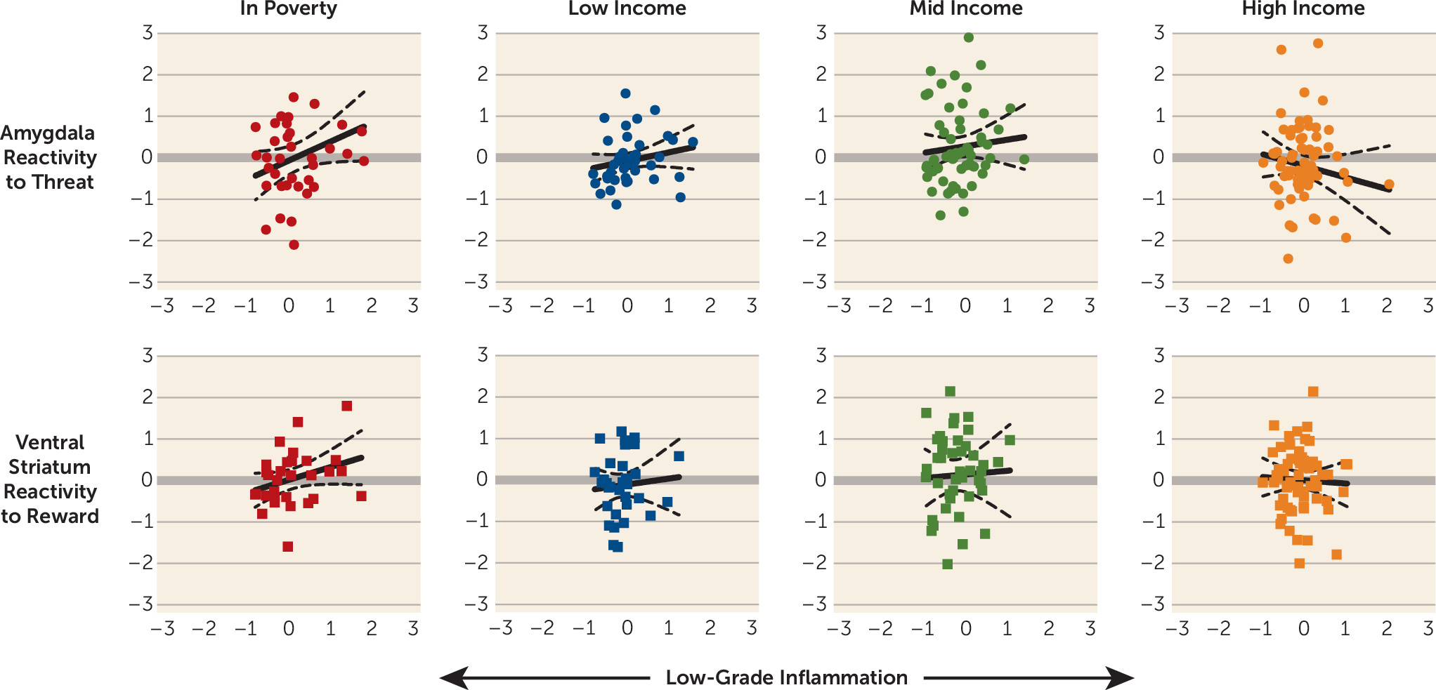
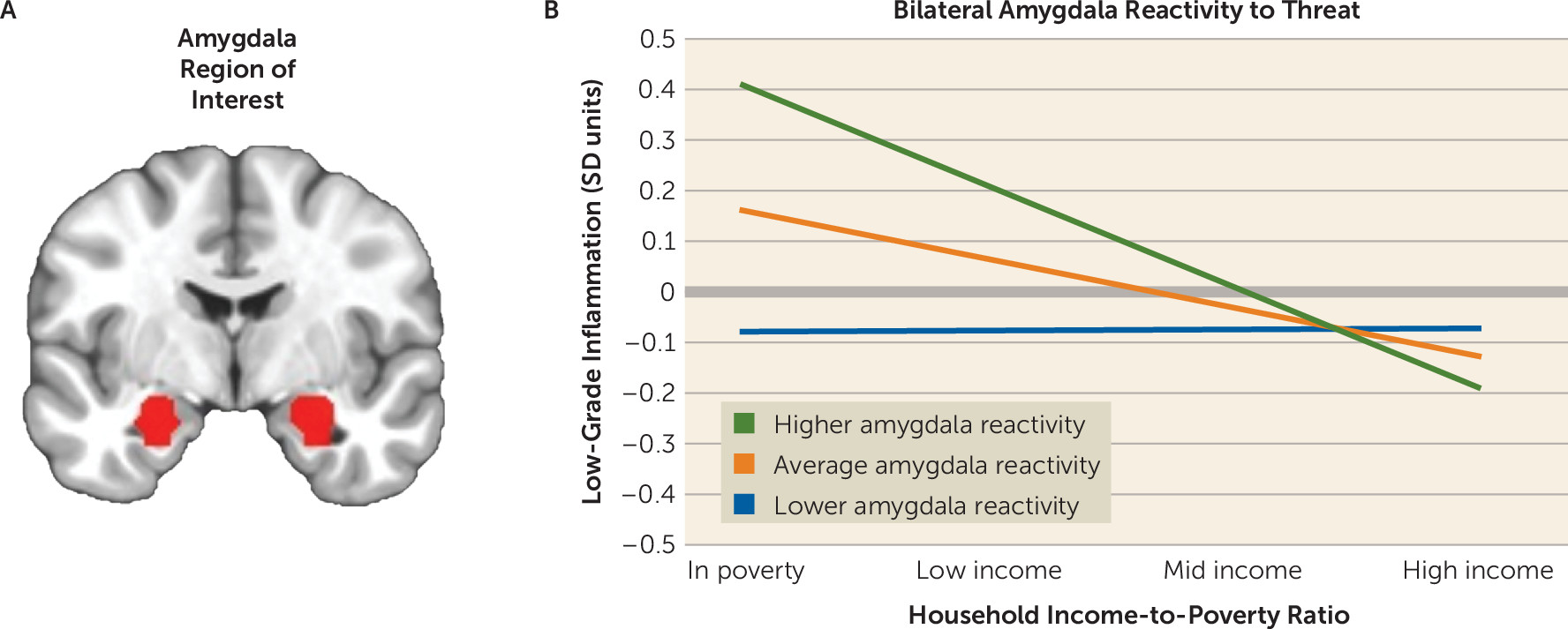
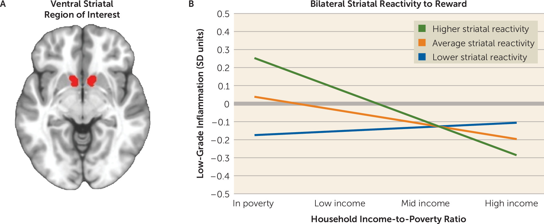
To decipher the interactions, inflammation scores were estimated at lower (−1 SD) and higher values (+1 SD) of the relevant neural responsivity distributions (31). These values were used to plot slopes relating neural responsivity and inflammatory activity for children in each of the four categories of income-to-poverty ratio. As illustrated in Figure 1 and Figure 2, among children living in poverty, amygdala threat reactivity was positively associated with inflammation composite scores, and the same was true for ventral striatum reward reactivity. As household socioeconomic conditions became more favorable, these associations became progressively weaker, and in some cases reversed direction. Figure 3 illustrates these associations in a more granular manner, with scatterplots of the raw individual data.
FIGURE 1. Relationship between household income-to-poverty ratio and inflammation as a function of bilateral amygdala reactivity to angry faces
FIGURE 2. Relationship between household income-to-poverty ratio and inflammation as a function of bilateral ventral striatal reactivity to monetary reward
FIGURE 3. Associations between the inflammation composite score and amygdala reactivity to angry faces and ventral striatal reactivity to monetary reward, stratified by household income-to-poverty ratioa
a Scatterplots are in standard deviation units. Each scatterplot depicts a best-fitting regression line with 95% confidence intervals around it.
To confirm visual interpretations of the interactions, we estimated the value of the slopes relating neural reactivity and inflammation scores (31), separately for children in each category. As shown in Table 2, the slopes were significantly greater than zero for children living in poverty. Among these children, each 1-SD increase in amygdala reactivity was associated with a 0.22 SD increase in inflammation score. The parallel value for ventral striatum reactivity was 0.34 SD. Among children living in more favorable socioeconomic conditions, the simple slopes relating neural reactivity and inflammation scores were not significantly different from zero.

Sensitivity Analyses

To evaluate the sensitivity of these observations, we reconducted analyses with two alternative definitions of socioeconomic status: a continuous indicator of household income-to-poverty ratio, natural-log-transformed to stabilize variance, and the MacArthur Scale of Subjective Social Status (33), where parents reported their family’s standing relative to other Americans. In models using continuous income-to-poverty ratio, the interactions remained significant for amygdala threat reactivity (ΔR2=0.02, p=0.05) and were marginally significant for ventral striatum reward reactivity (ΔR2=0.02, p=0.08). In models that used the MacArthur scale, both interactions continued to be significant, and in fact were slightly larger in magnitude (for amygdala threat reactivity, ΔR2=0.02, p=0.03; for ventral striatum reward reactivity, ΔR2=0.05, p=0.004). To further evaluate the sensitivity of the observed findings, we retested the primary hypotheses in generalized linear models, which do not require conventional assumptions about the normality of distributions. Mirroring the findings above, income-to-poverty ratio interacted with both amygdala threat reactivity and striatal reward reactivity in these models (p=0.03 in both cases).

Specificity Analyses

Our final analysis considered specificity, so we tested for interactions in contexts where the neuroimmune network hypothesis does not offer any predictions about how the brain will respond. These included the amygdala’s reactivity to neutral facial expressions and the ventral striatum’s reactivity to monetary losses. In neither of these conditions was there evidence of an interaction between income-to-poverty ratio and neural reactivity (p values 0.11 and.26, for amygdala and striatum, respectively). Moreover, the threat reactivity interaction remained significant when the amygdala’s response to neutral expressions was included as a covariate (ΔR2=0.04, p=0.04). Similarly, the reward reactivity interaction remained significant when ventral striatum responses to monetary losses were included as a covariate (ΔR2=0.05, p=0.04).

Specific Inflammatory Biomarkers

Although the inflammation composite score has advantages, some readers will be curious about the role of specific biomarkers. Thus, Figure S1 in the online supplement presents a heat map summarizing the covariate-adjusted relationship between each inflammatory biomarker and neural responsivity. Several patterns are apparent. First, the inflammation composite score generally had stronger associations with neural responsivity than individual biomarkers. Second, of the biomarkers, CRP and IL-6 had the strongest associations with neural responsivity. Third, some task-related variability was evident, with IL-8 relating more strongly to amygdala compared with striatal responsivity, while the reverse was true for IL-10 and TNF-α.

Discussion

To explain the health consequences of childhood adversity, the neuroimmune network hypothesis postulates that early stress amplifies crosstalk between peripheral inflammatory cells and networked brain regions involved in threat and reward processing (4). Although individual components of this hypothesis have been established in the literature, no published studies to date have integrated the data sources necessary to evaluate its core prediction. Here we sought to fill that gap in knowledge, using a diverse sample of urban children. In line with the core predictions of the framework, amygdala threat responsivity was positively associated with low-grade inflammation among children living in poverty. Intriguingly, the same pattern was evident for striatal reward responsivity. In households with more favorable socioeconomic conditions, these brain-inflammation associations were smaller in magnitude. The observed relationships were independent of demographic confounders and were robust to alternative measures of socioeconomic status. They also were condition specific: inflammation was associated with amygdala responsivity to threat stimuli, even when adjusting for activity during neutral trials, and with striatal responsivity to monetary rewards, even when adjusting for activity during losses.
The findings related to amygdala threat reactivity converge with previous evidence. Studies indicate that childhood disadvantage is associated with exaggerated behavioral, cardiovascular, and amygdala responses to threatening stimuli, a pattern thought to be adaptive in contexts where stressors can arise suddenly and unpredictably (22, 34). Disadvantaged youths also exhibit indications of a proinflammatory phenotype, as reflected in higher circulating inflammatory biomarkers, and leukocytes that mount larger cytokine responses to microbial challenge and are relatively insensitive to inhibition by glucocorticoids (20, 35). Our study’s findings bridge these distinct literatures and extend them by showing that the relationship between amygdala reactivity and inflammatory biomarkers is strongest among children living in poverty. Assuming this association reflects the crosstalk envisioned in the neuroimmune network hypothesis, it might help explain the excess health risks among disadvantaged children (3). However, because of the study’s observational design, we cannot determine whether these associations reflect causal effects. With that said, experimental studies have illustrated the plausibility of causal effects, revealing bidirectional signaling between the amygdala threat circuitry and peripheral inflammatory cells (14, 36). Such crosstalk is thought to be the substrate of an integrated network that detects threats involving microbial invasion and tissue damage, and subsequently mobilizes behavioral, physiologic, and immunologic resources for coping (37).
The findings involving reward responsivity in the ventral striatum were more complex. As predicted, children living in poverty displayed the strongest relationship between striatal responsivity and inflammatory activity. However, the direction of this relationship was positive, a pattern the original framework had not anticipated. With that said, the framework was formulated on the basis of studies from a decade ago, which showed that reward sensitivity dampens after administration of inflammatory agents (13, 38). More recent studies indicate that this formulation is overly simplistic, as inflammation’s effects on striatal reward responsivity are context dependent (8, 13, 39, 40). In situations where a reward matches what is motivationally salient to an individual, inflammation heightens striatal responsivity to that reward—for example, receiving social assistance when one is ill. In situations where there is a mismatch between a potential reward and what is motivationally salient, inflammation has the opposite effect, dampening striatal responsivity. This context dependence could explain the patterns seen here, because monetary rewards would be a highly salient reward for children living in poverty. Indeed, studies indicate that disadvantaged children display higher sensitivity to many appetitive stimuli, as reflected in temporal discounting, decision making, and self-control paradigms (1). Finally, at the trait level, mounting evidence suggests a U-shaped relationship, with individuals at both ends of the reward-sensitivity continuum showing inflammation. This pattern could reflect the tendency of individuals with high reward sensitivity to engage in inflammation-provoking behaviors—for example, consumption of foods rich in trans fats and refined sugars. Together, these observations suggest that the neuroimmune network hypothesis should be elaborated to reflect the context dependence of reward-immune signaling.
The study has several limitations. First, its cross-sectional design precludes inferences about causality, a problem that could be partially addressed in multiwave longitudinal studies. Second, there is currently no technology to directly measure neural-immune crosstalk, so our inferences about its occurrence are necessarily indirect. The brain’s resident immune cells—microglia—appear to be central to this crosstalk (5). As methods to image these cells’ functions develop, they will allow for direct evaluation of the framework’s key tenets. In the meantime, it would be valuable to more thoroughly explicate the neural basis of these results, using functional connectivity analyses to probe the broader cortico-amygdala and cortico-striatal circuits supporting threat and reward processing. It would also be valuable to replicate these findings with different paradigms, to determine whether they generalize to other threat and reward stimuli. Third, while consistent with the framework’s predictions, the interactions we observed were small to medium in terms of effect size, suggesting that to understand the health risks associated with poverty, a more thorough assessment of relevant exposures will be needed in future research. Of likely importance in this regard are the timing and duration of poverty, as well as presumptive mediators of its sequelae (e.g., stressors such as deprivation, violence, and discrimination). Finally, the study did not consider clinical outcomes, so it is unclear whether the hypothesized crosstalk accounts for health problems in the manner the framework suggests.
To summarize, these results provide initial support for the hypothesis that childhood stress amplifies crosstalk between peripheral inflammatory cells and brain regions involved in threat and reward. If substantiated, these patterns will have implications for understanding how early stressors, acting through neural-immune pathways, contribute to the development of a diverse set of health problems. With regard to mental health, this might take the form of a two-hit scenario, where stress-related inflammatory signaling increases risk for fear-related symptoms (e.g., vigilance, worry, rumination) by modulating amygdala circuity, and motivation-related symptoms by modulating striatal circuitry (e.g., substance misuse, anhedonia, mania). As the neuroimmune network hypothesis suggests, these neural and behavioral changes might, in turn, initiate a positive feedback loop that worsens psychiatric symptoms and extends them into other realms. Of course, longitudinal studies are needed to evaluate this scenario’s validity, but in the meantime it provides a framework for conceptualizing how vulnerability arises. In the long term, this work could facilitate a next generation of interventions that improve psychiatric outcomes by targeting brain-to-immune and/or immune-to-brain signaling.
Supplementary Material

File (appi.ajp.2020.20050635.ds001.pdf)

References

1.
Miller GE, Chen E, Parker KJ: Psychological stress in childhood and susceptibility to the chronic diseases of aging: moving toward a model of behavioral and biological mechanisms. Psychol Bull 2011; 137:959–997
2.
Danese A, J Lewis S: Psychoneuroimmunology of early-life stress: the hidden wounds of childhood trauma? Neuropsychopharmacology 2017; 42:99–114
3.
Hertzman C, Boyce T: How experience gets under the skin to create gradients in developmental health. Annu Rev Public Health 2010; 31:329–347
4.
Nusslock R, Miller GE: Early-life adversity and physical and emotional health across the lifespan: a neuroimmune network hypothesis. Biol Psychiatry 2016; 80:23–32
5.
Weber MD, Godbout JP, Sheridan JF: Repeated social defeat, neuroinflammation, and behavior: monocytes carry the signal. Neuropsychopharmacology 2017; 42:46–61
6.
Miller AH, Raison CL: The role of inflammation in depression: from evolutionary imperative to modern treatment target. Nat Rev Immunol 2016; 16:22–34
7.
Dantzer R, O’Connor JC, Freund GG, et al: From inflammation to sickness and depression: when the immune system subjugates the brain. Nat Rev Neurosci 2008; 9:46–56
8.
Eisenberger NI, Moieni M, Inagaki TK, et al: In sickness and in health: the co-regulation of inflammation and social behavior. Neuropsychopharmacology 2017; 42:242–253
9.
Sheridan MA, McLaughlin KA: Dimensions of early experience and neural development: deprivation and threat. Trends Cogn Sci 2014; 18:580–585
10.
Farah MJ: Socioeconomic status and the brain: prospects for neuroscience-informed policy. Nat Rev Neurosci 2018; 19:428–438
11.
Tottenham N, Galván A: Stress and the adolescent brain: amygdala-prefrontal cortex circuitry and ventral striatum as developmental targets. Neurosci Biobehav Rev 2016; 70:217–227
12.
Fagundes CP, Glaser R, Kiecolt-Glaser JK: Stressful early life experiences and immune dysregulation across the lifespan. Brain Behav Immun 2013; 27:8–12
13.
Eisenberger NI, Berkman ET, Inagaki TK, et al: Inflammation-induced anhedonia: endotoxin reduces ventral striatum responses to reward. Biol Psychiatry 2010; 68:748–754
14.
Inagaki TK, Muscatell KA, Irwin MR, et al: Inflammation selectively enhances amygdala activity to socially threatening images. Neuroimage 2012; 59:3222–3226
15.
Harrison NA, Brydon L, Walker C, et al: Inflammation causes mood changes through alterations in subgenual cingulate activity and mesolimbic connectivity. Biol Psychiatry 2009; 66:407–414
16.
Kraynak TE, Marsland AL, Wager TD, et al: Functional neuroanatomy of peripheral inflammatory physiology: a meta-analysis of human neuroimaging studies. Neurosci Biobehav Rev 2018; 94:76–92
17.
Koball H, Jiang Y: Basic facts about low-income children, 2016 (Fact Sheet). New York, National Center for Children in Poverty, Mailman School of Public Health, Columbia University, 2018
18.
Evans GW: The environment of childhood poverty. Am Psychol 2004; 59:77–92
19.
Broyles ST, Staiano AE, Drazba KT, et al: Elevated C-reactive protein in children from risky neighborhoods: evidence for a stress pathway linking neighborhoods and inflammation in children. PLoS One 2012; 7:e45419
20.
Pietras SA, Goodman E: Socioeconomic status gradients in inflammation in adolescence. Psychosom Med 2013; 75:442–448
21.
Muscatell KA: Socioeconomic influences on brain function: implications for health. Ann N Y Acad Sci 2018; 1428:14–32
22.
Kim P, Evans GW, Angstadt M, et al: Effects of childhood poverty and chronic stress on emotion regulatory brain function in adulthood. Proc Natl Acad Sci USA 2013; 110:18442–18447
23.
Muscatell KA, Morelli SA, Falk EB, et al: Social status modulates neural activity in the mentalizing network. Neuroimage 2012; 60:1771–1777
24.
Gianaros PJ, Horenstein JA, Hariri AR, et al: Potential neural embedding of parental social standing. Soc Cogn Affect Neurosci 2008; 3:91–96
25.
Gianaros PJ, Manuck SB, Sheu LK, et al: Parental education predicts corticostriatal functionality in adulthood. Cereb Cortex 2011; 21:896–910
26.
Marsh AA, Finger EC, Mitchell DG, et al: Reduced amygdala response to fearful expressions in children and adolescents with callous-unemotional traits and disruptive behavior disorders. Am J Psychiatry 2008; 165:712–720
27.
White SF, Geraci M, Lewis E, et al: Prediction error representation in individuals with generalized anxiety disorder during passive avoidance. Am J Psychiatry 2017; 174:110–117
28.
Cox RW: AFNI: software for analysis and visualization of functional magnetic resonance neuroimages. Comput Biomed Res 1996; 29:162–173
29.
Amunts K, Kedo O, Kindler M, et al: Cytoarchitectonic mapping of the human amygdala, hippocampal region, and entorhinal cortex: intersubject variability and probability maps. Anat Embryol (Berl) 2005; 210:343–352
30.
Destrieux C, Fischl B, Dale A, et al: Automatic parcellation of human cortical gyri and sulci using standard anatomical nomenclature. Neuroimage 2010; 53:1–15
31.
Hayes AF: Introduction to Mediation, Moderation, and Conditional Process Analysis, 2nd ed. New York, Guilford, 2018
32.
Petersen AC, Crockett L, Richards M, et al: A self-report measure of pubertal status: reliability, validity, and initial norms. J Youth Adolesc 1988; 17:117–133
33.
Adler NE, Epel ES, Castellazzo G, et al: Relationship of subjective and objective social status with psychological and physiological functioning: preliminary data in healthy white women. Health Psychol 2000; 19:586–592
34.
Chen E, Langer DA, Raphaelson YE, et al: Socioeconomic status and health in adolescents: the role of stress interpretations. Child Dev 2004; 75:1039–1052
35.
Miller GE, Chen E, Fok AK, et al: Low early-life social class leaves a biological residue manifested by decreased glucocorticoid and increased proinflammatory signaling. Proc Natl Acad Sci USA 2009; 106:14716–14721
36.
Wohleb ES, Powell ND, Godbout JP, et al: Stress-induced recruitment of bone marrow-derived monocytes to the brain promotes anxiety-like behavior. J Neurosci 2013; 33:13820–13833
37.
Irwin MR, Cole SW: Reciprocal regulation of the neural and innate immune systems. Nat Rev Immunol 2011; 11:625–632
38.
Capuron L, Pagnoni G, Drake DF, et al: Dopaminergic mechanisms of reduced basal ganglia responses to hedonic reward during interferon alfa administration. Arch Gen Psychiatry 2012; 69:1044–1053
39.
Muscatell KA, Moieni M, Inagaki TK, et al: Exposure to an inflammatory challenge enhances neural sensitivity to negative and positive social feedback. Brain Behav Immun 2016; 57:21–29
40.
Inagaki TK, Muscatell KA, Irwin MR, et al: The role of the ventral striatum in inflammatory-induced approach toward support figures. Brain Behav Immun 2015; 44:247–252

Information & Authors

Information

Published In

Go to American Journal of Psychiatry
American Journal of Psychiatry
Pages: 313 - 320
PubMed: 33207936

History

Received: 11 May 2020
Revision received: 10 August 2020
Accepted: 8 September 2020
Published online: 19 November 2020
Published in print: April 01, 2021

Keywords

  1. Stress
  2. Cytokines
  3. Amygdala
  4. Ventral Striatum
  5. Children

Authors

Details

Gregory E. Miller, Ph.D. [email protected]
Institute for Policy Research and Department of Psychology, Northwestern University, Evanston, Ill. (Miller, Chen, Nusslock); Boys Town National Research Hospital, Boys Town, Neb. (White).
Stuart F. White, Ph.D.
Institute for Policy Research and Department of Psychology, Northwestern University, Evanston, Ill. (Miller, Chen, Nusslock); Boys Town National Research Hospital, Boys Town, Neb. (White).
Edith Chen, Ph.D.
Institute for Policy Research and Department of Psychology, Northwestern University, Evanston, Ill. (Miller, Chen, Nusslock); Boys Town National Research Hospital, Boys Town, Neb. (White).
Robin Nusslock, Ph.D.
Institute for Policy Research and Department of Psychology, Northwestern University, Evanston, Ill. (Miller, Chen, Nusslock); Boys Town National Research Hospital, Boys Town, Neb. (White).

Notes

Send correspondence to Dr. Miller ([email protected]).

Competing Interests

The authors report no financial relationships with commercial interests.

Funding Information

Supported by NIMH grant R01 MH100117, NIDA grant P30 DA0278270, and National Heart, Lung, and Blood Institute grant R01 HL 122328.

Metrics & Citations

Metrics

Citations

Export Citations

If you have the appropriate software installed, you can download article citation data to the citation manager of your choice. Simply select your manager software from the list below and click Download.

For more information or tips please see 'Downloading to a citation manager' in the Help menu.

Format
Citation style
Style
Copy to clipboard

View Options

View options

PDF/EPUB

View PDF/EPUB

Login options

Already a subscriber? Access your subscription through your login credentials or your institution for full access to this article.

Personal login Institutional Login Open Athens login
Purchase Options

Purchase this article to access the full text.

PPV Articles - American Journal of Psychiatry

PPV Articles - American Journal of Psychiatry

Not a subscriber?

Subscribe Now / Learn More

PsychiatryOnline subscription options offer access to the DSM-5-TR® library, books, journals, CME, and patient resources. This all-in-one virtual library provides psychiatrists and mental health professionals with key resources for diagnosis, treatment, research, and professional development.

Need more help? PsychiatryOnline Customer Service may be reached by emailing [email protected] or by calling 800-368-5777 (in the U.S.) or 703-907-7322 (outside the U.S.).

Media

Figures

Other

Tables

Share

Share

Share article link

Share