Abstract

Alzheimer’s disease is a neuropsychiatric disorder with devastating clinical and socioeconomic consequences. Since the original description of the neuropathological correlates of the disorder, neuritic plaques and neurofibrillary tangles have been presumed to be critical to the underlying pathophysiology of the illness. The authors review the clinical and neuropathological origins of Alzheimer’s disease and trace the evolution of modern biomarkers from their historical roots. They describe how technological innovations such as neuroimaging and biochemical assays have been used to measure and quantify key proteins and lipids in the brain, cerebrospinal fluid, and blood and advance their role as biomarkers of Alzheimer’s disease. Together with genomics, these approaches have led to the development of a thematic and focused science in the area of degenerative disorders. The authors conclude by drawing distinctions between legitimate biomarkers of disease and molecular targets for therapeutic intervention and discuss future approaches to this complex neurobehavioral illness.
In 2017, someone in the United States developed Alzheimer’s disease (AD) every 66 seconds ( 1). By 2050, someone will develop AD every 33 seconds ( 1). It is estimated that 5.5 million Americans have Alzheimer’s dementia. By midcentury, that number is projected to grow to 13.8 million individuals ( 1). In 2014, the direct cost of AD to payers in the United States was estimated to be $214 billion ( 2). Societal costs from AD will increase substantially as the global burden increases. In its most common clinical presentation, AD manifests as an amnestic syndrome in later life, with impairments in language, spatial and executive functions, and behavior emerging with disease progression.
Research over the past few decades ( Box 1) has yielded important insights into the neurobiology of AD ( 2, 3). These include the identification of specific mutations associated with early-onset familial forms of the disease and a better understanding of the molecular and clinical risk factors for the far more common late-onset form, typically defined as onset of the illness after age 65 ( 2, 3). Nonetheless, one of the most distinguishing features of AD is the presence of distinct neuropathological correlates. These make it relatively unique in the realm of neuropsychiatric and behavioral disorders and form the basis of valid biomarkers that may be useful in the screening and early diagnosis of the disorder ( 4).

HISTORY

In 1901, the German psychiatrist Alois Alzheimer was asked to examine a 51-year-old woman, Auguste Deter, in the insane asylum of Frankfurt am Main ( 5). The first symptom displayed by the patient was suspiciousness of her husband, and this was followed by a decline in memory, loud crying, and a belief that someone was trying to kill her. Auguste Deter died on April 8, 1906, and after obtaining her family’s permission, Alzheimer performed an autopsy of her brain ( 5). Macroscopically, he observed atrophy of the cortex. When he examined thin slices of brain tissue under the microscope after staining them with silver salts, he observed two types of deposits in and in between neurons. On November 4, 1906, at the 37th annual conference of German Psychiatrists in Tübingen, Germany, Alzheimer presented the case as “a particular malady of the cerebral cortex.” The following year, he published the single case report as an article ( 6). The intracellular deposits were neurofibrillary tangles—intraneuronal aggregates of hyperphosphorylated and misfolded tau proteins. The extraneuronal deposits were called neuritic plaques. Those early neuropathological observations remain salient to this day, and postmortem evidence of plaques and tangles in the brain is required for a definitive diagnosis of AD ( 6, 7).

MOLECULAR NEUROPATHOLOGY

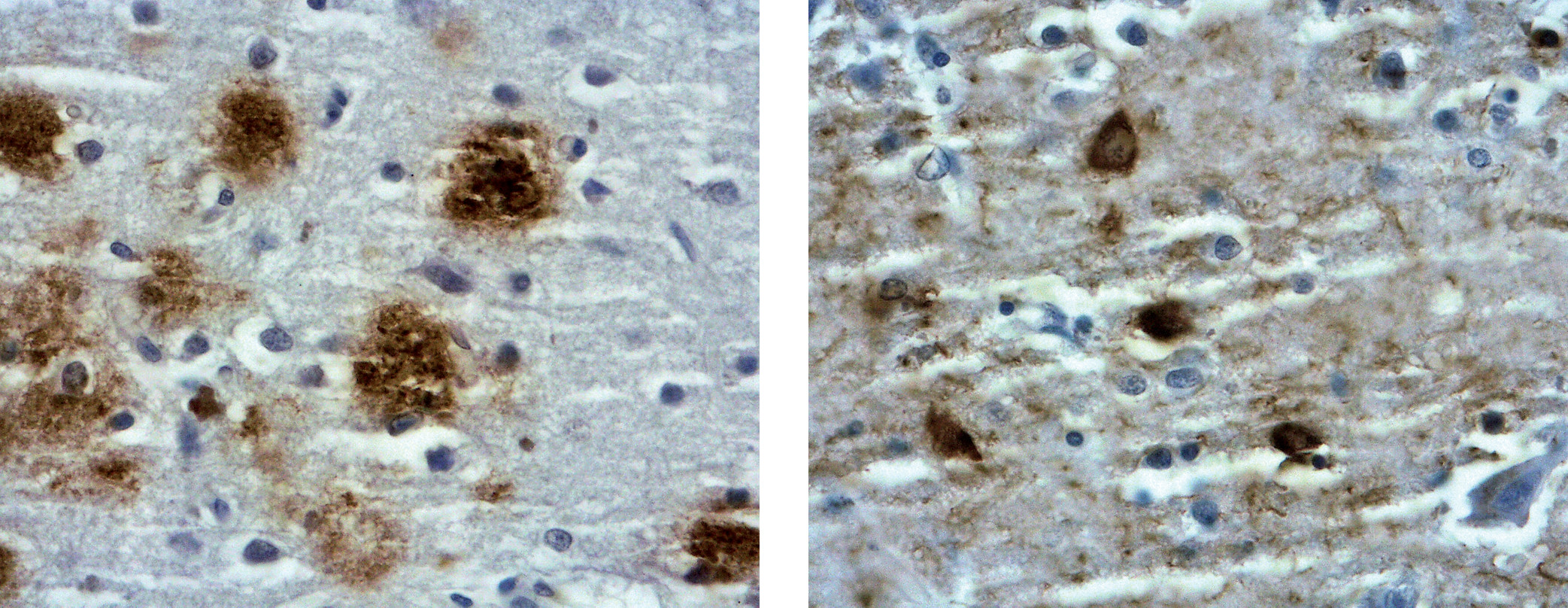
The protein originally purified from the plaque was a 40-amino-acid protein (4 kDa) that is better known today as amyloid beta (Aβ) ( 4, 7). Aβ is a component of the amyloid precursor protein APP, a transmembrane protein that is partially embedded in the plasma membrane and cleaved by two putative enzymes, beta secretase and gamma secretase, to yield the Aβ fragment. Because of its insolubility and higher rates of fibrillization, the 42-amino-acid form of Aβ (Aβ42) is more abundant than Aβ40 within the plaques ( 4, 7). Tangles are primarily made of paired helical filaments (PHFs)—fibrils of about 10 nm in diameter that form pairs with a helical tridimensional conformation at a regular periodicity of about 65 nm. The main constituent of tangles is tau, an intracellular microtubule-associated protein that is primarily located in the axons. It has six isoforms with 352–441 amino acids resulting from alternative splicing, with molecular weights ranging from 50 to 65 kDa ( 4, 7). Hyperphosphorylated tau protein has about three times more phosphorylation sites than normal tau. While normal tau stabilizes microtubules, hyperphosphorylated tau disrupts microtubules, thereby compromising axoplasmic flow and neuronal connectivity ( 4, 7). The combination of hyperphosphorylation and misfolding of the protein contributes to the underlying neuronal compromise presumed to underlie the disorder ( Figure 1). While the original conceptualization of AD was that it is a distinct disease with unique pathological correlates, more recent neuropathological studies indicate that the neuropathology of late-onset AD is more heterogeneous than originally characterized and includes the alpha-synuclein protein and microvascular disease ( 8). Nonetheless, Aβ and tau remain the primary molecular features of AD and define the disease neuropathologically ( Figure 2). They form the basis of the clinical biomarkers in AD and remain the neurobiological link between the molecular and clinical domains—a link that eludes much of the rest of neurobehavioral disorders and clinical psychiatry.
FIGURE 1. Diagrammatic representation of intraneuronal neurofibrillary tangles and extraneuronal plaques in Alzheimer’s disease a
a Paired helical filament (blue) and the extraneuronal neuritic plaque (brown). Note the presence of tangles within the neuron and amyloid beta outside the neuron.
FIGURE 2. Histological section of a plaque and a tangle in Alzheimer’s disease a
a Stained low-magnification slide depicting a neuritic plaque (left) and a neurofibrillary tangle (right). Note the neuropathological components surrounding the amyloid core of the plaque. (Photo courtesy of Harry Vinters, Department of Pathology, UCLA.)

BIOMARKERS

In 1998, the National Institutes of Health Biomarkers Definition Working Group defined a biomarker as “a characteristic that is objectively measured and evaluated as an indicator of normal biological processes, pathogenic processes, or pharmacologic responses to a therapeutic intervention” ( 9). In a broader definition of a biomarker, the World Health Organization stated that a true definition of biomarkers includes “almost any measurement reflecting an interaction between a biological system and a potential hazard, which may be chemical, physical, or biological. The measured response may be functional and physiological, biochemical at the cellular level, or a molecular interaction” ( 9). Modern biomarkers are laboratory-based, quantifiable extensions of the underlying biological aberrations that are believed to be responsible for the clinical manifestations of the disease process. Optimally, the core or primary biomarker should be closely linked to the primary pathophysiological mechanism of the disease.

NEUROIMAGING

Both structural and physiological neuroimaging have been used to identify changes in the brain that facilitate a diagnosis of AD ( 10, 11). MRI has been used to identify atrophy in focal, circumscribed areas such as the hippocampus—an area that is typically involved very early in the disease process. [ 18F]fluorodeoxyglucose (FDG) positron emission tomography (PET), a proxy for neuronal activity, helped identify widespread reductions in glucose utilization in the brain in AD with relative sparing of the sensorimotor areas, the visual cortices, and subcortical nuclei ( 10). While glucose metabolism in the brain is neurobiologically nonspecific, FDG PET methodology is well established and reliably separates patients with a diagnosis of AD from control subjects. It is also helpful in differentiating AD from other degenerative disorders. While these structural and physiological changes and patterns may not represent the presumed primary molecular abnormalities in AD, they remain useful in identifying patients with early AD and have a potential role in monitoring patients diagnosed with AD and in assessing the impact of therapeutic agents.

Molecular Neuroimaging

Imaging Aβ.

The development of ligands that target the underlying neuropathology of AD, most notably Aβ and tau, has resulted in major advances in PET imaging in AD ( 1220). A number of PET tracers that target Aβ were developed and are now used clinically in the diagnosis of AD ( Figure 3). Pittsburgh compound B (PiB) was the first in vivo radiotracer used to image Aβ in the brains of patients with AD and control subjects ( 12). PiB is a thioflavin analogue that binds with low nanomolar affinity to aggregated fibrillar deposits of the Aβ peptide, is detectable by PET, and clears rapidly from normal brain tissue ( 12). At low concentrations used in PET imaging, PiB selectively binds to fibrillary Aβ deposits in postmortem brain tissue. Moreover, in cortical areas, PiB retention was found to be inversely correlated with regional glucose utilization, determined using FDG ( 14). A more recent report examining the relationship of antemortem 11C-PiB binding to postmortem estimate of plaques and tangles found general agreement between regional measures of Aβ, but not tangles, and in vivo PiB retention in the anterior and posterior cingulate gyri and the precuneus ( 15).
FIGURE 3. Three 18F-labeled ligands that bind to the amyloid beta protein in patients diagnosed with Alzheimer’s disease (AD) and healthy volunteers a
a Regions in orange and red indicate areas of high uptake of ligand and therefore higher density of plaques. (Images courtesy of John P. Seibyl, M.D.)
PiB retention values help separate patients diagnosed with AD and mild cognitive impairment from healthy control subjects cross-sectionally. Further, longitudinal studies indicate that PiB-positive subjects with mild cognitive impairment at baseline are significantly more likely to convert to AD status than PiB-negative subjects ( 16). In addition, Morris et al. ( 17) have reported that elevated PiB binding in cognitively normal older adults predicts the development of symptoms of dementia. Newer 18F radiotracers that bind to Aβ have been developed over time and include florbetapir, florbetaben, and flutemetamol ( 18). They have been approved by the U.S. Food and Drug Administration (FDA) and the European Medicines Agency for use in the clinical assessment of cognitive disorders in order to exclude AD. They are comparable to one another and PiB in important respects and have the added advantage of being labeled with 18F, which, because of a longer half-life than 11C, facilitates imaging in clinical settings ( 18, 19).
[ 18F]FDDNP is a small molecule that binds to Aβ and tau ( 13, 20, 21). It is therefore less specific than the aforementioned radiotracers that bind exclusively to amyloid, although it more completely images the neuropathological changes observed in AD. Using [ 18F]FDDNP in clinical studies, Shoghi-Jadid et al. ( 20) showed greater radiotracer retention in several brain regions in the mild AD group when compared with control subjects. In the AD group, retention was greater in the hippocampus, the amygdala, and the entorhinal cortex, where retention was 30% higher than in the pons, which served as the reference region. Like PiB, [ 18F]FDDNP helps separate patients with AD from control subjects and also tracks conversion from normal cognitive status to mild cognitive impairment, and from mild cognitive impairment to clinical dementia ( 13). In addition, [ 18F]FDDNP imaging may help in differentiating different forms of tauopathies ( 22, 23). Amyloid and tau imaging done concurrently may help determine the relative contributions of both pathophysiological mechanisms to AD and assess the impact of therapies based on antiamyloid and antitau compounds.

Selective tau imaging.

Because a definitive diagnosis of AD requires neuropathological evidence of both Aβ and tau, precise in vivo tau imaging will greatly advance the field, both from the diagnostic and the therapeutic perspectives. The ideal PET tau tracer would have the following characteristics: high affinity and selective binding to paired helical filaments and phosphorylated tau over Aβ; high blood-brain barrier permeability; and low binding to non-tau proteins in the brain ( 24, 25). Unlike the widespread neocortical distribution of amyloid tracers in the neocortex, tau tracers are retained more notably in the inferior temporal and parietal regions of the brain. While tau deposition is widely believed to be a “downstream” event in the pathogenesis of AD, occurring after neocortical deposition of Aβ, the relationship between the two is complex and could be part of a feedback loop ( 26). While Aβ imaging shows a modest relationship between amyloid load and cognitive compromise, PET imaging demonstrates a closer relationship between tau pathology and the clinical severity of the dementia ( 24, 25). Optimally, comprehensive in vivo neuroimaging in AD should include imaging of both Aβ and tau, as they are both required for a definitive diagnosis of AD, and complementary neuroimaging will increase the accuracy of the diagnosis of AD.
Despite these challenges, several first-generation tau radiotracers were developed and used in clinical research studies in well-characterized patients. Several quinoline derivatives were first identified as candidates for tau binding ( 24, 25). 18F-linked compounds, including [ 18F]THK-5105 and [ 18F]THK-5117, and 11C-related agents, such as PBB3, were developed to improve binding PHF tau over Aβ. Interpretation of some of the THK ligand binding is confounded by the fact that monoamine oxidase B is an off-target binding site ( 27). Despite limitations, proof-of-concept studies demonstrate that these tracers separate AD patients from healthy control subjects, and tracer retention correlates significantly with the clinical severity of dementia ( 28, 29).
Second-generation tau radiotracers have been developed to overcome some of the earlier limitations, including limited sensitivity and selectivity to neurofibrillary tangles. While the future of in vivo tau neuroimaging in AD is promising, it is fair to say that unlike the Aβ-related radiotracers, the tau tracers have not been completely validated by autopsy studies. Imaging-autopsy correlations are necessary to determine whether tau neuroimaging accurately reflects the PHF tau in the brain. The complexity and variety of tau deposits in the brain in different diseases have also posed a challenge for the development of optimal PET ( 30). While the growing number of studies investigating tau PET holds promise in AD research and therapy monitoring, more in-depth work is needed to further understand the role of tau in AD and other tauopathies.

AMYLOID AND TAU IN CSF

The neuropathological hallmarks of AD are reflected in CSF. Specifically, the 42-amino-acid form of Aβ (Aβ42) is found at low levels in the CSF in an inverse relationship to amyloid plaque load ( 31, 32), likely related to how readily Aβ42 deposits in the brain; total tau (t-tau) is elevated in relationship with neuronal degeneration and pathological staging of AD ( 24, 25), but is not specific to AD, and elevations are seen in other diseases with neuronal degeneration; and phosphorylated tau (p-tau) is elevated as a marker of neurofibrillary tangle burden ( 33). A recent meta-analysis has shown that CSF measures of t-tau, p-tau, and Aβ42 all reliably distinguish AD and control groups ( 34). Of currently available biomarkers, these CSF measures become positive at the earliest stage of illness ( Figure 4).
FIGURE 4. Scatterplots of CSF amyloid beta and tau levels in patients diagnosed with Alzheimer’s disease and control subjects a
a Note lower amyloid beta (Aβ42) levels and higher tau levels in patients with Alzheimer’s disease compared with control subjects. Boxes represent the 25th, 50th, and 75th percentiles of the data. The length of the box is the interquartile range, and the lower and upper whiskers represent the 25th and 75th percentile plus or minus 1.5 times the interquartile range, respectively. (From Sunderland et al., JAMA, April 23–30, 2003, vol. 289, no. 16, pp. 2094–2103. Reprinted with permission.)
As would be expected, there is generally a concordance between PiB amyloid imaging and CSF biomarkers of AD ( 35). However, there is some variability in this finding, which mostly depends on the clinical sample characteristics; CSF biomarkers become positive at an earlier stage of disease compared with amyloid imaging positivity ( 36, 37). While significantly less well studied, tau imaging also appears to correlate at least modestly with CSF t-tau and p-tau levels ( 38) but does not necessarily correlate with low Aβ42 levels ( 39).

AMYLOID AND TAU IN BLOOD

Based on the successes described above in developing amyloid and tau measures in CSF into clinically useful biomarkers, there have been efforts to translate this work to less invasive measures from blood. Blood-based biomarkers have the potential to increase accessibility and ease of testing and to reduce the financial cost of a dementia workup ( 4043). Early studies yielded mixed results, with many showing no consistent results between patients clinically diagnosed with AD and control subjects. However, more recent findings using highly sensitive mass spectrometric, single molecule arrays (Simoa), and fully automated immunoassays suggest that Aβ peptides and peptide ratios in plasma are both specific and sensitive markers in individuals with positive Aβ PET scans ( 41, 42).
Similarly, more accurate methods to detect p-tau 181 in plasma have recently been developed, and in early studies this biomarker was able to distinguish healthy older individuals from individuals with mild cognitive impairment who had positive Aβ PET scans ( 41). Plasma p-tau 181 was also closely correlated with CSF p-tau concentrations, and higher plasma p-tau 181 levels were closely associated with tau and Aβ PET positivity and predicted cognitive decline and hippocampal atrophy ( 41). These findings increase confidence that peripheral biomarkers, accurately characterized, reflect central biological processes and changes in behavior and cognition in clinical samples.
It should be noted, however, that compared with patients diagnosed with AD, there are far fewer plasma-based biomarker studies from patients diagnosed with non-AD dementias at this point ( 41). This makes it more difficult to comment on the utility of these biomarkers in the differential diagnosis of clinical dementia. Nonetheless, the search for plasma-based biomarkers is moving in the right direction, and as more accurate assays are developed and applied to well-characterized clinical populations in longitudinal studies, the prospects for more routine clinical use increase.
Core AD biomarkers are typically broken up into two categories. The first category includes markers of Aβ pathology. These include positive amyloid imaging in the brain and lower levels of Aβ in the CSF ( 4446). The second category includes biomarkers that are believed to reflect more downstream biochemical events. This category includes CSF measures of tau (t-tau and p-tau), decreased glucose utilization in the temporo-parietal region identified using FDG PET, and significant MRI-determined atrophy in the medial and lateral temporal lobes and parietal cortex ( 4446).

SECONDARY BIOMARKERS

While neuronal plaques and tangles are widely considered to reflect the primary pathophysiological features of AD, other biological processes, perhaps not as central to the underlying pathophysiology, have also been identified in AD. Investigators have studied several proteins and lipids in the brain, blood, and CSF of patients with AD and control subjects with the hope of identifying biomarkers germane to AD and to examine the relationship of these markers to amyloid and tau.

Inflammatory Markers

Inflammation has been posited as a potential mechanism that may contribute to AD pathology and its clinical features. In a CSF study designed to examine biomarkers reflecting microglial and astrocyte activation, neuroinflammation, and vascular changes ( 47), multiple relevant markers were examined in the CSF of AD patients and control subjects together with more standard measures of tau and Aβ. Levels of YKL-40 (chitinase-3-like protein 1), intercellular adhesion molecule 1 (ICAM-1), vascular adhesion molecule 1 (VCAM-1), interleukin-15 (IL-15), and Flt-1 (fms-related tyrosine kinase 1, a vascular endothelial growth factor related to the tyrosine kinase receptor family) were all increased in AD patients compared with control subjects. In addition, all five markers showed a close direct correlation with tau, a relationship that was more significant in Aβ-positive individuals. YKL-40 levels were also higher in cases of preclinical AD and were correlated with cortical thinning in the precuneus and the superior parietal cortex.

Neurotrophins

Neurotrophins are a family of proteins that play important roles in neurodevelopment and overall maintenance of the nervous system in adults, including in processes such as synaptogenesis, cell differentiation, and neuronal survival ( 48). Neurotrophins, including brain-derived neurotrophic factor (BDNF), nerve growth factor (NGF), and neurotrophin-3 and -4 have been investigated. A recent meta-analysis and review examined studies that measured neurotrophic factor levels in blood, CSF, and postmortem tissue and combined blood and CSF data from individual studies ( 48). The authors found that BDNF levels were significantly reduced in both the CSF and blood of AD patients compared with control subjects, while increased NGF levels were detected in the CSF of AD patients compared with control subjects. The authors concluded from their comprehensive literature review that BDNF levels are lower and NGF levels are higher in the neocortex and the hippocampus of brains from AD patients compared with control subjects ( 48). While these studies do not address the question of whether changes in neurotrophin levels are a cause or a consequence of AD, they suggest that CSF neurotrophin levels are promising biomarkers in AD and related dementias.

Synaptic Proteins

Synaptic dysfunction and axonal loss occur early in the course of AD, often preceding overt cognitive decline ( 49). Contactin-2 is a synaptic and axonal membrane protein that strongly correlates with related synaptic proteins such as neurogranin and contactin-2 processing enzymes. Chatterjee et al. ( 49) reported that in a CSF study of two cohorts that included patients with AD and non-AD control subjects, CSF contactin-2 was significantly lower in the AD group compared with the control group. In postmortem tissue from AD patients, contactin-2 levels were also lower in and around amyloid plaques in the hippocampus and the temporal cortex. CSF contactin-2 strongly correlates with Aβ40, suggesting a relationship between contactin-2 and Aβ production ( 49).

Neurofilament

Neurofilament light (NfL) is the light protein of neurofilament that, together with the medium and heavy counterparts, makes up neurofilament bundles that determine both axonal caliber and conduction velocity ( 50, 51). Axonal destruction occurs relatively early in AD, thereby releasing NfL. In some CSF studies, NfL shows a large effect size, together with p-tau and Aβ, in separating AD patients from control subjects. Plasma NfL levels are also higher in patients diagnosed with mild cognitive impairment and preclinical and prodromal AD compared with control subjects ( 50). In addition, higher baseline abnormalities of AD, including cognitive and brain imaging measures and CSF biomarkers, were associated with increases in plasma NfL levels. In a longitudinal study, faster increases in NfL levels were associated with greater changes in measures of neuronal injury, including atrophy, hypometabolism, and CSF biomarkers ( 50). CSF neurofilament may also be altered in frontotemporal dementia and other subcortical pathologies, making it less useful in the differential diagnosis of the pathological substrates underlying dementia ( 51).

METABOLOMICS

Metabolomics involves measurement of the downstream biochemical products of cellular processes, including genomic, transcriptomic, and proteomic systems ( 52, 53). It is an approach with considerable potential, as it can capture snapshots of complex metabolic pathways that intersect and play an important role in the pathophysiology of disease. Using targeted metabolomic approaches, Varma et al. ( 52) examined and identified metabolites associated with the sphingolipids and glycerophospholipids that were related to AD pathology in brain tissue. They also report that blood concentrations of sphingolipids represented in the brain signature of AD are associated with progression during the preclinical and prodromal phases of AD. In the case of glycerophospholipids, lower brain levels were associated with greater plaque and tangle severity ( 52, 53). Collectively, these findings indicate that perturbations in sphingolipid metabolism may be involved in AD pathology and contribute to the cognitive symptoms in elderly individuals.

GENOMICS

The genetics of AD has advanced substantially over the past two decades ( 2, 3). Broadly speaking, there are two categories of disease genes associated with AD: causative genes, where a specific genetic mutation causes the disease, and risk or susceptibility genes, where the presence of a gene increases the likelihood of developing the disease ( 2, 3). Less than 5% of cases of AD occur in individuals under age 65—early-onset AD. About half of these cases will have an autosomal dominant inheritance pattern in the family, in which a mutation in one gene with a major effect can explain the occurrence of the disease. Mutations in APP, presenilin 1 (PSEN1), and presenilin 2 (PSEN2) genes account for more than half of the cases of early-onset AD ( 2, 3). These mutations are usually inherited from an affected parent in an autosomal dominant manner, with high penetrance, reaching almost 100% lifetime risk. Presymptomatic genetic testing can be used to identify family members who are very likely to develop the disease, years or even decades before signs and symptoms appear. Testing for these mutations in asymptomatic individuals in families with early-onset dementia should only be initiated at the request of the individual, with appropriate genetic counseling and relevant safeguards ( 2, 3).
The first and only gene to be consistently associated with sporadic late-onset AD is the apolipoprotein E (APOE) gene ( 54). The APOE gene occurs in three alleles: E2, E3, and E4. While E2 is the protective allele for AD, E4 confers increased risk for AD. Recent studies indicate that the lifetime risk for AD without considering AD status at age 85 was 11% in men and 14% in women. The lifetime risk increases to 50% for men homozygous for APOE E4, and 60% for homozygous women. For heterozygotes—APOE E3-E4 carriers—the lifetime risk is 23% for men and 30% for women ( 2). These data indicate that despite the greater risk for homozygous carriers, the E4 allele is neither necessary nor sufficient to cause AD. It is also difficult to estimate an absolute individual risk of AD. Therefore, in the absence of effective therapeutic interventions, the use of APOE genotyping to predict AD risk is not recommended for routine clinical use. Genome-wide association studies have additionally helped identify more than 20 susceptibility loci associated with AD ( 55). These include genes responsible for microglial function and inflammatory responses, such as TREM 2 variants ( 56). None of these loci are ready for clinical testing in patients at risk for AD.

THE TRANSLATIONAL VALUE OF BIOMARKERS

Biomarkers have been defined as objective, quantifiable characteristics of biological processes ( 2). Biomarkers have multiple roles in clinical medicine, including aiding in establishing a diagnosis, monitoring a disease over time, and assessing the true impact of treatment on the underlying biological aberrations. The clinical utility of biomarkers in AD is limited to some degree by the lack of standardization of the technical metrics used in neuroimaging and in the laboratory. Sources of variance include differences in scanners and laboratory equipment. Data from the Imaging Dementia–Evidence for Amyloid Scanning (IDEAS) study indicate that evidence for PET-determined amyloid deposition in the brain was not found in 36% of patients with a clinical diagnosis of AD ( 2). Conversely, PET amyloid imaging was positive in 52% of patients with a pre-PET clinical diagnosis of dementia of non-AD etiology ( 57, 58). Nonetheless, there is a broad scientific consensus that the core biomarkers in AD—Aβ, t-tau, and hyperphosphorylated tau—have demonstrated value in research and selective clinical settings and are superior to several secondary biomarkers in separating patients diagnosed with AD from age-matched control subjects ( 51). In addition, p-tau and Aβ in CSF correlate with PHFs and plaques postmortem, thereby providing the essential link between peripheral biomarkers and the molecular neuropathology of the disease—a critical connection in the validation of biomarkers ( 5961).

CONTRIBUTIONS OF BIOMARKERS TO THE CLINICAL DIAGNOSIS OF AD

The diagnosis and classification of the dementias relies on clinical signs and symptoms. Neuropathological evidence of plaques and tangles—the gold standard—is required for a definitive diagnosis. A thorough workup of a patient with history and symptoms indicating cognitive decline should include a comprehensive physical, neurologic, and psychiatric assessment, and a neuropsychological assessment when required. While biomarkers in isolation are not sufficient for a diagnosis, they increase the certainty associated with a clinical diagnosis of AD in the differential diagnosis of dementia ( Table 1). Each of these biomarkers can differentiate dementia due to AD from healthy control subjects with a specificity and sensitivity of 80%–90%. When they are combined—for example, Aβ and t-tau—the sensitivity and specificity of the AD diagnosis increase to 86% and 97%, respectively ( 51).
TABLE 1. Differential diagnosis of Alzheimer’s disease and frontotemporal dementia using biomarkers a
BiomarkerAlzheimer’s DiseaseFrontotemporal Dementia
MRIWidespread atrophy including frontal, temporal, and parietal regionsLargely confined to “anterior” frontal and temporal regions
FDG PETHypometabolism more striking in temporal and parietal regionsHypometabolism more striking in the prefrontal and temporal regions
PiB bindingWidespread PiB retentionMinimal PiB retention
CSF tauHigh levels of tau and phosphorylated tauVariable
Tau/Aβ42HighLow
a
Aβ42=amyloid beta 42; FDG=[ 18F]fluorodeoxyglucose; PET=positron emission tomography; PiB=Pittsburgh compound B.
The presence of CSF biomarkers, Aβ and tau, has been incorporated into standard clinical criteria and in the differential diagnosis of AD. Cases with a clinical diagnosis consistent with probable AD, together with CSF or neuroimaging biomarkers, can now be characterized as probable AD with an increased level of certainty ( 44). In addition, patients who meet criteria for a non-AD dementia—e.g., Lewy body dementia—and have both Aβ and tau biomarkers may be characterized as having possible AD ( 44). These biomarkers may have an even more important role in the early identification of patients prior to the appearance of clinical manifestations, as the underlying neurobiological abnormalities precede the clinical manifestations by a decade or more. Low CSF Aβ levels can be identified 25 years before expected symptoms appear, and PET-determined increased Aβ binding in the brain can be identified about 15 years before clinical onset ( 3). In their attempt to stage categories for preclinical research, Sperling et al. ( 46) describe three stages, with stages 1 and 2 occurring in cases that are asymptomatic clinically but with evidence of Aβ (stage 1), and of tau and FDG PET and MRI (stage 2) biomarkers of AD. Markers of Aβ have been demonstrated to identify individuals with mild cognitive impairment before they convert to clinical AD ( 46). Despite some of the aforementioned limitations, the core AD biomarkers have a meaningful role in the clinical arena, a role that is likely to expand when more effective therapeutic agents become available over time, and patients might be selected for treatment during the early, possibly preclinical stages of the disease, based on biomarker identification.

CLINICAL VERSUS BIOMARKER APPROACH TO AD

More recently, investigators have introduced a descriptive system that categorizes individuals on the basis of the presence or absence of biomarkers independent of clinical status ( 62). This approach, known as the “A/T/N” system, divides biomarkers into three main categories based on the identification of specific biomarkers that represent distinct pathophysiological processes. In this approach, “A” refers to the value of an Aβ marker (amyloid PET or CSF Aβ42), “T” refers to the presence of tau biomarkers (CSF p-tau or tau PET), and “N” refers to markers of neurodegeneration (FDG PET, structural MRI, or CSF t-tau) ( 62). This approach may be better applied to individuals in the preclinical state as a predictor of conversion to cognitive impairment. A recent study that used the A/T/N approach to determine the association between biomarkers and conversion to memory impairment in a cohort of cognitively intact elderly individuals reported that this approach was only modestly, albeit statistically significantly, superior to the model that incorporated clinical and genetic variables in predicting conversion in cognitive status ( 63). Another group working on defining preclinical dementia more precisely has also utilized a combination of clinical signs and symptoms (or lack thereof) and established biomarkers to identify patients who have preclinical AD ( 64).
All such approaches need to be interpreted in the proper clinical context to be diagnostically meaningful and therapeutically relevant. The growing consensus is that in order to be effective clinically, therapies should be administered in the earlier stages of the disease as opposed to the more advanced stages, where the chances of cognitive reversal and functional recovery are low. Biomarkers become increasingly relevant in this context and may aid not only in early diagnosis, possibly even in the preclinical state, but also in characterizing biological subtypes of the disease that may respond more selectively to targeted intervention.
Even a neuropathologically defined entity such as AD is phenotypically heterogeneous, and some of the technical limitations and overlap in biological processes preclude a precise classification of subtypes of AD based on currently available biomarkers ( 6568).

AMYLOID: BIOMARKER OR THERAPEUTIC TARGET? ARE WE SHOOTING THE MESSENGER?

The amyloid cascade hypothesis posits that abnormal amyloid precursor protein processing and the consequent formation of amyloid plaques constitute the critical pathophysiological event in the development of the clinical features of AD ( 69). However, the amyloid cascade remains unconfirmed, and approximately 20%–30% of individuals have substantial amyloid burden postmortem without a history of clinically detectable cognitive decline ( 65). In a study of 176 neuropathologically examined cases of dementia, the overall concordance between the clinical and pathological diagnosis was limited. Only in 63% of cases did the clinical diagnosis fully correspond with the pathological findings ( 68). The underlying pathophysiology of AD is increasingly recognized as complex, and it includes several overlapping and intersecting biological systems ( 70). A single biomarker therefore may be inadequate to accurately reflect the phenotypic variability and the course of the disease. Modern approaches that make it possible to study biomarkers concurrently in complex biological systems that intersect at various points are just beginning to be appreciated ( 70).
In the context of clinical trials, biomarkers may be considered surrogate endpoints—that is, they may act as surrogates or substitutes for clinically meaningful endpoints. Not all biomarkers serve as surrogate endpoints, and in order to be considered one, there must be strong evidence that a biomarker consistently and accurately predicts a clinical outcome, either a benefit or a harm, on at least one clinical endpoint ( 9). To meet this requirement, the biomarker must measure either a product or a process of a key biological pathway or must be indirectly related to important pathways related to disease causation. Treatment with antiamyloid agents affects amyloid and tau in expected ways—as much as a 30% decrease in tau and phosphorylated tau and an increase in CSF Aβ together with a decrease in PiB retention in the brain ( 67). It is noteworthy that these biomarker changes were not associated with change in the clinical status of patients even after a 78-week trial with the antiamyloid agent bapineuzumab ( 71). Changes in the expected direction in biomarker levels were not accompanied by a corresponding clinical improvement. However, as previously stated, both Aβ- and tau-based biomarkers track conversion from the mild cognitive impairment state to clinical AD and help in separating AD patients from control subjects. AD-related biomarkers therefore meet some but not all of the criteria for surrogate biomarkers.

Failure of Experimental Therapeutics in AD

To date, attempts to substantially modify or reverse the course of cognitive impairment in AD have been a stunning failure. Most of the FDA-approved pharmacological interventions for AD, the acetylcholinesterase inhibitors, were developed on the basis of early neuropathological and neurochemical studies demonstrating lower acetylcholine concentrations in the neocortex, together with atrophy in the basal nucleus of Meynert—the forebrain region that provides the bulk of the cholinergic input to the neocortex and limbic regions—in AD postmortem studies ( 72). Depression has been consistently identified as a risk factor for AD, and plausible mechanisms include a stress-related increase in endogenous glucocorticoids that are amyloidogenic and corticotrophin receptors that promote tau phosphorylation ( 73, 74). In spite of considerable evidence from postmortem studies indicating that cholinergic, adrenergic, serotonergic, and corticotrophin-releasing factor (CRFergic) neurons degenerate in AD, neurotransmitter-based pharmacological approaches have been disappointing thus far and show no evidence of reversing the cognitive decline. Their widespread clinical use reflects a lack of therapeutic alternatives rather than a meaningful evidence base ( 75, 76).
Over the past two decades, the therapeutics of AD have largely focused on preventing the accumulation of and/or removing Aβ from the brain ( 77). Aβ and, more recently, tau are therefore used not merely as biomarkers, but as molecular targets for drug development and treatment. Results have been disappointing, and yet these studies continue under the presumption that Aβ and tau are central to the pathophysiology of the disease and not merely diagnostic markers that have some clinical utility ( 78, 79). Other branches of medicine are replete with evidence to the contrary ( 80). For example, C-reactive protein (CRP), an inflammatory biomarker, is a strong marker of ischemic heart disease, but it is not a critical biological mechanism in the pathophysiology of heart disease ( 80). CRP is therefore not a target for therapeutic drug development. A contrarian opinion in the AD field is that cell death, and not the presence of Aβ and tau, is the critical pathophysiological event in AD ( 7578). In this conceptual framework, the hallmark neuropathology—plaques and tangles—may be “defensive mechanisms or damage response protein,” and therefore targeting them experimentally is a misleading and potentially dangerous approach to AD therapeutics ( 7379). Alternative approaches, including the repurposing of older, off-patent drugs, are receiving renewed attention with the growing appreciation that classical biomarkers may not be suitable targets for drug development.

CONCLUSIONS

After Alois Alzheimer presented the case of Auguste Deter in Tübingen in 1906 before 88 prominent psychiatrists from across Europe, the chairman of the scientific session remarked, “So then, respected colleague Alzheimer, I thank you for your remarks; clearly there is no desire for discussion” ( 6). Since then, AD research has evolved from a single, interesting case report, over a century ago, to an interdisciplinary scientific endeavor that no longer rests in any given laboratory, institute, or country. While the incidence of dementia may be stabilizing in some high-income countries, it remains a major socioeconomic problem globally ( 81). Multiple consortia focusing on different aspects of the disease have contributed to new findings and approaches. Although we now have a much better grasp of the early clinical presentation of the disorder, its longitudinal course, its risk factors, its genetics, and valid biomarkers, translation from the preclinical domain to effective therapeutics has, to this point, been unattainable. Nonetheless, there is a better appreciation of the complexity of the disorder and the need for further research, especially in identifying other critical pathways and molecules that may serve as more effective therapeutic targets in this devastating disorder ( 82). We may also have reached a critical scientific fork in the road where important distinctions may need to be made between a valid biomarker and a molecular target for drug development.

Footnote

Dr. Kumar serves as a data safety monitoring board member for a National Institute on Aging study. Dr. Nemeroff has received grants or research support from NIH; he has served as a consultant for Acadia Pharmaceuticals, EMA Wellness, Gerson Lehrman Group, Intra-Cellular Therapies, Janssen Research and Development, Magstim, Navitor Pharmaceuticals, Signant Health, Sunovion Pharmaceuticals, Taisho Pharmaceutical, Takeda, TC MSO, and Xhale; he has served on scientific advisory boards of the American Foundation for Suicide Prevention, the Anxiety Disorders Association of America (ADAA), the Brain and Behavior Research Foundation, the Laureate Institute for Brain Research, Signant Health, Skyland Trail, and Xhale and on boards of directors for ADAA, Gratitude America, Smart, Inc., and Xhale; he is a stockholder in AbbVie, Antares, BI Gen Holdings, Celgene, Corcept Therapeutics, EMA Wellness, OPKO Health, Seattle Genetics, TC MSO, Trends in Pharma Development, and Xhale; he has income sources or equity of $10,000 or more from American Psychiatric Publishing, CME Outfitters, EMA Wellness, Intra-Cellular Therapies, Magstim, Signant Health, and Xhale; he holds patents on a method and devices for transdermal delivery of lithium (US 6,375,990B1), a method of assessing antidepressant drug therapy via transport inhibition of monoamine neurotransmitters by ex vivo assay (US 7,148,027B2), and compounds, compositions, methods of synthesis, and methods of treatment (CRF Receptor Binding Ligand) (US 8,551,996B2). Dr. Widge’s work on this project was supported in part by NIH grants R21 MH113101, UH3 NS100548, and R01 MH119384, the One Mind Institute, the MnDRIVE Brain Conditions initiative, and the University of Minnesota Medical Discovery Team on Additions. Dr. Rodriguez is Deputy Editor for the American Journal of Psychiatry; the editors’ disclosures appear in the April issue of the Journal. Dr. Carpenter has received research support from Affect Neuro, Janssen, NeoSync, Neuronetics, Neurolief, Nexstim, and Sunovion and has served as a consultant for Affect Neuro, Janssen, NeoSync, Neurolief, Neuronetics, Neuronix, Nexstim, Otsuka, Sage Therapeutics, and Sunovion. Dr. McDonald is a member of the APA Council on Research, representing ECT and neuromodulation therapies; he has received research support from Cervel Neurotherapeutics, the National Institute of Neurological Disease and Stroke, the National Institute on Aging, NeoSync, Neuronetics, NIMH, Soterix, and the Stanley Foundation; he has served as a consultant for Sage Therapeutics and Signant Health; he receives funding from the J.B. Fuqua Foundation; he serves on the boards of Skyland Trail and 3Keys; he serves as a data safety monitoring board member for a National Institute on Aging study; and he receives royalties from Oxford University Press. Dr. Cooper reports no financial relationships with commercial interests.

References

1.
Alzheimer’s Association : 2017 Alzheimer’s disease facts and figures. Alzheimers Dement 2017 ; 13 : 325 – 373
2.
Winblad B, Amouyel P, Andrieu S, et al : Defeating Alzheimer’s disease and other dementias: a priority for European science and society. Lancet Neurol 2016 ; 15 : 455 – 532
3.
Bateman RJ, Xiong C, Benzinger TL, et al : Clinical and biomarker changes in dominantly inherited Alzheimer’s disease. N Engl J Med 2012 ; 367 : 795 – 804
4.
Serrano-Pozo A, Frosch MP, Masliah E, et al : Neuropathological alterations in Alzheimer disease. Cold Spring Harb Perspect Med 2011 ; 1 : a006189
5.
Toodayan N : Professor Alois Alzheimer (1864–1915): lest we forget. J Clin Neurosci 2016 ; 31 : 47 – 55
6.
Alzheimer A, Stelzmann RA, Schnitzlein HN, et al : An English translation of Alzheimer’s 1907 paper, “Uber eine eigenartige Erkankung der Hirnrinde”. Clin Anat 1995 ; 8 : 429 – 431
7.
Blennow K, Zetterberg H : Biomarkers for Alzheimer’s disease: current status and prospects for the future. J Intern Med 2018 ; 284 : 643 – 663
8.
Gottesman RF, Schneider AL, Zhou Y, et al : Association between midlife vascular risk factors and estimated brain amyloid deposition. JAMA 2017 ; 317 : 1443 – 1450
9.
Strimbu K, Tavel JA : What are biomarkers? Curr Opin HIV AIDS 2010 ; 5 : 463 – 466
10.
Kumar A, Schapiro MB, Grady C, et al : High-resolution PET studies in Alzheimer’s disease. Neuropsychopharmacology 1991 ; 4 : 35 – 46
11.
Frisoni GB, Sabattoli F, Lee AD, et al : In vivo neuropathology of the hippocampal formation in AD: a radial mapping MR-based study. Neuroimage 2006 ; 32 : 104 – 110
12.
Klunk WE, Engler H, Nordberg A, et al : Imaging brain amyloid in Alzheimer’s disease with Pittsburgh compound-B. Ann Neurol 2004 ; 55 : 306 – 319
13.
Small GW, Kepe V, Ercoli LM, et al : PET of brain amyloid and tau in mild cognitive impairment. N Engl J Med 2006 ; 355 : 2652 – 2663
14.
Nordberg A, Rinne JO, Kadir A, et al : The use of PET in Alzheimer disease. Nat Rev Neurol 2010 ; 6 : 78 – 87
15.
Driscoll I, Troncoso JC, Rudow G, et al : Correspondence between in vivo (11)C-PiB-PET amyloid imaging and postmortem, region-matched assessment of plaques. Acta Neuropathol 2012 ; 124 : 823 – 831
16.
Okello A, Koivunen J, Edison P, et al : Conversion of amyloid positive and negative MCI to AD over 3 years: an 11C-PIB PET study. Neurology 2009 ; 73 : 754 – 760
17.
Morris JC, Roe CM, Grant EA, et al : Pittsburgh compound B imaging and prediction of progression from cognitive normality to symptomatic Alzheimer disease. Arch Neurol 2009 ; 66 : 1469 – 1475
18.
Herholz K, Ebmeier K : Clinical amyloid imaging in Alzheimer’s disease. Lancet Neurol 2011 ; 10 : 667 – 670
19.
Laforce R Jr, Soucy J-P, Sellami L, et al : Molecular imaging in dementia: past, present, and future. Alzheimers Dement 2018 ; 14 : 1522 – 1552
20.
Shoghi-Jadid K, Small GW, Agdeppa ED, et al : Localization of neurofibrillary tangles and beta-amyloid plaques in the brains of living patients with Alzheimer disease. Am J Geriatr Psychiatry 2002 ; 10 : 24 – 35
21.
Kumar A, Kepe V, Barrio JR, et al : Protein binding in patients with late-life depression. Arch Gen Psychiatry 2011 ; 68 : 1143 – 1150
22.
Barrio JR, Small GW, Wong KP, et al : In vivo characterization of chronic traumatic encephalopathy using [F-18]FDDNP PET brain imaging. Proc Natl Acad Sci USA 2015 ; 112 : E2039 – E2047
23.
Kepe V, Bordelon Y, Boxer A, et al : PET imaging of neuropathology in tauopathies: progressive supranuclear palsy. J Alzheimers Dis 2013 ; 36 : 145 – 153
24.
Okamura N, Harada R, Ishiki A, et al : The development and validation of tau PET tracers: current status and future directions. Clin Transl Imaging 2018 ; 6 : 305 – 316
25.
Johnson KA, Schultz A, Betensky RA, et al : Tau positron emission tomographic imaging in aging and early Alzheimer disease. Ann Neurol 2016 ; 79 : 110 – 119
26.
Bloom GS : Amyloid-β and tau: the trigger and bullet in Alzheimer disease pathogenesis. JAMA Neurol 2014 ; 71 : 505 – 508
27.
Ng KP, Pascoal TA, Mathotaarachchi S, et al : Monoamine oxidase B inhibitor, selegiline, reduces 18F-THK5351 uptake in the human brain. Alzheimers Res Ther 2017 ; 9 : 25
28.
Barrio JR : The irony of PET tau probe specificity. J Nucl Med 2018 ; 59 : 115 – 116
29.
Hall B, Mak E, Cervenka S, et al : In vivo tau PET imaging in dementia: pathophysiology, radiotracer quantification, and a systematic review of clinical findings. Ageing Res Rev 2017 ; 36 : 50 – 63
30.
Saint-Aubert L, Lemoine L, Chiotis K, et al : Tau PET imaging: present and future directions. Mol Neurodegener 2017 ; 12 : 19
31.
Tapiola T, Alafuzoff I, Herukka SK, et al : Cerebrospinal fluid β-amyloid 42 and tau proteins as biomarkers of Alzheimer-type pathologic changes in the brain. Arch Neurol 2009 ; 66 : 382 – 389
32.
Seppälä TT, Louhija UM, Appelberg B, et al : Comparison between clinical diagnosis and CSF biomarkers of Alzheimer disease in elderly patients with late onset psychosis: Helsinki Old Age Psychosis Study (HOPS). Am J Geratr Psychiatry 2014 ; 22 : 908 – 916
33.
Buerger K, Otto M, Teipel SJ, et al : Dissociation between CSF total tau and tau protein phosphorylated at threonine 231 in Creutzfeldt-Jakob disease. Neurobiol Aging 2006 ; 27 : 10 – 15
34.
Olsson B, Lautner R, Andreasson U, et al : CSF and blood biomarkers for the diagnosis of Alzheimer’s disease: a systematic review and meta-analysis. Lancet Neurol 2016 ; 15 : 673 – 684
35.
Schindler SE, Gray JD, Gordon BA, et al : Cerebrospinal fluid biomarkers measured by Elecsys assays compared to amyloid imaging. Alzheimers Dement 2018 ; 14 : 1460 – 1469
36.
Palmqvist S, Mattsson N, Hansson O ; Alzheimer’s Disease Neuroimaging Initiative: Cerebrospinal fluid analysis detects cerebral amyloid-β accumulation earlier than positron emission tomography. Brain 2016 ; 139 : 1226 – 1236
37.
Vlassenko AG, McCue L, Jasielec MS, et al : Imaging and cerebrospinal fluid biomarkers in early preclinical Alzheimer disease. Ann Neurol 2016 ; 80 : 379 – 387
38.
Mattsson N, Schöll M, Strandberg O, et al : 18F-AV-1451 and CSF T-tau and P-tau as biomarkers in Alzheimer’s disease. EMBO Mol Med 2017 ; 9 : 1212 – 1223
39.
Brier MR, McCarthy JE, Benzinger TLS, et al : Local and distributed PiB accumulation associated with development of preclinical Alzheimer’s disease. Neurobiol Aging 2016 ; 38 : 104 – 111
40.
Hampel H, O’Bryant SE, Molinuevo JL, et al : Blood-based biomarkers for Alzheimer disease: mapping the road to the clinic. Nat Rev Neurol 2018 ; 14 : 639 – 652
41.
Ashton NJ, Hye A, Rajkumar AP, et al : An update on blood-based biomarkers for non-Alzheimer neurodegenerative disorders. Nat Rev Neurol 2020 ; 16 : 265 – 284
42.
Nakamura A, Kaneko N, Villemagne VL, et al : High performance plasma amyloid-β biomarkers for Alzheimer’s disease. Nature 2018 ; 554 : 249 – 254
43.
Chouraki V, Beiser A, Younkin L, et al : Plasma amyloid-β and risk of Alzheimer’s disease in the Framingham Heart Study. Alzheimers Dement 2015 ; 11 : 249 – 57.e1
44.
McKhann GM, Knopman DS, Chertkow H, et al : The diagnosis of dementia due to Alzheimer’s disease: recommendations from the National Institute on Aging–Alzheimer’s Association workgroups on diagnostic guidelines for Alzheimer’s disease. Alzheimers Dement 2011 ; 7 : 263 – 269
45.
Albert MS, DeKosky ST, Dickson D, et al : The diagnosis of mild cognitive impairment due to Alzheimer’s disease: recommendations from the National Institute on Aging–Alzheimer’s Association workgroups on diagnostic guidelines for Alzheimer’s disease. Alzheimers Dement 2011 ; 7 : 270 – 279
46.
Sperling RA, Aisen PS, Beckett LA, et al : Toward defining the preclinical stages of Alzheimer’s disease: recommendations from the National Institute on Aging–Alzheimer’s Association workgroups on diagnostic guidelines for Alzheimer’s disease. Alzheimers Dement 2011 ; 7 : 280 – 292
47.
Janelidze S, Mattsson N, Stomrud E, et al : CSF biomarkers of neuroinflammation and cerebrovascular dysfunction in early Alzheimer disease. Neurology 2018 ; 91 : e867 – e877
48.
Du Y, Wu HT, Qin XY, et al : Postmortem brain, cerebrospinal fluid, and blood neurotrophic factor levels in Alzheimer’s disease: a systematic review and meta-analysis. J Mol Neurosci 2018 ; 65 : 289 – 300
49.
Chatterjee M, Del Campo M, Morrema THJ, et al : Contactin-2, a synaptic and axonal protein, is reduced in cerebrospinal fluid and brain tissue in Alzheimer’s disease. Alzheimers Res Ther 2018 ; 10 : 52
50.
Mattsson N, Cullen NC, Andreasson U, et al : Association between longitudinal plasma neurofilament light and neurodegeneration in patients with Alzheimer disease. JAMA Neurol 2019 ; 76 : 791 – 799
51.
Blennow K, Hampel H, Weiner M, et al : Cerebrospinal fluid and plasma biomarkers in Alzheimer disease. Nat Rev Neurol 2010 ; 6 : 131 – 144
52.
Varma VR, Oommen AM, Varma S, et al : Brain and blood metabolite signatures of pathology and progression in Alzheimer disease: a targeted metabolomics study. PLoS Med 2018 ; 15 : e1002482
53.
Toledo JB, Arnold M, Kastenmüller G, et al : Metabolic network failures in Alzheimer’s disease: a biochemical road map. Alzheimers Dement 2017 ; 13 : 965 – 984
54.
Corder EH, Saunders AM, Strittmatter WJ, et al : Gene dose of apolipoprotein E type 4 allele and the risk of Alzheimer’s disease in late onset families. Science 1993 ; 261 : 921 – 923
55.
Lambert JC, Ibrahim-Verbaas CA, Harold D, et al : Meta-analysis of 74,046 individuals identifies 11 new susceptibility loci for Alzheimer’s disease. Nat Genet 2013 ; 45 : 1452 – 1458
56.
Ulrich JD, Ulland TK, Colonna M, et al : Elucidating the role of TREM2 in Alzheimer’s disease. Neuron 2017 ; 94 : 237 – 248
57.
Rabinovici GD, Gatsonis C, Apgar C, et al : Association of amyloid positron emission tomography with subsequent change in clinical management among Medicare beneficiaries with mild cognitive impairment or dementia. JAMA 2019 ; 321 : 1286 – 1294
58.
Jack CR Jr, Petersen RC : Amyloid PET and changes in clinical management for patients with cognitive impairment. JAMA 2019 ; 321 : 1258 – 1260
59.
Buerger K, Ewers M, Pirttilä T, et al : CSF phosphorylated tau protein correlates with neocortical neurofibrillary pathology in Alzheimer’s disease. Brain 2006 ; 129 : 3035 – 3041
60.
Strozyk D, Blennow K, White LR, et al : CSF A(beta) 42 levels correlate with amyloid-neuropathology in a population-based autopsy study. Neurology 2003 ; 60 : 652 – 656
61.
Tapiola T, Overmyer M, Lehtovirta M, et al : The level of cerebrospinal fluid tau correlates with neurofibrillary tangles in Alzheimer’s disease. Neuroreport 1997 ; 8 : 3961 – 3963
62.
Jack CR Jr, Bennett DA, Blennow K, et al : A/T/N: an unbiased descriptive classification scheme for Alzheimer disease biomarkers. Neurology 2016 ; 87 : 539 – 547
63.
Jack CR Jr, Wiste HJ, Therneau TM, et al : Associations of amyloid, tau, and neurodegeneration biomarker profiles with rates of memory decline among individuals without dementia. JAMA 2019 ; 321 : 2316 – 2325
64.
Dubois B, Hampel H, Feldman HH, et al : Preclinical Alzheimer’s disease: definition, natural history, and diagnostic criteria. Alzheimers Dement 2016 ; 12 : 292 – 323
65.
Blennow K, de Leon MJ, Zetterberg H : Alzheimer’s disease. Lancet 2006 ; 368 : 387 – 403
66.
Knopman DS, DeKosky ST, Cummings JL, et al : Practice parameter: diagnosis of dementia (an evidence-based review): report of the Quality Standards Subcommittee of the American Academy of Neurology. Neurology 2001 ; 56 : 1143 – 1153
67.
Neuropathology Group of the Medical Research Council Cognitive Function and Aging Study : Pathological correlates of late-onset dementia in a multicentre, community-based population in England and Wales. Lancet 2001 ; 357 : 169 – 175
68.
Brunnström H, Englund E : Clinicopathological concordance in dementia diagnostics. Am J Geriatr Psychiatry 2009 ; 17 : 664 – 670
69.
Hardy JA, Higgins GA : Alzheimer’s disease: the amyloid cascade hypothesis. Science 1992 ; 256 : 184 – 185
70.
Chen H, Zhu Z, Zhu Y, et al : Pathway mapping and development of disease-specific biomarkers: protein-based network biomarkers. J Cell Mol Med 2015 ; 19 : 297 – 314
71.
Rinne JO, Brooks DJ, Rossor MN, et al : 11C-PiB PET assessment of change in fibrillar amyloid-beta load in patients with Alzheimer’s disease treated with bapineuzumab: a phase 2, double-blind, placebo-controlled, ascending-dose study. Lancet Neurol 2010 ; 9 : 363 – 372
72.
Coyle JT, Price DL, DeLong MR : Alzheimer’s disease: a disorder of cortical cholinergic innervation. Science 1983 ; 219 : 1184 – 1190
73.
Libro R, Bramanti P, Mazzon E : Endogenous glucocorticoids: role in the etiopathogenesis of Alzheimer’s disease. Neuroendocrinol Lett 2017 ; 38 : 1 – 12
74.
Mather M, Harley CW : The locus coeruleus: essential for maintaining cognitive function and the aging brain. Trends Cogn Sci 2016 ; 20 : 214 – 226
75.
Mullane K, Williams M : Alzheimer’s disease (AD) therapeutics, 2: beyond amyloid: re-defining AD and its causality to discover effective therapeutics. Biochem Pharmacol 2018 ; 158 : 376 – 401
76.
Mullane K, Williams M : Alzheimer’s disease (AD) therapeutics, 1: repeated clinical failures continue to question the amyloid hypothesis of AD and the current understanding of AD causality. Biochem Pharmacol 2018 ; 158 : 359 – 375
77.
Ricciarelli R, Fedele E : The amyloid cascade hypothesis in Alzheimer’s disease: it’s time to change our mind. Curr Neuropharmacol 2017 ; 15 : 926 – 935
78.
Lee H-G, Castellani RJ, Zhu X, et al : Amyloid-beta in Alzheimer’s disease: the horse or the cart? Pathogenic or protective? Int J Exp Pathol 2005 ; 86 : 133 – 138
79.
The amyloid cascade hypothesis has misled the pharmaceutical industry. Biochem Soc Trans 2011 ; 39 : 920 – 923
80.
Ridker PM : A test in context: high-sensitivity C-reactive protein. J Am Coll Cardiol 2016 ; 67 : 712 – 723
81.
Roehr S, Pabst A, Luck T, et al : Is dementia incidence declining in high-income countries? A systematic review and meta-analysis. Clin Epidemiol 2018 ; 10 : 1233 – 1247
82.
Scheltens P, Blennow K, Breteler MM, et al : Alzheimer’s disease. Lancet 2016 ; 388 : 505 – 517
83.
Fischer O : Miliare Nekrosen mit drusigen Wucherungen der Neurofibrillen, eine regelmässige Veränderung der Hirnrinde bei seniler Demenz. Monatsschr Psychiatr Neurol 1907 ; 22 : 361 – 372
84.
Goedert M : Oskar Fischer and the study of dementia. Brain 2009 ; 132 : 1102 – 1111
85.
Davies P : Neurotransmitter-related enzymes in senile dementia of the Alzheimer type. Brain Res 1979 ; 171 : 319 – 327
86.
Foster NL, Chase TNP, Fedio P, et al : Alzheimer’s disease: focal cortical changes shown by positron emission tomography. Neurology 1983 ; 33 : 961 – 965
87.
St George-Hyslop PH, Tanzi RE, Polinsky RJ, et al : The genetic defect causing familial Alzheimer’s disease maps on chromosome 21. Science 1987 ; 235 : 885 – 890
88.
Schellenberg GD, Bird TD, Wijsman EM, et al : Genetic linkage evidence for a familial Alzheimer’s disease locus on chromosome 14. Science 1992 ; 258 : 668 – 671
89.
St George-Hyslop P, Haines J, Rogaev E, et al : Genetic evidence for a novel familial Alzheimer’s disease locus on chromosome 14. Nat Genet 1992 ; 2 : 330 – 334
90.
Rogaev EI, Sherrington R, Rogaeva EA, et al : Familial Alzheimer’s disease in kindreds with missense mutations in a gene on chromosome 1 related to the Alzheimer’s disease type 3 gene. Nature 1995 ; 376 : 775 – 778

Information & Authors

Information

Published In

Go to American Journal of Psychiatry
American Journal of Psychiatry
Pages: 1014 - 1025
PubMed: 34734743

History

Received: 26 August 2019
Revision received: 26 May 2020
Revision received: 13 October 2020
Revision received: 10 June 2021
Accepted: 22 June 2021
Published in print: November 2021
Published online: 4 November 2021

Keywords

  1. Dementia
  2. Alzheimer’s Disease

Authors

Details

Anand Kumar, M.D., M.H.A. [email protected]
Department of Psychiatry, University of Illinois at Chicago (Kumar, Cooper); Department of Psychiatry and Behavioral Sciences, University of Texas Dell Medical School in Austin, and Mulva Clinic for the Neurosciences, UT Health Austin (Nemeroff); Department of Psychiatry, University of Minnesota, Minneapolis (Widge); Department of Psychiatry and Behavioral Sciences, Stanford University, Stanford, Calif. (Rodriguez); Department of Psychiatry and Human Behavior, Warren Alpert Medical School at Brown University, Providence, R.I. (Carpenter); Department of Psychiatry and Behavioral Sciences, Emory University School of Medicine, Atlanta (McDonald).
Charles B. Nemeroff, M.D., Ph.D. https://orcid.org/0000-0001-7867-1160
Department of Psychiatry, University of Illinois at Chicago (Kumar, Cooper); Department of Psychiatry and Behavioral Sciences, University of Texas Dell Medical School in Austin, and Mulva Clinic for the Neurosciences, UT Health Austin (Nemeroff); Department of Psychiatry, University of Minnesota, Minneapolis (Widge); Department of Psychiatry and Behavioral Sciences, Stanford University, Stanford, Calif. (Rodriguez); Department of Psychiatry and Human Behavior, Warren Alpert Medical School at Brown University, Providence, R.I. (Carpenter); Department of Psychiatry and Behavioral Sciences, Emory University School of Medicine, Atlanta (McDonald).
Carolyn Rodriguez, M.D., Ph.D.
Department of Psychiatry, University of Illinois at Chicago (Kumar, Cooper); Department of Psychiatry and Behavioral Sciences, University of Texas Dell Medical School in Austin, and Mulva Clinic for the Neurosciences, UT Health Austin (Nemeroff); Department of Psychiatry, University of Minnesota, Minneapolis (Widge); Department of Psychiatry and Behavioral Sciences, Stanford University, Stanford, Calif. (Rodriguez); Department of Psychiatry and Human Behavior, Warren Alpert Medical School at Brown University, Providence, R.I. (Carpenter); Department of Psychiatry and Behavioral Sciences, Emory University School of Medicine, Atlanta (McDonald).
Linda Carpenter, M.D.
Department of Psychiatry, University of Illinois at Chicago (Kumar, Cooper); Department of Psychiatry and Behavioral Sciences, University of Texas Dell Medical School in Austin, and Mulva Clinic for the Neurosciences, UT Health Austin (Nemeroff); Department of Psychiatry, University of Minnesota, Minneapolis (Widge); Department of Psychiatry and Behavioral Sciences, Stanford University, Stanford, Calif. (Rodriguez); Department of Psychiatry and Human Behavior, Warren Alpert Medical School at Brown University, Providence, R.I. (Carpenter); Department of Psychiatry and Behavioral Sciences, Emory University School of Medicine, Atlanta (McDonald).
William M. McDonald, M.D.
Department of Psychiatry, University of Illinois at Chicago (Kumar, Cooper); Department of Psychiatry and Behavioral Sciences, University of Texas Dell Medical School in Austin, and Mulva Clinic for the Neurosciences, UT Health Austin (Nemeroff); Department of Psychiatry, University of Minnesota, Minneapolis (Widge); Department of Psychiatry and Behavioral Sciences, Stanford University, Stanford, Calif. (Rodriguez); Department of Psychiatry and Human Behavior, Warren Alpert Medical School at Brown University, Providence, R.I. (Carpenter); Department of Psychiatry and Behavioral Sciences, Emory University School of Medicine, Atlanta (McDonald).

Notes

Send correspondence to Dr. Kumar ( [email protected]).

Metrics & Citations

Metrics

Citations

Export Citations

If you have the appropriate software installed, you can download article citation data to the citation manager of your choice. Simply select your manager software from the list below and click Download.

For more information or tips please see 'Downloading to a citation manager' in the Help menu.

Format
Citation style
Style
Copy to clipboard

View Options

View options

PDF/EPUB

View PDF/EPUB

Login options

Already a subscriber? Access your subscription through your login credentials or your institution for full access to this article.

Personal login Institutional Login Open Athens login
Purchase Options

Purchase this article to access the full text.

PPV Articles - American Journal of Psychiatry

PPV Articles - American Journal of Psychiatry

Not a subscriber?

Subscribe Now / Learn More

PsychiatryOnline subscription options offer access to the DSM-5-TR® library, books, journals, CME, and patient resources. This all-in-one virtual library provides psychiatrists and mental health professionals with key resources for diagnosis, treatment, research, and professional development.

Need more help? PsychiatryOnline Customer Service may be reached by emailing [email protected] or by calling 800-368-5777 (in the U.S.) or 703-907-7322 (outside the U.S.).

Media

Figures

Other

Tables

Share

Share

Share article link

Share