Full access
Departments
Published Online: 29 April 2015

Reversible Mirror Clock Drawing After Acute Right Frontoparietal Lobe Infarct

Publication: The Journal of Neuropsychiatry and Clinical Neurosciences
To the Editor: Visuospatial disorientation, with variable clinical presentation, has been associated with acute vascular insult in the right hemisphere. Here the authors report a case of a patient who experienced a unique visuospatial problem: mirror clock drawing after an acute right frontoparietal subcortical infarct.

Case Report

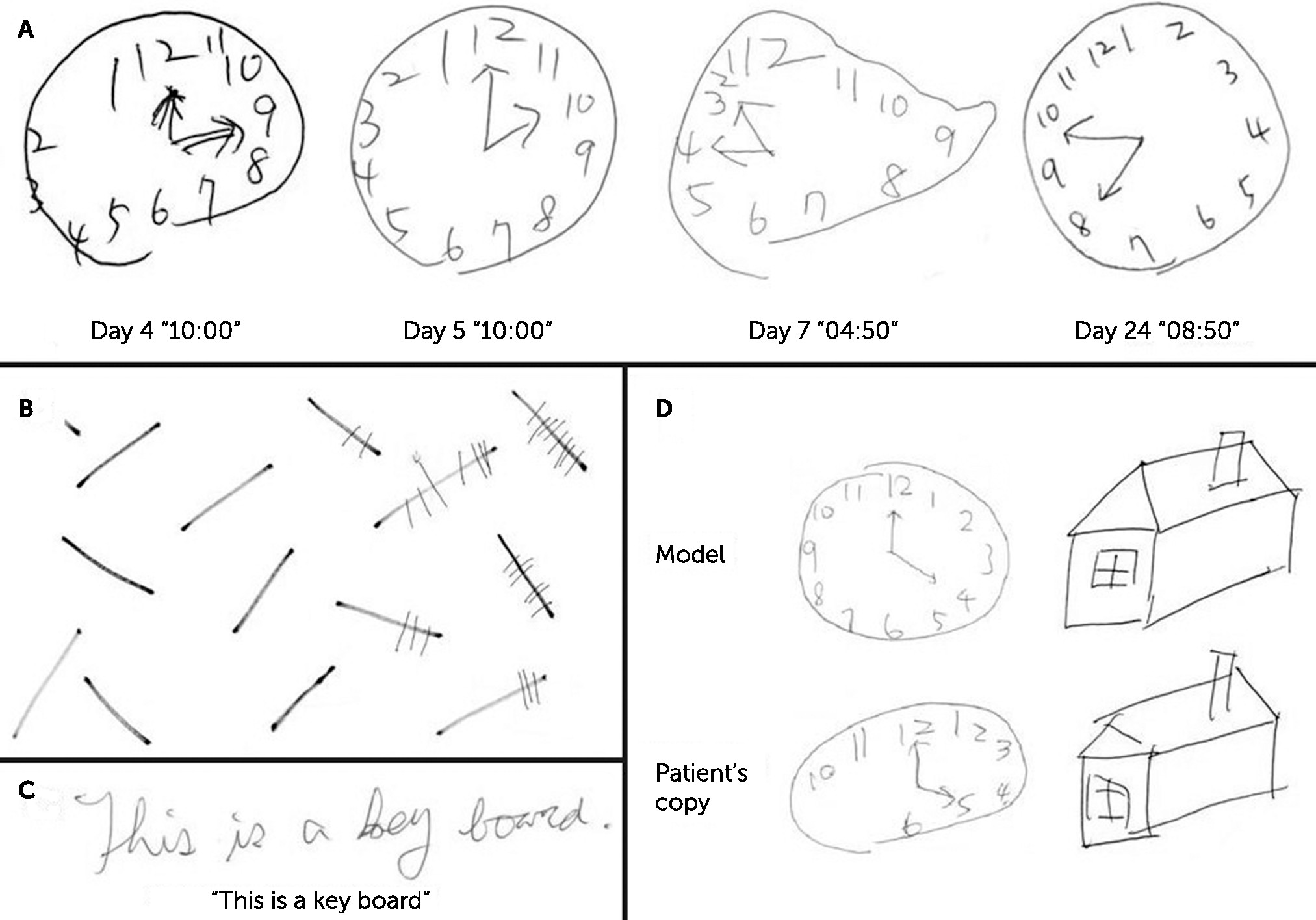
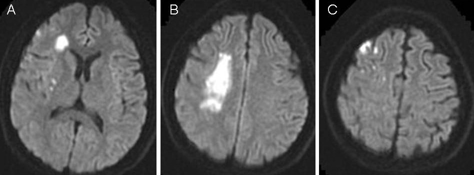
A 30-year-old right-handed woman experienced acute onset left hemiparesis and visuospatial problems during her first experience with scuba diving. Before this event, she worked as an illustrator of three-dimensional computer graphics and was generally healthy, except that she had experienced two episodes of transient left arm numbness and weakness while under acute emotional stress: the first event occurred when she was 6 years old, and the other occurred when she was 12 years old. The left hemiparesis developed within 30 minutes after the scuba diving, with some fluctuation for the following 3 days. Meanwhile, she got lost in the Mass Rapid Transit station, lost her ability to touch type, and became unable to find the toolbox in the computer graphics program she used for her work. On admission to our Stroke Center on the third day after onset, the patient manifested left central–type facial palsy, left hemiparesis, left hypesthesia, and left-sided unilateral spatial neglect. Interestingly, mirror clock drawing (Figure 1, panel A) in addition to the unilateral spatial neglect (Figure 1, panel B) was noted, whereas spatial orientation was preserved on her handwriting and picture copying (Figure 1, panels C and D). Brain MRI scans showed recent infarcts in the right frontoparietal subcortical regions (Figure 2, panels A and B) and small cortical areas (Figure 2, panel C). MR angiography and further conventional angiography revealed bilateral steno-occlusive middle cerebral arteries with formation of multiple tiny collateral vessels, indicating moyamoya disease. Her clock drawing became normalized at 3 weeks after stroke onset, along with improvements in the left hemiparesis and spatial neglect.
FIGURE 1. Patient's Performance on Clock Drawing and Other Visuospatial Tasksa
a (A) Mirror drawing on the clock drawing test persisted for 3 weeks after stroke onset. (B) Line cancellation test revealed left hemispatial neglect. (C) The patient's handwriting was normal. (D) Picture copying test showed correct left-right orientation and some hemispatial neglect. The line cancellation and picture copying tests were performed on the fourth day after stroke onset.
FIGURE 2. Diffusion-Weighted MRI Scan Showing Acute Infarcts at the Right Frontoparietal Lobe, Involving Mainly the Subcortical and Small Cortical Regions

Discussion

Visuospatial disorientation has long been associated with right hemispheric lesions, especially in the right parietal lobe.1,2 It commonly presents as hemispatial neglect, spatial disorientation, constructional apraxia, or impairments of topographic memory.3 In our patient, the hemispatial neglect could be demonstrated by the line cancellation test. The patient's history of getting lost in a familiar place and the inability to perform her regular computer graphics program suggested impairment in topographic memory. Importantly, the patient demonstrated a consistent mirror clock drawing with intact abilities of picture copying and writing words during the acute stroke stage. The reversibility of her visuospatial symptoms in the subacute phase suggested a direct link with the acute ischemic process rather than underlying moyamoya disease.
The discrepancy between free clock drawing and picture copying indicated a specific deficit in visuospatial memory, with preserved spatial perceptual and remapping functions. To the best of our knowledge, our patient is the first to demonstrate the particular mirror clock drawing after a right hemispheric infarct, which provides novel insight about the delicate and complicated processes involved in visuospatial perception, recall, and remapping.

References

1.
Ungerleider LG, Mishkin M: Two cortical visual systems, in Analysis of Visual Behavior. Edited by Ingle DJ, Mansfield RJW, Goodale MD. Cambridge, UK, MIT Press, 1982, pp 549–586
2.
Turunen KE, Kauranen TV, Laari SP, et al: Cognitive deficits after subcortical infarction are comparable with deficits after cortical infarction. Eur J Neurol 2013; 20:286–292
3.
Russell C, Deidda C, Malhotra P, et al: A deficit of spatial remapping in constructional apraxia after right-hemisphere stroke. Brain 2010; 133:1239–1251

Information & Authors

Information

Published In

Go to The Journal of Neuropsychiatry and Clinical Neurosciences
The Journal of Neuropsychiatry and Clinical Neurosciences
Pages: e156 - e157
PubMed: 25923866

History

Published in print: Spring 2015
Published online: 29 April 2015

Authors

Affiliations

Yu-Wen Cheng, M.D.
Stroke Center and Dept. of Neurology, National Taiwan University Hospital, Taipei, Taiwan (Y-WC, S-CT, J-SJ); and the Division of Clinical Psychology, Department of Occupational Therapy, College of Medicine, Chang-Gung University, Taoyuan, Taiwan (C-CY).
Chi-Cheng Yang, M.S., Ph.D.
Stroke Center and Dept. of Neurology, National Taiwan University Hospital, Taipei, Taiwan (Y-WC, S-CT, J-SJ); and the Division of Clinical Psychology, Department of Occupational Therapy, College of Medicine, Chang-Gung University, Taoyuan, Taiwan (C-CY).
Sung-Chun Tang, M.D., Ph.D.
Stroke Center and Dept. of Neurology, National Taiwan University Hospital, Taipei, Taiwan (Y-WC, S-CT, J-SJ); and the Division of Clinical Psychology, Department of Occupational Therapy, College of Medicine, Chang-Gung University, Taoyuan, Taiwan (C-CY).
Jiann-Shing Jeng, M.D., Ph.D.
Stroke Center and Dept. of Neurology, National Taiwan University Hospital, Taipei, Taiwan (Y-WC, S-CT, J-SJ); and the Division of Clinical Psychology, Department of Occupational Therapy, College of Medicine, Chang-Gung University, Taoyuan, Taiwan (C-CY).

Notes

Send correspondence to Sung-Chun Tang, M.D., Ph.D.; e-mail: [email protected]

Metrics & Citations

Metrics

Citations

Export Citations

If you have the appropriate software installed, you can download article citation data to the citation manager of your choice. Simply select your manager software from the list below and click Download.

For more information or tips please see 'Downloading to a citation manager' in the Help menu.

Format
Citation style
Style
Copy to clipboard

There are no citations for this item

View Options

View options

PDF/ePub

View PDF/ePub

Get Access

Login options

Already a subscriber? Access your subscription through your login credentials or your institution for full access to this article.

Personal login Institutional Login Open Athens login
Purchase Options

Purchase this article to access the full text.

PPV Articles - Journal of Neuropsychiatry and Clinical Neurosciences

PPV Articles - Journal of Neuropsychiatry and Clinical Neurosciences

Not a subscriber?

Subscribe Now / Learn More

PsychiatryOnline subscription options offer access to the DSM-5-TR® library, books, journals, CME, and patient resources. This all-in-one virtual library provides psychiatrists and mental health professionals with key resources for diagnosis, treatment, research, and professional development.

Need more help? PsychiatryOnline Customer Service may be reached by emailing [email protected] or by calling 800-368-5777 (in the U.S.) or 703-907-7322 (outside the U.S.).
Media

Figures

Other

Tables

Share

Share

Share article link

Share