Full access
Regular Articles
Published Online: 17 April 2017

“Top-Down” Mu-Opioid System Function in Humans: Mu-Opioid Receptors in Ventrolateral Prefrontal Cortex Mediate the Relationship Between Hedonic Tone and Executive Function in Major Depressive Disorder

Publication: The Journal of Neuropsychiatry and Clinical Neurosciences

Abstract

Cognitive dysfunction and anhedonia, the reduced ability to experience pleasure, are commonly comorbid symptoms that are persistent following successful resolution of negative affect in major depressive disorder (MDD). Little is known about whether they share common etiology. In the present study, the relationship between ventrolateral prefrontal cortex (VLPFC) activity, cognitive dysfunction (i.e., executive dysfunction), and positive emotionality was investigated in conjunction with mu-opioid neurotransmission in a sample of 39 MDD patients. Results suggest that increased endogenous mu-opioid tone in the VLPFC mediates the relationship between increased trait positive emotionality and more efficient executive functioning.
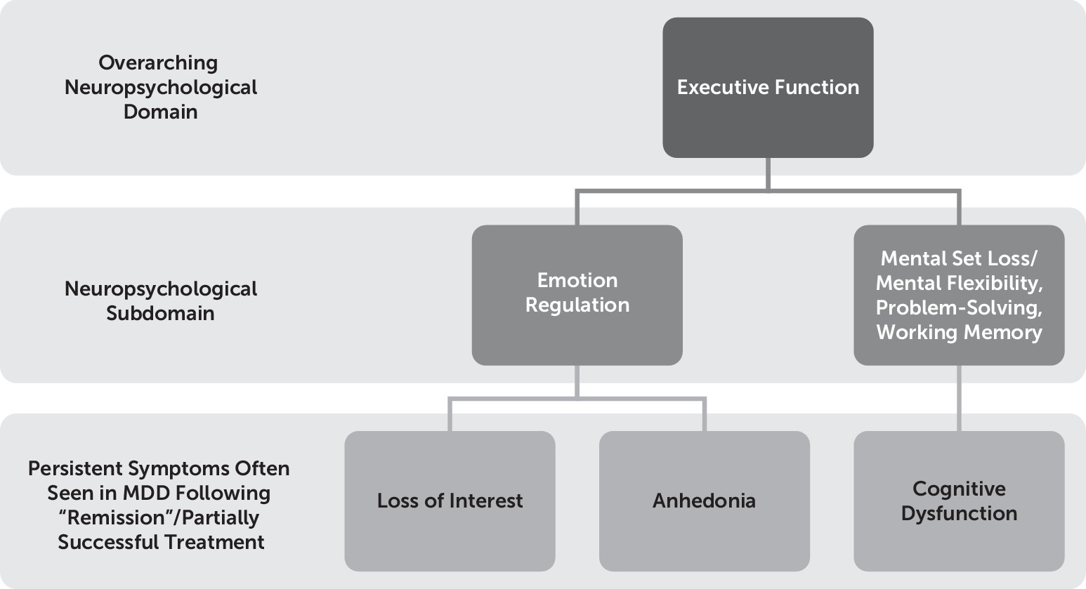
The DSM-51 identifies cognitive dysfunction (i.e., the “diminished ability to think or concentrate, or indecisiveness, nearly every day” for 2 weeks) and anhedonia (i.e., the reduced ability to experience positive emotion) as criteria for major depressive episode. We consider these symptoms together in the present study because we hypothesize that cognitive dysfunction, particularly executive dysfunction (an umbrella term used to describe a set of skills—including working memory, abstract reasoning, multitasking, problem solving, and planning—that enable us to learn, make choices, be creative, solve novel problems, and carry out everyday tasks), is related to one’s inability to experience pleasure, given their shared persistent nature following resolution of other symptoms of depression, and putatively shared neuroanatomical correlates owing to an underlying mechanistic relationship between positive emotionality and executive function. Specifically, the following two aspects of executive function are hypothesized here to be subserved by the same prefrontal region, namely, the ventrolateral prefrontal cortex (VLPC): 1) mental flexibility/mental set shifting and inhibition (constructs that contribute to the cognitive dysfunction typically observed in major depressive disorder [MDD])2 and 2) emotion regulation processes (which typically are associated with negative affect and anhedonia)3 (Figure 1). Of course, this is not the only region that plays a role in these processes, but it seems to be a key node in both of these processes. Therefore, we hypothesized that endogenous mu-opioid system function in the VLPC may be an important integrative site for these processes, in part given its histological and functional connectivity with the orbitofrontal prefrontal cortex on the one hand, a region known to be key for the subjective experience of pleasure4 due to its classification as a “hedonic hotspot” and an area dense in mu-opioid receptors relative to some other prefrontal regions, and its dual and distinct role in slightly “colder” executive function processes, such as response inhibition and mental flexibility,2,5 on the other hand (owing to its collaborative functions with other lateral prefrontal regions such as the dorsolateral prefrontal cortex [DLPFC]). First, the construct of cognitive dysfunction and anhedonia are described separately, and then they are integrated.
FIGURE 1. Hypothesized Relationship Between Anhedonia and Cognitive Dysfunction in Major Depressive Disorder (MDD)

Cognitive Dysfunction in MDD

Individuals with MDD commonly complain of cognitive dysfunction.6 Overall, the most replicable cognitive deficits in MDD patients are seen in the domain of executive function,7 even when full remission is achieved and correction for residual mood symptoms is made. This suggests that researchers seeking to uncover abnormalities in the neural systems underlying cognitive deficits in affective disorders such as MDD should particularly focus on executive function.

Anhedonia in MDD

Anhedonia is a key feature of MDD.810 The lifetime prevalence of anhedonia is 5.2%,11 and recent reports estimate that approximately 37% of individuals diagnosed with MDD experience clinically significant anhedonia.12 Significant focus has been placed on the symptom of sadness or depressed mood, and certainly the work done to date investigating the neurobiological basis of this symptom in MDD has been fruitful, as all antidepressants on the market tend to reduce the experience of negative emotions. However, anhedonia has only recently begun to be vigorously investigated. For example, the DSM-5 states that individuals meeting criteria for anhedonia may report feeling “less interested in hobbies, ‘not caring anymore,’ or not feeling any enjoyment in activities that were previously considered pleasurable,” and “family members often notice social withdrawal or neglect of pleasurable avocations” 1 (p. 163). This definition is very broad and does not discriminate between a decrease in motivation and a reduction in experienced pleasure, which have been found to be separable phenomena, with differing neurophysiological and neurotransmitter correlates13,14. “Consummatory” pleasure (pleasure derived during a pleasurable task) is thought to be mediated by opioid neurotransmission, whereas “anticipatory” pleasure (pleasure derived before the onset of a pleasurable task) is thought to be mediated by dopamine neurotransmission. Thus, there is significant heterogeneity at the level of symptom definition,15 which can cause problems when trying to devise treatments for specific symptoms. Certain treatments may affect certain aspects of a symptom (e.g., “anticipatory” anhedonia) and not others (e.g., “consummatory” anhedonia). Current treatments do not adequately address consummatory anhedonia in particular,16,17 and thus this form of anhedonia should be a target of fervent research, as medications do seem to target anticipatory anhedonia to some degree via dopaminergic and noradrenergic mechanisms.16
As we have alluded to, opioid functioning may play a role in mediating hedonic responses in humans, and top-down lateral prefrontal cortex activity may play a particularly important modulating role on opioid-rich subcortical reward circuitry (e.g., the amygdala and nucleus accumbens). To date, most of the work on the role of opioids in hedonic reactivity has come from animal studies. However, addiction research has lent itself to the study of the relationship between opioid neurotransmission and mood in humans. It is well known, for example, that substances that bind to mu-opioid receptors, such as morphine and heroin, produce feelings of euphoria and also reduce the conscious experience of pain. Furthermore, animal research has also provided us with information regarding sensory pleasure; for example, opioid release and pleasure related to sweet tastes is a well-documented phenomenon.14 Prior work has also illustrated the role of the opioid system in top-down modulation of the placebo pain response,18 which arguably has implications for the experience of positive emotions given that pain and pleasure may be opposite extremes on a continuum. Specifically, Zubieta et al.18 demonstrated that increases in endogenous opioid activity occurring in the DLPFC depended on participants’ expectation of analgesic effects. This suggests that lateral prefrontal cortex activity may play a particularly important role in aspects of cognitively mediated analgesia responses. By extension, analgesia is a construct that is closely related to pleasure and reward, as the absence of pain is generally thought to be necessary, but not sufficient, for the experience of pleasure/positive affect. In this manner, extending the findings of Zubieta et al.18 by studying the relationship between lateral prefrontal regions and the putative top-down regulation of emotional experience related to analgesia/pleasure in more detail seems important for further delineation of the function of the opioid system in humans.
Overall, it was hypothesized that those same brain regions that mediate the persistence of certain cognitive deficits (i.e., executive dysfunction) in MDD during and following treatment likely also mediate the persistence of affective deficits (i.e., anhedonia) in MDD, despite overall reduction in depression severity following treatment.
In the present study, we aimed to relate opioid functioning in the prefrontal cortex to trait positive emotion in general, extending our understanding of the opioid system’s involvement in analgesia/positive emotion at the human level in a sample of depressed patients, a group of people known to show deficient positive affect and to be at increased risk for pain syndromes and the deleterious effects of stress. Overall, the present project was designed to assess the role of endogenous mu-opioids in higher-order pleasure (i.e., top-down), such as those forms of pleasure involved in our conscious experience of feelings of well-being versus anhedonia.
Regarding the brain region of interest, prior research has strongly implicated the lateral prefrontal cortex in the pathophysiology of depression.19,20 Specifically, dorsolateral and ventrolateral prefrontal regions have been implicated in eudemonia (i.e., well-being)10 and anhedonia,3 respectively, particularly when attempting to use cognitive means to up-regulate or down-regulate positive affect, respectively. Thus, the DLPFC and ventrolateral prefrontal cortex (VLPFC) are promising regions to study in relation to cognition and positive affect. Specifically, given prior work3 suggesting a role of the VLPFC in positive affect and cognition (particularly the regulation of positive emotion3), it seemed reasonable to focus on determining whether this region, and its likely interactions with subcortical structures such as the nucleus accumbens (a key brain region involved in “liking”), is a valid substrate for conceptualizing the co-occurrence of symptoms of anhedonia and executive dysfunction in humans.

Integrating the Study of Cognitive Dysfunction and Anhedonia in MDD

In the present work, we aimed to specifically investigate the shared neurobiological correlates of executive dysfunction and positive emotionality deficits/anhedonia in a sample of MDD patients, with a particular focus on elucidating underlying neurotransmitter patterns (i.e., opioid functioning) in selected brain regions thought to be involved in both executive function and anhedonia (e.g., VLPFC3). Although the relationship between dopamine and the DLPFC and 1) learning and 2) motivation/reward is well established, the relation between cognitive functioning and the experience of pleasure and opioid function is not well mapped out in humans, though there is a very well described animal model of opioid involvement in hedonic capacity.4,13,14 Therefore, we hoped to extend the well-mapped animal model of pleasure capacity and opioid functioning and specifically investigate the role of opioid neurotransmission in the VLPFC in humans and relate that to anhedonia and executive functioning in a sample of MDD patients. We had two hypotheses. First, if executive function and hedonic capacity are related constructs and are mediated by similar brain regions, we expected to observe a relationship between cognitive test scores of executive function and behavioral ratings of anhedonia/positive emotionality measured using the NEO Personality Inventory21 “positive emotions” facet. Second, it was hypothesized that mu-opioid binding (quantified by Positron Emission Tomography-PET) in the VLPFC would mediate the relationship between executive function difficulties and anhedonia/positive emotionality deficits in MDD.

Methods

Sample

Thirty-nine volunteers with MDD were recruited for the present study. Volunteers were characterized using the Structured Clinical Interview for DSM-IV Axis I Disorders, a neuropsychological test battery, and the NEO Personality Inventory-3, a personality questionnaire. The mean age of participants was 33.4 years (SD=13.22), with a mean educational level of 14.97 years (SD=1.99), and mean Hamilton Depression Rating Scale (HAM-D) score of 20.80 (SD=4.51). There were 25 women who participated in the study.
Only 26 participants had complete neuropsychological data. The mean age of these 26 participants was 35.64 years (SD=13.15), with a mean educational level of 15.20 years (SD=2.20), and mean HAM-D score of 21.36 (SD=4.65).
Symptoms of depression were assessed biweekly before, during, and after antidepressant treatment for 10 weeks. Each volunteer completed a baseline positron emission tomography (PET) session with [11C]carfentanil, a mu-opioid receptor selective radiotracer, as well as functional MRI (fMRI).

Wisconsin Card Sorting Test (WCST)

The WCST is a neuropsychological measure that assesses problem-solving ability and several other facets of executive function.22 During the task, the participant is presented with a deck of cards and is asked to match each card to one of four key cards. The task calls for the patient to engage in new learning, plan ahead, make hypotheses (and update/change them as necessary), separate critical from irrelevant information, and benefit from trial-and-error. Prior research indicates that administration of an acute mu-opioid receptor agonist (in humans) does not hinder performance on the WCST,22 and there are studies suggesting that executive function may be enhanced by opioid functioning.2325 However, performance on the WCST is affected by psychiatric status. For example, Grant et al.26 found that performance on this measure was impaired in a young ambulatory depressed patient population even when other measures of cognition were normal. Furthermore, Martin et al.27 demonstrated that individuals with MDD or dysthymia perseverated more on the WCST and had more mental set lapses compared with a healthy control group matched on IQ. Therefore, we included this measure in our analyses given its documented association with MDD, as well as the necessary recruitment of executive functioning to complete the task.

NEO Personality Inventory-3

The NEO Personality Inventory-328 is a 240-item questionnaire that assesses the Big Five personality traits (extraversion, neuroticism, openness, conscientiousness, and agreeableness). Each personality trait (e.g., extraversion) is subdivided into six facets, with eight items making up each facet. The six extraversion facets include warmth, gregariousness, assertiveness, activity, excitement-seeking, and positive emotions. The facet of “positive emotions” is more related to life satisfaction than the aggregate extraversion,25 with a correlation of about 0.40 in a healthy adult sample. In fact, Schimmack29 suggested that aggregated experiences of happiness alone predicted life satisfaction better than an aggregation of several positive affective states (e.g., happiness, affection, and/or pride). Overall, the extant literature on positive affectivity suggests that when extraversion is decomposed, it is one’s tendency to experience positive emotions that enhances well-being, and this tendency signifies a stable disposition. Thus, the “positive emotions” facet from the NEO Personality Inventory-3 was the primary measure of trait positive emotionality in the present study.

PET Session Overview

A full description of the PET methods and protocols has been published elsewhere in a study by Peciña et al.30 Briefly, scans were acquired with a Siemens HR+ scanner in three-dimensional mode (reconstructed FWHM resolution approximately 5.5 mm in-plane and 5.0 mm axially) with septa retracted and scatter correction. Participants positioning uses the orbito-medial line as reference, and markings are placed on the participant’s scalp using the gantry laser lights. A light forehead restraint was used to minimize intrascan movement. The intravenous line was used for the administration of the radiotracer.
Images were reconstructed using iterative algorithms (brain mode; FORE/OSEM four iterations, 16 subsets; no smoothing) into a 128×128 pixel matrix, with 28.8-cm diameter field of view. Attenuation correction was performed through a 6-minute transmission scan (68Ge source) obtained prior to the PET study, also with iterative reconstruction of the blank/transmission data followed by segmentation of the attenuation image. Small head motions during emission scans were corrected by an automated computer algorithm for each participant before analysis, and the images were coregistered to each other with the same software.31 Time points were then decay-corrected during reconstruction of the PET data.
A total of 10–15 mCi was administered in the scan, with a maximum injected mass of <0.03 µg/kg to ensure that the compound was administered in tracer quantities, without significant receptor occupancy. Fifty percent of the [11C]carfentanil dose was administered as a bolus, and the remainder as a continuous infusion. Parametric binding potential (BP=Bmax/Kd; receptor concentration over affinity) data were calculated on a voxel-by-voxel basis using a modified Logan graphical analysis3234 with the occipital cortex as the reference region (a region devoid of mu-opioid receptors).

Statistical Analysis

Hypothesis-driven volume of interest analyses were used to evaluate selected brain regions previously implicated in pleasure and cognition in MDD using a mask inclusive of bilateral VLPFC, basal ganglia, and amygdala. For this approach, a general linear model was used (Pearson correlation for continuous variables) with a corrected significance threshold of p<0.05. The binding potential values for percentage of change calculations were extracted from image data by averaging the values of voxels contained in areas where significant differences were obtained in the voxel-by-voxel analysis down to a threshold of p<0.05. In other words, significant differences were detected using a p<0.05 family-wise error-corrected threshold.35 Significant regional data were extracted using MarsBaR36 for quantification of regional changes in binding potential and graphing and determination of correlation coefficients (Pearson/Spearman correlations at p<0.05). Using SPSS statistical software (version 21), these values were then used to plot the data and perform correlation and regression analyses. A whole-brain analysis was also run, using a general linear model with a corrected significance threshold of p<0.05.
When region of interest analyses are used in PET, our interest is in testing whether specific a priori regions show significant effects. We are not “cherry-picking” regions and then claiming these represent the effects for the whole brain. What we did is test a subset of spots in the brain (i.e., “voxels”) to see whether average mu-opioid binding potential in these regions from one subject to the next reliably related to differences in positive emotionality and executive functioning. This procedure is entirely valid. It is important to understand that our goal is not to find the average effect in the entire brain, but to find out whether activity in these particular a priori regions behave as predicted. The procedure we used is exactly the right one to do in order to answer that particular question. Although spurious correlations can occur, as in any behavioral research, they will be rare in typical neuroimaging studies involving sample sizes greater than 18.37
A mediation analysis was conducted with these data. The central idea was that the effects of an independent variable (here, positive emotionality) on executive function (behavior) would be intervened upon by various processes internal to the organism (i.e., mu-opioid functioning in the VLPFC). In other words, mediators help “explain how external physical events take on internal psychological significance”38 (p. 1176).
The most common mediator model is depicted in Figure 2.
FIGURE 2. Mediator Model
In this model, variation in the level of the independent variable significantly accounts for variations in the mediator (i.e., path “a”), variations in the mediator significantly account for variations in the dependent variable (i.e., path “b”), and when paths “a” and “b” are controlled, a previously significant relation between the independent and dependent variable is no longer significant, with the strongest demonstration of mediation occurring when path “c” is zero.

Results

Participants with complete neuropsychological data (N=26) versus participants with incomplete neuropsychological data did not differ in terms of age, education, or depression score (all p values >0.10)
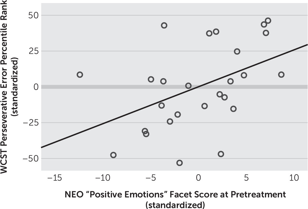
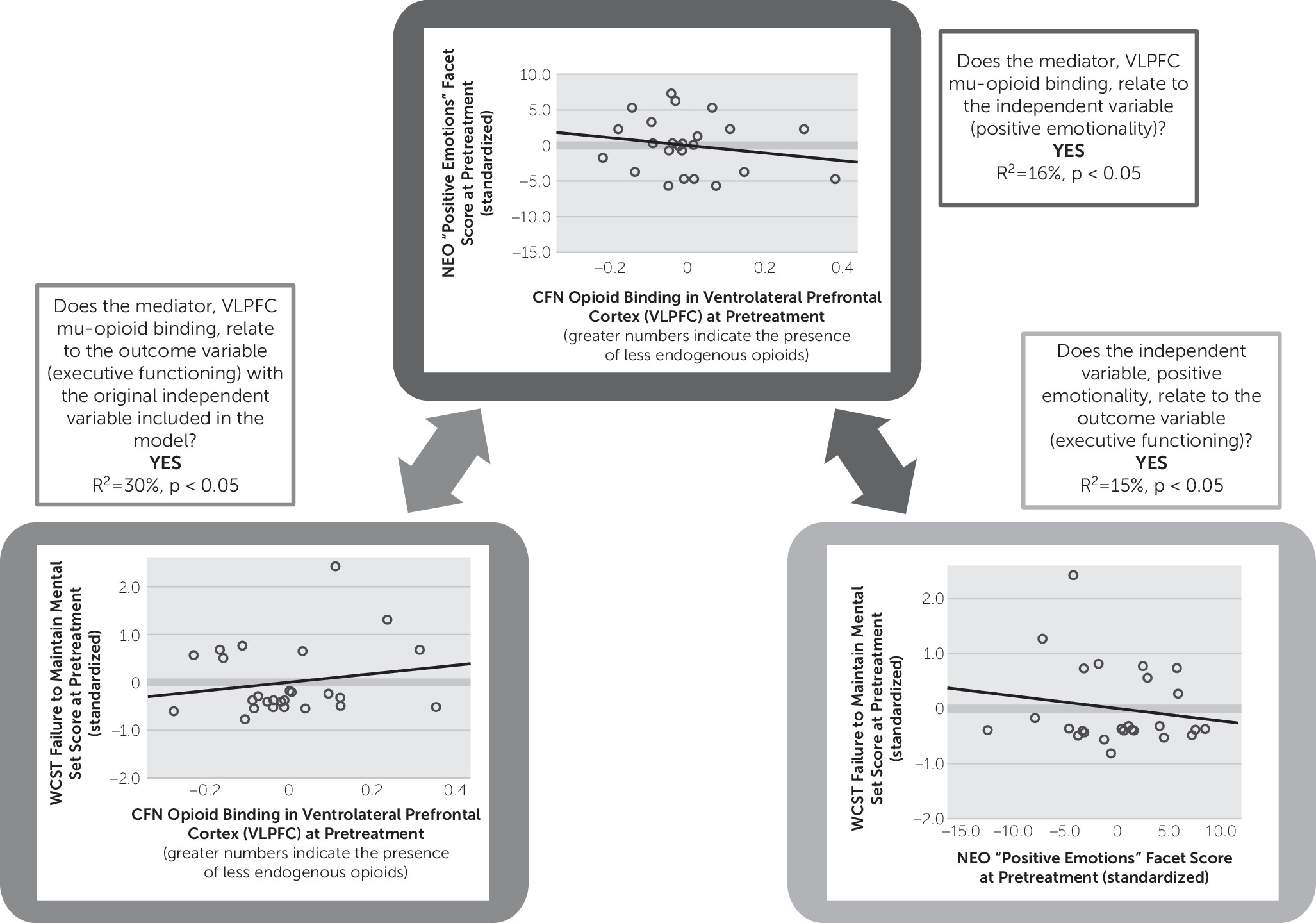
Next, we examined correlational relationships between self-reported positive emotionality, as measured by the NEO Personality Inventory and executive function measured by WCST at baseline (i.e., pretreatment). All aspects of performance on the WCST, a measure of executive functioning, related positively and significantly to positive emotionality as measured by the NEO Personality Inventory. Specifically, fewer total errors (represented by higher percentile scores, r=0.409, p<0.05), fewer perseverative responses (represented by higher percentile scores, r=0.442, p<0.05) (Figure 3), total number of categories deduced (r=0.421, p<0.05), and fewer number of failures to maintain mental set (r=–0.446, p<0.05) (Figure 4) were indicators of the greater the individual’s baseline tonic positive emotionality.
FIGURE 3. Greater Positive Emotionality Predicting Fewer Perseverative Responses on the Wisconsin Card Sorting Test (WCST)a
a The scatterplot shows results with the Hamilton Depression Rating Scale for Depression total score entered as a covariate (β=2.59, R2=21%, p=0.02). NEO=NEO Personality Inventory.
FIGURE 4. Mediation Analysisa
a CFN=[11C]carfentanil; NEO=NEO Personality Inventory; WCST=Wisconsin Card Sorting Test.
Mu-opioid receptor binding potential in the VLPFC was negatively related to positive emotionality as measured by the NEO Personality Inventory (R2=15%, p<0.05) (Figure 4) and executive function as measured by the WCST (R2=16%, p<0.05) (Figure 4), suggesting that either lower receptor concentration or, alternatively, higher levels of endogenous opioid activating receptor sites in the VLPFC region are related to greater trait positive emotionality and sharper executive functioning in this sample of MDD patients.
A mediation analysis based on these data collected from the 26 MDD patients with complete neuropsychological data examining the relationship between positive emotion, executive function, and mu-opioid receptor binding potential in the VLPFC showed that when entered into the same model, VLPFC mu-opioid receptor binding potential remained significant (p=0.049), and the NEO “positive emotions” facet became a marginal predictor (p=0.065) of executive function (i.e., WCST mental set loss percentile rank); the overall model was significant (R2=30%, p=0.015). This analysis suggests that VLPFC strongly mediates the relationship between trait positive emotionality and mental set loss/executive function.
We also ran analyses with the HAM-D total score as a covariate in our model to rule out depression severity as an explanatory factor for our mediation results. When the HAM-D total score was entered into the mediation model, the NEO positive emotions score remained a marginal positive predictor of the executive function score (p=0.07), and the overall model remained significant (R2=31%, p=0.04). The VLPFC mu-opioid receptor binding remained a negative and significant predictor of executive function even when the HAM-D score was entered into the model (β=–7.0, p=0.05), suggesting that VLPFC mu-opioid functioning is a strong mediator of the effect between positive emotionality and executive functioning, even when accounting for depression severity.
The VLPFC mu-opioid receptor binding also marginally predicted WCST categories completed (percentile rank) (r=–0.27, p=0.06) and positively and significantly predicted raw errors on the WCST (r=0.28, p<0.05).
No significant relationships were obtained between basal ganglia or amygdala activation and neuropsychological measures.

Discussion

Better performance on the WCST, a measure that taps general executive functioning, related to greater trait positive emotion (i.e., hedonic tone) as measured by the NEO “positive emotions” facet. Specifically, reduced mental set loss, fewer perseverative responses, lower total error rate, and increased total problem solving (i.e., categories completed) related to greater trait positive emotion. The relationship between positive emotionality and mental set loss errors was found to be specifically mediated by mu-opioid receptor availability in the VLPFC. These are the first data, to our knowledge, to directly suggest that the endogenous mu-opioid system in the prefrontal cortex relates directly to positive emotionality and executive functioning in humans with MDD. Furthermore, these findings replicate previous work illustrating a relationship between positive emotionality and executive functioning35 and add to this literature by providing a plausible neurobiological substrate for this association.
The data acquired in the VLPFC is consistent with its role in affective processes (e.g., emotional experience and emotion regulation) and cognition (e.g., response inhibition, mental flexibility, and complex problem solving). In emotion regulation studies, reduced VLPFC activity relates to better down-regulation of negative emotion39,40 and resilience against down-regulation of positive emotion.3 Our results indicate that there is meaningful endogenous mu-opioid system influence in the VLPFC relating to both executive function and positive emotionality. Ultimately, these results extend the work of Kent Berridge and colleagues4,13,14 on “liking” by suggesting that mu-opioid functioning in the VLPFC relates to complex, conscious positive emotionality at the human level.
Why is there a link between executive functioning and positive emotionality? Or more specifically, what is the real-world significance of the “mental set loss” score on the WCST, and what is its relevance to positive emotionality, and why is this aspect of the WCST mediated by mu-opioid functioning? There is debate in the literature related to whether mental set loss reflects distractibility or mental flexibility (the ability to change ideas). For example, Figueroa and Youmans41 suggest that the mental set loss score from the WCST is more related to distractibility. They propose that this distractibility may relate to loss of focus on the task because of boredom, mind wandering, or an inability to maintain task-relevant goals. Each of these possibilities is relevant to positive emotionality. A tendency to lose focus or become bored could negatively affect a person’s ability to adequately take in hedonic information when it is presented to them, rendering it impossible to experience subsequent positive emotion. Similarly, mind wandering may also prevent a person from being able to fully relish in a positive experience. Furthermore, an inability to maintain task-relevant goals, or the inability to sustain positive emotion once it is initially generated, may represent another facet of trait-like low positive emotionality/anhedonia.8
Regarding the question of why mu-opioid functioning in the VLPFC would mediate the observed effect, it is important to recognize the unique anatomic and functional characteristics of the VLPFC. The VLPFC is part of the paleocortical aspect of the prefrontal cortex emerging from the caudal orbitofrontal cortex. It is intimately connected with limbic nuclei involved in emotional processing.42 As such, the VLPFC is intimately connected with more primitive limbic nuclei involved in emotional processing42 and processes information about basic drives and rewards that inform and direct high-level decision making. Thus, when combined with known conceptions of mu-opioid functioning, namely, the role of mu-opioids in modulating pleasure,4,14,15 it becomes clear why neurotransmitter function in this region may be a central player in translating/building basic pleasure kernels into higher-order positive emotional states and action tendencies (i.e., via prefrontal decision making and problem-solving processes).
Overall, our results provide evidence that executive function and positive emotionality are linked to mu-opioid functioning in the VLPFC and suggest that deficits in executive functioning may have real downstream repercussions for an individual’s ability to experience positive emotions.
In conclusion, we suggest a circumscribed effect, whereby mu-opioid functioning in the VLPFC in particular mediates the relationship between executive function and positive emotionality. The VLPFC has already been associated with executive function, particularly response inhibition2 and maintaining mental set.5 Our prior fMRI work with a clinically depressed adult sample has shown this region to also be involved in the symptom of anhedonia.3 Here, we have combined these two lines of evidence by presenting data to suggest that these constructs (i.e., executive function and low positive emotionality) are related via mu-opioid functioning in the VLPFC. Overall, greater mu-opioid endogenous tone (lower binding potential) in the VLPFC mediates the relationship between greater positive emotionality and better executive functioning.
Although the opioid theory of depression that was most popular up through the 1950s has been replaced by the monoamine hypothesis of depression,43 the lack of widespread success of monoamine pharmacotherapy for “treatment-resistant depression” in particular with first-line antidepressants such as selective serotonin reuptake inhibitors (SSRIs) has prompted the resurgence of consideration of a more calibrated opioid modulation therapy for MDD. We know that reduced enthusiasm for use of mu-opioid agonists in the treatment of MDD stems from their abuse/addiction potential, but here we suggest that failure in the past of these drugs to be first-line antidepressant treatments may have resulted from the fact that opioid agonists developed in the past were not designed to target specific prefrontal versus subcortical brain regions. Similar to the recent research uncovering the mechanisms of cognitive enhancement of stimulant medication used in the treatment of attention deficit hyperactivity disorder,44 our data suggest that a general acting mu-opioid agonist would be less effective at relieving anhedonic depression because it does not specifically target the VLPFC. Thus, we suggest that MDD characterized by low positive emotion and executive dysfunction is associated specifically with decreased VLPFC mu-opioid tone, and treatments that specifically target mu-opioid functioning in this particular prefrontal region may be more efficacious for this subtype of MDD.
Two recent controlled trials conducted by Fava and colleagues45,46 utilizing a mu-opioid agonist, buprenorphine, in conjunction with a standard SSRI or selective norepinephrine reuptake inhibitor in a treatment-resistant sample of MDD patients provide supportive evidence. These were treatment studies investigating the efficacy of simultaneous administration of a mu-opioid agonist (buprenorphine) and an antagonist (i.e., to curb the rapid mood elevation associated with the “high” of the drug opposed to its antidepressant effects), with significant reductions in depression scores observed with 1 week45 and 446 weeks of treatment, respectively. These results support the idea that mu-opioids do in fact play a role in modulating depression symptomatology and can effectively be targeted with careful therapeutic approaches.
Limitations of the present study include lack of inclusion of a control group. Furthermore, we did not address treatment effects in this study, though future analyses will address this issue. Additionally, although three brain regions were investigated, only one region showed an effect. Further work is needed to determine whether treatment with antidepressant medication may indirectly regulate endogenous opioid functioning in the VLPFC and other areas of the brain such as the nucleus accumbens, as no studies, to our knowledge, have investigated the potentially procognitive effects of treatment with antidepressant medication.
Overall, this study provides the research community with a better understanding of how mu-opioid functioning in the human brain relates to higher-order cognitive-emotive functioning.
References

1.
American Psychiatric Association: Diagnostic and Statistical Manual of Mental Disorders, 5th ed. Washington, DC, American Psychiatric Publishing, 2013
2.
Aron AR, Robbins TW, Poldrack RA: Inhibition and the right inferior frontal cortex: one decade on. Trends Cogn Sci 2014; 18:177–185
3.
Light SN, Heller AS, Johnstone T, et al: Reduced right ventrolateral prefrontal cortex activity while inhibiting positive affect is associated with improvement in hedonic capacity after 8 weeks of antidepressant treatment in major depressive disorder. Biol Psychiatry 2011; 70:962–968
4.
Kringelbach ML, Berridge KC: The joyful mind. Sci Am 2012; 307:40–45
5.
Stuss DT, Levine B: Adult clinical neuropsychology: lessons from studies of the frontal lobes. Annu Rev Psychol 2002; 53:401–433
6.
Jaeger J, Berns S, Uzelac S, et al: Neurocognitive deficits and disability in major depressive disorder. Psychiatry Res 2006; 145:39–48
7.
Snyder HR: Major depressive disorder is associated with broad impairments on neuropsychological measures of executive function: a meta-analysis and review. Psychol Bull 2013; 139:81–132
8.
Heller AS, Johnstone T, Shackman AJ, et al: Reduced capacity to sustain positive emotion in major depression reflects diminished maintenance of fronto-striatal brain activation. Proc Natl Acad Sci USA 2009; 106:22445–22450
9.
Heller AS, Johnstone T, Light SN, et al: Relationships between changes in sustained fronto-striatal connectivity and positive affect in major depression resulting from antidepressant treatment. Am J Psychiatry 2013; 170:197–206
10.
Heller AS, van Reekum CM, Schaefer SM, et al: Sustained striatal activity predicts eudaimonic well-being and cortisol output. Psychol Sci 2013; 24:2191–2200
11.
First MB, Williams JBW, Gibbon M, et al: Structured Clinical Interview for DSM-III-R modified for the DSM-IV Dysthymia/Major Depression Field Trial. New York, State Psychiatric Institute, 1990
12.
Pelizza L, Ferrari A: Anhedonia in schizophrenia and major depression: state or trait? Ann Gen Psychiatry 2009; 8:22
13.
Kringelbach ML, Berridge KC: The joyful mind. Sci Am 2012; 307:40–45
14.
Castro DC, Berridge KC: Opioid hedonic hotspot in nucleus accumbens shell: mu, delta, and kappa maps for enhancement of sweetness “liking” and “wanting”. J Neurosci 2014; 34:4239–4250
15.
Ho N, Sommers M: Anhedonia: a concept analysis. Arch Psychiatr Nurs 2013; 27:121–129
16.
Shelton RC, Tomarken AJ: Can recovery from depression be achieved? Psychiatr Serv 2001; 52:1469–1478
17.
Nutt D, Demyttenaere K, Janka Z, et al: The other face of depression, reduced positive affect: the role of catecholamines in causation and cure. J Psychopharmacol 2007; 21:461–471
18.
Zubieta J-K, Bueller JA, Jackson LR, et al: Placebo effects mediated by endogenous opioid activity on mu-opioid receptors. J Neurosci 2005; 25:7754–7762
19.
Davidson RJ: What does the prefrontal cortex “do” in affect: perspectives on frontal EEG asymmetry research. Biol Psychol 2004; 67:219–233
20.
Taylor SF, Liberzon I: Neural correlates of emotion regulation in psychopathology. Trends Cogn Sci 2007; 11:413–418
21.
McCrae RR, Costa PT (ed): NEO Inventories. Lutz, Fla, Psychological Assessment Resources, Inc, 2010
22.
Heaton RK, Chelune GJ, Talley JL, et al: (ed): Wisconsin Card Sorting Test Manual. Lutz, Fla, Psychological Assessment Resources, Inc, 1993
23.
Primac DW, Mirsky AF, Rosvold HE: Effects of centrally acting drugs on two tests of brain damage. AMA Arch Neurol Psychiatry 1957; 77:328–332
24.
de Graaf J, van Lingen RA, Valkenburg AJ, et al: Does neonatal morphine use affect neuropsychological outcomes at 8 to 9 years of age? Pain 2013; 154:449–458
25.
O’Neill WM, Hanks GW, Simpson P, et al: The cognitive and psychomotor effects of morphine in healthy subjects: a randomized controlled trial of repeated (four) oral doses of dextropropoxyphene, morphine, lorazepam and placebo. Pain 2000; 85:209–215
26.
Grant MM, Thase ME, Sweeney JA: Cognitive disturbance in outpatient depressed younger adults: evidence of modest impairment. Biol Psychiatry 2001; 50:35–43
27.
Martin DJ, Oren Z, Boone K: Major depressives’ and dysthmics’ performance on the Wisconsin Card Sorting Test. J Clin Psychol 1991; 47:684–690
28.
Schimmack U, Oishi S, Furr RM, et al: Personality and life satisfaction: a facet-level analysis. Pers Soc Psychol Bull 2004; 30:1062–1075
29.
Schimmack U: Affect measurement in experience sampling research. J Happiness Stud 2003; 4:79–106
30.
Peciña M, Azhar H, Love TM, et al: Personality trait predictors of placebo analgesia and neurobiological correlates. Neuropsychopharmacology 2013; 38:639–646
31.
Minoshima S, Koeppe RA, Mintun MA, et al: Automated detection of the intercommissural line for stereotactic localization of functional brain images. J Nucl Med 1993; 34:322–329
32.
Titeler M, Lyon RA, Kuhar MJ, et al: Mu opiate receptors are selectively labelled by [3H]carfentanil in human and rat brain. Eur J Pharmacol 1989; 167:221–228
33.
Dannals RF, Ravert HT, Frost JJ, et al: Radiosynthesis of an opiate receptor binding radiotracer: [11C]carfentanil. Int J Appl Radiat Isot 1985; 36:303–306
34.
Jewett DM: A simple synthesis of [11C]carfentanil using an extraction disk instead of HPLC. Nucl Med Biol 2001; 28:733–734
35.
Friston KJ, Worsley KJ, Frackowiak RS, et al: Assessing the significance of focal activations using their spatial extent. Hum Brain Mapp 1994; 1:210–220
36.
Brett M, Anton JL, Valabregue R, et al: Region of interest analysis using an SPM toolbox. Presented at the 8th International Conference on Functional Mapping of the Human Brain, June 2–6, 2002, Sendai, Japan, 2002
37.
Lieberman MD, Berkman ET, Wager TD: Correlations in social neuroscience aren’t voodoo. Perspect Psychol Sci 2009; 4:299–307
38.
Baron RM, Kenny DA: The moderator-mediator variable distinction in social psychological research: conceptual, strategic, and statistical considerations. J Pers Soc Psychol 1986; 51:1173–1182
39.
Zubieta J-K, Ketter TA, Bueller JA, et al: Regulation of human affective responses by anterior cingulate and limbic mu-opioid neurotransmission. Arch Gen Psychiatry 2003; 60:1145–1153
40.
Wager TD, Davidson ML, Hughes BL, et al: Prefrontal-subcortical pathways mediating successful emotion regulation. Neuron 2008; 59:1037–1050
41.
Figueroa IJ, Youmans RJ: Failure to maintain set: a measure of distractibility or cognitive flexibility? Proc Hum Factors Ergon Soc Annu Meet 2013; 57:828–832
42.
Nauta WJ: The problem of the frontal lobe: a reinterpretation. J Psychiatr Res 1971; 8:167–187
43.
Tenore PL: Psychotherapeutic benefits of opioid agonist therapy. J Addict Dis 2008; 27:49–65
44.
Spencer RC, Devilbiss DM, Berridge CW: The cognition-enhancing effects of psychostimulants involve direct action in the prefrontal cortex. Biol Psychiatry 2015; 77:940–950
45.
Ehrich E, Turncliff R, Du Y, et al: Evaluation of opioid modulation in major depressive disorder. Neuropsychopharmacology 2015; 40:1448–1455
46.
Fava M, Memisoglu A, Thase ME, et al: Opioid modulation with buprenorphine/samidorphan as adjunctive treatment for inadequate response to antidepressants: A randomized double-blind placebo controlled trial. Am J Psychiatry 2016; 173:499–508

Information & Authors

Information

Published In

Go to The Journal of Neuropsychiatry and Clinical Neurosciences
The Journal of Neuropsychiatry and Clinical Neurosciences
Pages: 357 - 364
PubMed: 28412878

History

Received: 20 September 2016
Revision received: 1 December 2016
Accepted: 8 December 2016
Published online: 17 April 2017
Published in print: Fall 2017

Keywords

  1. PET
  2. Major Depressive Disorder
  3. Positive Emotionality
  4. Executive Function
  5. Anhedonia

Authors

Details

Sharee N. Light, Ph.D. [email protected]
From the Department of Psychology, Georgia State University, Atlanta (SNL); the Department of Psychiatry, Neuropsychology Section, University of Michigan, Ann Arbor, Mich. (LAB); and the Department of Psychiatry University of Utah, Salt Lake City, Utah (J-K Z).
Linas A. Bieliauskas, Ph.D.
From the Department of Psychology, Georgia State University, Atlanta (SNL); the Department of Psychiatry, Neuropsychology Section, University of Michigan, Ann Arbor, Mich. (LAB); and the Department of Psychiatry University of Utah, Salt Lake City, Utah (J-K Z).
Jon-Kar Zubieta, M.D., Ph.D.
From the Department of Psychology, Georgia State University, Atlanta (SNL); the Department of Psychiatry, Neuropsychology Section, University of Michigan, Ann Arbor, Mich. (LAB); and the Department of Psychiatry University of Utah, Salt Lake City, Utah (J-K Z).

Notes

Send correspondence to Dr. Light; e-mail: [email protected]

Competing Interests

The authors report no financial relationships with commercial interests.

Metrics & Citations

Metrics

Citations

Export Citations

If you have the appropriate software installed, you can download article citation data to the citation manager of your choice. Simply select your manager software from the list below and click Download.

For more information or tips please see 'Downloading to a citation manager' in the Help menu.

Format
Citation style
Style
Copy to clipboard

View Options

View options

PDF/EPUB

View PDF/EPUB

Get Access

Login options

Already a subscriber? Access your subscription through your login credentials or your institution for full access to this article.

Personal login Institutional Login Open Athens login
Purchase Options

Purchase this article to access the full text.

PPV Articles - Journal of Neuropsychiatry and Clinical Neurosciences

PPV Articles - Journal of Neuropsychiatry and Clinical Neurosciences

Not a subscriber?

Subscribe Now / Learn More

PsychiatryOnline subscription options offer access to the DSM-5-TR® library, books, journals, CME, and patient resources. This all-in-one virtual library provides psychiatrists and mental health professionals with key resources for diagnosis, treatment, research, and professional development.

Need more help? PsychiatryOnline Customer Service may be reached by emailing [email protected] or by calling 800-368-5777 (in the U.S.) or 703-907-7322 (outside the U.S.).

Media

Figures

Other

Tables

Share

Share

Share article link

Share