Open access
RESEARCH ARTICLES
Published Online: 20 January 2023

Interactions Between Neuropsychiatric Symptoms and Alzheimer's Disease Neuroimaging Biomarkers in Predicting Longitudinal Cognitive Decline

Publication: Psychiatric Research and Clinical Practice

Abstract

Objective

To examine interactions between Neuropsychiatric symptoms (NPS) with Pittsburgh Compound B (PiB) and fluorodeoxyglucose positron emission tomography (FDG‐PET) in predicting cognitive trajectories.

Methods

We conducted a longitudinal study in the setting of the population‐based Mayo Clinic Study of Aging in Olmsted County, MN, involving 1581 cognitively unimpaired (CU) persons aged ≥50 years (median age 71.83 years, 54.0% males, 27.5% APOE ɛ4 carriers). NPS at baseline were assessed using the Neuropsychiatric Inventory Questionnaire (NPI‐Q). Brain glucose hypometabolism was defined as a SUVR ≤ 1.47 (measured by FDG‐PET) in regions typically affected in Alzheimer's disease. Abnormal cortical amyloid deposition was measured using PiB‐PET (SUVR ≥ 1.48). Neuropsychological testing was done approximately every 15 months, and we calculated global and domain‐specific (memory, language, attention, and visuospatial skills) cognitive z‐scores. We ran linear mixed‐effect models to examine the associations and interactions between NPS at baseline and z‐scored PiB‐ and FDG‐PET SUVRs in predicting cognitive z‐scores adjusted for age, sex, education, and previous cognitive testing.

Results

Individuals at the average PiB and without NPS at baseline declined over time on cognitive z‐scores. Those with increased PiB at baseline declined faster (two‐way interaction), and those with increased PiB and NPS declined even faster (three‐way interaction). We observed interactions between time, increased PiB and anxiety or irritability indicating accelerated decline on global z‐scores, and between time, increased PiB and several NPS (e.g., agitation) showing faster domain‐specific decline, especially on the attention domain.

Conclusions

NPS and increased brain amyloid deposition synergistically interact in accelerating global and domain‐specific cognitive decline among CU persons at baseline.

Highlights

In cognitively unimpaired older adults.
Neuropsychiatric symptoms and brain amyloid synergistic interaction.
Lead to accelerated cognitive decline.
Neuropsychiatric symptoms (NPS) are very common in Alzheimer's Disease (AD). Some estimate that up to 97% of AD patients develop NPS at some point in the course of their illness (1). We and others (2, 3) have demonstrated that the prevalence of NPS ranges between 25% in cognitively unimpaired (CU) persons to approximately 50% in persons with mild cognitive impairment (MCI), with the most frequent NPS being depression, irritability, and apathy (3). As expected, the frequency of NPS is even higher in persons with AD with up to 80% exhibiting at least one NPS (2).
We and others have reported that NPS are associated with an increased risk of new onset of MCI (4, 5, 6) or dementia (7, 8, 9, 10, 11) as well as decline in cognitive trajectories (12, 13). NPS in the context of AD spectrum lead to accelerated cognitive and functional decline, profound caregiver distress, early institutionalization, and increased mortality. However, despite the enormous impact of NPS on patients, caregivers, and society at large, the mechanism linking AD biomarkers to NPS and cognitive decline over a longitudinal follow up remains unclear.
Biomarkers can identify persons on the AD spectrum early in the course of the disease and before the onset of clinical signs (14). Beta‐amyloid (Aβ) deposition can be visualized in vivo by amyloid brain imaging using various types of tracers (15). Amyloid imaging is a biomarker of AD, and brain glucose hypometabolism as measured by fluorodeoxyglucose positron emission tomography (FDG‐PET) is a biomarker of neurodegeneration (14).
While we and others have examined anxiety, depression and their interaction with neuroimaging biomarkers in predicting MCI (16) and cognitive decline (17), studies involving a broad spectrum of NPS are lacking.
Therefore, we sought to examine interactions between NPS as measured by the Neuropsychiatric Inventory Questionnaire (NPI‐Q) and neuroimaging biomarkers, that is, amyloid imaging and FDG‐PET in predicting global and domain‐specific cognitive decline in community‐dwelling older adults. The primary question was if any NPS, as measured by NPI‐Q, interacted with amyloid deposition or glucose hypometabolism in predicting cognitive decline. In addition, we examined specific NPS.
We hypothesized that there would be an interaction between NPS and neuroimaging biomarkers in increasing the rate of cognitive decline in community‐dwelling individuals.

METHODS

Study Design and Sample

We conducted a prospective cohort study in the setting of the population‐based Mayo Clinic Study of Aging (MCSA) in Olmsted County, MN, USA. Details of the study procedures have been reported elsewhere (18).
We included 1581 CU participants ≥50 years who underwent baseline NPS assessment and amyloid‐PET and FDG‐PET neuroimaging, with the majority having repeated cognitive testing after approximately every 15 months.
Participants were followed forward in time for a median of 6.2 years to examine interactions between baseline NPS and amyloid‐PET as well as FDG‐PET with longitudinal changes in global and domain specific (memory, attention, language, visuospatial) cognitive z‐scores.
The study was approved by the Mayo Clinic and Olmsted Medical Center institutional review boards, and informed consent for participation was obtained from every participant.

Cognitive Evaluation

MCSA participants underwent face‐to‐face evaluations including risk factor ascertainment (including NPI‐Q) and baseline evaluation (including Clinical Dementia Rating Scale) (19) performed by a nurse or study coordinator; a neurologic evaluation including a neurologic interview, Short Test of Mental Status (20), and neurologic examination performed by behavioral neurologists; and neuropsychological evaluation of four cognitive domains: memory (delayed recall trials from the Auditory Verbal Learning Test (21) and the Wechsler Memory Scale–Revised (22), Logical Memory and Visual Reproduction subtests); language (Boston Naming Test (23) and category fluency); visuospatial (Wechsler Adult Intelligence Scale–Revised (23), Picture Completion and Block Design subtests); and executive function (Trail Making Test Part B (24) and the Wechsler Adult Intelligence Scale–Revised (25), Digit Symbol subtest). All tests were administered by psychometrists and supervised by neuropsychologists. An expert consensus panel of physicians, neuropsychologists, and nurses or study coordinators reviewed the data and determined if a participant was CU, had MCI (based on the revised Mayo Clinic criteria (26) or dementia. In this analysis we included only individuals who were CU; participants with MCI or dementia were excluded for the current analysis at baseline. Classification of CU was based on normative data developed in this community (27, 28, 29, 30).
We further created domain‐specific cognitive z‐scores by z‐scoring the averages of the test‐specific z‐scores, and additionally created a global z‐score by z‐scoring the averages of the domain‐specific z‐scores. The outcome of interest for the linear mixed‐effect model analyses was the longitudinal change in global and domain‐specific (i.e., memory, attention/executive function, language, visuospatial skills) cognitive z‐scores.

Measurement of Neuropsychiatric Symptoms

NPS were measured by using the NPI‐Q (31) which was administered as a structured interview to an informant, usually the spouse. The NPI‐Q is a shorter version of the Neuropsychiatric Inventory (NPI) and is a clinical instrument that is cross‐validated with the standard NPI (31). We considered the NPI‐Q an appropriate screening instrument because it assesses a broad variety of neuropsychiatric symptoms and was also selected by the Uniform Data Set Initiative of the National Institute on Aging (32). The NPI‐Q is designed to obtain information on 12 behaviors (i.e., agitation, delusion, hallucination, depression, anxiety, euphoria, apathy, disinhibition, irritability, aberrant motor behavior, sleep, and eating/appetite). A severity scale has scores ranging from 1 to three points (1 = mild; 2 = moderate; and 3 = severe) and a scale for assessing caregiver distress has scores ranging from 0 to five points (0 = no distress; 1 = minimal distress; 2 = mild distress; 3 = moderate distress; 4 = severe distress; and 5 = extreme distress).

PiB‐PET Acquisition

Amyloid PET imaging was performed using the Pittsburgh Compound B (PiB) tracer. Details on PiB‐PET imaging in the MCSA have been published elsewhere (33, 34). Briefly, PiB scans, consisting of four 5‐min dynamic frames, were acquired 40–60 min after intravenous injection with 292–728 MBq of 11C‐PiB. We used an in‐house, fully automated image processing pipeline to analyze images. Herein, image voxel values were extracted from automatically labeled regions of interest (ROI) propagated from regions defined on each participant's own magnetic resonance imaging (MRI). The prefrontal, orbitofrontal, parietal, temporal, anterior cingulate, and posterior cingulate/precuneus ROI were normalized to the cerebellar gray matter to form a global amyloid PET standardized uptake value ratio (SUVR). We defined abnormal PiB‐PET retention (PiB‐PET+) by an SUVR ≥1.48, which is the current cut‐off used in the MCSA (33, 35). We ran the linear‐mixed effects models with continuous, z‐scored PiB‐PET SUVR.

FDG‐PET Acquisition

FDG‐PET imaging which consisted of four 2‐min dynamic frames, was performed 30 min after injecting 366–399 MBq of 18fluorodeoxyglucose intravenously. Images were analyzed using our in‐house fully automated image processing pipeline (36) in which image voxel values were extracted from automatically labeled cortical ROI (37) After combining the left and right regions from the Atlas, there were 19 ROI and the meta‐region of interest consisted of bilateral angular gyrus, posterior cingulate/precuneus, and inferior temporal cortical regions from both hemispheres and was identified as AD signature ROI (38, 39). SUVR was formed by the ratio of this AD signature ROI and two reference regions, namely the pons and the cerebellar vermis which have preserved glucose metabolism in AD (40). Participants were classified as having glucose hypometabolism, which is a measure of neurodegeneration as defined by NIA‐AA criteria (N+) (14) based on SUVR of ≤1.47 (33). We ran the linear‐mixed effects models with continuous, z‐scored FDG‐PET SUVR. We additionally flipped the sign of the z‐score so that higher values would correspond with a worsening of the biomarker, thereby allowing for a similar interpretation as for the PiB‐PET analysis.

Statistical Analysis

We conducted linear mixed‐effect models with random participant‐specific intercepts and slopes over time to examine the associations and interactions between baseline NPS with brain amyloid deposition (as measured by PiB‐PET) or glucose hypometabolism (as measured by FDG‐PET) in predicting longitudinal change in global and domain‐specific (i.e. attention/executive function, memory, visuospatial, language) cognitive z‐scores over time. We ran the models with continuous, z‐scored PiB‐PET as well as FDG‐PET SUVR (with sign reversed for interpretation purposes for the FDG‐PET SUVR). All models included NPS at baseline, PET imaging at baseline, time in years from baseline and their interactions. All models were adjusted for age at baseline, sex, education, and previous cognitive testing experience (Yes/No). We conducted this analysis separately for the 12 NPS as assessed by the NPI‐Q, and for presence of any NPS as well as NPS severity. For each model, we computed beta coefficients, 95% confidence intervals (CIs), and p‐values.
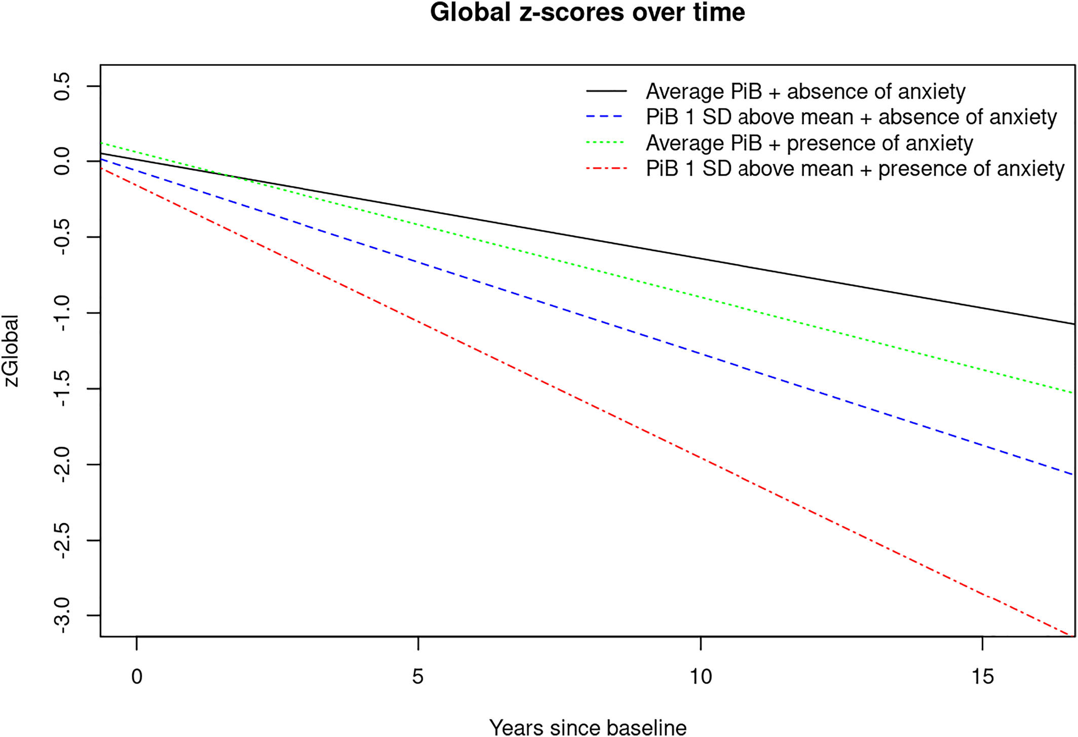
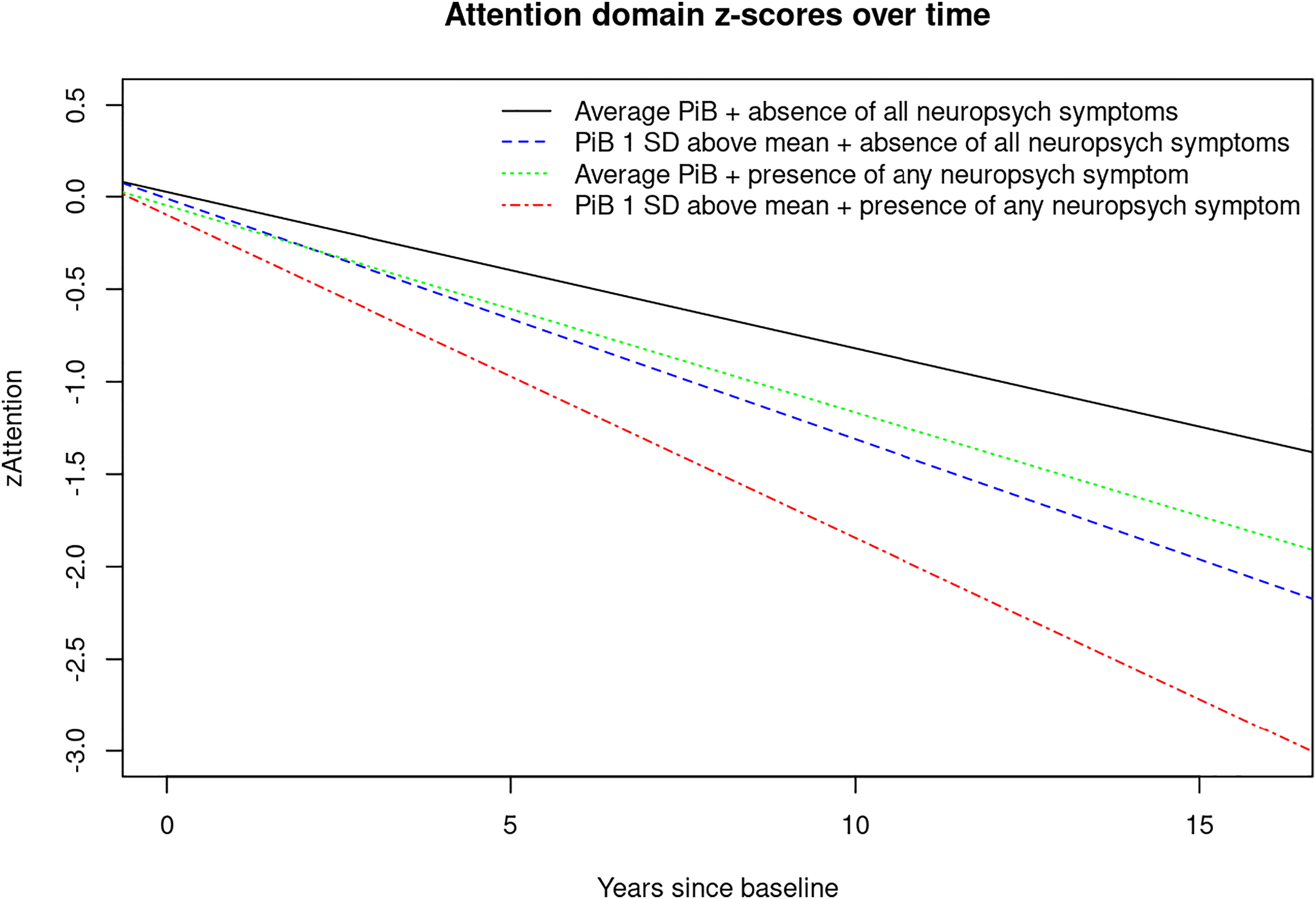
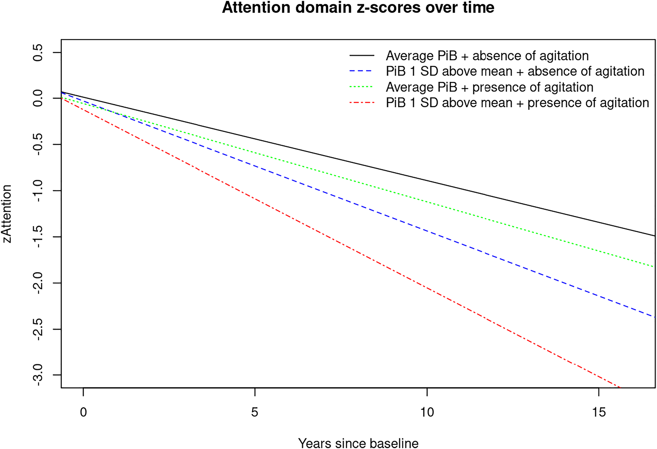
For visual display of data, we plotted the linear mixed effects model for PiB‐PET SUVR (average vs. 1 standard deviation (SD) above the mean) and presence of any NPS (Yes/No) predicting the attention z‐score, as well as presence of anxiety predicting the global cognition z‐score to show the trajectories over time for individuals in these groups (Figures 1, 2, 3). Statistical testing was performed at the conventional two‐tailed alpha level of 0.05. All analyses were performed using SAS System, version 9.4 software (SAS Institute, Cary, NC) and R, version 4.1.2 (R Foundation for Statistical Computing, Vienna, Austria).
image
FIGURE 1. Plot of linear mixed effects model for PiB‐PET‐SUVR (average vs. 1 SD above the mean) and presence of any neuropsychiatric symptoms (Yes/No) predicting attention z‐score. Abbreviations: PiB, brain amyloid deposition as measured by PiB‐PET; SD, standard deviation; zAttention, Attention z‐score.
image
FIGURE 2. Plot of linear mixed effects model for PiB‐PET‐SUVR (average vs. 1 SD above the mean) and presence of agitation (Yes/No) predicting attention z‐score. Abbreviations: PiB, brain amyloid deposition as measured by PiB‐PET; SD, standard deviation; zAttention, Attention z‐score.
image
FIGURE 3. Plot of linear mixed effects model for PiB‐PET‐SUVR (average vs. 1 SD above the mean) and presence of anxiety (Yes/No) predicting global cognition z‐score. Abbreviations: PiB, brain amyloid deposition as measured by PiB‐PET; SD, standard deviation; zGlobal, global cognition z‐score.

RESULTS

Demographics

We included 1581 CU community‐dwelling older adults. One thousand one hundred and twenty‐one (70.9%) were PiB‐PET‐, and 460 (29.1%) were PiB‐PET+; 1139 (72.0%) were N‐, and 442 (28.0%) were N+. The mean (SD) age was 71.1 (9.84) years, 54.0% were males, the mean (SD) education was 14.9 (2.59) years and 27.5% were APOEɛ4 carriers. The complete demographic characteristics are summarized in Table 1.
TABLE 1. Characteristics of the study cohort at baselinea
VariableTotal (N = 1581)
Age in years, mean (SD) [range]71.1 (9.84) [50.20–95.12]
Males854 (54.0)
Education in years, mean (SD)14.9 (2.59)
APOE Ɛ4 carrier425 (27.5)
PiB‐PET SUVR, mean (SD)1.5 (0.33)
PiB‐PET+460 (29.1)
FDG‐PET SUVR, mean (SD)1.6 (0.14)
N+442 (28.0)
Agitation28 (1.8)
Anxiety69 (4.4)
Apathy63 (4.0)
Appetite change47 (3.0)
Nighttime behaviorb72 (5.1)
Delusions1 (0.1)
Depression168 (10.6)
Disinhibition12 (0.8)
Euphoria7 (0.4)
Hallucinations1 (0.1)
Irritability109 (6.9)
Motor behavior13 (0.8)
Any NPS332 (21.0)
Sum of the 12 NPI‐Q severity scores; 0–36, mean (SD)0.5 (1.38)
Converted to MCI/dementia during follow‐up246 (15.6)
1 visit171 (10.8)
2 visits168 (10.6)
3 visits136 (8.6)
4 visits140 (8.9)
5 visits167 (10.6)
6 visits229 (14.5)
7 visits233 (14.7)
8 visits164 (10.4)
9 visits119 (7.5)
10 visits45 (2.8)
11 visits6 (0.4)
12 visits1 (0.1)
13 visits2 (0.1)
a
Data are presented as N (%) unless indicated otherwise. SD, standard deviation; PiB‐PET SUVR, global amyloid PET standardized uptake value ratio; PiB‐PET+, participants with elevated brain amyloid deposition; FDG‐PET SUVR, AD‐signature FDG‐PET standardized uptake value ratio, N+, participants with neurodegeneration, that is, brain glucose hypometabolism as measured by FDG‐PET.
b
Data missing on 164 participants.

Interactions of Amyloid positivity and Glucose Hypometabolism with Neuropsychiatric Symptoms in predicting Cognitive Decline

Interactions between biomarkers, that is, PiB and NPS in predicting cognitive decline were examined through linear‐mixed effects models (Tables 2, 3, 4, 5, 6, 7, 8, 9, 10, 11, 12, 13, 14, 15).
TABLE 2. Results of linear mixed‐effects models examining predictors of cognitive change (global cognition z‐scores) including PiB and anxietya
 EstimateLower CIUpper CIP value
Time−0.06521−0.07146−0.05896<0.0001
Time * z‐score PiB−0.05545−0.06204−0.04886<0.0001
Time * anxiety−0.03040−0.05884−0.001950.0362
Time*z‐score PiB* anxiety−0.02856−0.05464−0.002480.0318
a
Bold values denote statistical significance at the p < 0.05 level. Abbreviation: PiB, brain amyloid deposition as measured by PiB‐PET.
TABLE 3. Results of linear mixed‐effects models examining predictors of cognitive change (global cognition z‐scores) including PiB and irritabilitya
 EstimateLower CIUpper CIP value
Time−0.06567−0.07200−0.05933<0.0001
Time * z‐score PiB−0.05561−0.06223−0.04899<0.0001
Time * irritability−0.01673−0.039790.0063410.1552
Time*z‐score PiB* irritability−0.02721−0.05174−0.002680.0297
a
Bold values denote statistical significance at the p < 0.05 level. Abbreviation: PiB, brain amyloid deposition as measured by PiB‐PET.
TABLE 4. Results of linear mixed‐effects models examining predictors of cognitive change (attention z‐scores) including PiB and any NPSa
 EstimateLower CIUpper CIP value
Time−0.08454−0.09213−0. 07,696<0.0001
Time * z‐score PiB−0.04548−0.05405−0.03691<0.0001
Time * any NPS−0.02735−0.04387−0.010830.0012
Time*z‐score PiB* any NPS−0.01724−0.03269−0.001800.0286
a
Bold values denote statistical significance at the p < 0.05 level. Abbreviations: NPS, any neuropsychiatric symptom as measured by Neuropsychiatric Inventory Questionnaire; PiB, brain amyloid deposition as measured by PiB‐PET.
TABLE 5. Results of linear mixed‐effects models examining predictors of cognitive change (attention z‐scores) including PiB and NPS severitya
 EstimateLower CIUpper CIP value
Time−0.08703−0.09423−0.07983<0.0001
Time * z‐score PiB−0.04750−0.05517−0.03983<0.0001
Time * NPS severity−0.00672−0.01155−0.001890.0064
Time*z‐score PiB* NPS severity−0.00504−0.00887−0.001200.0101
a
Bold values denote statistical significance at the p < 0.05 level. Abbreviations: NPS, neuropsychiatric symptom as measured by Neuropsychiatric Inventory Questionnaire; PiB, brain amyloid deposition as measured by PiB‐PET.
TABLE 6. Results of linear mixed‐effects models examining predictors of cognitive change (attention z‐scores) including PiB and agitationa
 EstimateLower CIUpper CIP value
Time−0.09028−0.09722−0.08334<0.0001
Time * z‐score PiB−0.05044−0.05776−0.04311<0.0001
Time * agitation−0.01613−0.069510.037260.5538
Time*z‐score PiB* agitation−0.03609−0.07175−0.000420.0473
a
Bold values denote statistical significance at the p < 0.05 level. Abbreviation: PiB, brain amyloid deposition as measured by PiB‐PET.
TABLE 7. Results of linear mixed‐effects models examining predictors of cognitive change (attention z‐scores) including PiB and appetite changea
 EstimateLower CIUpper CIP value
Time−0.08969−0.09666−0.08273<0.0001
Time * z‐score PiB−0.04944−0.05679−0.04209<0.0001
Time * appetite−0.02161−0.060720.017510.2789
Time*z‐score PiB* appetite−0.04345−0.07470−0.012190.0065
a
Bold values denote statistical significance at the p < 0.05 level. Abbreviation: PiB, brain amyloid deposition as measured by PiB‐PET.
TABLE 8. Results of linear mixed‐effects models examining predictors of cognitive change (attention z‐scores) including PiB and euphoriaa
 EstimateLower CIUpper CIP value
Time−0.09011−0.09695−0.08328<0.0001
Time * z‐score PiB−0.05120−0.05831−0.04409<0.0001
Time * euphoria−0.1215−0.2247−0.018210.0211
Time*z‐score PiB* euphoria−0.1495−0.2575−0.041400.0067
a
Bold values denote statistical significance at the p < 0.05 level. Abbreviation: PiB, brain amyloid deposition as measured by PiB‐PET.
TABLE 9. Results of linear mixed‐effects models examining predictors of cognitive change (attention z‐scores) including PiB and irritabilitya
 EstimateLower CIUpper CIP value
Time−0.08816−0.09523−0.08109<0.0001
Time * z‐score PiB−0.04892−0.05627−0.04157<0.0001
Time * irritability−0.03885−0.06445−0.013240.0029
Time*z‐score PiB* irritability−0.04956−0.07828−0.020840.0007
a
Bold values denote statistical significance at the p < 0.05 level. Abbreviation: PiB, brain amyloid deposition as measured by PiB‐PET.
TABLE 10. Results of Linear mixed‐effects models examining predictors of cognitive change (memory z‐scores) including PiB anxietya
 EstimateLower CIUpper CIP value
Time−0.04263−0.04909−0.03616<0.0001
Time * z‐score PiB−0.04887−0.05561−0.04213<0.0001
Time * anxiety−0.02832−0.057590.0009390.0578
Time*z‐score PiB* anxiety−0.04337−0.06990−0.016840.0014
a
Bold values denote statistical significance at the p < 0.05 level. Abbreviation: PiB, brain amyloid deposition as measured by PiB‐PET.
TABLE 11. Results of linear mixed‐effects models examining predictors of cognitive change (memory z‐scores) including PiB and depressiona
 EstimateLower CIUpper CIP value
Time−0.04100−0.04763−0.03436<0.0001
Time * z‐score PiB−0.04891−0.05579−0.04202<0.0001
Time * depression−0.03109−0.05144−0.010730.0028
Time*z‐score PiB* depression−0.02623−0.04735−0.005110.0150
a
Bold values denote statistical significance at the p < 0.05 level. Abbreviation: PiB, brain amyloid deposition as measured by PiB‐PET.
TABLE 12. Results of linear mixed‐effects models examining predictors of cognitive change (memory z‐scores) including PiB and motor behaviora
 EstimateLower CIUpper CIP value
Time−0.04371−0.05008−0.03735<0.0001
Time * z‐score PiB−0.05153−0.05807−0.04499<0.0001
Time * motor behavior−0.09548−0.1656−0.025400.0076
Time*z‐score PiB* motor behavior−0.1023−0.1930−0.011520.0272
a
Bold values denote statistical significance at the p < 0.05 level. Abbreviation: PiB, brain amyloid deposition as measured by PiB‐PET.
TABLE 13. Results of linear mixed‐effects models examining predictors of cognitive change (language z‐scores) including PiB and euphoriaa
 EstimateLower CIUpper CIP value
Time−0.05721−0.06383−0.05059<0.0001
Time * z‐score PiB−0.05154−0.05831−0.04476<0.0001
Time * euphoria−0.07485−0.17490.025240.1427
Time*z‐score PiB* euphoria−0.1579−0.2551−0.060800.0014
a
Bold values denote statistical significance at the p < 0.05 level. Abbreviation: PiB, brain amyloid deposition as measured by PiB‐PET.
TABLE 14. Results of linear mixed‐effects models examining predictors of cognitive change (visuospatial z‐scores) including PiB and euphoriaa
 EstimateLower CIUpper CIP value
Time−0.02353−0.02880−0.01826<0.0001
Time * z‐score PiB−0.02755−0.03295−0.02214<0.0001
Time * euphoria−0.00508−0.085490.075330.9015
Time*z‐score PiB* euphoria−0.07986−0.1590−0.000750.0479
a
Bold values denote statistical significance at the p < 0.05 level. Abbreviation: PiB, brain amyloid deposition as measured by PiB‐PET.
TABLE 15. Results of linear mixed‐effects models examining predictors of cognitive change (visuospatial z‐scores) including PiB and appetite changea
 EstimateLower CIUpper CIP value
Time−0.02269−0.02801−0.01736<0.0001
Time * z‐score PiB−0.02638−0.03193−0.02084<0.0001
Time * appetite change−0.02540−0.054970.0041830.0924
Time*z‐score PiB* appetite change−0.02510−0.04915−0.001040.0409
a
Bold values denote statistical significance at the p < 0.05 level. Abbreviation: PiB, brain amyloid deposition as measured by PiB‐PET.
Our analyses showed that those at the average for PiB‐PET SUVR at baseline without the given NPS tended to decrease over time in all cognitive domains. Two‐way interactions revealed that those with higher PiB‐PET SUVR tended to decrease even faster over time for all cognitive z‐scores. Two‐way interactions also revealed that some NPS are associated with increased decline in cognitive z‐scores over time. Additionally, two‐way interactions showed that glucose hypometabolism was associated with faster cognitive decline (Supplemental Material S1).
Most interesting though, are the three‐way interactions we observed. There were significant interactions between years since baseline, increased PiB‐PET SUVR and anxiety or irritability indicating accelerated decline on global cognitive z‐scores, and between years since baseline, increased PiB‐PET SUVR and several NPS indicating faster domain‐specific decline, especially on the attention domain. For example, there were three‐way interactions between years since baseline, increased PiB‐PET SUVR and agitation, appetite change, euphoria, irritability, and any NPI‐Q assessed NPS as well as NPS‐severity showing accelerated decline in attention z‐scores (Tables 4, 5, 6, 7, 8, 9).
Models including PiB‐PET SUVR and NPS in predicting memory z‐scores showed that those with increased PiB‐PET SUVR and anxiety, depression or aberrant motor behavior declined faster (Tables 10, 11, 12). Additionally, there were few interactions between PiB‐PET SUVR and NPS in predicting longitudinal visuospatial and language z‐scores (Tables 13, 14, 15).
These results tell us that NPS can give us extra information beyond PiB‐PET for predicting cognitive decline.
For example, two‐way interactions showed, that participants with any NPS tended to decrease faster over time in attention z‐scores (β [95% CI], −0.0274 [−0.0439, −0.0108], P = 0.0012); and participants with higher PiB‐PET SUVR tended to decrease faster over time in attention z‐scores (−0.0455 [−0.0541, −0.0369], P < 0.001). Furthermore, three‐way interactions revealed that those with increased PiB‐PET SUVR and any NPS declined even faster in the attention domain (−0.0172, [−0.0327, −0.0018], P = 0.0286) (Table 2).
However, we did not observe significant three‐way interactions between years since baseline, lower FDG‐PET‐SUVR and NPS in predicting cognitive z‐score trajectory in our sample of CU. Interactions between FDG‐PET SUVR and NPS were only significant in a sample that also included cognitively impaired individuals (i.e., MCI and dementia) (data not shown).

DISCUSSION

Here we report interactions (two‐way and three‐way) between NPS, PiB and time since baseline with accelerated cognitive decline. Most novel to the present study are the three‐way interactions we observed showing that having NPS and elevated brain amyloid deposition are associated with even further accelerated global and domain‐specific cognitive decline, especially in the attention/executive function domain. For example, there were interactions between time since baseline, PiB, and agitation, appetite change, euphoria, irritability, any NPI‐Q assessed NPS and NPS‐severity indicating faster decline on attention z‐scores. We also observed interactions between time since baseline, PiB, and anxiety, depression and aberrant motor behavior with accelerated decline on memory z‐scores and a few three‐way interactions that reveal accelerated decline on visuospatial and language z‐scores.
While NPS and neuroimaging biomarkers have been shown to be independent predictors of cognitive decline, little is known about the underlying etiologic mechanisms. Our team has previously proposed four possible theoretical explanations for the link between NPS and cogitive decline (41). For example, the etiologic pathway, meaning that NPS may have a direct deleterious effect on the brain leading to cognitive decline. Further theoretical constructs are the shared risk factor or confounding pathway, reverse causality and synergistic interaction. In the current study, we examined the theory of synergistic interaction. Thus, we examined the possibility of NPS interacting with AD pathology, as measured by PiB‐PET, in accelerating cognitive decline.
While several studies have reported associations between NPS and brain amyloid deposition (42, 43, 44, 45) as well as glucose hypometabolism (46, 47), few have examined associations between multi‐modal amyloid and synaptic imaging with cognitive outcomes. For example, we and others have observed that clinically relevant anxiety interacts with amyloid pathology in predicting cognitive decline (16, 17). In the current study we observed interactions between PiB and NPI‐Q assessed anxiety symptoms in accelerating cognitive decline on global cognition and in the memory domain.
When it comes to depression, previous studies have observed longitudinal associations between amyloid imaging and depression (45, 48). Investigators from the Harvard Aging Brain Study have also examined cognitive outcomes and reported a significant interaction between baseline amyloid deposition with higher depressive symptoms on cognitive decline (49). While we previously found that CU persons with both depression (as measured by BDI‐II) and PiB+ were at increased risk of developing MCI, we did not observe significant interactions between these risk factors in predicting MCI (16). In the current study, we observed interactions between NPI‐Q‐assessed depressive symptoms and amyloid deposition in predicting faster cognitive decline in terms of memory.
While findings on depression and anxiety seem to be inconsistent in the previous literature, in the current study, we observed significant interactions between elevated PiB and anxiety with accelerated decline on global cognition and the memory domain; PiB and depression interacted to accelerate decline in the memory domain. However, one should keep in mind the differences in methodology across various studies. For example, it should be noted that in this study we examined CU individuals; participants with cognitive impairment at baseline, (i.e., MCI or dementia) were excluded. Furthermore, the outcome of interest for our analyses was cognitive z‐scores and not clinical syndromes like MCI or dementia. In addition, current research suggests that NPS can fluctuate and unlike cognition do not necessarily proceed in a straight line in a single direction.
To our knowledge, studies investigating interactions between amyloid deposition and a broad spectrum of NPS in accelerating cognitive decline in community‐dwelling individuals are lacking.
While in the current study we found three‐way interactions between PiB‐PET, NPS, and time since baseline in predicting cognitive z‐scores, we did not observe significant three‐way interactions between FDG‐PET, NPS, and time since baseline in predicting the same outcomes. However, it should be noted that while PiB‐PET can identify individuals on the AD‐spectrum even before cognitive decline occurs, FDG‐PET reflects neurodegeneration which is not necessarily specific for AD (14). In this analysis we included only CU at baseline. When we conducted the same analysis in a sample that also included cognitively impaired individuals (i.e. MCI and dementia), we found significant three‐way interactions between neurodegeneration, that is, lower FDG‐PET, various NPS, and time since baseline indicating faster decline on attention z‐scores (data not shown). In addition, our team previously observed that the combined presence of FDG‐PET and NPS increased the risk of incident MCI (50). However, as mentioned above, the differences in study methodology should be noted.
The strengths of our study include the large population‐based sample of CU older adults, and a relatively long follow‐up time of 6.2 years. Furthermore, we examined a broad spectrum of NPS.
Our study also has limitations. Some of the NPI‐Q assessed NPS were rare, thus potentially limiting statistical power (e.g., only one participant had delusions, seven had euphoria, and one had hallucinations). However, considering that our study sample consisted of community‐dwelling persons, low numbers in some strata are expected. Furthermore, data on nighttime behavior was missing for 164 participants. In addition, we did not adjust for multiplicity of testing which could be interpreted as a limitation by some. However, it is also important to note that some investigators do not recommend Bonferroni correction to avoid type 2 error. Also, in light of the previous literature, the results seem plausible and build a foundation to understanding and examining potential mechanisms that may underly the association between NPS and AD. Furthermore, our sample is relatively highly educated and 98% of study participants are of Caucasian decent. However, it has been shown that data from Olmsted County are generalizable to the U.S. population of Minnesota and the Upper Midwest (51), even though, generalization to ethnic minorities is still limited.
In summary, our study shows that NPS can give us extra information beyond PiB‐PET for predicting faster global and domain‐specific cognitive decline, especially in the attention domain. Based on our findings, it is possible that NPS interact with AD pathology, as measured by PiB‐PET, in accelerating global and domain specific cognitive decline in CU community‐dwelling older adults.
Furthermore, this study emphasizes the importance of NPS in AD‐research, and underlines the clinical importance of NPS in the early stages of AD, that is, individuals on the AD spectrum without cognitive impairment. More studies are needed to confirm our findings.
Footnotes

Support for this research was provided by NIH grants: National Institute on Aging (R01AG069453; R01AG057708; U01AG006786; P50 AG016574; R01AG034676; R01AG011378; R01AG041851), National Institute of Mental Health (K01 MH068351), and National Institute of Neurological Disorders and Stroke (R01 NS097495). This project was also supported by the Robert Wood Johnson Foundation, the Robert H. and Clarice Smith and Abigail Van Buren Alzheimer's Disease Research Program, the GHR Foundation, the Mayo Foundation for Medical Education and Research, Project LQ1605 from the National Program of Sustainability II (MEYS CR), the Edli Foundation and the Arizona Alzheimer's Consortium.
Janina Krell‐Roesch receives funding from the NIH. Maria Vassilaki has received research funding from Roche and Biogen and currently receives research funding from NIH and has equity ownership in Abbott Laboratories, Johnson and Johnson, Medronic and Amgen. Val J. Lowe serves on scientific advisory boards for AVID Radiopharmaceuticals, Eisai Co. Inc., GE Healthcare, Bayer Schering Pharma, Piramal Life Sciences, and Merck Research, and receives research support from GE Healthcare, Siemens Molecular Imaging, AVID Radiopharmaceuticals, and the NIH. Prashanthi Vemuri receives funding from the NIH. M.M.Mi. has consulted for Eli Lilly and Lysosomal Therapeutics, Inc. and receives unrestricted research grants from Biogen, and Lundbeck, and research funding from the NIH and the Department of Defense. M.M.Ma. receives research funding from the NIH. Walter K. Kremers receives research funding from the Department of Defense, the NIH, Astra Zeneca, Biogen, and Roche. Clifford R. Jack serves on scientific advisory board for Eli Lilly, and IDSMB for Roche. But, he receives no compensation from any commercial entity. He receives research support from the NIH and the Alexander Family Alzheimer's Disease Research Professorship of the Mayo Foundation. David S. Knopman serves on a Data Safety Monitoring Board for the DIAN study. He is an investigator in clinical trials sponsored by Lilly Pharmaceuticals, Biogen, and the Alzheimer's Treatment and Research Institute at USC, and receives research support from the NIH. Ronald C. Petersen is a consultant for Roche, Biogen, Merck, Eli Lilly, and Genentech. He receives publishing royalties from Mild Cognitive Impairment (Oxford University Press, 2003) and research support from the NIH. Yonas E. Geda receives funding from the NIH and Roche, and served on Lundbeck Advisory Board. Anna Pink, Jeremy A. Syrjanen, Luke R. Christenson, Julie A. Fields, Eugene L. Scharf and Gorazd B. Stokin report no disclosures.

Supplementary Material

File (rcp21057-sup-0001-table_s1.dotx)
Supplemental Material S1

REFERENCES

1.
Steinberg M, Shao H, Zandi P, Lyketsos CG, Welsh‐Bohmer KA, Norton MC, et al. Point and 5‐year period prevalence of neuropsychiatric symptoms in dementia: the Cache County Study. Int J Geriatr Psychiatry. 2008;23(2):170–7. https://doi.org/10.1002/gps.1858
2.
Lyketsos CG, Lopez O, Jones B, Fitzpatrick AL, Breitner J, DeKosky S. Prevalence of neuropsychiatric symptoms in dementia and mild cognitive impairment: results from the cardiovascular health study. JAMA. 2002;288(12):1475–83. https://doi.org/10.1001/jama.288.12.1475
3.
Geda YE, Roberts RO, Knopman DS, Petersen RC, Christianson TJH, Pankratz VS, et al. Prevalence of neuropsychiatric symptoms in mild cognitive impairment and normal cognitive aging: population‐based study. Arch Gen Psychiatry. 2008;65(10):1193–8. https://doi.org/10.1001/archpsyc.65.10.1193
4.
Geda YE, Roberts RO, Mielke MM, Knopman DS, Christianson TJ, Pankratz VS, et al. Baseline neuropsychiatric symptoms and the risk of incident mild cognitive impairment: a population‐based study. Am J Psychiatry. 2014;171(5):572–81. https://doi.org/10.1176/appi.ajp.2014.13060821
5.
Geda YE, Knopman DS, Mrazek DA, Jicha GA, Smith GE, Negash S, et al. Depression, apolipoprotein E genotype, and the incidence of mild cognitive impairment: a prospective cohort study. Arch Neurol. 2006;63(3):435–40. https://doi.org/10.1001/archneur.63.3.435%5B
6.
Wilson RS, Schneider JA, Boyle PA, Arnold SE, Tang Y, Bennett DA. Chronic distress and incidence of mild cognitive impairment. Neurology. 2007;68(24):2085–92. https://doi.org/10.1212/01.wnl.0000264930.97061.82
7.
Rosenberg PB, Mielke MM, Appleby BS, Oh ES, Geda YE, Lyketsos CG. The association of neuropsychiatric symptoms in MCI with incident dementia and Alzheimer disease. Am J Geriatr Psychiatry. 2013;21(7):685–95. https://doi.org/10.1016/j.jagp.2013.01.006
8.
Palmer K, Di Iulio F, Varsi AE, Gianni W, Sancesario G, Caltagirone C, et al. Neuropsychiatric predictors of progression from amnestic‐mild cognitive impairment to Alzheimer's disease: the role of depression and apathy. J Alzheimers Dis. 2010;20(1):175–83. https://doi.org/10.3233/jad-2010-1352
9.
Pink A, Stokin GB, Bartley MM, Roberts RO, Sochor O, Machulda MM, et al. Neuropsychiatric symptoms, APOE ε4, and the risk of incident dementia: a population‐based study. Neurology. 2015;84(9):935–43. https://doi.org/10.1212/wnl.0000000000001307
10.
Ramakers IH, Visser PJ, Aalten P, Kester A, Jolles J, Verhey FRJ. Affective symptoms as predictors of Alzheimer's disease in subjects with mild cognitive impairment: a 10‐year follow‐up study. Psychol Med. 2010;40(7):1193–201. https://doi.org/10.1017/s0033291709991577
11.
Kassem AM, Ganguli M, Yaffe K, Hanlon JT, Lopez OL, Wilson JW, et al. Anxiety symptoms and risk of dementia and mild cognitive impairment in the oldest old women. Aging Ment Health. 2018;22(4):474–82. https://doi.org/10.1080/13607863.2016.1274370
12.
Krell‐Roesch J, Syrjanen JA, Machulda MM, Christianson TJ, Kremers WK, Mielke MM, et al. Neuropsychiatric symptoms and the outcome of cognitive trajectories in older adults free of dementia: the Mayo Clinic Study of Aging. Int J Geriatr Psychiatry. 2021;36(9):1362–9. https://doi.org/10.1002/gps.5528
13.
Burhanullah MH, Tschanz JT, Peters ME, Leoutsakos JM, Matyi J, Lyketsos CG, et al. Neuropsychiatric symptoms as risk factors for cognitive decline in clinically normal older adults: the Cache County study. Am J Geriatr Psychiatry. 2020;28(1):64–71. https://doi.org/10.1016/j.jagp.2019.03.023
14.
Jack CR, Bennett DA, Blennow K, Carrillo MC, Dunn B, Haeberlein SB, et al. NIA‐AA research framework: toward a biological definition of Alzheimer's disease. Alzheimers Dement. 2018;14(4):535–62. https://doi.org/10.1016/j.jalz.2018.02.018
15.
Klunk WE, Engler H, Nordberg A, Wang Y, Blomqvist G, Holt DP, et al. Imaging brain amyloid in Alzheimer's disease with Pittsburgh Compound‐B. Ann Neurol. 2004;55(3):306–19. https://doi.org/10.1002/ana.20009
16.
Pink A, Krell‐Roesch J, Syrjanen JA, Vassilaki M, Lowe VJ, Vemuri P, et al. A longitudinal investigation of Aβ, anxiety, depression, and mild cognitive impairment. Alzheimers Dement; 2022 Oct;18(10):1824–31. https://doi.org/10.1002/alz.12504. Epub 2021 Dec 8. 34877794; PMCID: PMC9174347.
17.
Pietrzak RH, Lim YY, Neumeister A, Ames D, Ellis KA, Harrington K, et al. Amyloid‐β, anxiety, and cognitive decline in preclinical Alzheimer disease: a multicenter, prospective cohort study. JAMA Psychiatry. 2015;72(3):284–91. https://doi.org/10.1001/jamapsychiatry.2014.2476
18.
Roberts RO, Geda YE, Knopman DS, Pankratz VS, Boeve BF, et al. The Mayo Clinic Study of Aging: design and sampling, participation, baseline measures and sample characteristics. Neuroepidemiology. 2008;30(1):58–69. https://doi.org/10.1159/000115751
19.
Morris JC. The Clinical Dementia Rating (CDR): current version and scoring rules. Neurology. 1993;43(11):2412–4. https://doi.org/10.1212/wnl.43.11.2412-a
20.
Kokmen E, Smith GE, Petersen RC, Tangalos E, Ivnik RC. The short test of mental status. Correlations with standardized psychometric testing. Arch Neurol. 1991 Jul;48(7):725–8. https://doi.org/10.1001/archneur.1991.00530190071018. 1859300.
21.
Rey A. L'examen clinique en psychologie. Paris: Presses Universitaires de France; 1964.
22.
Wechsler D. Wechsler memory scale‐revised. New York: The Psychological Corporation; 1987.
23.
Kaplan E, Goodglass H, Brand S. Boston naming test. Philadelphia: Lea & Febiger; 1983.
24.
Reitan RM. Validity of the Trail Making Test as an indicator of organic brain damage. Percept Mot Skills. 1958;8(3):271–6. https://doi.org/10.2466/pms.8.7.271-276
25.
Wechsler D. Wechsler adult intelligence scale‐revised. New York: Psychological Corporation; 1981.
26.
Petersen RC. Mild cognitive impairment as a diagnostic entity. J Intern Med. 2004;256(3):183–94. https://doi.org/10.1111/j.1365-2796.2004.01388.x
27.
Ivnik RJ, Malec JF, Smith GE, Tangalos EG, Petersen RC, Kokmen E, et al. Mayo's older Americans normative studies: WAIS‐R norms for ages 56 to 97. Clin Neuropsychol. 1992;6(sup001):1–30. https://doi.org/10.1080/13854049208401877
28.
Ivnik RJ, Malec JF, Smith GE, Tangalos EG, Petersen RC, Kokmen E, et al. Mayo's older Americans normative studies: WMS‐R norms for ages 56 to 94. Clin Neuropsychol. 1992;6(sup001):49–82. https://doi.org/10.1080/13854049208401879
29.
Ivnik RJ, Malec JF, Smith GE, Tangalos EG, Petersen RC, Kokmen E, et al. Mayo's older Americans normative studies: updated AVLT norms for ages 56 to 97. Clin Neuropsychol. 1992;6(sup001):83–104. https://doi.org/10.1080/13854049208401880
30.
Malec JF, Ivnik RJ, Smith GE, Tangalos EG, Petersen RC, Kokmen E, et al. Mayo's older Americans normative studies: utility of corrections for age and education for the WAIS‐R. Clin Neuropsychol. 1992;6(sup001):31–47. https://doi.org/10.1080/13854049208401878
31.
Kaufer DI, Cummings JL, Ketchel P, Smith V, MacMillan A, Shelley T, et al. Validation of the NPI‐Q, a brief clinical form of the Neuropsychiatric Inventory. J Neuropsychiatry Clin Neurosci. 2000;12(2):233–9. https://doi.org/10.1176/jnp.12.2.233
32.
Morris JC, Weintraub S, Chui HC, Cummings J, DeCarli C, Ferris S, et al. The Uniform data Set (UDS): clinical and cognitive variables and descriptive data from Alzheimer disease Centers. Alzheimer Dis Assoc Disord. 2006;20(4):210–6. https://doi.org/10.1097/01.wad.0000213865.09806.92
33.
Jack CR, Wiste HJ, Weigand SD, Therneau TM, Lowe VJ, Knopman DS, et al. Defining imaging biomarker cut points for brain aging and Alzheimer's disease. Alzheimers Dement. 2017;13(3):205–16. https://doi.org/10.1016/j.jalz.2016.08.005
34.
Lowe VJ, Kemp BJ, Jack CR, Senjem M, Weigand S, Shiung M, et al. Comparison of 18F‐FDG and PiB PET in cognitive impairment. J Nucl Med. 2009;50(6):878–86. https://doi.org/10.2967/jnumed.108.058529
35.
Jack CR, Wiste HJ, Therneau TM, Weigand SD, Knopman DS, Mielke MM, et al. Associations of amyloid, tau, and neurodegeneration biomarker profiles with rates of memory decline among individuals without dementia. JAMA. 2019;321(23):2316–25. https://doi.org/10.1001/jama.2019.7437
36.
Jack CR, Lowe VJ, Senjem ML, Weigand SD, Kemp BJ, Shiung MM, et al. 11C PiB and structural MRI provide complementary information in imaging of Alzheimer's disease and amnestic mild cognitive impairment. Brain. 2008;131(Pt 3):665–80. https://doi.org/10.1093/brain/awm336
37.
Tzourio‐Mazoyer N, Landeau B, Papathanassiou D, Crivello F, Etard O, Delcroix N, et al. Automated anatomical labeling of activations in SPM using a macroscopic anatomical parcellation of the MNI MRI single‐subject brain. Neuroimage. 2002;15(1):273–89. https://doi.org/10.1006/nimg.2001.0978
38.
Landau SM, Harvey D, Madison CM, Koeppe RA, Reiman EM, Foster NL, et al. Associations between cognitive, functional, and FDG‐PET measures of decline in AD and MCI. Neurobiol Aging. 2011;32(7):1207–18. https://doi.org/10.1016/j.neurobiolaging.2009.07.002
39.
Jagust WJ, Bandy D, Chen K, Foster NL, Landau SM, Mathis CA, et al. The Alzheimer's disease neuroimaging initiative positron emission tomography core. Alzheimers Dement. 2010;6(3):221–9. https://doi.org/10.1016/j.jalz.2010.03.003
40.
Minoshima S, Frey KA, Foster NL, Kuhl DE. Preserved pontine glucose metabolism in Alzheimer disease: a reference region for functional brain image (PET) analysis. J Comput Assist Tomogr. 1995;19(4):541–7. https://doi.org/10.1097/00004728-199507000-00006
41.
Geda YE, Schneider LS, Gitlin LN, Miller DS, Smith GS, Bell J, et al. Neuropsychiatric symptoms in Alzheimer's disease: past progress and anticipation of the future. Alzheimers Dement. 2013;9(5):602–8. https://doi.org/10.1016/j.jalz.2012.12.001
42.
Marshall GA, Donovan NJ, Lorius N, Gidicsin CM, Maye J, Pepin LC, et al. Apathy is associated with increased amyloid burden in mild cognitive impairment. J Neuropsychiatry Clin Neurosci. 2013;25(4):302–7. https://doi.org/10.1176/appi.neuropsych.12060156
43.
Mori T, Shimada H, Shinotoh H, Hirano S, Eguchi Y, Yamada M, et al. Apathy correlates with prefrontal amyloid β deposition in Alzheimer's disease. J Neurol Neurosurg Psychiatry. 2014;85(4):449–55. https://doi.org/10.1136/jnnp-2013-306110
44.
Bensamoun D, Guignard R, Furst AJ, Derreumaux A, Manera V, Darcourt J, et al. Associations between neuropsychiatric symptoms and cerebral amyloid deposition in cognitively impaired elderly people. J Alzheimers Dis. 2016;49(2):387–98. https://doi.org/10.3233/jad-150181
45.
Babulal GM, Ghoshal N, Head D, Vernon EK, Holtzman DM, Benzinger TL, et al. Mood changes in cognitively normal older adults are linked to Alzheimer disease biomarker levels. Am J Geriatr Psychiatry. 2016;24(11):1095–104. https://doi.org/10.1016/j.jagp.2016.04.004
46.
Krell‐Roesch J, Ruider H, Lowe VJ, Stokin GB, Pink A, Roberts RO, et al. FDG‐PET and neuropsychiatric symptoms among cognitively normal elderly persons: the Mayo clinic study of aging. J Alzheimers Dis. 2016;53(4):1609–16. https://doi.org/10.3233/jad-160326
47.
Ng KP, Pascoal TA, Mathotaarachchi S, Chung CO, Benedet AL, Shin M, et al. Neuropsychiatric symptoms predict hypometabolism in preclinical Alzheimer disease. Neurology. 2017;88(19):1814–21. https://doi.org/10.1212/wnl.0000000000003916
48.
Donovan NJ, Locascio JJ, Marshall GA, Gatchel J, Hanseeuw BJ, Rentz DM, et al. Longitudinal association of amyloid beta and anxious‐depressive symptoms in cognitively normal older adults. Am J Psychiatry. 2018;175(6):530–7. https://doi.org/10.1176/appi.ajp.2017.17040442
49.
Gatchel JR, Rabin JS, Buckley RF, Locascio JJ, Quiroz YT, Yang HS, et al. Longitudinal association of depression symptoms with cognition and cortical amyloid among community‐dwelling older adults. JAMA Netw Open. 2019;2(8):e198964. https://doi.org/10.1001/jamanetworkopen.2019.8964
50.
Krell‐Roesch J, Syrjanen JA, Vassilaki M, Lowe VJ, Vemuri P, Mielke MM, et al. Brain regional glucose metabolism, neuropsychiatric symptoms, and the risk of incident mild cognitive impairment: the Mayo clinic study of aging. Am J Geriatr Psychiatry. 2021;29(2):179–91. https://doi.org/10.1016/j.jagp.2020.06.006
51.
St Sauver JL, Grossardt BR, Leibson CL, Yawn BP, Melton LJ, Rocca WA. Generalizability of epidemiological findings and public health decisions: an illustration from the Rochester Epidemiology Project. Mayo Clin Proc. 2012;87(2):151–60. https://doi.org/10.1016/j.mayocp.2011.11.009

Information & Authors

Information

Published In

Go to Psychiatric Research and Clinical Practice
Psychiatric Research and Clinical Practice
Pages: 4 - 15

History

Received: 1 November 2022
Revision received: 13 December 2022
Accepted: 16 December 2022
Published online: 20 January 2023
Published in print: Spring 2023

Authors

Details

Anna Pink, MD
First Department of Medicine, Paracelsus Medical University, Salzburg, Austria (A. Pink); Department of Quantitative Health Sciences, Mayo Clinic Rochester, Rochester, Minnesota, USA (J. Krell‐Roesch, J. A. Syrjanen, L. R. Christenson, W. K. Kremers, R. C. Petersen, M. Vassilaki); Institute of Sports and Sports Science, Karlsruhe Institute of Technology, Karlsruhe, Germany (J. Krell‐Roesch); Department of Radiology, Mayo Clinic Rochester, Rochester, Minnesota, USA (V. J. Lowe, P. Vemuri, C. R. Jack); Department of Psychiatry and Psychology, Mayo Clinic Rochester, Rochester, Minnesota, USA (J. A. Fields); International Clinical Research Center/St. Anne Hospital, Brno, Czech Republic (G. B. Stokin); Department of Neurology, Mayo Clinic Rochester, Rochester, Minnesota, USA (E. L. Scharf, D. S. Knopman, R. C. Petersen); Department of Neurology, Franke Global Neuroscience Education Center, Barrow Neurological Institute, Phoenix, Arizona, USA (Y. E. Geda)
Janina Krell‐Roesch, PhD
First Department of Medicine, Paracelsus Medical University, Salzburg, Austria (A. Pink); Department of Quantitative Health Sciences, Mayo Clinic Rochester, Rochester, Minnesota, USA (J. Krell‐Roesch, J. A. Syrjanen, L. R. Christenson, W. K. Kremers, R. C. Petersen, M. Vassilaki); Institute of Sports and Sports Science, Karlsruhe Institute of Technology, Karlsruhe, Germany (J. Krell‐Roesch); Department of Radiology, Mayo Clinic Rochester, Rochester, Minnesota, USA (V. J. Lowe, P. Vemuri, C. R. Jack); Department of Psychiatry and Psychology, Mayo Clinic Rochester, Rochester, Minnesota, USA (J. A. Fields); International Clinical Research Center/St. Anne Hospital, Brno, Czech Republic (G. B. Stokin); Department of Neurology, Mayo Clinic Rochester, Rochester, Minnesota, USA (E. L. Scharf, D. S. Knopman, R. C. Petersen); Department of Neurology, Franke Global Neuroscience Education Center, Barrow Neurological Institute, Phoenix, Arizona, USA (Y. E. Geda)
Jeremy A. Syrjanen, MS
First Department of Medicine, Paracelsus Medical University, Salzburg, Austria (A. Pink); Department of Quantitative Health Sciences, Mayo Clinic Rochester, Rochester, Minnesota, USA (J. Krell‐Roesch, J. A. Syrjanen, L. R. Christenson, W. K. Kremers, R. C. Petersen, M. Vassilaki); Institute of Sports and Sports Science, Karlsruhe Institute of Technology, Karlsruhe, Germany (J. Krell‐Roesch); Department of Radiology, Mayo Clinic Rochester, Rochester, Minnesota, USA (V. J. Lowe, P. Vemuri, C. R. Jack); Department of Psychiatry and Psychology, Mayo Clinic Rochester, Rochester, Minnesota, USA (J. A. Fields); International Clinical Research Center/St. Anne Hospital, Brno, Czech Republic (G. B. Stokin); Department of Neurology, Mayo Clinic Rochester, Rochester, Minnesota, USA (E. L. Scharf, D. S. Knopman, R. C. Petersen); Department of Neurology, Franke Global Neuroscience Education Center, Barrow Neurological Institute, Phoenix, Arizona, USA (Y. E. Geda)
Luke R. Christenson, MS
First Department of Medicine, Paracelsus Medical University, Salzburg, Austria (A. Pink); Department of Quantitative Health Sciences, Mayo Clinic Rochester, Rochester, Minnesota, USA (J. Krell‐Roesch, J. A. Syrjanen, L. R. Christenson, W. K. Kremers, R. C. Petersen, M. Vassilaki); Institute of Sports and Sports Science, Karlsruhe Institute of Technology, Karlsruhe, Germany (J. Krell‐Roesch); Department of Radiology, Mayo Clinic Rochester, Rochester, Minnesota, USA (V. J. Lowe, P. Vemuri, C. R. Jack); Department of Psychiatry and Psychology, Mayo Clinic Rochester, Rochester, Minnesota, USA (J. A. Fields); International Clinical Research Center/St. Anne Hospital, Brno, Czech Republic (G. B. Stokin); Department of Neurology, Mayo Clinic Rochester, Rochester, Minnesota, USA (E. L. Scharf, D. S. Knopman, R. C. Petersen); Department of Neurology, Franke Global Neuroscience Education Center, Barrow Neurological Institute, Phoenix, Arizona, USA (Y. E. Geda)
Val J. Lowe, MD
First Department of Medicine, Paracelsus Medical University, Salzburg, Austria (A. Pink); Department of Quantitative Health Sciences, Mayo Clinic Rochester, Rochester, Minnesota, USA (J. Krell‐Roesch, J. A. Syrjanen, L. R. Christenson, W. K. Kremers, R. C. Petersen, M. Vassilaki); Institute of Sports and Sports Science, Karlsruhe Institute of Technology, Karlsruhe, Germany (J. Krell‐Roesch); Department of Radiology, Mayo Clinic Rochester, Rochester, Minnesota, USA (V. J. Lowe, P. Vemuri, C. R. Jack); Department of Psychiatry and Psychology, Mayo Clinic Rochester, Rochester, Minnesota, USA (J. A. Fields); International Clinical Research Center/St. Anne Hospital, Brno, Czech Republic (G. B. Stokin); Department of Neurology, Mayo Clinic Rochester, Rochester, Minnesota, USA (E. L. Scharf, D. S. Knopman, R. C. Petersen); Department of Neurology, Franke Global Neuroscience Education Center, Barrow Neurological Institute, Phoenix, Arizona, USA (Y. E. Geda)
Prashanthi Vemuri, PhD
First Department of Medicine, Paracelsus Medical University, Salzburg, Austria (A. Pink); Department of Quantitative Health Sciences, Mayo Clinic Rochester, Rochester, Minnesota, USA (J. Krell‐Roesch, J. A. Syrjanen, L. R. Christenson, W. K. Kremers, R. C. Petersen, M. Vassilaki); Institute of Sports and Sports Science, Karlsruhe Institute of Technology, Karlsruhe, Germany (J. Krell‐Roesch); Department of Radiology, Mayo Clinic Rochester, Rochester, Minnesota, USA (V. J. Lowe, P. Vemuri, C. R. Jack); Department of Psychiatry and Psychology, Mayo Clinic Rochester, Rochester, Minnesota, USA (J. A. Fields); International Clinical Research Center/St. Anne Hospital, Brno, Czech Republic (G. B. Stokin); Department of Neurology, Mayo Clinic Rochester, Rochester, Minnesota, USA (E. L. Scharf, D. S. Knopman, R. C. Petersen); Department of Neurology, Franke Global Neuroscience Education Center, Barrow Neurological Institute, Phoenix, Arizona, USA (Y. E. Geda)
Julie A. Fields, PhD
First Department of Medicine, Paracelsus Medical University, Salzburg, Austria (A. Pink); Department of Quantitative Health Sciences, Mayo Clinic Rochester, Rochester, Minnesota, USA (J. Krell‐Roesch, J. A. Syrjanen, L. R. Christenson, W. K. Kremers, R. C. Petersen, M. Vassilaki); Institute of Sports and Sports Science, Karlsruhe Institute of Technology, Karlsruhe, Germany (J. Krell‐Roesch); Department of Radiology, Mayo Clinic Rochester, Rochester, Minnesota, USA (V. J. Lowe, P. Vemuri, C. R. Jack); Department of Psychiatry and Psychology, Mayo Clinic Rochester, Rochester, Minnesota, USA (J. A. Fields); International Clinical Research Center/St. Anne Hospital, Brno, Czech Republic (G. B. Stokin); Department of Neurology, Mayo Clinic Rochester, Rochester, Minnesota, USA (E. L. Scharf, D. S. Knopman, R. C. Petersen); Department of Neurology, Franke Global Neuroscience Education Center, Barrow Neurological Institute, Phoenix, Arizona, USA (Y. E. Geda)
Gorazd B. Stokin, MD, PhD
First Department of Medicine, Paracelsus Medical University, Salzburg, Austria (A. Pink); Department of Quantitative Health Sciences, Mayo Clinic Rochester, Rochester, Minnesota, USA (J. Krell‐Roesch, J. A. Syrjanen, L. R. Christenson, W. K. Kremers, R. C. Petersen, M. Vassilaki); Institute of Sports and Sports Science, Karlsruhe Institute of Technology, Karlsruhe, Germany (J. Krell‐Roesch); Department of Radiology, Mayo Clinic Rochester, Rochester, Minnesota, USA (V. J. Lowe, P. Vemuri, C. R. Jack); Department of Psychiatry and Psychology, Mayo Clinic Rochester, Rochester, Minnesota, USA (J. A. Fields); International Clinical Research Center/St. Anne Hospital, Brno, Czech Republic (G. B. Stokin); Department of Neurology, Mayo Clinic Rochester, Rochester, Minnesota, USA (E. L. Scharf, D. S. Knopman, R. C. Petersen); Department of Neurology, Franke Global Neuroscience Education Center, Barrow Neurological Institute, Phoenix, Arizona, USA (Y. E. Geda)
Walter K. Kremers, PhD
First Department of Medicine, Paracelsus Medical University, Salzburg, Austria (A. Pink); Department of Quantitative Health Sciences, Mayo Clinic Rochester, Rochester, Minnesota, USA (J. Krell‐Roesch, J. A. Syrjanen, L. R. Christenson, W. K. Kremers, R. C. Petersen, M. Vassilaki); Institute of Sports and Sports Science, Karlsruhe Institute of Technology, Karlsruhe, Germany (J. Krell‐Roesch); Department of Radiology, Mayo Clinic Rochester, Rochester, Minnesota, USA (V. J. Lowe, P. Vemuri, C. R. Jack); Department of Psychiatry and Psychology, Mayo Clinic Rochester, Rochester, Minnesota, USA (J. A. Fields); International Clinical Research Center/St. Anne Hospital, Brno, Czech Republic (G. B. Stokin); Department of Neurology, Mayo Clinic Rochester, Rochester, Minnesota, USA (E. L. Scharf, D. S. Knopman, R. C. Petersen); Department of Neurology, Franke Global Neuroscience Education Center, Barrow Neurological Institute, Phoenix, Arizona, USA (Y. E. Geda)
Eugene L. Scharf, MD
First Department of Medicine, Paracelsus Medical University, Salzburg, Austria (A. Pink); Department of Quantitative Health Sciences, Mayo Clinic Rochester, Rochester, Minnesota, USA (J. Krell‐Roesch, J. A. Syrjanen, L. R. Christenson, W. K. Kremers, R. C. Petersen, M. Vassilaki); Institute of Sports and Sports Science, Karlsruhe Institute of Technology, Karlsruhe, Germany (J. Krell‐Roesch); Department of Radiology, Mayo Clinic Rochester, Rochester, Minnesota, USA (V. J. Lowe, P. Vemuri, C. R. Jack); Department of Psychiatry and Psychology, Mayo Clinic Rochester, Rochester, Minnesota, USA (J. A. Fields); International Clinical Research Center/St. Anne Hospital, Brno, Czech Republic (G. B. Stokin); Department of Neurology, Mayo Clinic Rochester, Rochester, Minnesota, USA (E. L. Scharf, D. S. Knopman, R. C. Petersen); Department of Neurology, Franke Global Neuroscience Education Center, Barrow Neurological Institute, Phoenix, Arizona, USA (Y. E. Geda)
Clifford R. Jack Jr., MD
First Department of Medicine, Paracelsus Medical University, Salzburg, Austria (A. Pink); Department of Quantitative Health Sciences, Mayo Clinic Rochester, Rochester, Minnesota, USA (J. Krell‐Roesch, J. A. Syrjanen, L. R. Christenson, W. K. Kremers, R. C. Petersen, M. Vassilaki); Institute of Sports and Sports Science, Karlsruhe Institute of Technology, Karlsruhe, Germany (J. Krell‐Roesch); Department of Radiology, Mayo Clinic Rochester, Rochester, Minnesota, USA (V. J. Lowe, P. Vemuri, C. R. Jack); Department of Psychiatry and Psychology, Mayo Clinic Rochester, Rochester, Minnesota, USA (J. A. Fields); International Clinical Research Center/St. Anne Hospital, Brno, Czech Republic (G. B. Stokin); Department of Neurology, Mayo Clinic Rochester, Rochester, Minnesota, USA (E. L. Scharf, D. S. Knopman, R. C. Petersen); Department of Neurology, Franke Global Neuroscience Education Center, Barrow Neurological Institute, Phoenix, Arizona, USA (Y. E. Geda)
David S. Knopman, MD
First Department of Medicine, Paracelsus Medical University, Salzburg, Austria (A. Pink); Department of Quantitative Health Sciences, Mayo Clinic Rochester, Rochester, Minnesota, USA (J. Krell‐Roesch, J. A. Syrjanen, L. R. Christenson, W. K. Kremers, R. C. Petersen, M. Vassilaki); Institute of Sports and Sports Science, Karlsruhe Institute of Technology, Karlsruhe, Germany (J. Krell‐Roesch); Department of Radiology, Mayo Clinic Rochester, Rochester, Minnesota, USA (V. J. Lowe, P. Vemuri, C. R. Jack); Department of Psychiatry and Psychology, Mayo Clinic Rochester, Rochester, Minnesota, USA (J. A. Fields); International Clinical Research Center/St. Anne Hospital, Brno, Czech Republic (G. B. Stokin); Department of Neurology, Mayo Clinic Rochester, Rochester, Minnesota, USA (E. L. Scharf, D. S. Knopman, R. C. Petersen); Department of Neurology, Franke Global Neuroscience Education Center, Barrow Neurological Institute, Phoenix, Arizona, USA (Y. E. Geda)
Ronald C. Petersen, MD, PhD
First Department of Medicine, Paracelsus Medical University, Salzburg, Austria (A. Pink); Department of Quantitative Health Sciences, Mayo Clinic Rochester, Rochester, Minnesota, USA (J. Krell‐Roesch, J. A. Syrjanen, L. R. Christenson, W. K. Kremers, R. C. Petersen, M. Vassilaki); Institute of Sports and Sports Science, Karlsruhe Institute of Technology, Karlsruhe, Germany (J. Krell‐Roesch); Department of Radiology, Mayo Clinic Rochester, Rochester, Minnesota, USA (V. J. Lowe, P. Vemuri, C. R. Jack); Department of Psychiatry and Psychology, Mayo Clinic Rochester, Rochester, Minnesota, USA (J. A. Fields); International Clinical Research Center/St. Anne Hospital, Brno, Czech Republic (G. B. Stokin); Department of Neurology, Mayo Clinic Rochester, Rochester, Minnesota, USA (E. L. Scharf, D. S. Knopman, R. C. Petersen); Department of Neurology, Franke Global Neuroscience Education Center, Barrow Neurological Institute, Phoenix, Arizona, USA (Y. E. Geda)
Maria Vassilaki, MD, PhD
First Department of Medicine, Paracelsus Medical University, Salzburg, Austria (A. Pink); Department of Quantitative Health Sciences, Mayo Clinic Rochester, Rochester, Minnesota, USA (J. Krell‐Roesch, J. A. Syrjanen, L. R. Christenson, W. K. Kremers, R. C. Petersen, M. Vassilaki); Institute of Sports and Sports Science, Karlsruhe Institute of Technology, Karlsruhe, Germany (J. Krell‐Roesch); Department of Radiology, Mayo Clinic Rochester, Rochester, Minnesota, USA (V. J. Lowe, P. Vemuri, C. R. Jack); Department of Psychiatry and Psychology, Mayo Clinic Rochester, Rochester, Minnesota, USA (J. A. Fields); International Clinical Research Center/St. Anne Hospital, Brno, Czech Republic (G. B. Stokin); Department of Neurology, Mayo Clinic Rochester, Rochester, Minnesota, USA (E. L. Scharf, D. S. Knopman, R. C. Petersen); Department of Neurology, Franke Global Neuroscience Education Center, Barrow Neurological Institute, Phoenix, Arizona, USA (Y. E. Geda)
Yonas E. Geda, MD, MSc
First Department of Medicine, Paracelsus Medical University, Salzburg, Austria (A. Pink); Department of Quantitative Health Sciences, Mayo Clinic Rochester, Rochester, Minnesota, USA (J. Krell‐Roesch, J. A. Syrjanen, L. R. Christenson, W. K. Kremers, R. C. Petersen, M. Vassilaki); Institute of Sports and Sports Science, Karlsruhe Institute of Technology, Karlsruhe, Germany (J. Krell‐Roesch); Department of Radiology, Mayo Clinic Rochester, Rochester, Minnesota, USA (V. J. Lowe, P. Vemuri, C. R. Jack); Department of Psychiatry and Psychology, Mayo Clinic Rochester, Rochester, Minnesota, USA (J. A. Fields); International Clinical Research Center/St. Anne Hospital, Brno, Czech Republic (G. B. Stokin); Department of Neurology, Mayo Clinic Rochester, Rochester, Minnesota, USA (E. L. Scharf, D. S. Knopman, R. C. Petersen); Department of Neurology, Franke Global Neuroscience Education Center, Barrow Neurological Institute, Phoenix, Arizona, USA (Y. E. Geda)

Notes

Send correspondence to Dr. Geda ([email protected])

Funding Information

Robert H. and Clarice Smith
National Institute on Aging: P50 AG016574, R01AG069453, R01AG011378, R01AG034676, R01AG041851, R01AG057708, U01AG006786
Franke Barrow Global Neuroscience Program

Metrics & Citations

Metrics

Citations

Export Citations

If you have the appropriate software installed, you can download article citation data to the citation manager of your choice. Simply select your manager software from the list below and click Download.

For more information or tips please see 'Downloading to a citation manager' in the Help menu.

Format
Citation style
Style
Copy to clipboard

View Options

View options

PDF/EPUB

View PDF/EPUB

Login options

Already a subscriber? Access your subscription through your login credentials or your institution for full access to this article.

Personal login Institutional Login Open Athens login

Not a subscriber?

Subscribe Now / Learn More

PsychiatryOnline subscription options offer access to the DSM-5-TR® library, books, journals, CME, and patient resources. This all-in-one virtual library provides psychiatrists and mental health professionals with key resources for diagnosis, treatment, research, and professional development.

Need more help? PsychiatryOnline Customer Service may be reached by emailing [email protected] or by calling 800-368-5777 (in the U.S.) or 703-907-7322 (outside the U.S.).

Media

Figures

Other

Tables

Share

Share

Share article link

Share