Full access
Special Article
Published Online: February 1998

Neuropsychiatric Aspects of the Adult Variant of Tay-Sachs Disease

Publication: The Journal of Neuropsychiatry and Clinical Neurosciences

Abstract

Tay-Sachs disease (a GM2 gangliosidosis) is an inherited neuronal storage disease that can affect individuals across the age spectrum. Psychosis is reported in 30% to 50% of adult-onset patients, and many are misdiagnosed with schizophrenia. Mood disorders are present in more than 25% and cognitive impairment in more than 20%. Treatment of psychosis with neuroleptics may not have a favorable risk/benefit ratio, but treatment with benzodiazepines or electroconvulsive therapy may be efficacious. Metabolic diseases such as gangliosidosis are probably under-recognized as causes of neuropsychiatric illness. Increased awareness of these disorders will lead to accurate diagnosis, appropriate treatment selection, and genetic counseling.
Tay-Sachs disease (TSD) is an autosomal, recessively inherited lipid storage disease in which gangliosides accumulate within neurons as a result of a deficiency of the enzyme β-hexosaminidase A (HEX A). It is now clear that this disorder encompasses a very wide age and clinical spectrum.1,2 The classic or infantile form3,4 is a rapidly progressive neurodegenerative disorder characterized by motor weakness, myoclonic jerking, loss of developmental milestones, a characteristic macular cherry-red spot, and death within the first few years of life. A late infantile form of HEX A deficiency (also called juvenile onset)57 has been described, with the onset ranging from 1 to 9 years. There are no typical neonatal problems, and early milestones are often normally achieved. Neurological deficits then develop and are prominent. They commonly include seizures, choreiform and dystonic movement disorders, incontinence, speech problems leading to complete mutism, dysphagia, and severe gait disturbances with eventual immobility. Dementia appears to supervene in all cases, and death occurs by age 15.
More recently, a GM2 gangliosidosis variant designated “adult onset”(AGG),8,9 has been described in which both psychiatric and neurologic disorders occur. Afflicted individuals often exhibit mild or no symptoms until young adulthood. Although the most common features include disturbances of speech and coordination, psychosis and mood disturbances can be the presenting features, so that a psychiatrist may be the first health professional to assess such a patient. The age of onset can be difficult to determine in the adult variant. Even when one can date signs and symptoms back to childhood, the more benign course of illness leads to its being classified as adult onset. In the following, we review the etiology and pathophysiology of AGG. We then summarize a literature review of 64 cases of AGG, describing the neuropsychiatric symptoms and treatment options.

GENETICS OF AGG

The major isoenzymes of hexosaminidase in humans are HEX A and HEX B. HEX A is a trimer composed of alpha and beta chains; HEX B is a homopolymer composed of beta subunits. A mutation on chromosome 15 in the alpha chain region will result in a selective deficiency of HEX A. A mutation on chromosome 5, which alters the region coding for the beta subunit, will affect both HEX A and HEX B, producing what is termed the Sandhoff variant of the disease, characterized by visceral organ involvement as well as neuropsychiatric disturbances.
The infantile form of Tay-Sachs is associated with severe mutations on chromosome 15 resulting in formation of no, or highly unstable, mRNA and thus a total absence of the protein (alpha subunit) coded by that mRNA. In the adult variant of the illness, a glycine-serine point mutation within the protein coding regions leads to stable mRNA and production of the corresponding protein subunits, but the subunits are defective.1013 Patients with juvenile forms of the illness have point mutations at a different site on the gene than that associated with the adult form, suggesting that the clinical division of the GM2 gangliosidoses into infantile, juvenile, and adult forms may correspond to variable mutation sites within the gene. It has been proposed that within the realm of the adult variant specific mutations may be correlated with certain symptom patterns, but this has yet to be empirically confirmed.1,14

PATHOGENESIS OF TSD AND VARIANTS

Gangliosides are sialic acid-containing glycophospholipids that are integral components of all plasma membranes. In the brain they are found predominantly in neurons, although they are also present in glia and myelin.15 Gangliosides are the major lipid components of neuronal plasma membranes and are composed of a lipophilic portion, which is embedded in the membrane, and a hydrophilic portion, which protrudes onto the surface. They are thought to be involved in neuritogenesis, synaptogenesis, neuronal differentiation and regeneration, and cell-to-cell interactions, and to act as receptors for certain hormones and bacterial toxins.1517
In the normal process of catabolism, gangliosides are removed from the membrane by an activator protein with which it forms a complex. This ganglioside–protein activator complex constitutes the substrate that is transported to the lysosome within the cell body and degraded, in the case of the GM2 ganglioside, by HEX A and HEX B. Inability to process the ganglioside may result from an abnormality of either the isoenzyme or an activator protein. There is some evidence of a correlation between the level of enzyme activity and the severity of the disease.18
It appears that neuronal function is compromised in AGG as the residual enzyme ultimately falls behind in the processing of the gangliosides and unmetabolized substrate accumulates within lysosomes.18,19 These depositions are not believed to be directly cytotoxic, and they are present long before there is any evidence of cell death. In some neurons the storage materials simply increase cell size, whereas in others they give rise to unique morphological abnormalities. In 1976 Purpura and Suzuki20 described outgrowths in the region between the cell body and the axon of cortical pyramidal neurons taken from a brain biopsy of a 14-month-old child with GM2 gangliosidosis. These “meganeurites“20 were associated with aberrant dendritic, neuritic, and synaptic growth. Purpura studied these morphological changes further in mutant cat models of GM1 and GM2 gangliosidoses. He noted the selectivity of these changes for certain neuronal populations, particularly the medium spiny neurons of the caudate nucleus and the small and medium cortical pyramidal neurons.2124 The relationship between the excessive abnormal neuronal connectivity and the clinical nature of the disease remains to be elucidated.
Siegel and Walkley25 have recently demonstrated a correlation between the amount of GM2 ganglioside accumulation and the extent of ectopic dendritic growth in cortical pyramidal neurons in a number of neuronal storage diseases. These data suggest that abnormalities in the processing of GM2 gangliosides may be a final common pathway for degeneration, regardless of the primary metabolic defect. The finding that gangliosides are potent inhibitors of protein kinase C,26 important in the transduction of neurotransmitter signals, suggests another possible link between the abnormal accumulation of gangliosides and neuronal dysfunction. More recent studies have shown that in a number of storage diseases, including the gangliosidoses, GABAergic neurons develop axonal enlargements termed spheroids.23
Overall, it is not clear why neurons are targeted in the absence of hexosaminidase and, beyond that, why subpopulations of neurons are characteristically (but variably) affected. Several potentially important factors include higher production rates of substrate in certain cells, different levels of residual enzyme in specific cell populations, and differential ability of the mutated residual enzyme to function within variable intracellular environments.19 Which, if any, of these factors actually contribute to the observed pattern of symptoms in the gangliosidoses remains unclear.

CASES IN LITERATURE

A review of the literature was conducted by using online search methods, review of citations in relevant papers, and personal correspondence with key investigators in this field. The search revealed 63 cases of AGG.1,9,2748 We recently reported an additional case, which we have included in this analysis.49

Demographics

Gender:

Fifty-seven case reports (89%) included information on the patient's gender. Of these, 25 were female, 32 male. Results of a chi-square test to assess for a gender bias were nonsignificant (n=56, df=1, χ2=3.0).

Ethnic Origin:

All reports provided information on the ethnic background of the patients. Forty-one (64%) of the cases described were of Jewish origin. Of these, 35 patients were of Ashkenazi descent, and another 3 were of mixed Ashkenazi/Sephardic origin. Three patients were described only as “of Jewish descent.”
Twenty-two (36%) of the cases were not of known Jewish descent. Of these, the ethnic origins included Portuguese, French Canadian, German/Irish, and Polish/Ukrainian.
It is possible that a portion of the increased frequency of cases in Ashkenazi Jewish families has, in fact, been related to testing bias. Several of the cases were identified during screening for infantile Tay-Sachs carriers, which occurs predominantly in the Ashkenazi population.29,33

Age at First Symptoms:

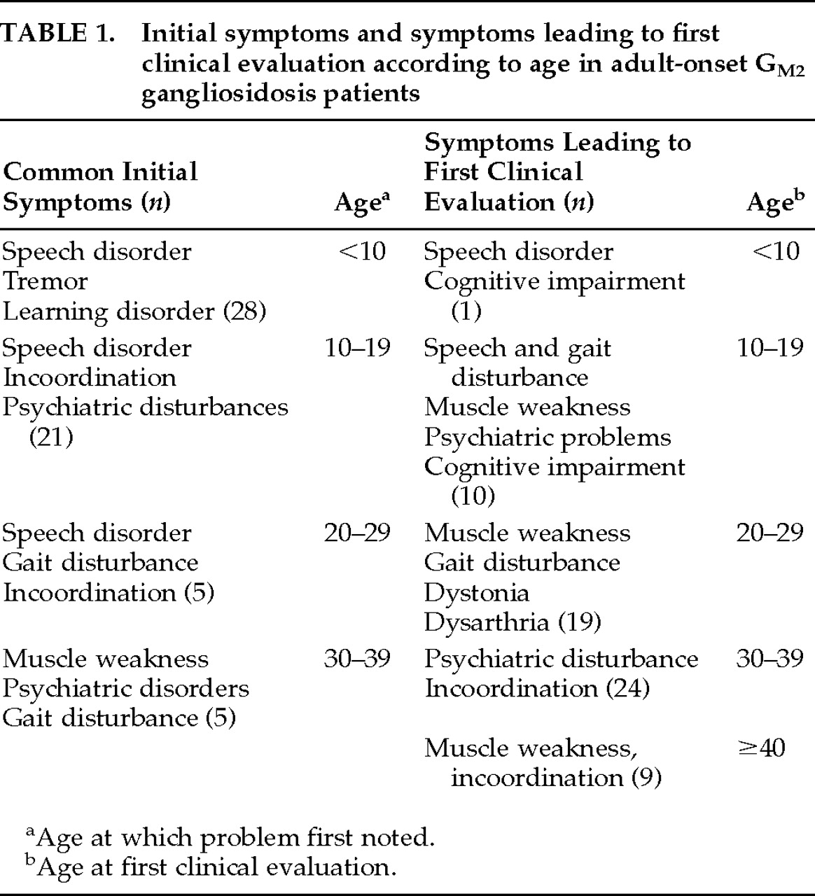
Sixty case reports (93%) included an approximate age at which symptoms were first noted. Table 1 lists these symptoms and the patient's age at the time. It was often difficult to ascertain the precise age at which clinical expression of the illness first appeared. Individuals were retrospectively described in many of the cases as having been “always clumsy” or “never a good athlete,” but these observations had not been recognized at the time as being signs of a disease process, and they were not included as first symptoms because of the vague dating and possibility of retrospective bias. Similarly, early signs such as falls or stuttering may have been overlooked or attributed to other causes. Nevertheless, symptoms were noted by the patient or family before the age of 20 in more than 80% of cases. Thus, often the denotation adult-onset gangliosidosis actually refers to patients who have manifested symptoms in childhood or adolescence.
There is great variability in the reported ages of symptom appearance, ranging 1.5 to 42 years of age. Within families there is some correspondence in the age of symptom onset. Thirty-nine families accounted for the 60 individuals for which age at onset was provided; 14 families had more than one affected member. Of these, 12 families had members whose first symptoms appeared within 5 years of the age at which other affected family members first manifested symptoms. While we acknowledge the problem of retrospective reporting, it appears that families present with similar patterns of onset, suggesting that time of onset may be related to the specific mutation underlying the illness.

Age at Evaluation:

The age at which patients were initially evaluated was reported for all cases. For patients followed over time, we have reported their age at the time they were examined for the purpose of the case report as well as the symptoms and signs apparent at that time (Table 1). The majority of patients (67%) were assessed between the ages of 20 and 40. There was usually a history of gradual progression of symptoms, and often the precipitating factor that brought the patient to medical attention was not identified. One-third of cases were discovered after an affected family member had come to attention,9,28,30,31,33,3538,40,43,46 and a few patients came to attention during routine screening for infantile Tay-Sachs carriers.29,33,41

Neurological Features

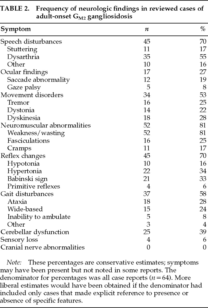
The nature and frequency of neurological signs is shown in Table 2. These signs reflect involvement of upper and lower motor neurons, basal ganglia, and cerebellum, with apparent sparing of sensory systems and cranial nerves. Even within commonly affected systems, however, the extent to which each system is affected varies greatly between individuals.50 For example, some cases have presented as spinal muscular atrophy and others as Friedreich's ataxia, motor neuron disease, or spinocerebellar degeneration.42 It is noteworthy that movement disorders such as dystonia and dyskinesia occur in almost 50% of all patients. In the small number of case reports that focus on the psychiatric disturbances, there is a very high rate of movement disorders (75%), including catatonia, raising questions about the relationships among movement disorders, psychiatric illness, and neuroleptic use in this subgroup. 49

Psychiatric Features

Psychosis:

A mental status examination was reported for 39 patients, and psychosis was noted in 21 (54%) of these. A more conservative estimate of 33% is obtained by dividing the number of occurrences of psychosis by all case reports, including those that made no specific reference to mental status. Given the available data, therefore, psychosis appears to occur in one-third to one-half of patients with AGG. This is consistent with previous estimates; although Navon and colleagues38 estimated a prevalence of about 30% from the literature, psychosis was present in 50% of patients from her own series.38 Another review39 reported a prevalence of about 30%, and in a more recently reported series of patients, psychosis was present in 60% of AGG patients.52
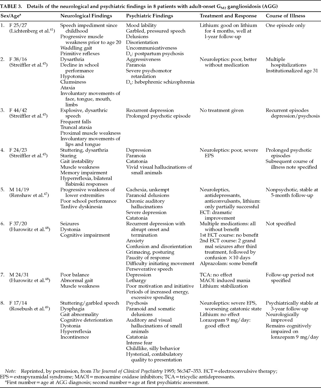
Of the 21 patients who developed psychosis, 10 (48%) were diagnosed with schizophrenia, usually of the hebephrenic type.30,39,41,49 Signs and symptoms included auditory and visual hallucinations, paranoia, grimacing, posturing, and regression to childlike behavior. Impaired consciousness, memory disturbance, and inability to perform self-care were reported to accompany the psychosis in several cases.39,45 Psychotic episodes of a more transient nature have also been described during the postpartum period,41 following the use of tryptophan,37 and secondary to trauma.9 Another patient had three discrete episodes of paranoid psychosis, all of which resolved with treatment.43
In a recent case report we described a patient who received a diagnosis of schizophrenia at the age of 14 and was treated over 3 years with a variety of typical neuroleptic medications in doses of up to 1,000 mg/day in chlorpromazine equivalents, with no appreciable benefit.49 When first assessed by us, he was catatonic and was accordingly treated with lorazepam. As the catatonia resolved and he began to talk, he described visual hallucinations of a threatening nature, including sharks, fires, and knives, and, in an interesting parallel to a previously described case,45 snakes and insects. His presentation had a regressive, childlike quality; for example, he would pretend to be driving a sports car, moving his hands as though on a steering wheel and making car noises.

Mood Disorders:

Eighteen case reports (27%) described alterations in mood, and in several patients the mood disturbance was a prominent and early manifestation of the illness.33,38,45,47,48 Of the total number of patients with reported abnormalities of mood, there was documentation of mania or euphoria in 7 (41%).31,33,37,46,48 One patient was psychotic during the period of euphoria,31 and psychosis was associated with a transient mood elevation after the administration of tryptophan37 in another patient. Unipolar depression was described in 8 patients,29,38,4548 3 of whom developed psychosis.45,47 Emotional lability was prominent in 2 patients, both of whom were also described as psychotic.9,41 One patient received a diagnosis of atypical manic-depressive psychosis, but the reported clinical features included only agitated depression, making it difficult to confidently classify this case.46
We could find only 8 published cases that detail the neuropsychiatric findings, treatment response, and course of illness for patients with clear psychiatric syndromes. These are summarized in Table 3.

Cognitive Function

Cognitive functioning was commented on in 35 (55%) of the case reports, and 21 (60%) of these patients were noted to be normal.1,28,29,31,32,35,37,43,46,48 For those in whom impaired cognitive status was documented (40% of those with a reported mental status exam), the severity of the impairment ranged from mild memory deficits9,31,36,40,43 to significant global cognitive decline9,39,48,49 and dementia.27,38 A low score on the Wechsler Memory Scale was the most typical abnormality reported on neuropsychologic testing.9,31 Little detail is provided in the cases to indicate whether other abilities such as visuospatial ability or new learning remained intact. Indeed, we could find only 15 individuals for whom IQ results were actually reported,9,31,36,39,40,48,49 and only 4 for whom IQ was measured serially over time.9,49 Three of these 4 patients had a decline in IQ, and 1 showed no change from age 9 to age 24.Our patient49 had an IQ in the normal range at age 8, at which time he had a speech disturbance. At age 14, two years after he became psychiatrically ill, his IQ was 57, placing him in the “mentally defective” range. He was untestable at age 17. Repeat testing at age 18 revealed persistent severe cognitive limitations in language, visuospatial abilities, psychomotor coordination, and memory, with a full scale IQ of 60. There was evidence of significant frontal lobe dysfunction, with perseveration, confabulation, poor judgment and insight, and complete inability to perform the Wisconsin Card Sorting Test.

FAMILIAL INVOLVEMENT

As discussed above, AGG is transmitted by an autosomal recessive pattern of inheritance. Relatives of affected individuals may therefore be normal, they may be carriers, or they may have the illness. Beyond the recognition that the carrier states tend to have intermediate levels of hexosaminidase, little attention had been given to the potential for phenotypic expression of the carrier state. Several reports have included hexosaminidase levels of patients' families,44,45 but reports of clinical findings in these people are lacking.
Hexosaminidase levels as well as clinical and neurophysiological changes have been reported for 14 carriers in a family with a beta subunit mutation.50 Upper and lower motor neuron signs, as well as psychiatric manifestations (depression, anxiety, behavioral changes) were present to a variable degree in 13 of these heterozygotes. Although we recognize the difficulty in quantifying “behavioral change”or severity of “cramps”for a numerical analysis, there appeared to be some relationship between hexosaminidase level and extent of symptoms. We assigned point values to the symptoms described by Federico et al.,50 based on the described severity of the symptoms. We then totaled the symptoms present for each carrier that had a hexosaminidase level provided (n=13) and did a correlational analysis plotting hexosaminidase level against symptom totals. There was a significant correlation between hexosaminidase level and expression of symptoms in the carrier state (n=13, r=0.75, r2=0.56, t=5.5 where t[df=12, α=0.01]=2.68). It is well recognized that females who are heterozygotes for inherited illnesses such as adrenoleukodystrophy and fragile X syndrome can develop clinical syndromes that range from mild to severe neuropsychiatric impairment.5255 It appears that phenotypic expression of the heterozygous state occurs in AGG as well; it is thus important that all family members of AGG patients receive a thorough neuropsychiatric examination in addition to measurement of hexosaminidase levels.

DIFFERENTIAL DIAGNOSIS

The differential diagnosis of AGG is extensive because of the variability in clinical presentation. In any patient with psychiatric illness, the presence of neurologic symptoms or cognitive impairment should raise the index of suspicion and lead to further evaluation.49,52 Several storage diseases are likely under-recognized as illnesses that may present in adulthood with psychiatric features. In addition to AGG, as outlined here, X-linked adrenoleukodystrophy,56 metachromatic leukodystrophy,5763 ceroid neuronal lipofuscinosis,6366 hepatolenticular degeneration,6769 Niemann-Pick type C,70,71 and cerebrotendinous xanthomatosis72,73 should be included in the differential when unusual or refractory neuropsychiatric symptom clusters are present.
There are no pathognomonic physical findings associated with AGG. Neither the characteristic macular cherry-red spot apparent in the infantile form of Tay-Sachs nor the visceral organ involvement seen in infantile Sandhoff's disease are present in the adult variants of these diseases. Dysmorphism, characteristic of the GM1 gangliosidoses, is not found in the GM2 gangliosidoses. The neurological signs and symptoms outlined above may or may not be present in the early stages of the disease.
Typical screening investigations will usually provide no evidence of an underlying metabolic defect such as AGG. Although there are reports of elevated lactate dehydrogenase and creatinine phosphokinase in AGG patients,1,31 these are nonspecific findings, and the majority of case reports describe normal serum chemistries. Similarly, EEGs are commonly normal. Cranial CT and MRI studies often show cerebellar atrophy, although they too can be normal.38,49 In a recent study, no correlation between clinical signs and radiologic changes (CT and MRI) in AGG was demonstrated.74 We were similarly unable to demonstrate a correlation between the presence of cerebellar signs in the clinical summary and the reports of cerebellar atrophy in the cases reviewed.
Electromyographic (EMG) studies were abnormal in 89% of the 35 patients for whom results were reported. Although Mitsumoto et al.36 have claimed to find a unique pattern of complex repetitive discharges in their AGG patients, there was no characteristic abnormality reliably documented in association with AGG in other EMG studies.
Thus, although imaging and EMG studies may document the presence of an underlying neurologic process, there are no findings in standard investigations to suggest a diagnosis of AGG. It is important, therefore, to order hexosaminidase levels from serum, leukocytes, or cultured skin fibroblasts to diagnose AGG when it is suspected. Rectal biopsy may confirm the presence of myenteric plexus neurons swollen with cytoplasmic membrane bodies, but this is not a necessary test for diagnostic confirmation.

TREATMENT

There is no specific treatment for AGG. Neurologic sequelae are managed by supportive measures, along with orthopedic and rehabilitative measures where indicated.
Treatment of the psychiatric manifestations of AGG is controversial. Although Rubin et al.43 reported successful treatment of an AGG patient in an acute paranoid state, most reports suggest that neuroleptic medications are rarely efficacious. Streifler et al.45 described 3 psychotic patients with AGG who demonstrated poor response to psychoactive medication. Hurowitz et al.48 reported no benefit from medications in 2 other patients with prominent mood disorders, and Renshaw et al.47 noted little effectiveness of traditional antipsychotics and antidepressants in an individual with psychosis and depression. Our patient49 received a long trial of high-dose neuroleptic medication (mean daily dose 1,000 mg in chlorpromazine equivalents) with no benefit. Thus, from the data available it would seem that the efficacy of neuroleptics in these patients is limited.
A further concern regarding the use of antipsychotic medication in AGG patients comes from reports that amphiphilic drugs, including phenothiazines and tricyclic antidepressants, increase lipidosis.75,76 It has been reported that patients with psychiatric symptoms and AGG have a more severe illness course, with the assumption being that treatment with these drugs worsened the underlying illness. While this theoretical concern is valid, there may be other ways to account for the observed worsening of symptoms in AGG patients treated with neuroleptic medication. Drugs such as neuroleptics can produce many of the extrapyramidal symptoms commonly described in AGG patients independent of worsening of the underlying disease process, and the anticholinergic properties of neuroleptics can cause sedation and cognitive impairment. Thus, a patient with AGG on phenothiazines might appear more neurologically impaired, but this may represent drug side effects rather than acceleration of the illness.
Individuals with a variety of brain lesions or abnormalities are, in general, believed to be more sensitive to psychoactive medication. The presence of movement disorders in untreated patients with AGG implicates the basal ganglia and suggests that these patients might be especially vulnerable to the extrapyramidal complications associated with the use of neuroleptics. Streifler et al.45 noted that attempts to treat the psychosis of an AGG patient resulted in “severe adverse reactions, mainly of extrapyramidal type” and eventually led to a catatonic state that resolved only after the medication was discontinued. Thus, although AGG patients may well be especially vulnerable to the effects of neuroleptic drugs, there are explanations for this sensitivity that do not require an actual effect of the drug on the underlying disease process.
In order to demonstrate such an effect, one would have to have evidence of prolonged impairment of a kind not typically produced by neuroleptics and persisting after neuroleptics are discontinued. A worsening of symptoms such as ataxia, dysphagia, dysarthria, or muscle weakness with neuroleptic treatment is stronger evidence for an association of drug and disease process because these are not typical side effects of such medications. Our patient developed catatonia, gait disturbance, severe dysarthria and dysphagia, inability to perform tandem gait, and muscle weakness, all of which corresponded temporally to neuroleptic use. Furthermore, withdrawal of the neuroleptics led to improvement in speech and complete resolution of these neurological abnormalities over 24 months without psychiatric decompensation. We could find no other reports of reversible worsening of disease by medication use. At this point it is unclear whether our patient's improvement reflects a resolution of atypical side effects with neuroleptic withdrawal, improvement in the underlying illness secondary to neuroleptic withdrawal, or merely fluctuation in the natural disease progression.
The use of benzodiazepines in the patient we described also raises the possibility of a therapeutic response to benzodiazepines, particularly with respect to his ongoing neurological and psychiatric improvement long after there would have been complete washout of neuroleptics. The efficacy of lorazepam in treating catatonia has been documented.77,78 Although we know of no other trials of benzodiazepine alone in the treatment of psychiatric symptoms in AGG, our experience with one patient suggests that benzodiazepines deserve consideration as potentially effective therapy in AGG.
There are variable reports of the efficacy of lithium carbonate in treating the psychiatric manifestations of AGG patients. Treatment of postpartum psychosis in an AGG patient with lithium carbonate brought about complete resolution of the delusions within 18 days.41 Given that this patient had no prior or subsequent psychiatric manifestations, however, it is possible that her psychosis might have resolved over 3 weeks regardless of the addition of lithium. Lithium in combination with either tranylcypromine or carbamazepine has been reported to be effective in several cases.47,48
Use of specific serotonergic reuptake inhibitors has been reported in only 1 patient with AGG; fluoxetine was initiated after electroconvulsive therapy (ECT) to prevent relapse.47 The long-term benefit of this intervention was not reported.
There are reports suggesting that ECT is efficacious in treating the psychiatric symptoms of AGG.47,48 An AGG patient with depression had been treated with multiple antipsychotics and antidepressants without benefit.48 Although a good response to initial ECT was not observed, there was a marked improvement following a further course of ECT. Similarly, another patient with psychosis and depression had only a partial response to multiple medications.47 Improvement, including resolution of chronic auditory hallucinations, was observed after a course of seven unilateral ECT treatments.47 Although reports of its use in AGG patients are limited, the available literature suggests that ECT may represent a valuable nonpharmacologic approach to treatment in these patients.
SUMMARY

Although this review has focused on the neuropsychiatric aspects of AGG, the most important point to be made is that in the presence of a mixed neuropsychiatric picture or refractory psychiatric symptoms, consideration should be given to unusual causes of psychiatric illness, such as metabolic diseases. Metabolic defects may underlie many more cases of atypical neuropsychiatric presentations than have been previously recognized.79 Awareness and accurate diagnosis of diseases such as AGG has significant implications for providing prognostic information and for family genetic counseling. Diagnosis should also influence treatment selection, since neuroleptics appear to have a higher than usual risk/benefit ratio in these patients. Benzodiazepines and ECT, alternatively, may be safe and efficacious therapies for the psychiatric manifestations in AGG.
TABLE 1.
TABLE 2.
TABLE 3.

References

1.
Johnson WG: The clinical spectrum of hexosaminidase deficiency diseases. Neurology 1981; 31:1453–1456
2.
Gravel RA, Clarke JTR, Kaback MM, et al: The GM2 gangliosidoses, in The Metabolic and Molecular Basis of Inherited Disease, 7th edition, edited by Scriver CR, Beaudet AL, Sly WS, et al. New York, McGraw-Hill, 1995, pp 2839–2882
3.
Tay W: Symmetrical changes in the region of the yellow spot in each eye of an infant. Transactions of the Ophthalmological Societies of the United Kingdom 1881; 1:55
4.
Sachs B: On arrested cerebral development with special reference to its cortical development. J Nerv Ment Dis 1887; 14:541–553
5.
Suzuki K, Suzuki K, Chen GC: Morphological, histochemical and biochemical studies on a case of systemic late infantile lipidosis. J Neuropathol Exp Neurol 1968; 27:15–38
6.
Suzuki K, Suzuki K, Rapin I, et al: Juvenile GM2 gangliosidosis: clinical variant of Tay-Sachs disease or a new disease? Neurology 1970; 20:190–204
7.
Meek D, Wolfe LS, Andermann F, et al: Juvenile progressive dystonia: a new phenotype of GM2 gangliosidosis. Ann Neurol 1984; 15:348–352
8.
Navon R, Padeh B, Adam A: Apparent deficiency of hexosaminidase A in healthy members of a family with Tay-Sachs disease. Am J Hum Genet 1973; 25:287–292
9.
Rapin I, Suzuki K, Suzuki K, et al: Adult (chronic) GM2 gangliosidosis: atypical spinocerebellar degeneration in a Jewish sibship. Arch Neurol 1976; 33:120–130
10.
Navon R: Molecular and clinical heterogeneity of adult GM2 gangliosidosis. Dev Neurosci 1991; 13:295–298
11.
Navon R, Proia RL: The mutations in Ashkenazi Jews with adult GM2 gangliosidosis, the adult form of Tay-Sachs disease. Science 1989; 243:1471–1474
12.
Ohna K, Suzuki K: A splicing defect due to an exon-intron junction mutation results in abnormal beta-hexosaminidase alpha-chain mRNAs in Ashkenazi Jewish patients with Tay-Sachs disease. Biochem Biophys Res Commun 1988; 153:463–469
13.
Paw BH, Kaback MM, Lim J, et al: Frequency of three HEX A mutation alleles among Jewish and non-Jewish carriers identified in a Tay-Sachs screening program. Am J Hum Genet 1990; 47:697–704
14.
Suzuki K, Vanier MT: Biochemical and molecular aspects of late-onset GM2 gangliosidosis: B1 variant as a prototype. Dev Neurosci 1991; 13:288–294
15.
Ledeen R: Gangliosides of the neuron. Trends Neurosci 1985; 8:169–174
16.
Ando S: Gangliosides of the nervous system. Neurochem Int 1983; 5:507–536
17.
Nobile-Orazio E, Carpo M, Scarlato G: Gangliosides: their role in clinical neurology. Drugs 1994; 47:576–585
18.
Leinekugel P, Michel S, Conzelmann E, et al: Qualitative correlation between the residual activity of beta-hexosaminidase A and arylsulfatase A and the severity of the resulting liposomal storage disease. Hum Genet 1992; 88:513–523
19.
Conzelmann E, Sandhoff K: Biochemical basis of late-onset neurolipidoses. Dev Neurosci 1991; 13:197–204
20.
Purpura DP, Suzuki K: Distortion of neuronal geometry and formation of aberrant synopsis in neuronal storage disease. Brain Res 1976; 116:1–21
21.
Purpura DP, Baker H: Meganeurites and other aberrant processes of neurons in feline GM1 gangliosis: a golgi study. Brain Res 1977; 143:13–26
22.
Purpura DP, Walkley SU: Aberrant neurite and spine generation in mature neurons in the gangliosidoses, in Gangliosides in Neurological and Neuromuscular Function, edited by Rapport MM, Gorio A. New York, Raven, 1991, pp 1–16
23.
Walkley SU: Pathobiology of neuronal storage disease. Int Rev Neurobiol 1988; 29:191–244
24.
Walkley SU, March PA: Biology of neuronal dysfunction in storage disorders. J Inherit Metab Dis 1993; 16:284–287
25.
Siegel DA, Walkley SU: Growth of ectopic dendrites on cortical pyramidal neurons in neuronal storage diseases correlates with abnormal accumulation of GM2 ganglioside. J Neurochem 1994; 62:1852–1862
26.
Hannun YA, Bell RA: Lysosphingolipids inhibit protein kinase C: implications for the sphinolipidoses. Science 1987; 235:670–673
27.
O'Neill B, Butler AB, Young E, et al: Adult onset GM2 gangliosidosis: seizures, dementia and normal pressure hydrocephalus associated with glycolipid storage in the brain and arachnoid granulation. Neurology 1978; 28:1117–1123
28.
Oonk JGW, van der Helm HJ, Martin JJ: Spinocerebellar degeneration: hexosaminidase A and B deficiency in two adult sisters. Neurology 1979; 29:380–384
29.
Yaffe MG, Kaback M, Goldberg M, et al: An amyotrophic lateral sclerosis-like syndrome with hexosaminidase-A deficiency: a new type of GM2 gangliosidosis (abstract). Neurology 1979; 29:611
30.
Navon R, Argov Z, Brand N, et al: Adult GM2 gangliosidosis in association with Tay-Sachs disease: a new phenotype. Neurology 1981; 31:1397–1401
31.
Willner JP, Grabowski GA, Gordon RE, et al: Chronic GM2 gangliosidosis masquerading as atypical Friedreich ataxia: clinical, morphologic and biochemical studies of nine cases. Neurology 1981; 31:787–798
32.
Johnson WG, Wigger HJ, Karp HR, et al: Juvenile spinal muscular atrophy: a new hexosaminidase deficiency phenotype. Ann Neurol 1982; 11:11–16
33.
Kolodny EH, Lyerl T, Raghavan SS, et al: Significance of hexosaminidase A deficiency in adults (abstract). Neurology 1982; 322:A81
34.
Dale AJD, Engel AG, Rudd NL: Familial hexosaminidase A deficiency with Kugelberg-Wellander phenotype and mental change (abstract). Ann Neurol 1983; 14:109
35.
Barbeau A, Plasse L, Cloutier T, et al: Lysosmal enzymes in ataxia: discovery of two new cases of late onset hexosaminidase A and B deficiency (adult Sandhoff disease) in French Canadians. Can J Neurol Sci 1984; 11:601–606
36.
Mitsumoto H, Sliman R, Schafer IA, et al: Motor neuron disease and adult hexosaminidase A deficiency in two families: evidence for multisystem degeneration. Ann Neurol 1985; 17:378–385
37.
Parnes S, Karpati G, Carpenter S, et al: Hexosaminidase-A deficiency presenting as atypical juvenile-onset spinal muscular atrophy. Arch Neurol 1985; 42:1176–1180
38.
Navon R, Argov Z, Frisch A: Hexosaminidase deficiency in adults. Am J Med Genet 1986; 24:179–196
39.
Oates CE, Bosch EP, Hart MN: Movement disorders associated with chronic GM2 gangliosidosis: case report and review of the literature. Eur Neurol 1986; 25:154–159
40.
Harding E, Young EP, Schon F: Adult onset supranuclear ophthalmoplegia, cerebellar ataxia, and neurogenic proximal muscle weakness in a brother and sister: another hexosaminidase deficiency syndrome. J Neurol Neurosurg Psychiatry 1987; 50:687–690
41.
Lichtenberg P, Navon R, Wertman E, et al: Post-partum psychosis in adult GM2 gangliosidosis: a case report. Br J Psychiatry 1988; 153:387–389
42.
Karni A, Navon R, Sadeh M: Hexosaminidase A deficiency manifesting as spinal muscular atrophy of late onset. Ann Neurol 1988; 24:451–453
43.
Rubin M, Karpati G, Wolfe LS, et al: Adult onset motor neuronopathy in the juvenile type of hexosaminidase A and B deficiency. J Neurol Sci 1988; 87:103–119
44.
Thomas PK, Young E, King RHM: Sandhoff disease mimicking adult-onset bulbospinal neuronopathy. J Neurol Neurosurg Psychiatry 1989; 52:1103–1106
45.
Streifler J, Golomb M, Gadoth N: Psychiatric features of adult GM2 gangliosidosis. Br J Psychiatry 1989; 155:410–413
46.
Specola N, Vanier MT, Goutieres F, et al: The juvenile and chronic forms of GM2 gangliosidosis: clinical and enzymatic heterogeneity. Neurology 1990; 40:145–150
47.
Renshaw PF, Stern TA, Welch C, et al: Electroconvulsive therapy treatment of depression in a patient with adult GM2 gangliosidosis. Ann Neurol 1992; 31:342–344
48.
Hurowitz GI, Silver JM, Brin MF, et al: Neuropsychiatric aspects of adult onset Tay-Sachs disease: two case reports with several new findings. J Neuropsychiatry Clin Neurosci 1993; 5:30–36
49.
Rosebush PI, MacQueen G, Mazurek MF: Late-onset Tay-Sachs disease presenting as catatonic schizophrenia: diagnostic and treatment issues. J Clin Psychiatry 1995; 56:347–353
50.
Federico A, Palmeri S, Malandrini A, et al: The clinical aspects of adult hexosaminidase deficiencies. Dev Neurosci 1991; 13:280–287
51.
Reference number not used.
52.
Coker SB: The diagnosis of childhood degenerative disorders presenting as dementia in adults. Neurology 1991; 41:794–798
53.
Johnson WG: Lysosomal diseases and other storage diseases, in Merritt's Textbook of Neurology, 8th edition, edited by Rowland LP. Philadelphia, Lea and Febiger, 1989, pp 500–537
54.
Grigsby JP, Kemper, MB, Hagerman RJ, et al: Neuropsychological dysfunction among affected heterozygous fragile X females. Am J Med Genet 1990; 35:28–35
55.
Reiss AL, Hagerman RJ, Vinogradov S, et al: Psychiatric disability in female carriers of the fragile X chromosome. Arch Gen Psychiatry 1988; 45:25–30
56.
Moser HW, Moser AE, Singh I, et al: Adrenoleukodystrophy: survey of 303 cases: biochemistry, diagnosis, and therapy. Ann Neurol 1984; 16:628–664
57.
Manowitz P, Kling A, Kohn H: Clinical course of adult metachromatic leukodystrophy presenting as schizophrenia: a report of two living cases in siblings. J Nerv Ment Dis 1978; 166:500–506
58.
Betts TA, Smith WT, Kelly RE: Adult metachromatic leukodystrophy (sulphatide lipidosis) simulating acute schizophrenia: report of a case. Neurology 1968; 18:1140–1142
59.
Finelli P: Metachromatic leukodystrophy manifesting as a schizophrenic disorder: computed tomographic correlation. Ann Neurol 1985; 18:94–95
60.
Mahon-Haft H, Stone RK, Johnson R, et al: Biochemical abnormalities of metachromatic leukodystrophy in an adult psychiatric population. Am J Psychiatry 1981; 138:1372–1374
61.
Austin J, Armstrong D, Fouch S, et al: Metachromatic leukodystrophy (MLD), VIII: MLD in adults—diagnosis and pathogenesis. Arch Neurol 1968; 18:225–239
62.
Wulff CH, Trojaborg W: Adult metachromatic leukodystrophy: neurophysiologic findings. Neurology 1985; 35:1776–1778
63.
Waldman AJ: Sometimes when you hear hoofbeats…two cases of inherited metabolic diseases with initial presentation of psychiatric symptoms (letter). J Neuropsychiatry Clin Neurosci 1992; 4:113–114
64.
Berkovic SF, Carpenter S, Andermann F, et al: Kufs' disease: a critical reappraisal. Brain 1988; 111:27–62
65.
Berkovic SF, Andermann F, Andermann E, et al: Clinical features and forms. Am J Med Genet 1988; 5(suppl):105–109
66.
Sandyk R: Adult neuronal ceroid lipofuscinosis (Kufs' disease): a sporadic case. S Afr Med J 1981; 7:754–755
67.
Starosta-Rubenstein S, Young AB, Kluin K, et al: Clinical assessment of thirty-one patients with Wilson's disease: correlations with structural changes on magnetic resonance imaging. Arch Neurol 1987; 44:365–370
68.
Dening TR, Berrios GE: Wilson's disease: psychiatric symptoms in 195 cases. Arch Gen Psychiatry 1989; 46:1126–1134
69.
Menkes JH: Disorders of metal metabolism, in Merritt's Textbook of Neurology, 8th edition, edited by Rowland LP. Philadelphia, Lea and Febiger, 1989, pp 538–544
70.
Vanier MT, Rodriguez-Lafrasse C, Rousson R, et al: Type C Niemann-Pick disease: biochemical aspects and phenotypic heterogeneity. Dev Neurosci 1991; 13:307–314
71.
Fink JK, Filling-Katz MR, Sokol J: Clinical spectrum of Niemann-Pick disease type C. Neurology 1989; 39:1040–1049
72.
Berginer VM, Foster NL, Sadowsky M, et al: Psychiatric disorders in patients with cerebrotendinous xanthomatosis. Am J Psychiatry 1988; 145:354–357
73.
Dotti MT, Salen G, Federico A: Cerebrotendinous xanthomatosis as a multisystem disease mimicking premature ageing. Dev Neurosci 1991; 13:371–376
74.
Streifler JY, Gornish M, Hadar H, et al: Brain imaging in late-onset GM2 gangliosidosis. Neurology 1993; 43:2055–2058
75.
Lullman H, Lullman-Rauch R, Wassermann D: Lipidosis induced by amphiphilic cationic drugs. Biochem Pharmacol 1978; 27:1103–1108
76.
Palmeri S, Mangano L, Battisti C, et al: Imipramine induced lipidosis and dexamethasone effect: morphological and biochemical study in normal and chronic GM2 gangliosidosis fibroblasts. J Neurol Sci 1992; 110:215–221
77.
Rosebush PI, Hildebrand AM, Furlong BG, et al: Catatonic syndrome in a general psychiatric inpatient population: frequency, clinical presentation, and response to lorazepam. J Clin Psychiatry 1990; 51:357–362
78.
Gaind GS, Rosebush PI, Mazurek MF: Lorazepam treatment of acute and chronic catatonia in two mentally retarded brothers. J Clin Psychiatry 1994; 55:20–23
79.
Adams RD, Victor M: Principles of Neurology, 5th edition. New York, McGraw-Hill, 1993, pp 799–849

Information & Authors

Information

Published In

Go to The Journal of Neuropsychiatry and Clinical Neurosciences
The Journal of Neuropsychiatry and Clinical Neurosciences
Pages: 10 - 19
PubMed: 9547461

History

Published in print: February 1998
Published online: 1 April 2006

Authors

Details

Glenda M. MacQueen, M.D., Ph.D., F.R.C.P.C.
Received August 13, 1996; revised January 10, 1997; accepted January 13, 1997. From the Departments of Psychiatry and Medicine, Division of Neurology, and Biomedical Sciences, Division of Neuroscience, McMaster University Medical Centre, Hamilton, Ontario, Canada. Address correspondence to Dr. MacQueen, Department of Psychiatry, McMaster University, 1200 Main Street West, Hamilton, Ontario, L8N 3Z5, Canada.
Patricia I. Rosebush, M.Sc.N., M.D., F.R.C.P.C.
Received August 13, 1996; revised January 10, 1997; accepted January 13, 1997. From the Departments of Psychiatry and Medicine, Division of Neurology, and Biomedical Sciences, Division of Neuroscience, McMaster University Medical Centre, Hamilton, Ontario, Canada. Address correspondence to Dr. MacQueen, Department of Psychiatry, McMaster University, 1200 Main Street West, Hamilton, Ontario, L8N 3Z5, Canada.
Michael F. Mazurek, M.D., F.R.C.P.C.
Received August 13, 1996; revised January 10, 1997; accepted January 13, 1997. From the Departments of Psychiatry and Medicine, Division of Neurology, and Biomedical Sciences, Division of Neuroscience, McMaster University Medical Centre, Hamilton, Ontario, Canada. Address correspondence to Dr. MacQueen, Department of Psychiatry, McMaster University, 1200 Main Street West, Hamilton, Ontario, L8N 3Z5, Canada.

Metrics & Citations

Metrics

Citations

Export Citations

If you have the appropriate software installed, you can download article citation data to the citation manager of your choice. Simply select your manager software from the list below and click Download.

For more information or tips please see 'Downloading to a citation manager' in the Help menu.

Format
Citation style
Style
Copy to clipboard

View Options

View options

PDF/EPUB

View PDF/EPUB

Login options

Already a subscriber? Access your subscription through your login credentials or your institution for full access to this article.

Personal login Institutional Login Open Athens login
Purchase Options

Purchase this article to access the full text.

PPV Articles - Journal of Neuropsychiatry and Clinical Neurosciences

PPV Articles - Journal of Neuropsychiatry and Clinical Neurosciences

Not a subscriber?

Subscribe Now / Learn More

PsychiatryOnline subscription options offer access to the DSM-5-TR® library, books, journals, CME, and patient resources. This all-in-one virtual library provides psychiatrists and mental health professionals with key resources for diagnosis, treatment, research, and professional development.

Need more help? PsychiatryOnline Customer Service may be reached by emailing [email protected] or by calling 800-368-5777 (in the U.S.) or 703-907-7322 (outside the U.S.).

Media

Figures

Other

Tables

Share

Share

Share article link

Share