Closed head injury (CHI) can result in subacute or chronic alterations in cognition, behavior, and emotions. The neuropsychiatric conditions commonly seen after CHI include mood disorders,
1,2 anxiety disorders,
3,4 and personality changes.
5,6 The frequency of neuropsychiatric symptoms following CHI ranges from 10% to 87%, depending on the severity of injury and multiple other factors.
7 Gualtieri and Cox
8 estimated the incidence of depression after CHI to range from 25% to 50%.
Mood disorders often respond to pharmacotherapy, but in some cases it is not effective or is poorly tolerated. In such cases, electroconvulsive therapy (ECT) may be considered a treatment option. ECT is an effective treatment modality for certain psychiatric conditions, such as major depression, mania, catatonia, and delirium. Many case reports and literature reviews have described the effectiveness and safety of ECT in neurologically impaired patients.
9–13 However, there are few recent case reports about the use of ECT in patients who have suffered CHI.
2,14–17 Clark and Davidson
2 described a case of mania following head injury that was treated successfully with six bilateral ECT treatments. Schnur et al.
16 described 2 patients with partial complex seizures, delusions, and postictal aggression following head injury. ECT was effective in reducing the severity of aggression and the frequency of seizures without any cognitive side effects. Kant et al.
17 have reported a case of chronic delirium after severe head injury, refractory to behavioral and pharmacological interventions over a period of 7 months. This patient was treated with six bilateral ECT treatments, leading to resolution of the delirium followed by dramatic cognitive and functional improvement.
17In this article, we report our experience of using ECT for the treatment of neuropsychiatric conditions in patients who had suffered CHI, employing standardized treatment and assessment protocols.
METHODS
Subjects
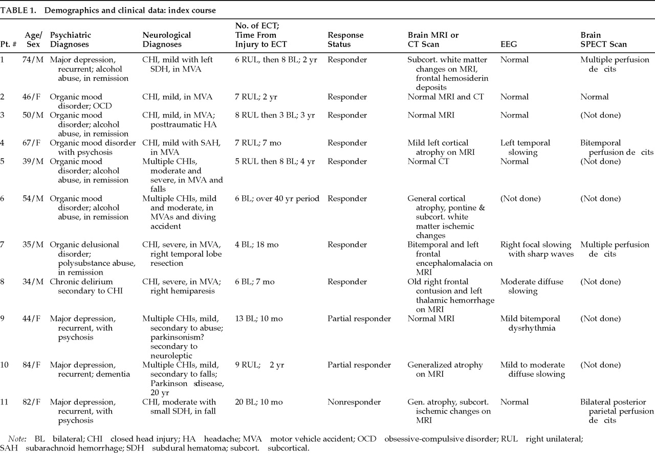
A retrospective review of the ECT Program computerized database at Allegheny Neuropsychiatric Institute identified 11 patients with a history of CHI who had received ECT between February 1992 and December 1994. There were 6 men and 5 women, with a mean age of 55 (SD=18) years. Diagnoses were determined by consensus between the ECT Program clinicians and the patients' attending psychiatrist by formal application of DSM-III-R criteria. Of the 11 patients, 4 had major depression, 5 had a mood disorder secondary to CHI, and 1 each had chronic delirium and delusional disorder secondary to CHI (
Table 1). The patients diagnosed with major depression had a history of depression antedating their CHI.
The etiologies of the CHI included one or more of the following: motor vehicle accident (MVA) (
n=8), fall (
n=4), and fight or physical abuse (
n=1). Four patients had histories of multiple head injuries. In every case, the severity of injury was determined from a detailed review of the medical records, together with interviews of the patient and/or significant other. A standard classification of injury severity
18 based on the Glasgow Coma Scale (GCS)
19 and/or duration of loss of consciousness (LOC) was used to rate the injury as “mild” (GCS score ≥13, LOC ≤20 minutes) in 6 patients, “moderate” (GCS score 9–12, LOC >20 minutes but <24 hours) in 2 patients, and “severe” (GCS score ≤8, LOC >24 hours) in 3 patients (
Table 1). Time interval between the most recent head injury and ECT treatment ranged from 7 to 48 months. All patients experienced complete neurological recovery from their injuries, except for 1 patient who had residual right hemiparesis.
ECT Technique
All patients were referred for ECT because of lack of response to medications and/or history of response to ECT in the past. All patients were free of psychotropic medications at the time of the ECT treatment, with the exception of labetalol for hypertension (#1), valproic acid as an adjunctive mood stabilizer (#6), as-needed droperidol for agitation (#8), molindone for agitation (#9), and carbidopa-levodopa and bromocriptine for Parkinson's disease (#10). The ECT treatments were administered three times per week. Typical modifications included anticholinergic premedication (glycopyrrolate or atropine sulfate), anesthesia (methohexital sodium, 1 mg/kg intravenously), and muscle relaxation (succinylcholine, 1 mg/kg intravenously). Patients received 100% oxygen via face mask throughout the procedure. All patients were monitored with electrocardiography, pulse oximetry, and two-channel (bilateral frontomastoid) electroencephalography (EEG).
The choice of stimulus electrode placement was determined by the ECT Program in consultation with the patient's treatment team. The standard bifrontotemporal and d'Elia electrode placements were employed for bilateral (BL) ECT and right unilateral (RUL) ECT, respectively. Three (50%) of the 6 patients who began on RUL ECT were switched to BL ECT (after the fifth to eighth treatment) because of inadequate therapeutic response to RUL ECT (see
Table 1).
The ECT stimulus was delivered by a customized brief pulse device (MECTA SR-1; MECTA, Lake Oswego, OR). ECT seizure threshold was determined at the first treatment by a stimulus dosing titration protocol.
20 For subsequent ECT treatments, stimulus dosage was set at a fixed percentage above initial seizure threshold (2.5 times initial seizure threshold for RUL ECT; 1.5 times initial seizure threshold for BL ECT).
21 During the course of ECT treatments, stimulus dosage settings were adjusted upward whenever necessary in order to maintain a seizure duration of at least 25 seconds by EEG, using standard criteria.
22Assessment of Clinical Response
Patients were evaluated for response to ECT by use of a 7-point Clinical Global Impression (CGI) severity scale administered 2 to 3 days after the last ECT treatment. The Global Assessment of Functioning (GAF) was administered at weekly intervals. This was supplemented by baseline and then weekly administration of the observer-rated Montgomery-Åsberg Depression Rating Scale (MADRS)
23 to assess symptom severity and response to treatment.
The following criteria were used to define the response to treatment for patients with depression: MADRS score ≤15; 50% reduction in pre-ECT MADRS score; and CGI score ≤3. For a patient to be considered a “responder,” all three criteria had to be met. If only one criterion was met, the patient was rated as “partial responder.” If none of the criteria were met, the patient was considered a “nonresponder,” even if some clinical improvement was observed.
Assessment of Adverse Effects
Cognitive side effects were assessed with the Neurobehavioral Cognitive Status Examination (NCSE)
24 and the Mini-Mental State Examination (MMSE).
25 The NCSE measures functioning in five major cognitive areas: language, construction, memory, calculations, and reasoning; attention and orientation are assessed independently. Both the NCSE and the MMSE were administered at baseline and again 2 to 3 days after the final ECT. The MMSE was repeated weekly during the ECT course.
All of the patients were asked nine orientation questions prior to each treatment and then asked the same questions again at 15, 30, 60, 90, and 120 minutes after treatment to assess the duration of post-ECT confusion. Patients were considered reoriented when they were able to answer as many questions correctly as they did prior to ECT.
Continuation ECT (cECT)
After completion of their index courses of ECT, 8 patients received cECT because they had histories of nonreponse to numerous medication trials. Typically the cECT course consisted of four weekly treatments, then two to four treatments on an alternate-week schedule, followed by monthly treatments. However, treatment intervals were shortened if breakthrough symptoms were noted. Clinical ratings and cognitive assessments (MMSE) were obtained prior to each treatment.
RESULTS
Therapeutic Response to ECT
Of the 11 patients, 8 (72.7 %) were classified as responders; 2 (18.2%) were considered partial responders and became responders during cECT; and 1 (9.1%) was a nonresponder. The patient who was a nonresponder showed improvement several times (MADRS as low as 14), but these gains were not sustained despite a long course of ECT (20 bilateral ECT treatments).
Both MADRS and GAF scores (mean±SD) showed robust improvement after treatment with ECT. MADRS scores (n=9; MADRS was not applicable in 2 patients) improved from 33.4±6.4 (median 31, range 28–48) to 11.2±8.5 (median 14, range 0–25, P<0.0002; lower scores indicate improvement). Similarly, GAF scores (n=11) improved from 33.2±14.2 (median 35; range 10–50) to 54.5±13.0 (median 55; range 30–70; P<0.00009).
Neurocognitive Test Results
The pre- and post-ECT NCSE was administered to 9 of the 11 patients. One patient was unable to participate in the pre-ECT testing because of constant agitation (#8), and another patient was discharged prior to completing post-ECT testing (#3).
No significant change was noted in post-ECT NCSE scores (t-test for dependent measures, P>0.70). Three patients showed improvement (median increase of 8 points, range 3–10); 5 patients showed decline (median decline of 2 points, range 1–23); and 1 patient had no change in NCSE scores. Of the 2 patients who had a decline of 5 or more points on the post-ECT NCSE, 1 was a partial responder (5 points) and the other a nonresponder (23 points).
Pre- and post-ECT MMSE scores (mean±SD; n=11) were 24.6±6.3 and 24.5±6.7, respectively (P>0.89). Four patients showed median improvement of 2 points (range 2–4) and the other 7 patients showed median decline of 1 point (range 1–4) in MMSE scores after the index course. Average MMSE scores improved by 1.23 in patients with mild CHI and declined by 2.5 and 1.0 in patients with moderate and severe CHI, respectively.
The most significant decline in NCSE scores (from 67 to 44) and MMSE scores (from 24 to 20) was noted in the only nonresponder. Follow-up data were not available on this patient. According to the progress notes, the confusion and functioning level improved after 1 week following the last ECT, and the patient was discharged home after having a successful therapeutic pass. This patient had responded to ECT 1 year earlier (prior to the head injury). At that time, her pre-ECT NCSE score was 69 and her post-ECT NCSE scores were 53 at 48 hours and 67 at 1 year. Brain MRI did not show any interval change over the year after the first course of ECT, but SPECT scan showed evidence of new areas of hypoperfusion in the right posterior parietal lobe in addition to a perfusion defect in the left posterior parietal lobe a year earlier, raising the possibility of early Alzheimer's disease.
None of the 11 patients suffered any medical complications from ECT. Despite baseline cognitive deficits, 10 of the patients showed only transient cognitive decline following each treatment. The reorientation time ranged from 30 to 120 minutes after each treatment. Only 1 patient (nonresponder) had prolonged reorientation times, beyond 120 minutes, from the fourth treatment onward.
cECT Neurocognitive Monitoring
Eight patients received cECT and were monitored with MMSE prior to each treatment. Mean MMSE scores (n=8) pre- and post-cECT were 26.62±3.9 and 25.62±3.8, respectively (P>0.29). Total number of cECTs ranged from 3 to 62 (mean=18) over a period of 18 months (range 2 weeks to 18 months; mean=5.7 months). Four patients received UL and 4 received BL treatments.
DISCUSSION
Our findings indicate that ECT is safe and effective in the treatment of neuropsychiatric symptoms in patients after CHI. In this group of 11 patients, all but 1 responded to the treatment, and no untoward events were noted except for significant confusion in the nonresponding patient (9%). Severity of head injury was not related to the cognitive side effects of ECT or the rate of recovery from post-ECT confusion. Similarly, no specific findings on brain MRI, SPECT scan, or EEG were noted to correlate with outcome in this group.
Cognitive deficits are a concern in considering the use of ECT in patients with a history of CHI. Transient disturbances in memory (both retrograde and anterograde), postictal confusion, and rare interictal delirium have been associated with ECT. Many earlier reports on this topic have suggested that preexisting cognitive deficits do not get worse after ECT and that some patients actually show improved cognition following resolution of depression.
13,26–28 Price and McAllister
26 reviewed the ECT treatments of 56 patients with diagnoses of organic dementia and pseudodementia of depression. One-third of the patients with dementia showed improvement in cognitive functioning. Mulsant et al.
27 treated 40 elderly depressed patients with ECT, of whom 47% had baseline cognitive deficits. They reported no significant change in MMSE scores after ECT. Zwil and Pelchat
13 noted in their review of literature on use of ECT in neurologically impaired patients that ECT is safe and effective in patients with chronic cognitive impairments. In our series, 10 of 11 patients suffered no lasting cognitive side effects secondary to ECT, irrespective of baseline cognitive status or severity of head injury.
These findings cannot be generalized because of the following limitations of this study: a small sample size, retrospective review of the data, and lack of any comparison group. However, in our experience of treating patients who had suffered closed head injury, it appears that a history of CHI is not necessarily a contraindication for use of ECT, even if patients have cognitive impairment. Further investigation is indicated, particularly on a prospective basis, before widespread use of ECT can be recommended in this patient population.


