Full access
Special Articles
Published Online: 1 July 2011

Pathogenic Mechanisms of Depression in Multiple Sclerosis

Publication: The Journal of Neuropsychiatry and Clinical Neurosciences

Abstract

This article reviews various theories and findings on mechanisms of depression in MS, describing the latest research approaches, as they relate to theories of disease-process. The authors have compiled data on lesion location, extensity, and severity; the effects of immune dysfunction and side effects of immunomodulatory drugs, including interferon therapy; and psychosocial stressors as they all relate to the common symptom of depression in MS.

Abstract

Patients with multiple sclerosis (MS) have an increased risk of developing depression as compared with healthy subjects and patients with many other chronic neurological conditions. The observation that depressive symptoms can precede the onset of neurological symptoms suggests that depression may be related to early disease-specific processes. Several pathogenic mechanisms have been proposed to explain the etiology of depression in patients with MS. This article reviews the current evidence for the contribution of lesional, autoimmune, iatrogenic, and psychosocial factors. It appears that the etiology of depression is multifactorial and varies in individual patients with MS.
Multiple sclerosis (MS) is a chronic and unpredictable neurological disease commonly associated with a host of symptoms such as sensory and motor deficits, fatigue, ataxia, blindness, pain, cognitive impairment, and depression. It is the commonest disabling neurological condition in adults of working age.1 MS is an autoimmune condition characterized by destruction of the myelin sheaths in the CNS. This condition affects twice as many women as men and is associated with accumulating levels of physical disability. However, there is a large variation between patients in the type, severity, and course of the illness. In the majority of cases, the disease begins with a relapsing–remitting course characterized by acute exacerbation of symptoms followed by periods of stability. This is typically followed by a phase of continuous deterioration, which is referred to as secondary progression.2
Symptoms of depression in patients with MS have been described from the days of Charcot, and they are currently recognized as a common finding.3,4 The lifetime risk of depression in MS ranges between 40% and 60%,5,6 and an annual prevalence figure of 20% has been reported.7 A large, cross-sectional, community study (N=1,374) among members of the Multiple Sclerosis Association reported a prevalence of clinically significant depressive symptoms in 41.8% of patients, of whom 29.1% had moderate or severe depression.8 These results are comparable with other neurological disorders such as Parkinson's disease (prevalence: 20%–45%) and stroke (prevalence: 10%–34%).9 Moreover, several studies have reported higher suicide rates among patients with MS, as compared with other chronic progressive diseases, suggesting that there may be a specific vulnerability for depressive symptoms in MS.10 An early study by Sadovnick et al. reported a suicide rate that was 7.5 times higher than among age-matched controls.11 Studies using more rigorous methodology have reported a rate of 1.8–2.3 times the expected rate.11 In a study by Feinstein, the lifetime prevalence for suicidal ideation was 28.6%, and the rate of actual suicidal attempts was 6.4%.12 Depression severity was identified as the strongest risk marker for suicidal ideation, which was more common in patients with secondary progressive MS.1
Historically, the most common explanation for the high prevalence of depression among patients with MS was that depression is a response to the physical and social challenges faced by the patients. At the beginning of the 20th century, Barbellion (who suffered from MS) observed that severe depression could precede and accompany neurological symptoms of MS.13 Since then, several authors reported a high incidence of severe depression among patients with MS and the occasional development of depression before the onset of neurological signs.1416 These early reports have been confirmed by later studies.17 Witlock and Siskind compared 30 patients with MS and 30 patients with other chronic neurological disorders causing a similar degree of disability.18 They found that patients with MS experienced more episodes of severe depression before and after the onset of neurological symptoms. Sullivan et al.19 reported that 52% of patients with MS experienced a depressive episode before the onset of MS, as compared with 17% of patients with chronic low-back pain.
Several models have been proposed to explain the etiology of depression in patients with MS. This article reviews the current evidence for the following four pathogenic mechanisms: 1) lesion-related factors; 2) autoimmune factors; 3) iatrogenic factors; and 4) psychosocial factors. We discuss the leading theories on the role of these factors in the development of depressive symptoms with reference to the main evidence supporting and refuting the pathogenic models.
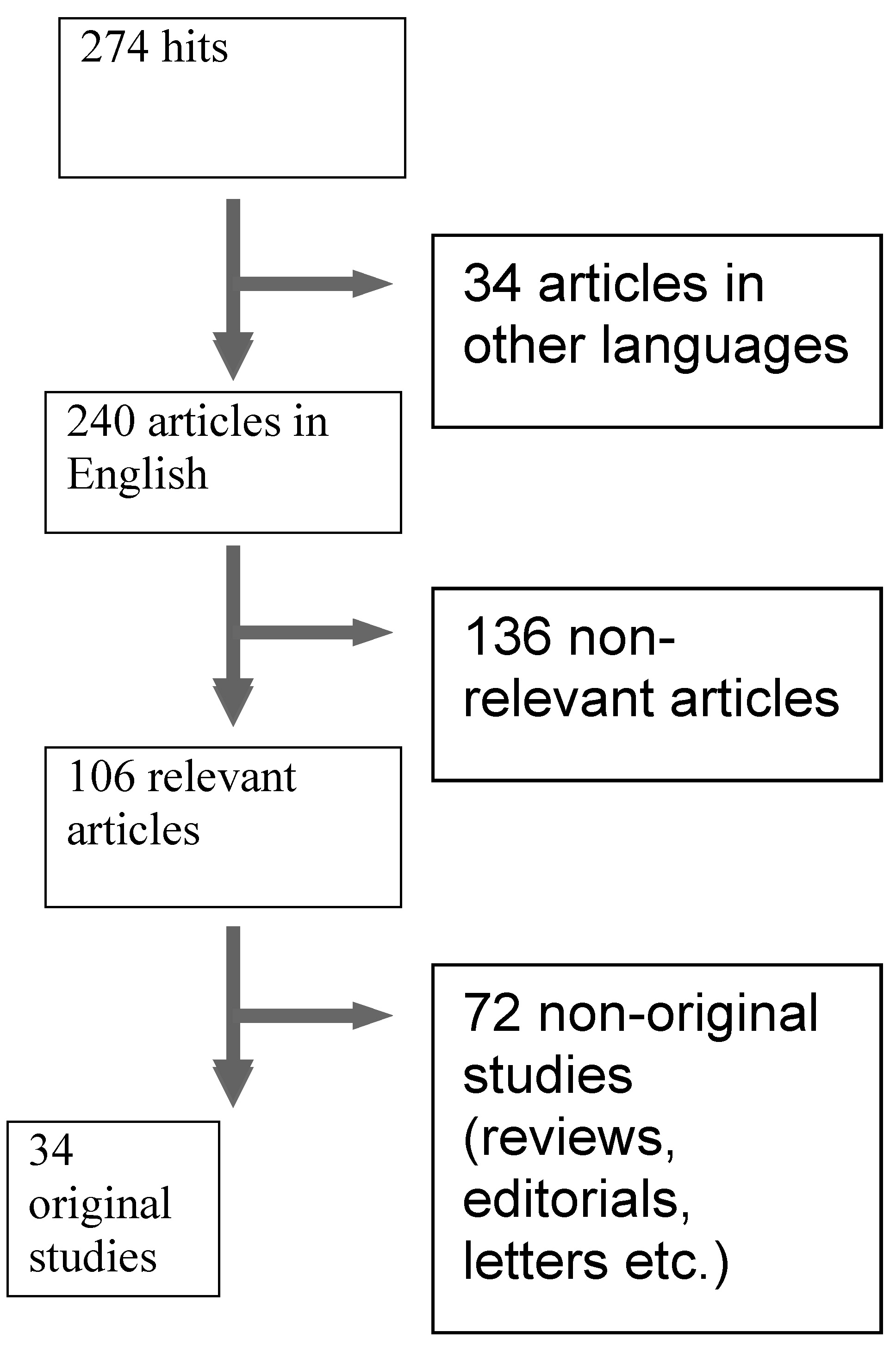
For this literature review, computerized searches were conducted using the Nature online journals, Cochrane Library, Thomson World of Science, PubMed, Ovid, EMBASE, PsycINFO, and Science Direct databases. The search terms “multiple sclerosis,” “demyelinating disease,” “depression,” “affective disorders,” “mood disorders,” and “suicide” were entered into the databases. Further, the table of contents of journals that regularly publish articles relevant to these topics were also reviewed; these included Multiple Sclerosis, Lancet Neurology, Brain, Neurology, Journal of Neurology Neurosurgery and Psychiatry, Journal of Neurology, and Journal of Neuropsychiatry and Clinical Neurosciences. Finally, the reference lists of pertinent articles were also scanned for related papers. All relevant articles published in English between 1960 and 2009 were included in this review. The articles included in the review did not differentiate between unipolar and bipolar depression. Figure 1 summarizes the results of the literature search as described in this section.
FIGURE 1. Flow-Chart Describing the Results of the Literature Search

Brain Lesions and Depression in MS

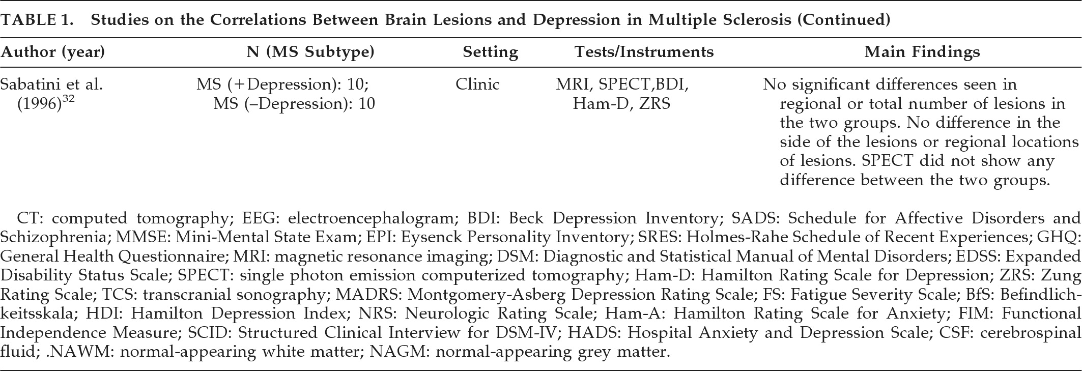
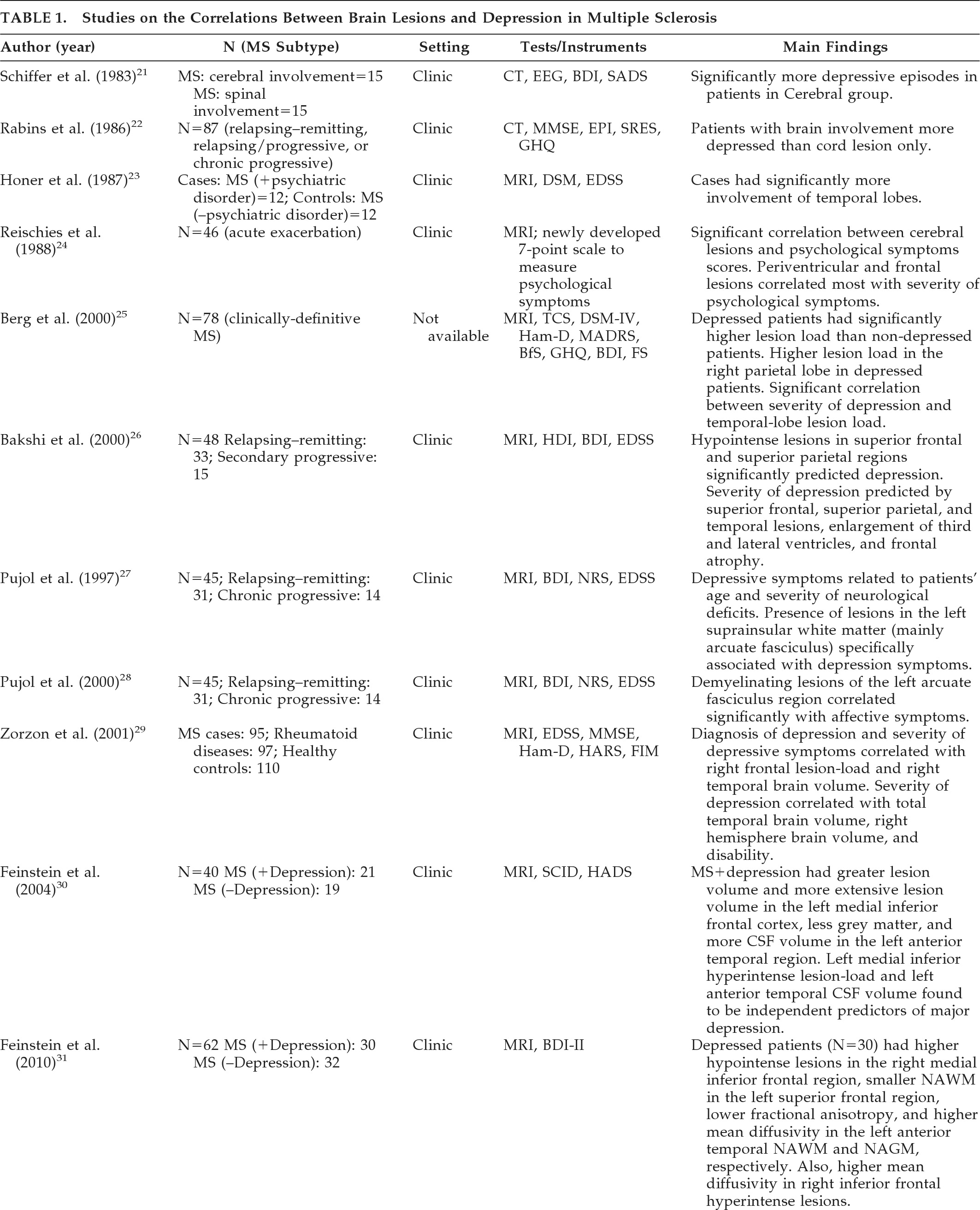
MS is believed to be caused by an autoimmune reaction in genetically susceptible individuals, with the initial attack possibly triggered by environmental factors. The persisting symptoms of MS are thought to be caused by chronic plaques of demyelination. As MS affects the myelin sheath in the CNS, it has been suggested that mood changes may be a direct neurological consequence of the disease.20 We have reviewed studies that investigated the association between structural brain lesions and depression in MS. These are summarized in Table 1.
TABLE 1. Studies on the Correlations Between Brain Lesions and Depression in Multiple Sclerosis
CT: computed tomography; EEG: electroencephalogram; BDI: Beck Depression Inventory; SADS: Schedule for Affective Disorders and Schizophrenia; MMSE: Mini-Mental State Exam; EPI: Eysenck Personality Inventory; SRES: Holmes-Rahe Schedule of Recent Experiences; GHQ: General Health Questionnaire; MRI: magnetic resonance imaging; DSM: Diagnostic and Statistical Manual of Mental Disorders; EDSS: Expanded Disability Status Scale; SPECT: single photon emission computerized tomography; Ham-D: Hamilton Rating Scale for Depression; ZRS: Zung Rating Scale; TCS: transcranial sonography; MADRS: Montgomery-Asberg Depression Rating Scale; FS: Fatigue Severity Scale; BfS: Befindlichkeitsskala; HDI: Hamilton Depression Index; NRS: Neurologic Rating Scale; Ham-A: Hamilton Rating Scale for Anxiety; FIM: Functional Independence Measure; SCID: Structured Clinical Interview for DSM-IV; HADS: Hospital Anxiety and Depression Scale; CSF: cerebrospinal fluid; .NAWM: normal-appearing white matter; NAGM: normal-appearing grey matter.
Schiffer et al.21 investigated 30 patients with MS; 15 patients fulfilled the criteria for cerebral involvement, and 15 had cerebellar or spinal cord involvement. The criteria for cerebral involvement included neurological findings on examination, abnormal EEG, and abnormal CT scans. According to assessment measures (the Beck Depression Inventory [BDI], and a psychiatric interview patterned after the Schedule for Affective Disorders and Schizophrenia [SADS]), there were significantly more depressive symptoms in the patient group with cerebral involvement. Rabins et al.22 studied 87 patients with MS. The treating neurologist characterized MS subtypes (relapsing–remitting, relapsing/progressive, or chronic-progressive) and determined whether the patient only suffered spinal cord involvement or had evidence of brain involvement. CT head scans were performed on 37 patients with brain involvement, and ventricle-to-brain ratio was determined. The General Health Questionnaire was used to assess patients' emotional states. Patients also received the Mini-Mental State Exam (MMSE), the Eysenck Personality Inventory (EPI), and the Holmes-Rahe Schedule of Recent Experiences (SRE). Patients with MS who had brain involvement were more depressed than patients with spinal cord lesion only. Moreover, euphoric patients (i.e., patients with inappropriately cheerful mood) were more likely to have progressive MS with brain involvement and enlarged ventricles, and were more likely to be cognitively impaired than the non-euphoric patients. It should be mentioned that the clinical picture of MS can be associated with both euphoria (exaggerated mood elation) and eutonia (increased sense of physical fitness). Honer et al.23 compared the brain MRI findings from 12 patients with MS and psychiatric disorders with 12 matched patients without psychiatric disorders. Psychiatric data were obtained from case notes, and diagnoses were made using Diagnostic and Statistical Manual of Mental Disorders, 3rd Edition (DSM-III) criteria. The Kurtzke/Expanded Disability Status Scale (EDSS) score was used to measure disability. Analysis of data showed that patients with MS and psychiatric disorders had significantly more pathological involvement of the temporal lobes. Of note, this study did not specifically investigate depression in MS, and the case group had patients with other affective disorders, organic personality disorders, and organic hallucinosis. Reischies et al.24 investigated 46 patients with MS, using brain MRI. All patients had psychological symptoms assessed using a newly-developed 7-point scale that measured symptoms of depressed mood, irritability, reduced drive, and disorders of judgment. The authors found a significant correlation between cerebral lesions and psychological-symptom scores. Periventricular and frontal lesions appeared to correlate most with severity of psychological symptoms. As in the previous study, they did not specifically investigate depression in MS, and the reliability and validity of the scale they developed to assess psychological symptoms is questionable. Berg et al.25 studied 78 patients who fulfilled diagnostic criteria for MS, using neurological, neuropsychiatric and neurophysiological assessment, MRI, and transcranial sonography (TCS). The study aimed to assess whether specific changes of the basal limbic system could be identified in patients with MS and depression; 39.7% of patients fulfilled DSM-IV criteria for depression. The authors did not find any significant alteration of the basal limbic system in the depressed group versus the nondepressed group. However, post-hoc analysis showed that depressed patients had significantly higher lesion-load than nondepressed patients, particularly in the right parietal lobe. A significant correlation was detected between the severity of depression (assessed using the BDI, the Hamilton Rating Scale for Depression [Ham-D], and the Montgomery-Asberg Depression Scale [MADRS]), and the lesion-load in the temporal lobe. Scores on the Fatigue Scale (FS) and lesion-load in right temporal lobe also showed a significant correlation. Bakshi et al.26 assessed the severity of depression in 48 patients with MS, using the BDI and the Hamilton Depression Inventory (HDI). Neurological disability was measured with the EDSS. Hypointense lesions on T1-weighted images in superior frontal and superior parietal regions significantly predicted the presence of depression, both before and after adjusting for EDSS scores. Severity of depression was predicted by superior frontal, superior parietal, and temporal lesions, enlargement of the third and lateral ventricles, and frontal atrophy. Pujol et al.27 studied 45 patients with a definite clinical diagnosis of MS and evidence of cerebral demyelinating lesions on routine brain MRI. All subjects were assessed during a stable period of their illness (at least 3 months before recruitment without relapse or progression). Depressive symptoms were measured with the BDI; neurological symptoms were assessed with the Neurologic Rating Scale (NRS); and disability was assessed with the EDSS. Patients were also assessed using a specific MRI protocol to quantify lesions separately in the basal, medial, and lateral-frontotemporal white matter. Depressive symptoms were weakly but significantly related to patients' age and severity of neurological deficits. The presence of lesions in the left suprainsular white matter (arcuate fasciculus) was specifically associated with depressive symptoms. Pujol et al. conducted a further study with the same group of patients to establish the significance of the previously reported association between depressive symptoms and demyelinating lesions in the region of the left arcuate fasciculus in patients with MS.28 Patients were requested to complete the BDI. The BDI was broken down into main symptom categories according to the results of factor-analytic studies, and correlation patterns between BDI subscores and lesion measurements were analyzed. Demyelinating lesions of the left arcuate fasciculus region correlated significantly with affective symptoms (sadness, pessimism, suicidal ideas, irritability, and social withdrawal) and somatic complaints (insomnia, loss of appetite, weight loss, and somatic preoccupation), with the former showing the highest correlation. The arcuate fasciculus is a major component of suprainsular white matter that is laterally located in the brain and includes several reciprocal connections of the frontal lobe with posterior cortices.
The left hemisphere appears to be more involved than the right hemisphere in the control of motivation-related emotional activity.28 Zorzon et al.29 investigated the relationship between involvement of specific brain areas and the occurrence of anxiety and depression. A group of 95 patients with clinically definitive MS were recruited; 97 patients with other chronic rheumatoid diseases, such as rheumatoid arthritis, systemic lupus erythematosus, ankylosing spondylitis, and psoriatic arthritis were recruited; 110 healthy subjects were also recruited as controls. All subjects underwent a full neurological examination, and assessments using EDSS, Functional Independence Measure (FIM), Ham-D, and Hamilton Anxiety Rating Scale (Ham-A). Diagnosis of depression was made using DSM-IV criteria. In all, 18% of participants with MS, 16% of controls with RA, and 4% of healthy volunteers fulfilled DSM-IV criteria for major depression. All the patients with a diagnosis of MS underwent MRI examinations. Regional and total lesion-loads were calculated. Both diagnosis of depression and severity of depressive symptoms correlated weakly with right frontal lesion-load and right temporal brain volume. The severity of depression correlated significantly with total temporal brain volume, right hemisphere brain volume, and disability. Anxiety did not correlate significantly with any regional and total lesion-loads, brain volume, or any considered clinical variables. The authors suggested that anxiety and depression may have different etiologies in patients with MS. Depression may be caused by brain damage related to MS, whereas anxiety may be a reactive response to psychosocial pressures. Feinstein et al. studied 40 patients with MS.30 All patients were initially screened with the Hospital Anxiety and Depression Rating Scale (HADS), and those who scored ≥10 were interviewed with the Structured Clinical Interview for DSM-IV (SCID); 19 patients had depression, and 21 did not have depression. Compared with euthymic patients, patients with depression had a greater T2-weighted lesion volume and more extensive T1-weighted lesion volume in the left medial inferior frontal cortex. They also had less gray matter and more CSF volume in the left anterior temporal region. Degree of contribution of brain abnormalities to the development of depression was calculated by a logistic-regression model. Hyperintense lesion-load in the left medial inferior frontal cortex and left anterior-temporal CSF volume (an index of regional cerebral atrophy) were found to be independent predictors of major depression.
There is growing interest in newer brain-imaging techniques, such as diffuse tensor and diffusion-weighted imaging studies in patients with MS. These imaging techniques are useful in extracting subtle pathological change in normal-appearing brain tissue. Feinstein et al. studied 62 subjects with MS, using BDI-II and MRI scans. Whole-brain and regional volumes were calculated for lesions and Normal-Appearing Gray Matter (NAGM) and White Matter (NAWM). Depressed patients (N=30) had more hypointense lesions in the right medial inferior frontal region, less NAWM in the left superior frontal region, and lower fractional anisotropy and higher mean diffusivity in the left anterior-temporal NAWM and NAGM, respectively. They also had higher mean diffusivity in right inferior frontal hyperintense lesions. These data suggest that patients with MS and depression have higher lesion volumes and brain atrophy in frontal and temporal regions. Changes in NAGM and NAWM suggest subtle brain pathology in frontal and temporal areas.31
Some studies have not found any association between brain lesions and depression in MS. Sabatini et al.32 investigated the relationship between depression and both brain-structural and blood-flow abnormalities in patients with MS. Ten depressed patients with MS were compared with 10 nondepressed patients with MS. All patients underwent neuropsychological assessment, MRI, and SPECT. Depressive symptoms were measured with the BDI and Ham-D. Anxiety symptoms were measured with the Zung Rating Scale (ZRS). The data from the MRI scans showed that all patients had several cerebral demyelinating lesions, but no significant differences were seen in the regional or total number of lesions in the two groups. The two groups did not differ significantly in the side of the lesions or regional locations of the lesions. SPECT did not show any significant difference between the two groups in any cerebral structure. It could be argued that the study failed to show any significant differences because the number of subjects was quite small. However, cerebral blood flow was significantly different in the two groups. There was relatively higher blood flow on the left side of depressed patients and on the right side of nondepressed patients. Depressed patients had significantly less CRF in the limbic system. Perfusion asymmetries in the limbic system correlated significantly with depression test scores.
In summary, imaging studies to-date vary widely in both their design and methodology. There are few, if any, unequivocal conclusions that can be made from these studies. Most studies used poor rating scales for depression, particularly the BDI, which has too much loading on somatic symptoms. The correlation between lesion location and depression in MS appears less precise than in poststroke depression.9 This may be because most of the older studies investigated the relationship between depressive symptoms and brain atrophy and demyelinating lesions that were clearly evident on the available imaging techniques, potentially missing more subtle disease. However, newer and more sophisticated brain imaging techniques have shown subtle neuropathological changes even in patients deemed to have benign disease. These changes have been linked to neurological and cognitive dysfunction and depression.31 Further research is needed to improve our understanding of the neuropathology of depression and how precisely this relates to MS. Better tools for future research include voxel-based morphometry and MRI techniques that examine connectivity between different areas. Better visualization of cortical and deep white-matter pathology, together with quantitative MRI techniques, such as diffuse tensor imaging, may improve our understanding of this association. Conclusions from earlier studies that subdivided patients with MS into those with and without brain pathology will have to be judged more critically, as the split is likely to be rendered less clear if current imaging techniques to visualize lesional and non-lesional brain pathology were available when the studies were performed. Nevertheless, the available evidence seems to suggest an association between depression in MS and greater neuropathology in frontal, temporal, and parietal regions.
Immune Dysfunction and Depression in MS

An increasing body of evidence suggests that patients with depression show changes in immunological markers. In depressed patients, peripheral markers of the inflammatory response system are often increased.33 Animal models of depression (“sickness behavior”) have shown a clear link between cytokines and these states.34 Direct evidence of a relationship between depression and inflammation has been reported in “vascular” or late-life depression, and postmortem studies of the brain point toward miniature infarcts and a subsequent inflammatory response.35 Several mechanisms have been suggested to explain the link between inflammation and depression. The neuropsychiatric side effects experienced when cytokines are administered for treatment of illness such as hepatitis C provide evidence that these inflammatory mediators can directly affect the brain. Proinflammatory cytokines have been shown to have several effects, such as decreased appetite and weight, sleep disturbance, psychomotor retardation, and anhedonia.34 Assessment of immunological parameters in major depressive disorder has shown that pretreatment levels of C-reactive protein (CRP) and TNF-α were significantly higher in patients with major depression than in controls, and a significant decrease in TNF-α was seen in a responder group during antidepressant treatment.35 It is hypothesized that depressive symptoms may be caused by direct effect of cytokines on the hypothalamus.36 At an experimental level, cytokines such as TNF-α and IL-2 have been found to significantly up-regulate the expression of SERT (serotonin transporter), thus reducing the synaptic availability of serotonin.37
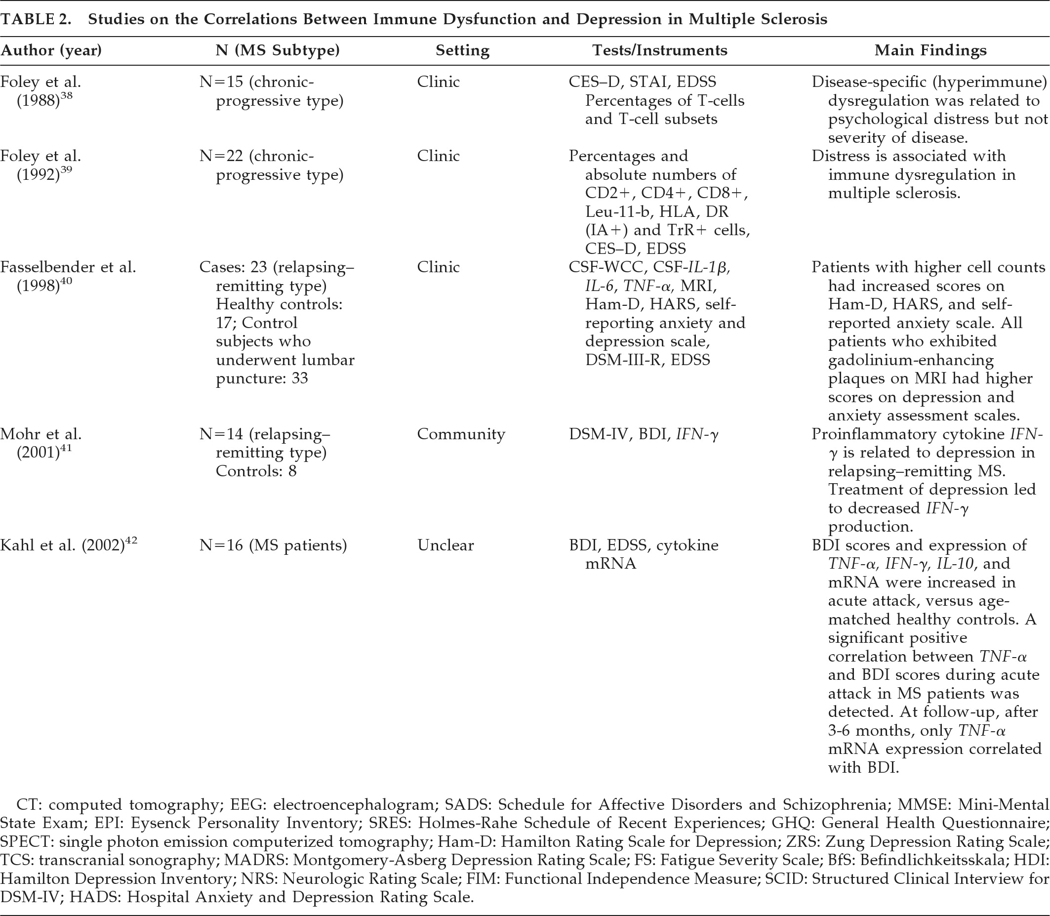
MS is characterized by inflammatory changes in the CNS, and increasing evidence from animal models suggests that some aspects of depression and fatigue in MS may be linked with inflammatory markers.34 Table 2 provides a summary of studies on the role of autoimmune mechanisms in the development of depression in patients with MS. Foley et al.38 studied the relationship between mood and immunity in 15 patients with MS (chronic-progressive type). The EDSS was used to assess physical impairment, and psychological states were measured with the CES–D (Center for Epidemiological Studies–Depression scale) and the STAI (State–Trait Anxiety Inventory). Direct immunofluorescence was used to determine percentages of T-cells and T-cell subsets. Patients were classified into Low-Depressed, High-Depressed, Low-Anxious, and High-Anxious groups. The High-Depressed group manifested a significantly higher number of T4+ helper/inducer cells in peripheral blood. The High-Anxious group had a higher number of T4+ cells, a low number of T8+ suppressor/cytotoxic cells, and a higher T4+/T8+ ratio than the Low Anxious-mood group. These results were not attributable to variation in disease severity. This finding is in contrast with psychologically-distressed, healthy individuals and corresponds to changes noted in MS exacerbations. The data suggest that psychologically distressed patients with MS show disease-related patterns to a greater extent than non-distressed patients with MS. Foley et al.39 examined psychological distress and immune functioning in 22 patients with MS, chronic-progressive type, participating in a placebo-controlled trial of cyclosporine. Immune measures included percentages and absolute numbers of CD2+, CD4+, CD8+, Leu-11-b, HLA-DR (IA+) and transferrin receptor-positive lymphocytes (TrR+). Depression was measured with the CES–D scale, and neurological disability was assessed with the EDSS. Subjects were followed up for 2 years, and High-Depressed episodes and Low-Depressed episodes were compared. Times of greater depression were associated with lower CD8+ percentage and a higher CD4/CD8 ratio. CD4+ cell numbers and percentage were higher when subjects were depressed (only in the placebo group). “CD4+'s” are considered to be helper/inducer cells, and CD8+ cells are thought to be suppressor/cytotoxic. There was no difference in EDSS scores when subjects were more depressed. The authors concluded that distress is associated with immune dysregulation in MS. Fassbender et al.40 compared 23 patients with definite relapsing–remitting MS with two age-matched control groups. The first group (N=17) comprised healthy volunteers. The second group (N=33) had a clinically-indicated lumbar puncture because of suspicion of neurologic or inflammatory disease, but this was excluded after extensive evaluation. Inflammatory activity was evaluated using routine indicators such as CSF white cell count (WCC) and experimental laboratory indicators such as serum and CSF levels of IL-1β, IL-6, and TNF-α. All MS patients underwent MRI imaging using T1-weighted, proton-density, T2-weighted, and gadolinium-pentetic acid (Gd-DTPA)-enhanced images. Depression and anxiety were evaluated with the Ham-D, Ham-A, Zung Self-Reporting Depression Scale, and Zung Self-Reporting Anxiety Scale. Major depression was diagnosed according to DSM-III-R criteria. Neurological impairment was quantified with the EDSS. As expected, patients with MS had higher levels of CSF cell count than controls. When subpopulations of MS patients were compared, those with higher cell counts had significantly increased scores on Hamilton Rating Scales for anxiety and depression and the self-reported anxiety scale. However, none of the experimental indicators correlated with scales for affective disorders. All patients who exhibited gadolinium-enhancing plaques on MRI had significantly higher scores on depression and anxiety assessment scales. Mohr et al.41 investigated the relationship between depression, treatment of depression, and production of interferon gamma (IFN-γ) by peripheral blood mononuclear cells in patients with comorbid diagnoses of depression and relapsing–remitting MS. Interferon-gamma (IFN-γ) is regarded as a major effector mechanism in the pathogenesis of MS, and administration of IFN-γ is reported to trigger exacerbations. The authors recruited 14 patients with clinically definite relapsing–remitting MS. They also recruited 8 nondepressed, healthy subjects. Depression was diagnosed by DSM-IV criteria and measured with the BDI. They used a randomized, comparative outcome trial design and patients had 16 weeks of treatment with sertraline, individual cognitive-behavioral therapy, or group therapy. Their findings suggest that the production of proinflammatory cytokine IFN-γ in relapsing–remitting MS is related to depression, and treatment of depression may decrease IFN-γ production. There was a significant improvement in depressive symptoms in all treatment arms. There was no significant difference between treatment modalities. The authors concluded that treatment of depression might be an important disease-modifying component of management of relapsing–remitting MS. Kahl et al.42 studied 16 patients with MS, using BDI and EDSS. Cytokine mRNA in whole blood was serially determined by a quantitative polymerase chain reaction (PCR) method. Disability scores were mild, and BDI scores did not correlate with extent of disability. BDI scores and expression of TNF-α, IFN-γ, and IL-10 mRNA were increased in acute attacks, as compared with age-matched healthy control subjects. The authors detected a significant positive correlation between TNF-α and BDI scores during acute attacks in patients with MS. At follow-up, after 3–6 months, only TNF-α mRNA expression correlated with BDI scores. These results suggest a contribution of TNF-α and IFN-γ in the development of depressive symptoms in MS.42
TABLE 2. Studies on the Correlations Between Immune Dysfunction and Depression in Multiple Sclerosis
CT: computed tomography; EEG: electroencephalogram; SADS: Schedule for Affective Disorders and Schizophrenia; MMSE: Mini-Mental State Exam; EPI: Eysenck Personality Inventory; SRES: Holmes-Rahe Schedule of Recent Experiences; GHQ: General Health Questionnaire; SPECT: single photon emission computerized tomography; Ham-D: Hamilton Rating Scale for Depression; ZRS: Zung Depression Rating Scale; TCS: transcranial sonography; MADRS: Montgomery-Asberg Depression Rating Scale; FS: Fatigue Severity Scale; BfS: Befindlichkeitsskala; HDI: Hamilton Depression Inventory; NRS: Neurologic Rating Scale; FIM: Functional Independence Measure; SCID: Structured Clinical Interview for DSM-IV; HADS: Hospital Anxiety and Depression Rating Scale.
In summary, evidence from studies in depression suggest a significant association between immune dysfunction and depression. How this precisely relates to MS is still unclear, although, hypothetically, this is possible in an autoimmune disease such as MS. There is only limited evidence that suggests a relationship between autoimmune factors and depression in MS. Small sample sizes are a major limitation of these studies. Because there is a paucity of studies in this field, it is difficult to come to any firm conclusions on the association between immune dysfunction and depression in MS.

Immunomodulatory Treatments and Depression in MS

Literature on the frequency and phenomenology of IFN-induced depression indicates that depression could occur among patients undergoing IFN therapy for variety of conditions, such as malignancies, hepatitis C, MS, and skin conditions.43 It is thought that proinflammatory cytokines and tumor necrosis factor affect serotonin metabolism directly or indirectly by inducing the enzyme indoleamine 2,3-dioxygenase. This leads to peripheral depletion of tryptophan, a precursor of serotonin. There is a large body of evidence linking serotonin and depression and the effectiveness of selective serotonin-reuptake inhibitor (SSRI) antidepressants in treating IFN-induced depression.43,44
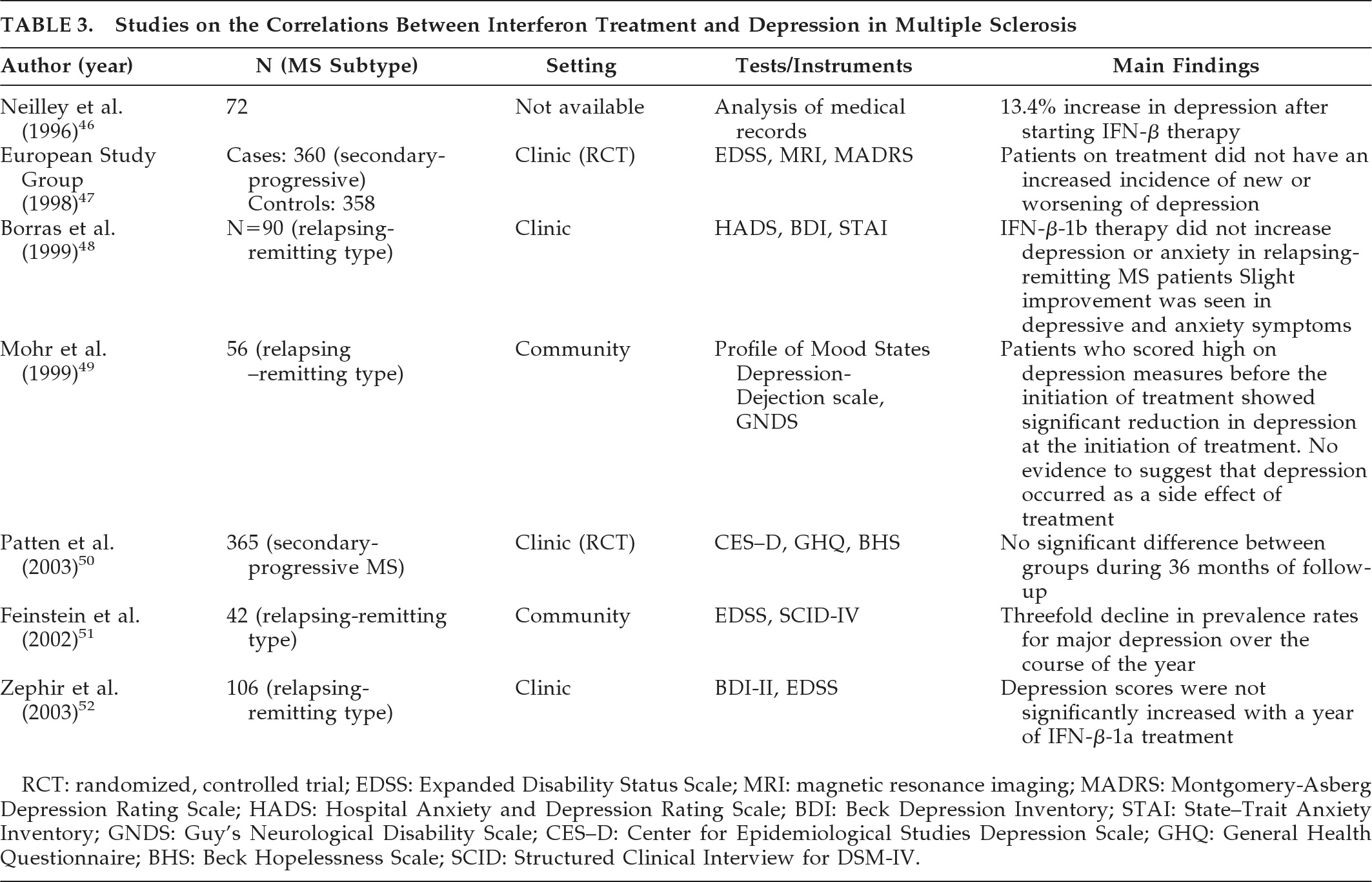
Initial suspicion of a link between IFN-β therapy and depression arose after reports of a suicide and an attempted suicide during the first trial of IFN-β-1b.45 Studies that have investigated the effect of interferon treatment on mood in patients with MS have been summarized in Table 3. Neilley et al.46 followed up 72 patients with MS who had begun IFN-β therapy.46 Side effects were noted in patient records. Analysis of records showed a 13.4% increase in depression after patients were commenced on IFN-β therapy. However, the results were not statistically significant. The authors did not describe how they evaluated depression in their study group. Later studies have not shown an association between IFN-β therapy and depression. The European study group on IFN-β-1b in secondary-progressive MS conducted a multicenter, double-blind, randomized, placebo-controlled trial to assess beneficial effects of IFN-β-1b in secondary-progressive MS;47 360 patients were allocated to treatment, and 358 patients were allocated to the placebo arm. Depressive symptoms were monitored with the MADRS every 3 months. Patients on treatment did not have an increase in incidence of new or worsening of depression. Suicide or suicidal acts were reported in five patients on placebo and three in the treatment arm. Borras et al.48 evaluated emotional states of 90 patients during IFN-β-1b therapy. They assessed their emotional states by Ham-D, BDI, and STAI. All patients underwent psychological testing at onset of treatment; 75 patients were evaluated at 12 months; and 56 patients at 24 months. They found that IFN-β-1b therapy did not increase depression or anxiety in patients with relapsing–remitting MS. On the contrary, slight improvement was seen in depressive and anxiety symptoms.
TABLE 3. Studies on the Correlations Between Interferon Treatment and Depression in Multiple Sclerosis
RCT: randomized, controlled trial; EDSS: Expanded Disability Status Scale; MRI: magnetic resonance imaging; MADRS: Montgomery-Asberg Depression Rating Scale; HADS: Hospital Anxiety and Depression Rating Scale; BDI: Beck Depression Inventory; STAI: State–Trait Anxiety Inventory; GNDS: Guy's Neurological Disability Scale; CES–D: Center for Epidemiological Studies Depression Scale; GHQ: General Health Questionnaire; BHS: Beck Hopelessness Scale; SCID: Structured Clinical Interview for DSM-IV.
Mohr et al.49 studied 56 patients with confirmed relapsing forms of MS. Levels of depression in patients with MS was assessed 2 weeks before initiation of IFN-β-1a treatment, at initiation of treatment, and at 2-month follow-up, using the Profile of Mood States Depression–Dejection scale. Neurological status was assessed with the Guy's Neurological Disability Scale (GNDS). Patients who scored high on depression measures before the initiation of treatment showed significant reduction in depression at the initiation of treatment. However, depression measures returned to initial levels within 2 months of treatment. There was no evidence to suggest that depression occurred as a side effect of treatment. Patten et al.50 analyzed data of 365 subjects treated with IFN-β-1a or placebo from a multicenter trial of IFN-β-1a in patients with secondary-progressive MS. The CES–D, General Health Questionnaire (GHQ) and Beck Hopelessness Scale (BHS) were used to evaluate psychological symptoms. The authors did not find any significant difference between groups during 36 months of follow-up. Feinstein et al.51 followed 42 patients with clinically-definite MS (relapsing–remitting type) for a year after starting IFN-β-1b treatment. Psychiatric symptoms were evaluated, using the Structural Clinical Interview for DSM-IV Axis I disorders (SCID-IV). Disability was assessed using the EDSS. The majority of subjects with major depression had a history of depression before initiation of treatment. There was a threefold decline in prevalence rates for major depression over the course of the year. Zephir et al.52 followed a cohort of 106 patients with relapsing–remitting MS during their first year of treatment with IFN-β-1a. Their depressive symptoms were evaluated with the BDI–II. Disability was assessed with the EDSS. Their results showed that depression scores were not significantly increased with a year of IFN-β-1a treatment.
IFN-α is well known to affect mood when used in treatment of hepatitis C, malignant melanoma, and other malignancies.43 It was thought that IFN-β might have a similar effect on mood when it was introduced for treatment of MS. However, despite the initial suspicion and concerns, rigorous studies have not confirmed an association between IFN-β therapy and depression. Studies have also failed to show higher rates of depression in patients with MS treated with other long-term immunomodulatory therapy, such as glatiramer acetate. Therefore, it is safe to conclude that other factors are more likely to cause depression in patients with MS.53,54 Nevertheless, clinicians must consider the possibility of a link between the two when dealing with patients with MS on interferon therapy.6,56

Psychosocial Effects of MS and Depression

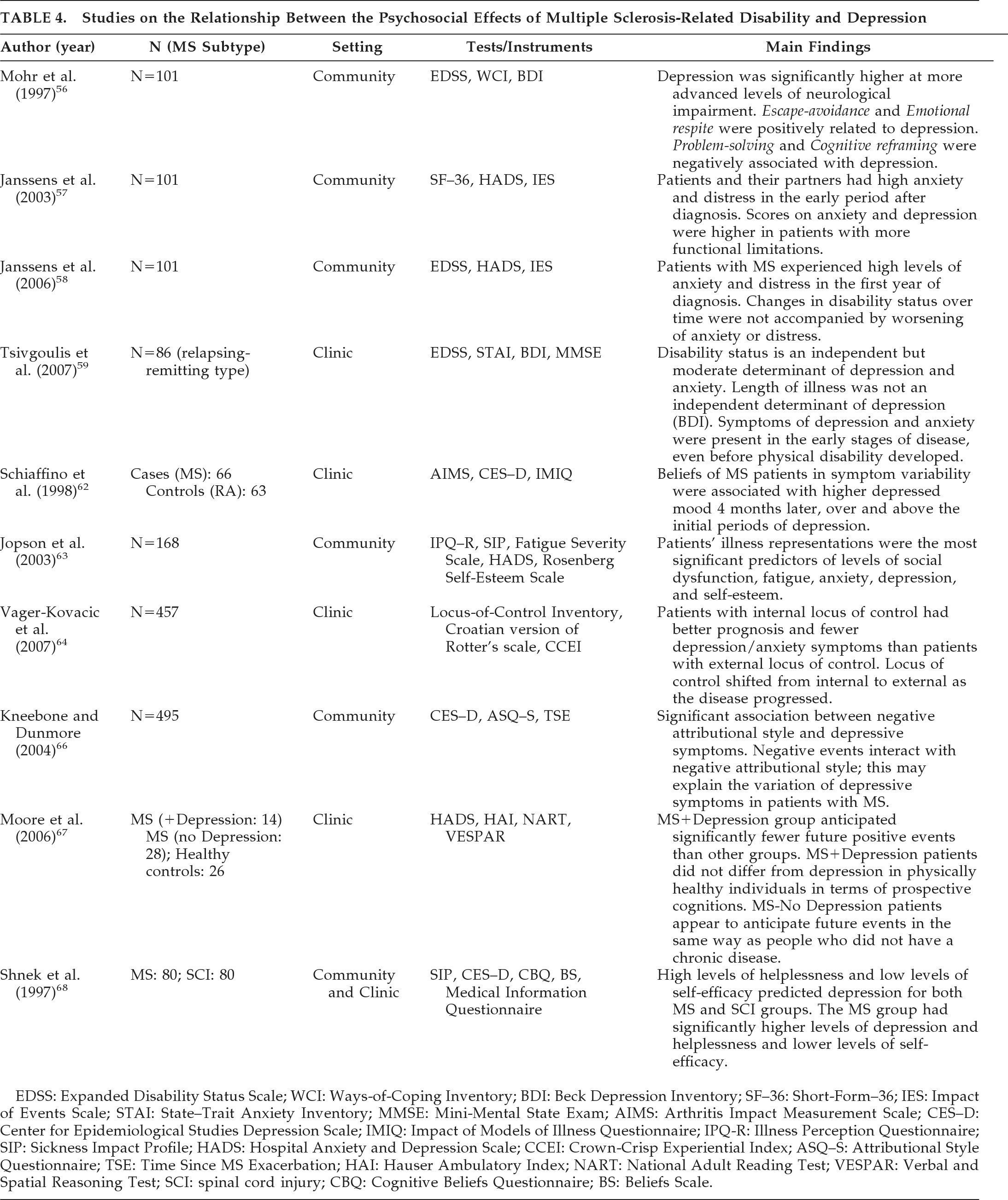
MS is often associated with loss of employment, decreased mobility, and change in social roles. The unpredictable nature of the illness may result in feelings of lack of control and hopelessness, which may in turn lead to depression.6 Several hypotheses have been proposed to explain psychosocial effects of MS; the studies that have investigated these hypotheses have been summarized in Table 4. One hypothesis suggests a causal link between severity of illness and depression. Studies have investigated the relationship between illness severity and depression/anxiety symptoms. The results are ambiguous, and recent studies have been inconclusive. Mohr et al.56 examined the relationship between depression, coping, and severity of neurological illness. A group of 101 patients with clinically definitive MS were assessed with the EDSS, the BDI, and the Ways-of-Coping Inventory (WCI). The authors reported that depression was significantly higher at more advanced levels of neurological impairment. The mechanisms of escape-avoidance and emotional respite (fantasy, daydreaming) were positively related to levels of depression; problem-solving and cognitive reframing were negatively associated with depression. Janssens et al.57 studied 101 recently-diagnosed patients (less than 2 years) with MS. Data were collected from patients and their partners on health-related quality of life (SF–36), anxiety, depression (HADS) and disease-related distress (Impact of Events Scale) measures. Functional limitations were assessed by the EDSS. This group of patients was followed up for 2 years.58 The authors reported that patients with MS experienced high levels of anxiety and distress in the first year of diagnosis. Scores on anxiety and depression were higher in patients with more functional limitations. Changes in disability status over time were not accompanied by worsening of anxiety or distress. Tsivgoulis et al.59 investigated the association between depression and disability in 86 patients with MS by use of a cross-sectional design. They concluded that disability status is an independent but moderate determinant of depression and anxiety. Length of illness was not an independent determinant of BDI scores, and pronounced symptoms of depression and anxiety were present in the early stages of disease, even before physical disability developed.
TABLE 4. Studies on the Relationship Between the Psychosocial Effects of Multiple Sclerosis-Related Disability and Depression
EDSS: Expanded Disability Status Scale; WCI: Ways-of-Coping Inventory; BDI: Beck Depression Inventory; SF–36: Short-Form–36; IES: Impact of Events Scale; STAI: State–Trait Anxiety Inventory; MMSE: Mini-Mental State Exam; AIMS: Arthritis Impact Measurement Scale; CES–D: Center for Epidemiological Studies Depression Scale; IMIQ: Impact of Models of Illness Questionnaire; IPQ-R: Illness Perception Questionnaire; SIP: Sickness Impact Profile; HADS: Hospital Anxiety and Depression Scale; CCEI: Crown-Crisp Experiential Index; ASQ–S: Attributional Style Questionnaire; TSE: Time Since MS Exacerbation; HAI: Hauser Ambulatory Index; NART: National Adult Reading Test; VESPAR: Verbal and Spatial Reasoning Test; SCI: spinal cord injury; CBQ: Cognitive Beliefs Questionnaire; BS: Beliefs Scale.
The model of “illness representations” suggests that people's illness representations influence their adjustment to illness through the impact of these representations on coping, regimen compliance, and self-evaluation.60 Illness representations include beliefs about personal identity and causes/consequences of diseases. An individual's ideas about his medical condition and its prognosis are thought to derive from personal experiences with illnesses, information from the medical care system, and messages from the broader culture.61 Schiaffino et al.62 assessed illness representations in 66 patients with MS and 63 patients with rheumatoid arthritis, exploring the relationship between concurrent and later mood. The beliefs of patients with MS in symptom variability were associated with higher depressed mood 4 months later, over and above the initial periods of depression. Jopson et al.63 investigated patients' illness representations' impact on their adjustment to MS, even when controlling for severity of the condition. Their results suggest that illness-severity accounted for the majority of the variance in physical and role dysfunction, whereas patients' illness representations were the most significant predictors of levels of social dysfunction, fatigue, anxiety, depression, and self-esteem.
Vager-Kovacic et al. investigated the relationship between locus-of-control and depression/anxiety symptoms in 457 Croatian patients.65 They reported that patients with internal locus of control (belief that they can control events in their lives) had better prognosis and fewer depression/anxiety symptoms than patients with external locus of control. They also found that the locus of control shifted from internal to external as the disease progressed. “Hopelessness theory” has been suggested to explain depression in MS. Negative life-events interact with negative attributional style in patients with MS, and this could explain the higher prevalence of depression in this patient population.65 Kneebone and Dunmore investigated this theory by collecting data via cross-sectional survey of 495 patients with MS.66 A significant association between negative attributional style and depressive symptoms was identified. These findings suggest that negative events interact with negative attributional style, and this may explain the variance of depressive symptoms in patients with MS. However, because the study had a cross-sectional design, the authors were unable to investigate a causal relationship between negative attributional style and depression. In other words, the high prevalence of negative attributional style in patients with MS and depression may simply be a reflection of negative thinking associated with depressive state of mind and not a causative factor, as proposed in the above-mentioned study. Moore et al. compared patients with MS and depression (N=14), MS without depression (N=28), and healthy controls (N=26) on their ability to generate possible future positive and negative experiences. They reported that the group of patients with MS and depression anticipated significantly fewer future positive events than other groups. This group also anticipated more MS-related negative events. Nondepressed patients with MS appeared to anticipate future events in the same way as people who did not have a chronic disease.67
Learned helplessness, low levels of self-efficacy, and cognitive distortions have all been associated with depression. Shnek et al.68 compared 80 patients with MS and 80 patients with spinal cord injury (SCI), using a cross-sectional design. Results indicated that high levels of helplessness and low levels of self-efficacy predicted depression for both MS and SCI groups. The MS group had significantly higher levels of depression and helplessness and lower levels of self-efficacy. The authors hypothesized that it may have been the combination of the unpredictable nature of MS and the possibility of being affected in several different ways that may have produced greater levels of depression, helplessness, and lower levels of self-efficacy in the MS group.
In summary, the role of psychosocial factors in the etiology of depression in MS is well acknowledged. However, the precise mechanism by which these factors affect mood in MS is still unclear. It appears that individuals' cognitive styles and circumstances play an important role in the etiology of depression in patients with MS.

CONCLUSIONS

The association between depression and MS was identified a long time ago and has been explored in several rigorous studies over the past few decades. It appears that the etiology of depression is multifactorial, and the cause of depression in individual patients might differ. Studies of depression in patients with MS are problematic for a variety of reasons. “Depressive symptoms” in this patient population may be easily mistaken for “major depressive illness.”69 Although prevalence of depression in MS has been estimated to be higher than in normal population and those with other chronic illness, these numbers could be overestimated because of sample selection, as most prevalence studies considered mainly patients attending specialist MS clinics, rather than those coping well at home.70
Some physical symptoms of MS, such as fatigue and sleep and appetite disturbance may overlap with symptoms of depression. Certain classic symptoms of depression, such as guilt or feelings of worthlessness, occur less frequently in depressed patients with MS. Pathological laughing or crying, known as “pseudobulbar affect,” occurs with varying severity in up to 10% of patients with MS.71 This may lead to an erroneous diagnosis of depression in these patients. Thus, depression in patients with MS is harder to diagnose and is often unrecognized and undertreated, as compared with their neurological disabilities.72 Nevertheless, it is imperative that clinicians do not miss depression, as it is a major problem faced by patients with MS and has significant effect on their quality of life. Unlike other aspects of MS, depression is a potentially preventable cause of death in MS.6 Several treatment options appear to be effective in treating depression. Antidepressant drugs, such as tricyclic antidepressants (TCAs), SSRIs, serotonin-noradrenaline reuptake inhibitors (SNRIs), and noradrenaline reuptake inhibitors (NARIs) have been reported to be effective in treating depression in patients with MS.73,74 Psychotherapeutic interventions such as group and individual cognitive-behavioral therapy and insight-oriented therapy have also been reported to be effective.55 Finally, ECT has been reported to be effective in severe cases.75
With advancements in research, it might be possible in the future to differentiate the precise etiology in individual patients and tailor specific treatment strategies with greater effectiveness. Early recognition and effective treatment of depression in patients with MS not only improves their psychological well-being, it also improves compliance with medical treatment and enhances health-related quality of life.76 Early recognition and management of depression in MS has also been identified as a key predictor of morbidity, mortality, and, possibly, physical outcome and disease exacerbations, further confirming that a comprehensive biopsychosocial neuropsychiatric approach is essential for the optimal care of patients with MS.77

References

1.
Turner AP, Williams RM, Bowe JD, et al.: Suicidal ideation and multiple sclerosis. Arch Phys Med Rehabil 2006; 87:1073–1077
2.
Moore AC, MacLeod AK, Barnes D, et al.: Future thinking and depression in relapsing–remitting multiple sclerosis. Br J Health Psychol 2006; 11:663–675
3.
Charcot JM: Lectures on Diseases of the Nervous System, delivered at La Salpetriere. London, UK, New Sydenham Society, 1887, p 194–195
4.
José M: Psychological aspects of multiple sclerosis. Clin Neurol Neurosurg 2008; 110:868–877
5.
Minden SL, Schiffer RB: Affective disorders in multiple sclerosis: review and recommendations for clinical research. Arch Neurol 1990; 47:98–104
6.
Wallin MT, Wilken JA, Turner AP, et al.: Depression and multiple sclerosis: review of a lethal combination. J Rehab Res Dev 2006; 43:45–62
7.
Sadovnick AD, Remick RA, Allen J, et al.: Depression and multiple sclerosis. Neurology 1996; 46:628–632
8.
Chwastiak L, Ehde DM, Gibbons LE, et al.: Depressive symptoms and severity of illness in multiple sclerosis: epidemiologic study of a large community sample. Am J Psychiatry 2002; 159:1862–1868
9.
Rickards H: Depression in neurological disorders: Parkinson's disease, multiple sclerosis, and stroke. J Neurol Neurosurg Psychiatry 2005; 76:i48–i52
10.
Lester K, Stepleman L, Hughes M: The association of illness severity, self reported cognitive impairment, and perceived illness-management with depression and anxiety in a multiple sclerosis clinic population. J Behav Med 2007; 30:177–186
11.
Sadovnick AD, Eisen K, Paty DW, et al.: Cause of death in patients attending multiple sclerosis clinics. Neurology 1991; 41:1193–1196
12.
Feinstein A: An examination of suicide intent in patients with multiple sclerosis. Neurology 2002; 59:674–678
13.
Barbellion WNP: The Journal of a Disappointed Man. London, UK, Chatto and Windus, 1919
14.
Pommé B, Girard J, Planche R: Forme depressive de début d'une sclerosis en plaques. Ann Med Psychol 1963; 121:133
15.
O' Malley PP: Severe mental symptoms in disseminated sclerosis: a neuropathological study. J Irish Med Ass 1966; 55:115–127
16.
Young AC, Saunders J, Ponford JR: Mental change as an early feature of multiple sclerosis. J Neurol Neurosurg Psychiatry 1976; 39:1008–1013
17.
Zarei M: Clinical characteristics of cortical multiple sclerosis. J Neurol Sci 2006; 245:53–58
18.
Whitlock FA, Siskind MM: Depression as a major symptom of multiple sclerosis. J Neurol Neurosurg Psychiatry 1980; 43:861–865
19.
Sullivan MJL, Weinshenker B, Mikail S, et al.: Depression before and after diagnosis of multiple sclerosis. Multiple Sclerosis 1995; 1:104–108
20.
Wallin MT, Wilken JA, Turner AP, et al.: Depression and multiple sclerosis: review of a lethal combination. J Rehab Res Dev 2006; 43:45–62
21.
Schiffer RB, Caine ED, Bamford KA, et al.: Depressive episodes in patients with multiple sclerosis. Am J Psychiatry 1983; 140:1498–1500
22.
Rabins PV, Benjamin RB, O' Donell P, et al.: Structural brain correlates of emotional disorder in multiple sclerosis. Brain 1986; 109:585–597
23.
Honer WG, Hurwitz T, Li DKB, et al.: Temporal lobe involvement in multiple sclerosis patients with psychiatric disorders. Arch Neurol 1987; 44:187–190
24.
Reischies FM, Baum K, Brau H, et al.: Cerebral magnetic resonance imaging findings in multiple sclerosis. Arch Neurol 1988; 45:1114–1116
25.
Berg D, Supprian T, Thomas J, et al.: Lesion patterns in patients with multiple sclerosis and depression. Multiple Sclerosis 2000; 6:156–162
26.
Bakshi R, Czarnecki D, Shaikh ZA, et al.: Brain MRI lesions and atrophy are related to depression in multiple sclerosis. Brain Imag 2000; 11:1153–1158
27.
Pujol J, Bello J, Deus J, et al.: Lesions in the left arcuate fasciculus region and depressive symptoms in multiple sclerosis. Neurology 1997; 49:1105–1110
28.
Pujol J, Bello J, Deus J, et al.: Beck Depression Inventory factors related to demyelinating lesions of the left arcuate fasciculus region. Psychiatr Res 2000; 99:151–159
29.
Zorzon M, de Masi R, Nasuelli D, et al.: Depression and anxiety in multiple sclerosis: a clinical and MRI study in 95 subjects. J Neurol 2001; 248:416–421
30.
Feinstein A, Roy P, Lobaugh N, et al.: Structural brain abnormalities in multiple sclerosis patients with major depression. Neurology 2004; 62:586–590
31.
Feinstein A, O'Connor P, Akbar N, et al.: Diffuse tensor imaging abnormalities in depressed multiple sclerosis patients. Multiple Sclerosis 2010; 16:189–196
32.
Sabatini U, Pozzilli C, Pantano P, et al.: Involvement of the limbic system in multiple sclerosis patients with depressive disorders. Biol Psychiatry 1996; 39:970–975
33.
Korf J, Klein HC, Versijpt J, et al.: Considering depression as a consequence of activation of the inflammatory response system. Acta Neuropsychiatr 2002; 14:1–10
34.
Gold SM, Irwin MR: Depression and immunity: inflammation and depressive symptoms in multiple sclerosis. Neurol Clin 2006; 24:507–519
35.
Thomas AJ, Ferrier IN, Kalaria RN, et al.: Evaluation in late-life depression of intercellular adhesion molecule-1 expression in the dorsal prefrontal cortex. Am J Psychiatry 2000; 157:1682–1684
36.
Lanquillon S, Krieg JC, Bening-Abu-Sach U, et al.: Cytokine production and treatment response in major depressive disorder. Neuropsychopharmacology 2000; 22:370–379
37.
Cavanagh J, Mathias C: Inflammation and its relevance to psychiatry. Adv Psychiatr Treat 2008; 14:248–255
38.
Foley FW, Miller AH, Traugott U, et al.: Psychoimmunological dysregulation in multiple sclerosis. Psychosomatics 1988; 29:398–403
39.
Foley FW, Traugott U, LaRocca NG, et al.: A prospective study of depression and immune dysregulation in multiple sclerosis. Arch Neurol 1992; 49:238–244
40.
Fesselbender K, Schmidt R, Mofsner R, et al.: Mood disorders and dysfunction of the hypothalamic-pituitary-adrenal axis in multiple sclerosis. Arch Neurol 1998; 55:66–72
41.
Mohr DC, Goodkin DE, Islar J, et al.: Treatment of depression is associated with suppression of non-specific and antigen-specific TH1 responses in multiple sclerosis. Arch Neurol 2001; 58:1081–1086
42.
Kahl KG, Kruse N, Faller H, et al.: Expression of tumor necrosis factor-α and interferon-γ mRNA in blood cells correlates with depression scores during an attack in patients with multiple sclerosis. Psychoneuroendocrinology 2002; 27:671–681
43.
Loftis JM, Hauser P: The phenomenology and treatment of interferon-induced depression. J Affect Disord 2004; 82:175–190
44.
Myint AM, Schwarz MJ, Steinbusch HWM, et al.: Neuropsychiatric disorders related to interferon and interleukins treatment. Metab Brain Dis 2009; 24:55–68
45.
Klapper JA: Interferon-beta treatment of multiple sclerosis. Neurology 1994; 44:188
46.
Neilley LK, Goodin DS, Goodkin DE, et al.: Side-effect profile of interferon β-1b in MS: results of an open-label trial. Neurology 1996; 46:552–554
47.
European Study Group on IFN-β-1b in secondary-progressive MS: placebo-controlled multicentre randomised trial of interferon-β-1b in treatment of secondary-progressive multiple sclerosis. Lancet 1998; 352:1491–1497
48.
Borras C, Rio J, Porcel J, et al.: Emotional state of patients with relapsing–remitting MS treated with interferon β-1b. Neurology 1999; 52:1636–1639
49.
Mohr DC, Likosky W, Dwyer P, et al.: Course of depression during the initiation of interferon β-1a treatment for multiple sclerosis. Arch Neurol 1999; 56:1263–1265
50.
Patten SB, Metz LM: Interferon β1a and depression in secondary-progressive MS: data from SPECTRIMS trial. Neurology 2003; 59:744–746
51.
Feinstein A, O'Connor P, Feinstein K: Multiple sclerosis, interferon β-1b, and depression: a prospective investigation. J Neurol 2002; 249:815–820
52.
Zephir H, De Seze J, Stojkovic T, et al.: Multiple sclerosis and depression: influence of interferon-β therapy. Multiple Sclerosis 2003; 9:284–288
53.
Pattern SB: Depressive symptoms in a treated multiple sclerosis cohort. Multiple Sclerosis 2003; 9:616–620
54.
Feinstein A: Multiple sclerosis, disease-modifying treatments, and depression: a critical methodological review. Multiple Sclerosis 2000; 6:343–348
55.
Walther EU, Hohlfeld R: Multiple sclerosis: side effects of interferon beta therapy and their management. Neurology 1999; 53:1622–1627
56.
Mohr DC, Goodkin DE, Gatto N, et al.: Depression, coping, and level of neurological impairment in multiple sclerosis. Multiple Sclerosis 1997; 3:254–258
57.
Janssens ACJW, Van Doorn PA, de Boer JB, et al.: Impact of recently-diagnosed multiple sclerosis on quality of life, anxiety, depression, and distress of patients and partners. Acta Neurol Scand 2003; 108:389–395
58.
Janssens ACJW, Buljevac D, Van Doorn PA, et al.: Prediction of anxiety and distress following diagnosis of multiple sclerosis: a two-year longitudinal study. Multiple Sclerosis 2006; 12:794–780
59.
Tsivgoulis G, Triantafyllou N, Papageorgiou C, et al.: Associations of the Expanded Disability Status Scale with anxiety and depression in multiple sclerosis outpatients. Acta Neurol Scand 2007; 115:67–72
60.
Nerenz DR, Leventhal H: Self-regulation theory in chronic illness, in Coping With Chronic Disease. Edited by, Burish TG, Bradley LA. New York, Academic Press, 1983, pp 13–37
61.
Bishop GD, Briede C, Cavazos L, et al.: Processing illness information: the role of disease prototypes. Basic Appl Soc Psychol 1987; 8:21–44
62.
Schiaffino KM, Shawaryn MA, Blum D: Examining the impact of illness representations on psychological adjustment to chronic illnesses. Health Psychol 1998; 17:262–268
63.
Jopson NM, Moss-Morris R: The role of illness severity and illness representations in adjusting to multiple sclerosis. J Psychosom Res 2003; 54:503–511
64.
Vuger-Kovacic D, Gregurek R, Kovacic D, et al.: Relationship between anxiety, depression, and locus of control of patients with multiple sclerosis. Multiple Sclerosis 2007; 13:1065–1067
65.
Abramson LY, Matasky GI, Alloy LB: Hopelessness depression: a theory-based subtype of depression. Psychol Rev 1989; 96:358–372
66.
Kneebone II, Dunmore E: Attributional style and symptoms of depression in persons with multiple sclerosis. Int J Behav Med 2004; 11:110–115
67.
Moore AC, MacLeod AK, Barnes D, et al.: Future thinking and depression in relapsing–remitting multiple sclerosis. Br J Health Psychol 2006; 11:663–675
68.
Shnek ZM, Foley FW, LaRocca NG, et al.: Helplessness, self-efficacy, cognitive distortions, and depression in multiple sclerosis and spinal cord injury. Ann Behav Med 1997; 19:287–294
69.
Sadovnick AD, Remick RA, Allen J, et al.: Depression and multiple sclerosis. Neurology 1996; 46:628–632
70.
Siegert RJ, Abernethy DA: Depression in multiple sclerosis: a review. J Neurol Neurosurg Psychiatry 2005; 76:469–475
71.
Feinstein A: Mood disorders in multiple sclerosis and effects on cognition. J Neurol Sci 2006; 245:63–66
72.
Rickards H: Depression in neurological disorders: an update. Curr Opin Psychiatry 2006; 19:294–298
73.
Schiffer RB, Winemann NM: Antidepressant pharmacology of depression associated with multiple sclerosis. Am J Psychiatry 1990; 147:1493–1497
74.
Scott TF, Nussbaum P, McConnell H, et al.: Measurement of treatment response to sertraline in depressed multiple sclerosis using the Carroll Scale. Neurol Res 1995; 17:421–422
75.
Corruble E, Awad H, Chouinard G, et al.: ECT in delusional depression with multiple sclerosis. Am J Psychiatry 2004; 161:1715
76.
Sallom AC, Kneebone II: Treatment of depression in people who have multiple sclerosis. Multiple Sclerosis 2007; 13:632–635
77.
Paparrigopoulos T, Ferentinos P, Kouzoupis A, et al.: The neuropsychiatry of multiple sclerosis: focus on disorders of mood, affect, and behaviour. Int Rev Psychiatry 2010; 22:14–21

Information & Authors

Information

Published In

Go to The Journal of Neuropsychiatry and Clinical Neurosciences
The Journal of Neuropsychiatry and Clinical Neurosciences
Pages: 261 - 276
PubMed: 21948887

History

Received: 18 July 2010
Revision requested: 3 October 2010
Accepted: 22 November 2010
Published online: 1 July 2011
Published in print: Summer 2011

Keywords

  1. Multiple Sclerosis
  2. Depression
  3. Cytokines

Authors

Details

Joe John Vattakatuchery, M.D.
From the Dept. of Psychiatry, Hollins Park Hospital, 5 Boroughs Partnership NHS Foundation Trust, Warrington, UK; the Dept. of Neuropsychiatry MTNRG, University of Birmingham, Birmingham, UK; and Sobell Department of Motor Neuroscience and Movement Disorders, Institute of Neurology, UCL, London, UK.
Hugh Rickards, M.D.
From the Dept. of Psychiatry, Hollins Park Hospital, 5 Boroughs Partnership NHS Foundation Trust, Warrington, UK; the Dept. of Neuropsychiatry MTNRG, University of Birmingham, Birmingham, UK; and Sobell Department of Motor Neuroscience and Movement Disorders, Institute of Neurology, UCL, London, UK.
Andrea Eugenio Cavanna, M.D., Ph.D.
From the Dept. of Psychiatry, Hollins Park Hospital, 5 Boroughs Partnership NHS Foundation Trust, Warrington, UK; the Dept. of Neuropsychiatry MTNRG, University of Birmingham, Birmingham, UK; and Sobell Department of Motor Neuroscience and Movement Disorders, Institute of Neurology, UCL, London, UK.

Notes

Correspondence: Dr. Andrea E. Cavanna, Department of Neuropsychiatry, Birmingham and Solihull Mental Health NHS Foundation Trust; [email protected] (e-mail).

Metrics & Citations

Metrics

Citations

Export Citations

If you have the appropriate software installed, you can download article citation data to the citation manager of your choice. Simply select your manager software from the list below and click Download.

For more information or tips please see 'Downloading to a citation manager' in the Help menu.

Format
Citation style
Style
Copy to clipboard

View Options

View options

PDF/EPUB

View PDF/EPUB

Get Access

Login options

Already a subscriber? Access your subscription through your login credentials or your institution for full access to this article.

Personal login Institutional Login Open Athens login
Purchase Options

Purchase this article to access the full text.

PPV Articles - Journal of Neuropsychiatry and Clinical Neurosciences

PPV Articles - Journal of Neuropsychiatry and Clinical Neurosciences

Not a subscriber?

Subscribe Now / Learn More

PsychiatryOnline subscription options offer access to the DSM-5-TR® library, books, journals, CME, and patient resources. This all-in-one virtual library provides psychiatrists and mental health professionals with key resources for diagnosis, treatment, research, and professional development.

Need more help? PsychiatryOnline Customer Service may be reached by emailing [email protected] or by calling 800-368-5777 (in the U.S.) or 703-907-7322 (outside the U.S.).

Media

Figures

Other

Tables

Share

Share

Share article link

Share