Full access
Regular Articles
Published Online: 1 July 2011

Dopaminergic Challenge With Bromocriptine One Month After Mild Traumatic Brain Injury: Altered Working Memory and BOLD Response

Publication: The Journal of Neuropsychiatry and Clinical Neurosciences

Abstract

Catecholamines, particularly dopamine, modulate working memory (WM). Altered sensitivity to dopamine might play a role in WM changes observed after traumatic brain injury (TBI). Thirty-one healthy controls (HC) and 26 individuals with mild TBI (MTBI) 1 month after injury were challenged with bromocriptine versus placebo before administration of a verbal WM functional MRI task. Bromocriptine was associated with improved WM performance in the HC but not the MTBI group. On bromocriptine, the MTBI group showed increased activation outside of a task-specific region of interest. Findings are consistent with the hypothesis that individuals with MTBI have altered responsivity to dopamine.
Initial and persistent cognitive deficits are the most common complaints after TBI1 and are the major hindrance to return to baseline functioning.2 There are several predictable areas of cognitive impairment after TBI;36 these include working memory (WM), which is the ability to hold information in mind and to manipulate that material in light of incoming information. We previously used functional MRI (fMRI) to demonstrate that 1 month after mild TBI (MTBI), performance on a moderately difficulty WM task was associated with significantly greater cerebral activation (“compensatory activation”) and increased memory complaints, despite the fact that WM task performance was similar to that seen in controls.7 We subsequently showed that MTBI patients were unable to further increase brain activation with a higher WM processing load.8 We interpreted these results as evidence for abnormalities in the activation and allocation of WM processing resources after MTBI. In essence, patients were “working harder” to achieve the same results with a moderately difficult task, and thus had fewer additional processing resources to recruit during a more difficult task condition. Other groups have subsequently shown alterations in cerebral activation after MTBI.9,10
The mechanism underlying these findings has not been addressed. A significant body of work suggests that catecholaminergic mechanisms play important roles in WM functioning, especially in the prefrontal cortex (see Arnsten et al.11). We have suggested12 that these abnormalities play a role in abnormal activation and allocation of WM processing resources after MTBI. Although it is difficult to test this hypothesis directly, it is possible to use pharmacological challenges to probe whether individuals with MTBI differ in their response to catecholaminergic agents. Altered behavioral (cognitive) and neurophysiological (task-related cerebral activation) responses to a catecholamine agonist would provide indirect support for our hypothesis. In this study, we hypothesized that individuals with MTBI would show altered cognitive and cerebral activation patterns in response to the dopamine D2 receptor agonist bromocriptine.

METHOD

This was a prospective, placebo-controlled, double-blind, crossover study of consecutive patients with MTBI referred to Dartmouth-Hitchcock Medical Center (DHMC), a Level 1 Trauma Center.

Participants

Diagnosis of MTBI was established by criteria of the American Congress of Rehabilitation Medicine.13 MTBI patients were excluded if they had a history of other neurologic disorders, significant systemic medical illness, or current DSM-IV Axis I diagnosis of psychiatric illness as assessed by the Structured Clinical Interview for DSM-IV.14 Healthy control subjects (HC) were recruited through community advertisements and were screened for neurologic, medical, or any past or current psychiatric illness. After complete description of the study to the participants, we obtained written informed consent. The study protocol and the informed-consent document were approved by the Dartmouth College Committee for the Protection of Human Subjects.

Clinical Assessment

Participants completed a neuropsychological test battery that assessed level of estimated general intellectual functioning (WRAT-3, Reading subtest;15 WAIS-III, Block Design subtest16), verbal memory (California Verbal Learning Test17 or California Verbal Learning Test-II18), psychomotor speed (WAIS-III, Digit Symbol-Coding subtest16), and WM, executive, and attentional functioning (Trail-Making Test, Parts A and B19 or Delis-Kaplan Executive Function System (D-KEFS), Trail-Making Test, Conditions 2 and 4;20 D-KEFS Color–Word Interference Test,20 Controlled Oral Word Association Test,21,22 Paced Auditory Serial Addition Test (PASAT),23 and Gordon Continuous Performance Test24).

Procedures

Participants were studied on two occasions, approximately 1 week apart. The order of bromocriptine/placebo was counterbalanced. Order of neuropsychological and fMRI task administration was also counterbalanced, using alternate forms when available.

Study Protocol

Participants had a line for intravenous access placed and had five blood samples drawn (Baseline and 1, 2, 3, and 4 hours after medication or placebo ingestion) to determine serial serum-prolactin levels. Dopamine agonists inhibit prolactin secretion; thus, decrease in serum prolactin serves as a measure of central dopaminergic effect. Participants and staff were blind to medication condition. Approximately 2½ hours after oral drug (1.25 mg bromocriptine) or placebo ingestion, participants were positioned in the MRI scanner, using laser alignment beams and a non-magnetic deformable foam headholder to stabilize head position. Stimuli for the cognitive tasks were programmed in Presentation (Neurobehavioral Systems, Inc., Albany, CA) and presented visually through an MRI-compatible goggle system (Resonance Technology, Van Nuys, CA).

Serum Prolactin Assay

Blood was collected in serum separator tubes and immediately sent to the Dartmouth Reference Laboratory. After clotting, samples were centrifuged at 3,000 rpm and then immediately frozen and stored at –80°C until ready for assay. The prolactin assay is based on a solid-phase, two-site, chemiluminescent, immunometric format, performed on the Immulite analyzer (Diagnostic Products Corp., Los Angeles, CA).

fMRI Scanning Procedure

All scans were acquired by use of the same GE Horizon 1.5T LX scanner. A gradient echo, echo-planar sequence was used to provide whole-brain coverage: TR=2,500 msec; TE=40 msec; FOV=24 cm; NEX=1, 29 interleaved 5-mm thick sagittal slices with no skip, yielding a 64 × 64 matrix with 3.75 mm2 in-plane resolution. Initial volumes before spin saturation were discarded.

WM Task

As in our previous studies,7,8,25 a visually presented verbal “N-back” task was used to test WM. During scanning, participants viewed a string of consonant letters (except L, W, and Y), presented at a rate of one every 3 sec, in a four-condition, blocked design. Conditions were 0-, 1-, 2-, and 3-back. For each consonant seen, participants used a button-press device (Photon Control Inc., Burnaby, B.C.) to signify whether the current letter was a match (i.e., was the same as the designated target or the letter presented 1, 2, or 3 back in the sequence, depending on the condition instructions) or was a non-match. The number of correct and incorrect responses was recorded, along with reaction times. Each task condition was presented in 27-sec epochs preceded by 3 sec of instruction (e.g., “the match is D” or “the match is one back”). The four experimental conditions were each presented three times in pseudo-random order, for a total of 12 task blocks. Participants rehearsed a practice version of the task on a laptop before scanning to ensure comprehension of task demands.

Statistical Analysis

Demographics and Cognitive Measures

Demographic, self-report, WM in-scanner performance data, and neuropsychological variables for both groups were compared by a repeated-measures analysis of variance model taking into account drug condition (bromocriptine versus placebo), diagnosis (MTBI versus HC), and order of administration (drug/placebo versus placebo/drug). Because gender26,27 and loss of consciousness (LOC) might affect the results, these factors were also included in supplemental analyses to determine whether there were main effects for these factors on cognitive outcome variables. For neuropsychological test data, raw or standard scores were used, as indicated, with the exception of Trail-Making. Because the version of the test changed during the protocol, raw scores for Trails A and B and D-KEFS Conditions 2 and 4 were z-transformed using the HC group means in the placebo condition, with z-scores utilized for group comparisons.

Serum Prolactin Levels

Repeated-measures analysis of covariance (ANCOVA) was used to assess the effect of bromocriptine versus placebo on time-trends for serum prolactin levels. A logarithmic transformation was applied to the dependent variables after inspection of initial plots of the data. A random interaction effect was included between individual and treatment period in the crossover design. A fixed effect was included for TBI status and the interaction between TBI status and treatment. All models were fit using Proc Mixed in SAS V.9 (SAS Institute, Cary, NC).

fMRI Analyses

Spatial realignment, using a six-parameter model, was performed on all raw scan data to remove any minor (subvoxel) motion-related signal change. All volumes for each subject were normalized into standardized Montreal Neurological Institute Atlas space using SPM5 (Wellcome Department of Cognitive Neurology, University College, London, UK). During spatial normalization, all scans were resampled to 2-mm3 isotropic voxels. Spatial smoothing to a full-width half maximum of 8 mm was performed before statistical analysis. fMRI analyses included statistical parametric mapping on a voxel-by-voxel basis, using a general-linear-model approach,28 as implemented in SPM5. Smoothed normalized scans for all subjects were entered into the model, and contrast images comparing pairs of the WM processing-load conditions (1-back > 0-back; 2-back > 0-back; 3-back > 0-back) were created for each subject. These contrast images were then used for the second-level multisubject/between-group random-effects analyses. The random-effects procedure performs a mixed-model analysis to account for both random effects (scan) and fixed effects (task condition).29
Random-effects analyses were conducted using ANCOVA to construct contrast maps of voxels in which brain activation differed between Group and Drug conditions (full factorial model in SPM5). Comparisons were conducted within an omnibus Group (two independent levels: MTBI, HC) x Drug (two non-independent levels: bromocriptine, placebo) ANCOVA, covarying for age, education, and order of drug/placebo administration. The design matrix therefore included both conditions for both groups, accounting for the repeated-measures nature of the Drug factor (i.e., the matrix included four columns, one for each group on placebo and bromocriptine). The critical significance threshold (pcrit) was set to 0.001. Only clusters of activated voxels with a whole-brain search cluster-level puncorrected <0.05 are discussed. For illustrative purposes, figures and tables also include results at a pcrit of 0.01, where noted. Within the omnibus SPM5 design matrix, between-group comparisons were conducted using weighted contrast vectors. For example, pairwise comparisons of brain activation on bromocriptine (MTBI versus HC) were conducted by entering values of 1 and –1 in the appropriate columns in the matrix. In this manner, determination of regions where HC showed greater activation than MTBI on bromocriptine would be identified by entering 1 in the HC Bromocriptine column and –1 in the MTBI Bromocriptine column. To address multiple comparison issues, significance levels are reported here for both voxel-level pFWE-corrected and cluster-level pcorrected results. Two-tailed correlations between brain activation and task performance were performed with SPSS.
To examine the potential effects of gender and LOC on brain activation patterns, supplemental analyses were performed, separating groups in the design matrix by the variable of interest. For gender, the matrix then included eight columns, one for each gender of each group on placebo and bromocriptine. For LOC, this comparison was conducted using only the MTBI group, in a four-column matrix, one column for LOC or no-LOC under each drug condition. Gender was also examined as a covariate in the original design matrix. As LOC was essentially confounded by Group (HC versus MTBI) status, this approach was not appropriate for this variable.

RESULTS

Demographics

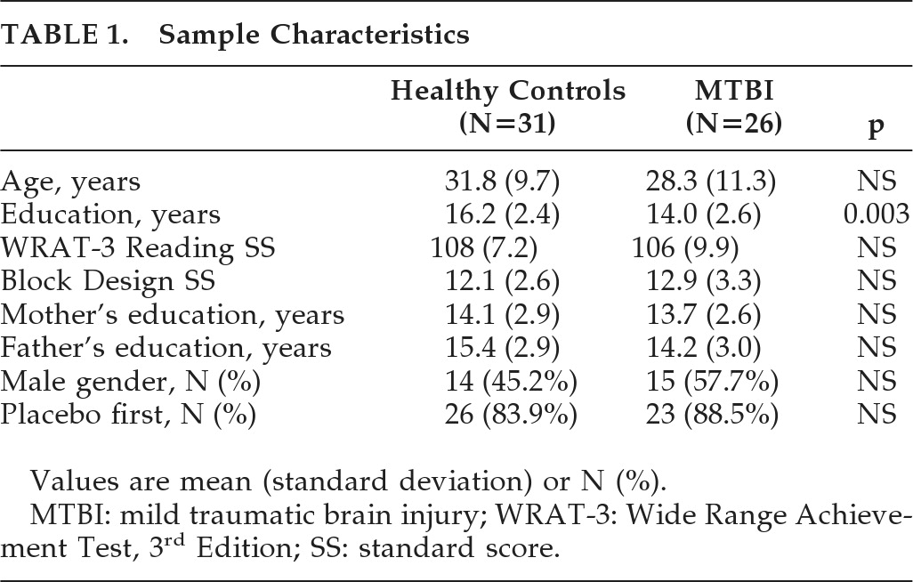
Twenty-six subjects with MTBI and 31 HCs completed the protocol (see Table 1). There were no significant group differences in WRAT-3 Reading or WAIS–III Block Design scores — estimates of baseline verbal and nonverbal intellectual ability, respectively. The groups did differ with respect to years of education, largely because several of the MTBI subjects were students at the time of study participation. Parental education did not differ between groups. Because the protocol called for repeated cognitive assessments over a short interval, performance and imaging results might be vulnerable to the influence of practice and exposure effects. Even when alternate forms of a task are given, there can be improvement in performance related to improved strategy/efficiency in task approach. This theoretical concern was confirmed by the finding of a main effect of drug order in our original model. Thus, we included order (drug or placebo first) as a covariate. The two groups did not differ significantly with respect to age. Nevertheless, the literature supports a robust relationship between age and performance across a variety of cognitive measures, including the age range spanned by our participants. Also, there is evidence that age affects task-related blood oxygen level-dependent (BOLD) response. Thus, the subsequent analyses reported used age, education, and order of drug administration as covariates. MTBI participants were studied for a mean of 39 days after injury (SD: 16.1; range: 14–78); 19 of the 26 MTBI participants had LOC, with a mean duration of 4.8 (SD: 10.1) minutes and a mean Glasgow Coma Scale score of 14.2 (SD: 1.6). The protocol was well tolerated by both groups.
TABLE 1. Sample Characteristics
Values are mean (standard deviation) or N (%).
MTBI: mild traumatic brain injury; WRAT-3: Wide Range Achievement Test, 3rd Edition; SS: standard score.

Serum Prolactin

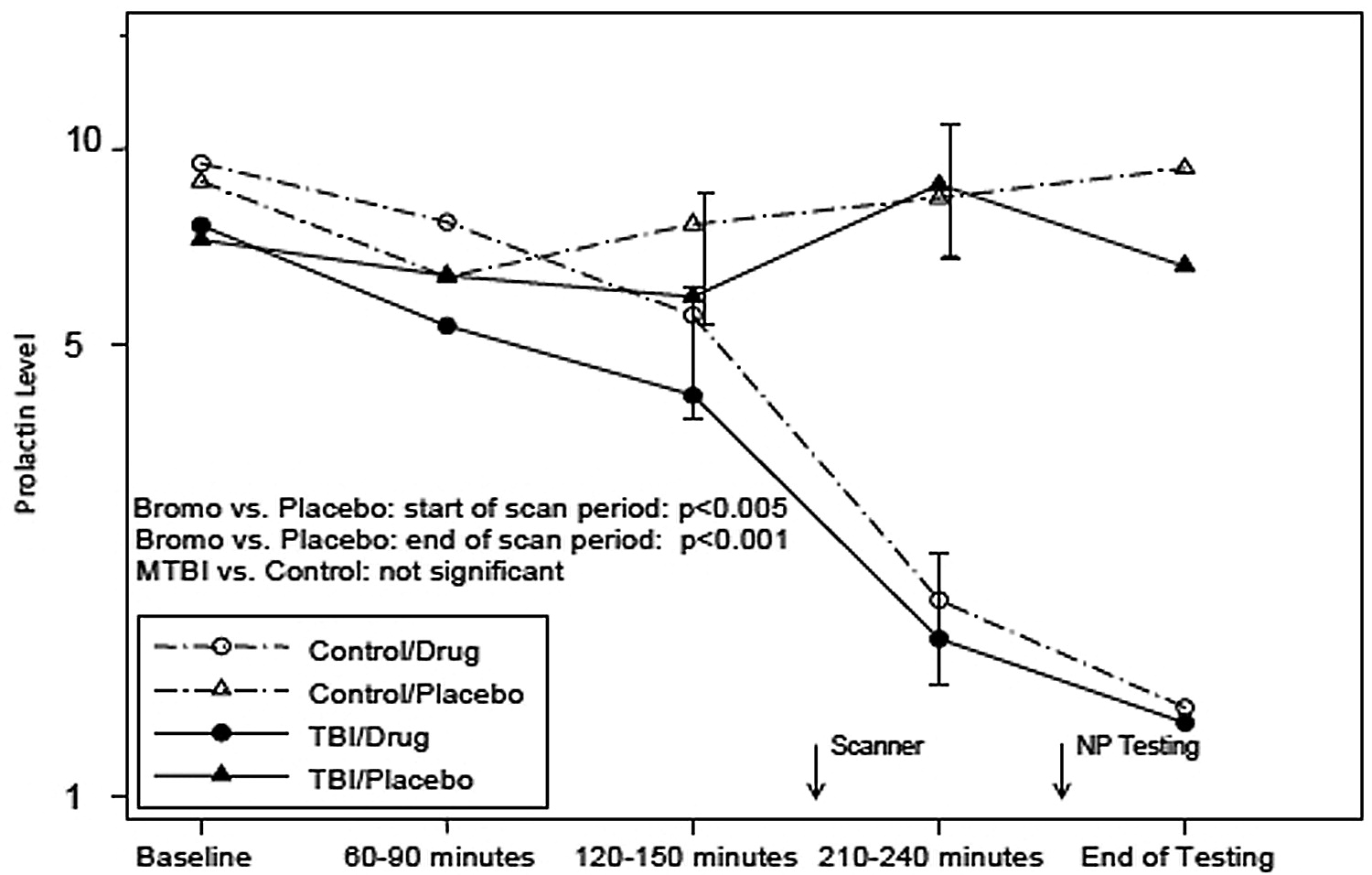
Figure 1 shows serum prolactin levels over time for participants while on placebo and on bromocriptine. As expected, blunting of serum prolactin levels while on bromocriptine started at about 2½ hours after ingestion and lasted for at least 5 hours after ingestion. This effect spanned the time when the participants were undergoing fMRI and the subsequent neuropsychological testing, and it confirmed that there was a central dopaminergic effect during this interval. The differences between bromocriptine and placebo were statistically significant (p=0.005 pre-scanner and p=0.001 post-scanner), but did not differ significantly between the MTBI and HC groups.
FIGURE 1. Serum Prolactin Levels
Time trends by treatment conditions (bromocriptine versus placebo) and participant groups (MTBI versus HC), with p values for overall treatment and difference in treatment effects between the groups, using repeated measures analysis of covariance. The targeted times for entering and leaving the MRI are marked on the figure. Prolactin suppression is evident during the scan and cognitive-assessment time periods. Degree of suppression does not differ between groups.

Cognitive Performance

Both groups showed the expected working memory-load effect on N-back performance, regardless of drug condition (see Table 2), with percentage of correct responses (adjusted for guessing) decreasing with increasing WM-load requirements. There was no main effect of gender in either group, nor was there a main effect of LOC in the MTBI group. The HC group showed no differences in performance between drug conditions, whereas the MTBI patients showed poorer 0-back (p=0.004), 3-back (p=0.047), and mean-back (p=0.009) performance on bromocriptine, as compared with placebo. No differences were apparent between diagnostic groups on placebo, whereas, on bromocriptine, MTBI patients performed worse than HCs for 0-back (p=0.010), 3-back (p=0.008), and mean-back (p<0.001) conditions, with a trend toward poorer performance for 1-back and 2-back (p=0.055, p=0.052). A main effect of drug was found on 0-back (p=0.039), attributable primarily to MTBI patients. A main effect of diagnosis was apparent for 2-back (p=0.038), 3-back (p=0.020), and mean-back (p=0.005), with MTBI patients performing significantly worse than HCs. A Drug × Diagnosis interaction was evident for the 0-back (p=0.018) and mean-back conditions (p=0.022), with MTBI patients showing declines in performance on bromocriptine relative to placebo, whereas HCs' performance was stable-to-improved across drug conditions. Few statistically significant differences were noted between groups or drug conditions on neuropsychological measures. For the Stroop Color-Naming Condition, there was a Drug effect, with both HCs and MTBI patients showing improved performance on bromocriptine (p=0.027). HCs also showed improvement on the Trail-Making subtest (Condition A or 2) when on bromocriptine (p=0.041), whereas MTBI patients performed more poorly on the Stroop Switching condition while on bromocriptine (p=0.031).
TABLE 2. N-Back Performance
Values are mean (standard error).
HC: healthy control participants; MTBI: mild traumatic brain injury.

fMRI

3-Back

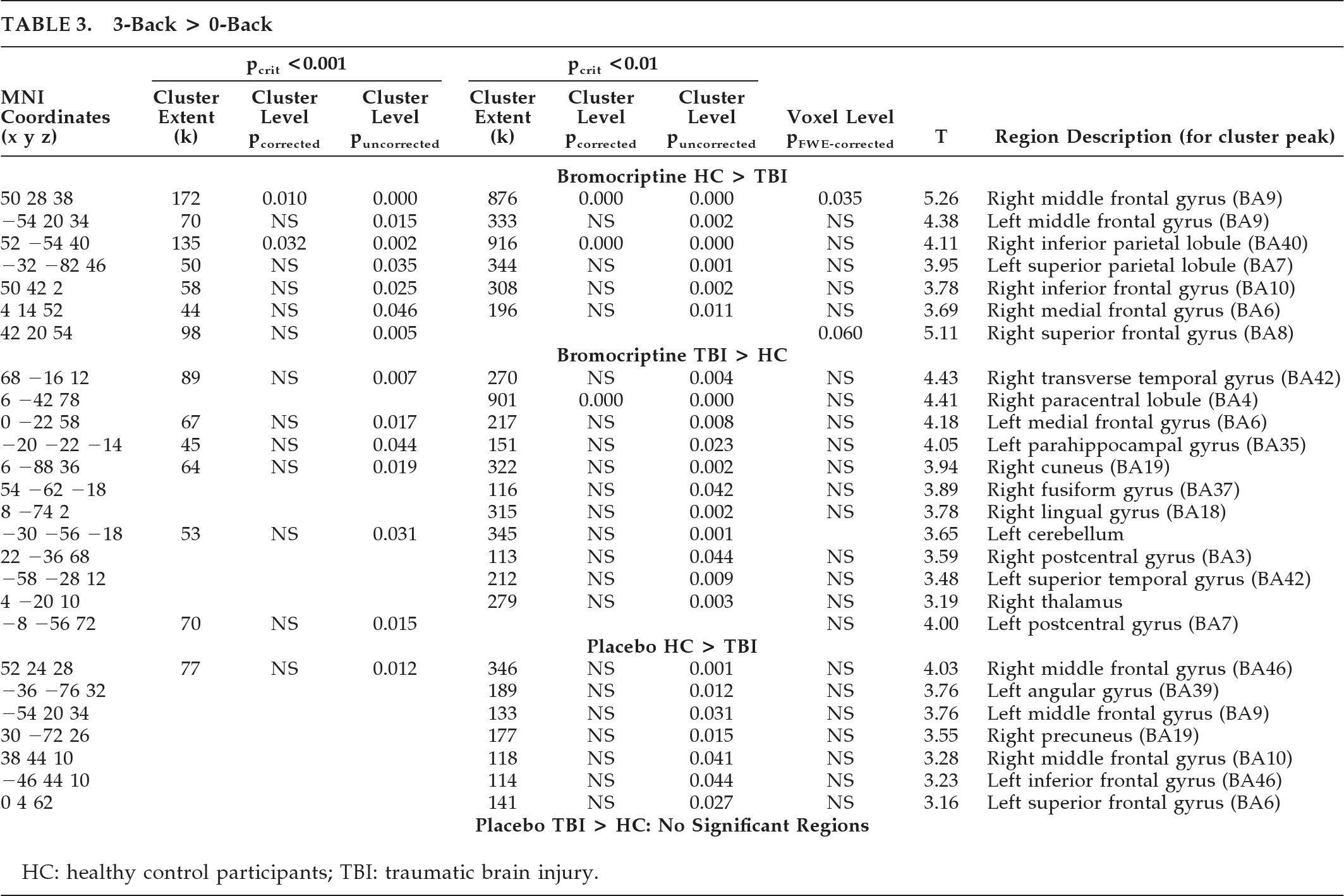
fMRI results are summarized in Figure 2 and Table 3. In both Drug conditions, both groups displayed activation of WM circuitry consistent with previous results with the N-back task.7,8 While performing the 3-back task relative to the 0-back task, both groups activated WM circuitry (Figure 2, shaded areas). However, consistent with our previous results,8 while on placebo, the HC group showed increased activation relative to the MTBI group within WM circuitry while doing the most difficult (3-back) task (Figure 2 top row, blue areas). Although the MTBI group showed activation on the 3-back task (relative to the 0-back), there were no regions where the MTBI group showed significantly greater activation than HCs while on placebo.
FIGURE 2. Group Differences in Brain Activation During 3-Back in Response to Pharmacological Challenge
The combined SPM5 activation map for the 3-back > 0-back contrast for both groups (MTBI and HC) in both drug conditions (placebo and bromocriptine) is displayed in red. Note the bilateral frontal, parietal, and cerebellar network typically activated during the N-back task. Group contrasts of interest are overlaid in blue (MRIcroGL software package).
Top Row: Blue areas show increased activation in HC relative to MTBI on placebo. Consistent with previous results,8 note increased activation within WM task-related circuitry while doing the most difficult (3-back) task. There were no regions where the MTBI group showed significantly greater activation than HC while on placebo.
Middle Row: Blue areas show increased activation in HC relative to MTBI on bromocriptine. Note increased activation within red WM task-related circuitry, greater than seen on placebo.
Bottom Row: Increased activation in MTBI relative to HC on bromocriptine. Note that increased activation occurs exclusively outside of WM task-related circuitry.
TABLE 3. 3-Back > 0-Back
HC: healthy control participants; TBI: traumatic brain injury.
On bromocriptine, the HC group again showed increased activation relative to the MTBI group in WM circuitry (Figure 2, middle row, blue areas), whereas the MTBI group showed increased activation in regions outside of WM circuitry (Figure 2, bottom row, blue areas), including bilateral postcentral and superior temporal gyri.
As shown in Figure 2 and Table 3, the areas of greatest group difference (HC > MTBI) in activation while on bromocriptine were the bilateral frontal and parietal regions typically activated during WM performance, with the greatest difference in the right middle frontal gyrus (pFWE-corrected=0.035). Furthermore, increased activation in this region correlated with improved performance on the 3-back task (r=0.266; p<0.05) across groups while on bromocriptine.

2-Back and 1-Back

The same pattern of differences in brain activation was observed at lower WM loads (supplemental materials available on request). The HC group showed greater activation than the MTBI group in WM-related circuitry, particularly in the frontal lobes. This effect was accentuated on bromocriptine, and became more pronounced with increasing WM load. In contrast, the MTBI group showed greater activation than HCs in regions not typically associated with WM processing. Additional analyses (not shown) of groups broken down by gender and presence or absence of LOC (MTBI group) showed no significant effects on activation pattern or on the above findings.

DISCUSSION

Experimental results indicate that individuals with MTBI have altered cognitive and cerebral activation patterns in response to the dopamine D2 receptor-agonist bromocriptine, consistent with the main study hypothesis. Of particular interest is that an overall N-back performance indicator, the mean back-accuracy score, showed a significant (p<0.02) Diagnosis × Drug condition interaction. Post-hoc analysis of this interaction shows this was due both to improvement in the HC group and poorer performance in the MTBI group on bromocriptine. Furthermore, the pattern of cerebral activation in response to bromocriptine differed between the two groups. Increased activation in WM circuitry in the HC group, including bilateral frontal and parietal regions, was more pronounced on bromocriptine, and greater right middle frontal activation also correlated with improved task performance across both groups, suggesting some functional significance to the finding. The increased activation of this region while on bromocriptine in HCs, coupled with the improved performance in HCs as compared with the MTBI group, raises the possibility that bromocriptine facilitates selective recruitment of task-related processing resources in the healthy brain. In contrast, MTBI patients on bromocriptine showed increased activation in brain regions not typically associated with WM processing, suggesting an altered neural response to bromocriptine challenge. This may suggest that the MTBI group is less efficient at deactivating brain regions outside of task-related neural circuitry during WM processing, or that there is compensatory activation of these resources in an effort to maintain WM performance, which, in this cohort, was unsuccessful. Taken together, these findings are consistent with the hypothesis that subtle changes in the central dopaminergic system may contribute to previously described alterations in WM processing after MTBI.7,8,25
Our results differ in some ways from related studies. For example, improved performance on a dual-task paradigm while on bromocriptine was noted in 24 individuals with TBI.30 However, participants in that study were of mixed injury severity, were studied at variable intervals after injury, and performed a different task from the one used in our study. Subsequent studies did not replicate the previous finding and, in fact, noted a trend for bromocriptine to be associated with poorer performance on a variety of attentional measures.31 Other discrepant findings have been reported in response to dopamine agonists,32 and dose–response effects with dopamine agonists have been noted: low doses can facilitate WM, whereas higher doses can impair performance.3335 It is possible that those with MTBI are more sensitive to the effects of dopamine agonists, and thus perform more poorly or do not show the same improvement as HCs at equivalent doses of bromocriptine. There may also be a biphasic pharmacokinetic response with bromocriptine. Pizzolato et al.36 suggested that initial effects of bromocriptine can be inhibitory, whereas later effects can be excitatory. Our study used different ingestion-to-fMRI intervals from previous studies (about 1.5 hours, versus about 2.5 hours). Previous work in rodents, using a controlled cortical impact model of TBI,26,27 suggests that TBI-associated changes in levels of frontal and basal ganglia dopamine transporter are more pronounced in males. Also, there may be gender-related differential responses to interventions such as environmental enrichment, perhaps related to neuroprotective effects of female sex hormones.27 This raises the possibility that similar gender differences might exist in human TBI, and thus response to dopaminergic challenges might differ in men and women. Although we did not find such differences in either the cognitive outcome or the BOLD response, it is possible that differences might be more apparent with a larger sample size. It is also interesting that we did not find differences within the MTBI group as a function of presence or absence of LOC at the time of injury. However, report of LOC in the MTBI group is not always reliable, and, even in those who did report LOC, the mean duration was brief (4.8 minutes). Again, larger sample sizes might reveal such differences.
The mechanism of dopamine system dysregulation is not clear. MTBI is associated with alterations of catecholaminergic systems (including dopamine) that can be prolonged, may be associated with alterations in catecholaminergic receptor density in damaged cortical areas, and can impair catecholaminergic function after trauma.37,38 The studies by Wagner et al.26,27 suggest that, at least in rodents, TBI is associated with reduced dopamine transporter levels in the striatum. Neuronal damage in the region of the anterior frontal cortex might also contribute to the changes in brain activation, with a relative absence of anterior frontal activation seen in the MTBI group. Although it is clear that neuronal loss does accompany MTBI,3941 it is generally considered to be greatly reduced in extent and significance relative to that accompanying more severe injuries.
Alternatively, dopaminergic dysregulation may be a downstream effect of dysfunction of other neurotransmitter systems, such as the cholinergic system. There is a significant body of work in animals and humans suggesting that TBI results in alterations to the cholinergic system (for reviews, see DeAngelis et al.,42 Verbois et al.,43 and Arciniegas44). Given the reciprocal hippocampal–prefrontal connections and their interplay in the regulation of memory,45 it is possible that subtle changes in cholinergic tone, stemming from excitotoxic injury to highly vulnerable regions of hippocampal cortex, result in apparent changes in prefrontal dopaminergic regulation.
Several limitations should be considered in the interpretation of these findings. We intentionally did not include MTBI participants with significant medical and psychiatric disorders; therefore these results may not generalize to all individuals with MTBI. This MTBI group had very mild injuries, and the results may not apply to those with moderate and severe injuries. It is also important to point out that the regions of increased activity evident on bromocriptine do not necessarily indicate the exact locations of dopaminergic effect. Although BOLD fMRI activation in these areas indicates local increased cerebral blood flow and metabolic activity, bromocriptine administration could indirectly modulate this activity through striatal or other interconnected subcortical or cortical sites. Also, given the behavioral/performance differences on bromocriptine between the HC and MTBI groups, we cannot rule out the possibility that the area of increased activation in the right middle frontal region (see Figure 2 and Table 3) may modulate other behavioral effects, which then secondarily affect WM performance. However, the fact that this region is embedded within a neural network known to play a key role in verbal WM circuitry is consistent with a drug-related WM modulatory effect.
Despite these limitations and caveats, the current results remain most consistent with the conclusion that MTBI is associated with subtle dysregulation of frontal dopaminergic systems in the first 4–6 weeks after injury and that simple augmentation strategies with a dopamine agonist that affects predominantly D2 receptors may not improve cognitive functioning. Further studies are needed to clarify the effects that different dosing strategies, severity of injury, and the injury-to-treatment interval may all have on outcome.

Acknowledgments

Location of Work: Section of Neuropsychiatry and Brain Imaging Laboratory, Departments of Psychiatry, Radiology, Community and Family Medicine (Biostatistics), Emergency Medicine, Dartmouth Medical School, Lebanon, NH.
The authors thank Brian Greenlee and Kaloyan Tanev, Neuropsychiatry Fellows James Ford and Heather Pixley, Department of Psychiatry Robert Ferranti, Alice Davison, Shreve Soule, and Robert Shaffer, for their assistance on this project.
This research was supported in part by NIDRR Grants H133G70031 and H133000136, and NIH Grant R01 NS40472-01.
References

1.
Whyte J, Schuster K, Polansky M, et al.: Frequency and duration of inattentive behavior after traumatic brain injury: effects of distraction, task, and practice. J Int Neuropsychol Soc 2000; 6:1–11
2.
Cicerone K, Dahlberg C, Kalmar K, et al.: Evidence-based cognitive rehabilitation: recommendations for clinical practice. Arch Phys Med Rehabil 2000; 81:1596–1615
3.
Levin HS: Cognitive function outcomes after traumatic brain injury. Curr Opin Neurol 1998; 11:643–646
4.
Levin HS, Mattis S, Ruff RM, et al.: Neurobehavioral outcome following minor head injury: a three-center study. J Neurosurg 1987; 66:234–243
5.
Binder LM, Rohling ML, Larrabee J: A review of mild head trauma, Part 1: meta-analytic review of neuropsychological studies. J Clin Exp Neuropsychol 1997; 19:421–431
6.
McMillan TM: Minor head injury. Curr Opin Neurol 1997; 10:479–483
7.
McAllister TW, Saykin AJ, Flashman LA, et al.: Brain activation during working memory 1 month after mild traumatic brain injury: a functional MRI study. Neurology 1999; 53:1300–1308
8.
McAllister TW, Sparling MB, Flashman LA, et al.: Differential working memory load effects after mild traumatic brain injury. Neuroimage 2001; 14:1004–1012
9.
Perlstein WM, Cole MA, Demery JA, et al.: Parametric manipulation of working memory load in traumatic brain injury: behavioral and neural correlates. J Int Neuropsychol Soc 2004; 10:724–741
10.
Chen K, Reiman EM, Alexander GE, et al.: An automated algorithm for the computation of brain volume change from sequential MRIs using an iterative, principal-component analysis and its evaluation for the assessment of whole-brain atrophy rates in patients with probable Alzheimer disease. Neuroimage 2004; 22:134–143
11.
Arnsten AF, Steere JC, Jentsch DJ, et al.: Noradrenergic influences on prefrontal cortical cognitive function: opposing actions at postjunctional alpha1- versus alpha2-adrenergic receptors. Adv Pharmacol (New York) 1998; 42:764–767
12.
McAllister TW, Flashman LA, Sparling MB, et al.: Working memory deficits after mild traumatic brain injury: catecholaminergic mechanisms and prospects for catecholaminergic treatment: a review. Brain Inj 2004; 18:331–350
13.
Kay T, et al.: Definition of mild traumatic brain injury. J Head Trauma Rehabil 1993; 8:86–87
14.
First MB, Spitzer RL, Gibbon M, et al.: Structured Clinical Interview for DSM-IV Axis I Disorders, Clinician Version. Washington, DC, American Psychiatric Press, 1997
15.
Wilkinson GS: The Wide Range Achievement Test (WRAT-3): Administration Manual, Wilmington, DE, Wide Range, Inc., 1993
16.
The Psychological Corporation: WAIS-III: WAIS, 3rd Edition; WMS-III: Wechsler Memory Scale, 3rd Edition Technical Manual, San Antonio, TX: The Psychological Corporation, 1997
17.
Delis DC: California Verbal Learning Test, Adult Version. New York, The Psychological Corporation, 1987
18.
Delis DC: California Verbal Learning Test, 2nd Edition: Adult Version Manual. San Antonio, TX, The Psychological Corporation, 2000
19.
Reitan RM, Wolfson D: The Halstead-Reitan Neuropsychological Test Battery: Theory and Clinical Interpretation, 2nd Edition.Tucson, AZ, Neuropsychology Press, 1993
20.
Dellis DC, Kaplan E, Kramer JF: Delis-Kaplan Executive Function System, San Antonio, TX, The Psychological Corporation, 2001
21.
Lezak MD: Neuropsychological Assessment, 3rd Edition. New York, Oxford University Press, 1995
22.
Spreen O, Strauss E: Controlled Oral Word Association, in A Compendium of Neuropsychological Tests, Oxford, England, New York, 1998, pp 447–467
23.
Gronwall D: Paced Auditory Serial Addition Task: a measure of recovery from concussion. Percept Mot Skills 1977; 44:367–373
24.
Gordon M, McClure FD, Aylward GP: Gordon Diagnostic System: Instruction Manual and Interpretive Guide. DeWitt, NY, Gordon Systems, Inc., 1986
25.
McAllister TW, Flashman LA, McDonald BC, et al.: Mechanisms of working memory dysfunction after mild and moderate TBI: evidence from functional MRI and neurogenetics. J Neurotrauma 2006; 23:1450–1467
26.
Wagner A, Sokoloski JE, Ren D, et al.: Controlled cortical impact injury affects dopaminergic transmission in the rat striatum. J Neurochem 2005; 95:457–465
27.
Wagner A, Chen X, Kline AE, et al.: Gender and environmental enrichment impact dopamine transporter expression after experimental traumatic brain injury. Exp Neurol 2005; 195:475–483
28.
Friston KJ, et al.: Statistical parametric maps in functional imaging: a general-linear approach. Hum Brain Mapping 1995; 2:189–210
29.
Holmes A, Friston K: Generalisability, random effects, and population inference. Neuroimage 1998; 7:S754
30.
McDowell S, Whyte J, D'Esposito M: Differential effect of a dopaminergic agonist on prefrontal function in traumatic brain injury patients. Brain 1998; 121:1155–1164
31.
Whyte J, Vaccaro M, Grieb-Neff B, et al.: The effects of bromocriptine on attention deficits after traumatic brain injury: a placebo-controlled pilot study. Am J Phys Med Rehabil 2008; 87:85–99
32.
Kimberg DY, D'Esposito M, Farah MJ: Effects of bromocriptine on human subjects depend on working memory capacity. NeuroReport 1997; 8:3581–3585
33.
Arnsten AFT: Catecholamine modulation of prefrontal cortical cognitive function. Trends Cog Sci 1998; 2:436–447
34.
Arnsten AFT, Cai JX, Murphy BL, et al.: Dopamine D1 receptor mechanisms in the cognitive performance of young adult and aged monkeys. Psychopharmacology 1994; 116:143–151
35.
Cai JX, Arnsten AF: Dose-dependent effects of the dopamine D1 receptor agonists A77636 or SKF81297 on spatial working memory in aged monkeys. J Pharmacol Exp Ther 1997; 283:183–189
36.
Pizzolato G, Soncrant TT, Rapoport SI: Time-course and regional distribution of the metabolic effects of bromocriptine in the rat brain. Brain Res 1985; 341:303–312
37.
McIntosh TK: Neurochemical sequelae of traumatic brain injury: therapeutic implications. Cerebrovasc Brain Metab Rev 1994; 6:109–162
38.
Prasad MR, Tzigaret CM, Smith D, et al.: Decreased alpha-adrenergic receptors after experimental brain injury. J Neurotrauma 1993; 9:269–279
39.
Oppenheimer DR: Microscopic lesions in the brain following head injury. J Neurol Neurosurg Psychiatry 1968; 31:299–306
40.
Blumbergs PC, Scott G, Manavis J, et al.: Staining of amyloid precursor protein to study axonal damage in mild head injury. Lancet 1994; 344:1055–1056
41.
Povlishock JT, Jenkins LW: Are the pathobiological changes evoked by traumatic brain injury immediate and irreversible? Brain Pathol 1995; 5:415–426
42.
DeAngelis MM, Hayes RL, Lyeth BG: Traumatic brain injury causes a decrease in M2 muscarinic cholinergic receptor binding in the rat brain. Brain Res 1994; 653:39–44
43.
Verbois SL, Scheff SW, Pauly JR: Time-dependent changes in rat brain cholinergic receptor expression after experimental brain injury. J Neurotrauma 2002; 19:1569–1585
44.
Arciniegas DB: The cholinergic hypothesis of cognitive impairment caused by traumatic brain injury. Curr Psychiatry Rep 2003; 5:391–399
45.
Seamans JK, Floresco SB, Phillips AG: D1 receptor modulation of hippocampal prefrontal cortical circuits integrating spatial memory with executive functions in the rat. J Neurosci 1998; 18:1613–1621

Information & Authors

Information

Published In

Go to The Journal of Neuropsychiatry and Clinical Neurosciences
The Journal of Neuropsychiatry and Clinical Neurosciences
Pages: 277 - 286
PubMed: 21948888

History

Received: 8 December 2010
Revision requested: 24 February 2011
Accepted: 31 March 2011
Published online: 1 July 2011
Published in print: Summer 2011

Keywords

  1. traumatic brain injury
  2. dopamine
  3. working memory
  4. functional MRI
  5. bromocriptine

Authors

Details

Thomas W. McAllister, M.D.
From the Section of Neuropsychiatry, Department of Psychiatry, Dartmouth Medical School, Lebanon, NH; Center for Neuroimaging, Department of Radiology and Imaging Sciences, Indiana University School of Medicine, Indianapolis, IN; Department of Community and Family Medicine, Biostatistics, Dartmouth Medical School; Section of Emergency Medicine, Dartmouth Medical School, Lebanon, NH.
Laura A. Flashman, Ph.D.
From the Section of Neuropsychiatry, Department of Psychiatry, Dartmouth Medical School, Lebanon, NH; Center for Neuroimaging, Department of Radiology and Imaging Sciences, Indiana University School of Medicine, Indianapolis, IN; Department of Community and Family Medicine, Biostatistics, Dartmouth Medical School; Section of Emergency Medicine, Dartmouth Medical School, Lebanon, NH.
Brenna C. McDonald, Psy.D.
From the Section of Neuropsychiatry, Department of Psychiatry, Dartmouth Medical School, Lebanon, NH; Center for Neuroimaging, Department of Radiology and Imaging Sciences, Indiana University School of Medicine, Indianapolis, IN; Department of Community and Family Medicine, Biostatistics, Dartmouth Medical School; Section of Emergency Medicine, Dartmouth Medical School, Lebanon, NH.
Richard B. Ferrell, M.D.
From the Section of Neuropsychiatry, Department of Psychiatry, Dartmouth Medical School, Lebanon, NH; Center for Neuroimaging, Department of Radiology and Imaging Sciences, Indiana University School of Medicine, Indianapolis, IN; Department of Community and Family Medicine, Biostatistics, Dartmouth Medical School; Section of Emergency Medicine, Dartmouth Medical School, Lebanon, NH.
Tor D. Tosteson, Sc.D.
From the Section of Neuropsychiatry, Department of Psychiatry, Dartmouth Medical School, Lebanon, NH; Center for Neuroimaging, Department of Radiology and Imaging Sciences, Indiana University School of Medicine, Indianapolis, IN; Department of Community and Family Medicine, Biostatistics, Dartmouth Medical School; Section of Emergency Medicine, Dartmouth Medical School, Lebanon, NH.
Norman N. Yanofsky, M.D.
From the Section of Neuropsychiatry, Department of Psychiatry, Dartmouth Medical School, Lebanon, NH; Center for Neuroimaging, Department of Radiology and Imaging Sciences, Indiana University School of Medicine, Indianapolis, IN; Department of Community and Family Medicine, Biostatistics, Dartmouth Medical School; Section of Emergency Medicine, Dartmouth Medical School, Lebanon, NH.
Margaret R. Grove, M.S.
From the Section of Neuropsychiatry, Department of Psychiatry, Dartmouth Medical School, Lebanon, NH; Center for Neuroimaging, Department of Radiology and Imaging Sciences, Indiana University School of Medicine, Indianapolis, IN; Department of Community and Family Medicine, Biostatistics, Dartmouth Medical School; Section of Emergency Medicine, Dartmouth Medical School, Lebanon, NH.
Andrew J. Saykin, Psy.D.
From the Section of Neuropsychiatry, Department of Psychiatry, Dartmouth Medical School, Lebanon, NH; Center for Neuroimaging, Department of Radiology and Imaging Sciences, Indiana University School of Medicine, Indianapolis, IN; Department of Community and Family Medicine, Biostatistics, Dartmouth Medical School; Section of Emergency Medicine, Dartmouth Medical School, Lebanon, NH.

Notes

Correspondence: Thomas W. McAllister, M.D., Dartmouth-Hitchcock Medical Center, One Medical Center Dr., Lebanon, NH 03756; [email protected] (e-mail).

Metrics & Citations

Metrics

Citations

Export Citations

If you have the appropriate software installed, you can download article citation data to the citation manager of your choice. Simply select your manager software from the list below and click Download.

For more information or tips please see 'Downloading to a citation manager' in the Help menu.

Format
Citation style
Style
Copy to clipboard

View Options

View options

PDF/EPUB

View PDF/EPUB

Get Access

Login options

Already a subscriber? Access your subscription through your login credentials or your institution for full access to this article.

Personal login Institutional Login Open Athens login
Purchase Options

Purchase this article to access the full text.

PPV Articles - Journal of Neuropsychiatry and Clinical Neurosciences

PPV Articles - Journal of Neuropsychiatry and Clinical Neurosciences

Not a subscriber?

Subscribe Now / Learn More

PsychiatryOnline subscription options offer access to the DSM-5-TR® library, books, journals, CME, and patient resources. This all-in-one virtual library provides psychiatrists and mental health professionals with key resources for diagnosis, treatment, research, and professional development.

Need more help? PsychiatryOnline Customer Service may be reached by emailing [email protected] or by calling 800-368-5777 (in the U.S.) or 703-907-7322 (outside the U.S.).

Media

Figures

Other

Tables

Share

Share

Share article link

Share