
Abstract

OBJECTIVE: To screen for dopaminergic abnormalities in tobacco smokers and patients with posttraumatic stress disorder (PTSD), the authors determined serial CSF and plasma concentrations of the dopamine metabolite homovanillic acid (HVA). METHOD: Continuous subarachnoid sampling was used to obtain 37 serial CSF samples over 6 hours in 13 normal volunteers and 11 patients with combat-related PTSD; 10 smoked and 14 had never smoked. The smokers were abstinent from tobacco for 11 to 17 hours. RESULTS: The smokers had markedly lower CSF, but not plasma, HVA levels. Their CSF HVA concentrations averaged only 54% of the concentrations of the nonsmokers, independent of diagnosis. CONCLUSIONS: Smokers’ low CSF concentrations of HVA may be associated either with chronic inhalation of nicotine or other constituents of tobacco smoke or with acute abstinence. Any possible basal dopaminergic abnormalities in patients with PTSD are small relative to the abnormalities associated with smoking.
The goal of the present investigation was to determine serial cerebrospinal fluid (CSF) and plasma concentrations of the dopamine metabolite homovanillic acid (HVA) in tobacco-smoking and nonsmoking combat veterans with posttraumatic stress disorder (PTSD) and in smoking and nonsmoking normal volunteers. It has been suggested that hyperresponsiveness of brain dopaminergic systems may contribute to the heightened arousal and increased sensitivity to both trauma-related and nonspecific stress seen in patients with PTSD (1). Stimulation of central nervous system (CNS) dopaminergic activity has been associated with nicotine administration in experimental animals (24), and chronic cigarette smoking has been linked with both peripheral (57) and brain (8, 9) monoamine oxidase inhibition in humans. We sought to extend these studies by examining a hypothesized association between tobacco cigarette smoking and perturbations in dopamine metabolism. We postulated that CSF concentrations of HVA would be abnormal in both smokers and patients with PTSD.

METHOD

The study was approved by the Institutional Review Board of the University of Cincinnati Medical Center and the Research Committee of the Cincinnati Department of Veterans Affairs Medical Center. Informed written consent was obtained from 13 healthy volunteers, without history of axis I psychiatric disorder, and 11 patients with combat-related PTSD. The lifetime-nonsmoking (N=9) and smoking (N=4) healthy volunteers had mean ages of 34 years (SD=10) and 42 years (SD=9), respectively, and weighed 76 kg (SD=9) and 70 kg (SD=13), respectively. The lifetime-nonsmoking (N=5) and smoking (N=6) patients with PTSD had mean ages of 43 years (SD=11) and 42 years (SD=8) and weighed 91 kg (SD=14) and 96 kg (SD=16), respectively. The smokers smoked 1.2 packets of cigarettes per day (SD=0.2, range=0.75–1.50). With the exception of one normal volunteer who had smoked for 13 years and one patient with PTSD who had smoked for 9 years, all had smoked for over 20 years. Six of the 11 patients with PTSD had histories of alcohol or drug abuse (excluding tobacco) but had been abstinent for 0.5 to 15.0 years. Seven of the 11 patients had taken psychoactive medication in the past but had discontinued taking all medications at least five half-lives before the study. The mean score of the patients on the Clinician-Administered PTSD Scale (10) was 82 (SD=22).
CSF was collected into iced test tubes from 11:00 a.m. to 5:00 p.m. at the rate of 0.1 ml/min through an indwelling 20-gauge subarachnoid catheter that had been placed at 8:00 a.m. as previously described (11), except that the seated rather than the lateral decubitus position was used. CSF samples were obtained at 10-minute intervals (37 samples total) for quantification of HVA. No smoking or oral intake was permitted after the midnight before the procedure. Blood was withdrawn at hourly intervals through an indwelling venous catheter, placed the night before, to obtain plasma for HVA quantification. CSF HVA was quantified by high-performance liquid chromatography with electrochemical detection, as previously described (12); slight modifications of the system were used for plasma HVA assay.
Separate two-way analyses of variance were used to assess the effects of smoking and diagnosis on CSF HVA and plasma HVA concentrations.

RESULTS

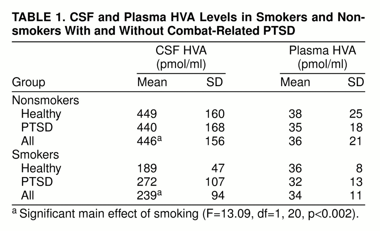
CSF HVA concentrations (table 1) were lower in the smokers than in the nonsmokers (F=13.09, df=1, 20, p<0.002), but there was no significant effect of a diagnosis of PTSD (F=0.39, df=1, 20, n.s.). No significant interaction between smoking and diagnosis was present (F=0.61, df=1, 20, n.s.).
With regard to plasma HVA concentrations (table 1), neither the main effects of diagnosis and smoking nor their interaction were significant (main effect of diagnosis: F=0.15, df=1, 16, n.s.; main effect of smoking: F=0.06, df=1, 16, n.s.; interaction: F=0.0004, df=1, 16, n.s.).

DISCUSSION

Using continuous sampling over 6 hours, we observed markedly low CSF levels of the major dopamine metabolite HVA in chronic tobacco-cigarette smokers, with CSF HVA concentrations averaging only 54% of those found in nonsmokers. Reduced dopamine synthesis or release, impaired metabolic conversion of dopamine to HVA, and enhanced efflux of HVA from CSF might all account for low levels of CSF HVA.
We cannot determine from the present data whether the low CSF HVA concentrations are related to cigarette smoking per se or to early withdrawal from the effects of tobacco smoke. Our patients were withdrawn for 11 hours at the time CSF sampling began and for 17 hours by the end of the sampling period. Thus, acute withdrawal of a dopaminergic stimulant (tobacco smoke) may have resulted in a rebound suppression of CNS dopaminergic activity. Accordingly, bupropion’s apparent efficacy in promoting smoking cessation (13) may be related in part to its pro-dopaminergic effects (14).
In contrast to the striking diminution in CSF HVA levels in cigarette smokers regardless of diagnosis, we failed to find any significant differences in CSF HVA concentrations between patients with PTSD and normal volunteers. It therefore appears that any possible basal abnormalities in CSF HVA concentrations in patients with PTSD, if present at all, are small relative to the dopaminergic abnormalities associated with tobacco smoking.
No significant differences in plasma HVA levels were detected in either patients with PTSD or smokers, again showing that plasma HVA concentrations alone cannot be used as a surrogate for CSF HVA concentrations. The majority of plasma HVA is derived from dopamine (also the precursor of norepinephrine) in noradrenergic neurons, and only about 25% of plasma HVA originates from brain dopaminergic pathways (15).
In light of our current findings, it should be noted that previous studies of possible alterations in brain-derived HVA concentrations in psychotic patients, who tend to be heavy smokers (16), did not control for tobacco use and thus may require reinterpretation in light of the major perturbations of the dopamine system associated with tobacco smoking.
Among the limitations of this study is the fact that the only dopamine metabolite quantified was HVA; we cannot rule out the possibility of abnormal metabolism of dopamine to 3,4-dihydroxyphenylacetic acid (DOPAC) or 3-O-methyldopa in cigarette smokers. However, the levels of those metabolites are an order of magnitude lower than those of HVA in lumbar CSF (17, 18). Also, only men were studied.
In conclusion, acutely abstinent tobacco smokers had greatly lower concentrations of the dopamine metabolite HVA in CSF, but not in plasma, than nonsmokers. The low CSF HVA levels may be the effects of smoking-related reductions in brain MAO concentrations (8, 9) on CNS dopamine neurotransmission or may be due to alterations in dopamine synthesis, release, or clearance from CSF. In any event, the present results indicate that future studies of brain dopaminergic systems must take into account the tobacco-smoking histories of all experimental subjects.
TABLE 1

Footnote

Received Dec. 22, 1997; revision received May 18, 1998; accepted July 10, 1998; From the Department of Psychiatry, University of Cincinnati Medical Center, and the Psychiatry Service, Cincinnati VA Medical Center. Address reprint requests to Dr. Geracioti, Psychiatry Service, VA Medical Center, 3200 Vine St., Cincinnati, OH 45220. Supported by Research Advisory Group and Merit Review grants from the Department of Veterans Affairs (to Dr. Geracioti). The authors thank Ann Bruce, M.D., Renee Hanna, Sean Stanton, and the nursing staff of ward 7S at the Cincinnati VA Medical Center for technical support and Robert M. Anthenelli, M.D., and Rose A. Maxwell, Ph.D., for statistical consultation.

References

1.
Charney DS, Deutch AY, Krystal JH, Southwick SM, Davis M: Psychobiologic mechanisms of posttraumatic stress disorder. Arch Gen Psychiatry 1993; 50:294–305
2.
DiChiara G, Imperato A: Drugs abused by humans preferentially increase synaptic dopamine concentrations. Proc Natl Acad Sci USA 1988; 85:5274–5278
3.
Imperato A, DiChiara G: Preferential stimulation of dopamine release in the nucleus accumbens of freely moving rats. J Pharmacol Exp Ther 1986; 239:219–228
4.
Benwell MEM, Balfour DJK: The effects of acute and repeated nicotine treatment on nucleus accumbens dopamine and locomotor activity. Br J Pharmacol 1992; 105:849–856
5.
Berlin I, Said S, Spreux-Varoquaux O, Olivares R, Launay J-M, Puech AJ: Monoamine oxidase A and B activities in heavy smokers. Biol Psychiatry 1995; 38:756–761
6.
Oreland L, Fowler CJ, Schalling D: Low platelet monoamine oxidase activity in cigarette smokers. Life Sci 1981; 29:2511–2518
7.
Norman TR, Chamberlain KG, French MA: Platelet monoamine oxidase activity: low activity in cigarette smokers. Psychiatry Res 1987; 20:199–205
8.
Fowler JS, Volkow ND, Wang GJ, Pappas N, Logan J, Shea C, Alexoff D, MacGregor RR, Schlyer DJ, Zezulkova I, Wolf AP: Brain monoamine oxidase A inhibition in cigarette smokers. Proc Natl Acad Sci USA 1996; 93:14065–14069
9.
Fowler JS, Volkow ND, Wang G-J, Pappas N, Logan J, MacGregor R, Alexoff D, Shea C, Schlyer D, Wolf AP, Warner D, Zezulkova I, Cilento R: Inhibition of monoamine oxidase B in the brains of smokers. Nature 1996; 379:733–736
10.
Blake DD, Weathers FW, Nagy LN, Kaloupek DG, Klauminzer G, Charney DS, Keane TM: A clinician rating scale for assessing current and lifetime PTSD: the CAPS-1. Behavior Therapy 1990; 18:187–188
11.
Geracioti TD, Orth DN, Ekhator NN, Blumenkopf B, Loosen PT: Serial cerebrospinal fluid corticotropin-releasing hormone concentrations in healthy and depressed humans. J Clin Endocrinol Metab 1992; 74:1325–1330
12.
Geracioti TD Jr, Keck PE Jr, Ekhator NN, West SA, Baker DG, Hill KK, Bruce AB, Wortman MD: Continuous covariability of dopamine and serotonin metabolites in human cerebrospinal fluid. Biol Psychiatry 1998; 44:228–233
13.
Hurt RD, Sachs DPL, Glover ED, Offord KP, Johnston JA, Dale LC, Khayrallah MA, Schroeder DR, Glover PN, Sullivan CR, Croghan IT, Sullivan PM: A comparison of sustained-release bupropion and placebo for smoking cessation. N Engl J Med 1997; 337:1195–1202
14.
Ascher JA, Cole JO, Colin J-N, Feighner JP, Ferris RM, Fibiger HC, Golden RN, Martin P, Potter WZ, Richelson E, Sulser F: Bupropion: a review of its mechanism of antidepressant activity. J Clin Psychiatry 1995; 56:395–401
15.
Kopin IJ, Bankiewicz KS, Harvey-White J: Assessment of brain dopamine metabolism from plasma HVA and MHPG during debrisoquin treatment: validation in monkeys treated with MPTP. Neuropsychopharmacology 1988; 1:119–125
16.
de Leon J, Dadvand M, Canuso C, White AO, Stanilla JK, Simp­son GM: Schizophrenia and smoking: an epidemiological survey in a state hospital. Am J Psychiatry 1995; 152:453–455
17.
Ebinger G, Michotte Y, Herregodts P: The significance of homovanillic acid and 3,4-dihydroxyphenylacetic acid concentrations in human lumbar cerebrospinal fluid. J Neurochem 1987; 48:1725–1729
18.
Tohgi H, Abe T, Yamazaki K, Saheki M, Takahashi S, Tsukamoto Y: Effects of the catechol-O-methyltransferase inhibitor tolcapone in Parkinson’s disease: correlations between concentrations of dopaminergic substances in the plasma and cerebrospinal fluid and clinical improvement. Neurosci Lett 1995; 192:165–168

Information & Authors

Information

Published In

Go to American Journal of Psychiatry
American Journal of Psychiatry
Pages: 130 - 132
PubMed: 9892309

History

Published online: 1 January 1999
Published in print: January 1999

Authors

Affiliations

Thomas D. Geracioti, Jr., M.D.
Dewleen G. Baker, M.D.
Matthew D. Wortman, B.S.
Paul E. Keck, Jr., M.D.
Andrew B. Norman, Ph.D.

Metrics & Citations

Metrics

Citations

Export Citations

If you have the appropriate software installed, you can download article citation data to the citation manager of your choice. Simply select your manager software from the list below and click Download.

For more information or tips please see 'Downloading to a citation manager' in the Help menu.

Format
Citation style
Style
Copy to clipboard

There are no citations for this item

View Options

View options

PDF/ePub

View PDF/ePub

Get Access

Login options

Already a subscriber? Access your subscription through your login credentials or your institution for full access to this article.

Personal login Institutional Login Open Athens login
Purchase Options

Purchase this article to access the full text.

PPV Articles - American Journal of Psychiatry

PPV Articles - American Journal of Psychiatry

Not a subscriber?

Subscribe Now / Learn More

PsychiatryOnline subscription options offer access to the DSM-5-TR® library, books, journals, CME, and patient resources. This all-in-one virtual library provides psychiatrists and mental health professionals with key resources for diagnosis, treatment, research, and professional development.

Need more help? PsychiatryOnline Customer Service may be reached by emailing [email protected] or by calling 800-368-5777 (in the U.S.) or 703-907-7322 (outside the U.S.).

Media

Figures

Other

Tables

Share

Share

Share article link

Share