Full access
Article
Published Online: 1 February 2002

Anterior Cingulate Activation During Stroop Task Performance: A PET to MRI Coregistration Study of Individual Patients With Schizophrenia

Publication: American Journal of Psychiatry

Abstract

OBJECTIVE: The authors used single-subject functional imaging analyses to 1) corroborate the findings of anterior cingulate hypoperfusion during an attentional task in schizophrenia and 2) examine whether anterior cingulate activation is associated with underlying morphology. METHOD: Five healthy subjects and six patients with schizophrenia underwent positron emission tomography scanning while they performed the Stroop task. The medial-frontal lobes were masked out for analysis, and activation peaks were individually coregistered to each subject’s magnetic resonance imaging scan. RESULTS: Healthy subjects showed activations in both limbic and paralimbic anterior cingulate regions. Patients with schizophrenia showed only paralimbic activations, and these were apparent only in patients having a paracingulate sulcus. CONCLUSIONS: These findings suggest that 1) patients with schizophrenia have limbic-anterior cingulate hypoperfusion during attentional tasks and 2) paralimbic activation is associated with underlying morphology.
Attentional deficits are a prominent feature of schizophrenia and have been ascribed, in part, to impairment of the anterior cingulate cortex (1). For example, the anterior cingulate of patients with schizophrenia is not activated during Stroop task performance (2). However, in spite of normalization into stereotaxic space, there is marked interindividual variability in anterior cingulate morphology (3), and this variation can lead to a failure to detect activations when data from individual subjects are pooled (4, 5). Further, we have shown (6) that a paracingulate sulcus occurs less frequently in chronically ill patients with schizophrenia than in healthy subjects. Such morphological anomalies may be relevant to understanding the functional differences between healthy subjects and patients with schizophrenia (2).
In the current study, we used single-subject analyses to examine the interindividual consistency of the previously reported group difference between healthy subjects and patients with schizophrenia in anterior cingulate activation during Stroop task performance. In addition, we examined whether the pattern of anterior cingulate activation was associated with the underlying morphological characteristics of the anterior cingulate.

Method

Subjects

All subjects were right-handed men with normal visual acuity and color vision. The mean age of the six patients was 33.8 years (SD=9.3); the mean age of the healthy subjects was 25.4 (SD=4.1). For patients, the mean age at illness onset was 21.8 years (SD=4.4) and the mean duration of illness was 11.5 years (SD=8.5). The six patients were diagnosed with chronic schizophrenia according to DSM-III-R criteria. Five were receiving stable doses of medication, and one was neuroleptic free. All subjects were screened for comorbid medical and psychiatric conditions, and all gave written informed consent after full explanation of the procedures. The institutional research and ethics committee approved the study.

Stroop Activation Task

The two Stroop task conditions were performed six times in an ABABABABABAB design, similar to that used by Pardo et al. (7). For each congruent and incongruent condition, 36 words were presented sequentially 3 mm above a central fixation point for 1300 msec with an interstimulus interval of 350 msec.

Image Acquisition, Reconstruction, and Analysis

Twelve positron emission tomography (PET) scans (six activation and six baseline) were acquired for each subject with a Siemens/CT1-951R ECAT PET scanner (Siemens CTI, Knoxville, Tenn.), producing a contiguous set of 31 transaxial images (voxel dimensions=2.43 × 2.43 × 3.375 mm3). After realignment, a mean PET image was calculated for each individual and coregistered to his T1-weighted magnetic resonance imaging (MRI) scan (TR=3.3 msec, TE=14.3 msec, flip angle=30°, voxel dimensions=0.938 × 0.938 × 1.5 mm3) by using the Woods automated image registration algorithm (8). Spatial normalization was accomplished by registering these data to a standard MRI that had been previously transformed to Talairach coordinates. Following a 12-mm full width at half maximum Gaussian filter smooth, the data were restricted to the hypothesized regions of interest (anterior cingulate and supplementary motor area). This was accomplished by using a mask (x, y, z level boundaries=±20, –20, and 0 mm) to include anterior cingulate and supplementary motor area gray matter. Voxels within the supplementary motor area were investigated as a comparison measure because this region is also activated during Stroop performance (7) but not thought to be dysfunctional in schizophrenia (8).
Using statistical parametric mapping (SPM 96, Wellcome Department of Cognitive Neurology, London), a voxel-based single-subject analysis of covariance was performed over the congruent and incongruent conditions, which removed differences in global cerebral blood flow (CBF). The factor of interest was the contrast between incongruent and congruent conditions, and only voxels with significant differences between the 12 regional CBF means (F>1.97, df=11, 65, p<0.05) were subjected to further analysis.
The contrast of interest was displayed as a statistical parametric map (9) for each individual. The critical threshold for this comparison was a z score of 2.32 (p=0.01, one-tailed) on the basis that the analyses were intrasubject, hypothesis-driven, and focused on the anterior cingulate/supplementary motor area (i.e., approximately 10% [or 10,000] of the available voxels were included in the SPM analyses) (2). The exact locations of activation peaks were identified with reference to sulcal/gyral anatomy. Anterior cingulate morphology was classified according to the presence or absence and anteroposterior extent of a paracingulate sulcus (details are available elsewhere [3]).

Results

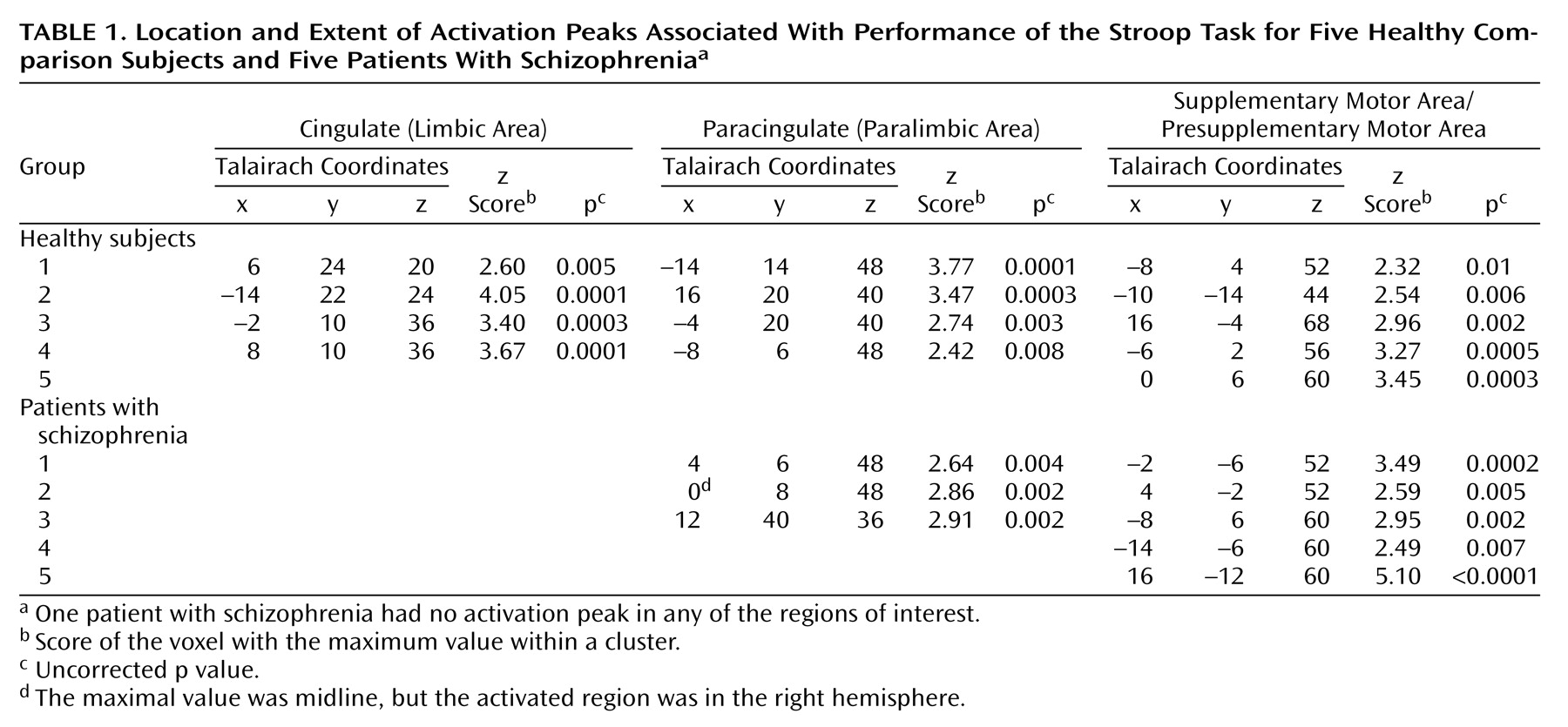
Table 1 shows the single highest task-related activation peak within the limbic anterior cingulate, paralimbic anterior cingulate, and supplementary motor area for each individual.
Four of the five healthy subjects had an activation peak in both the limbic and the paralimbic anterior cingulate. In contrast, none of the patients displayed an activation peak in the limbic anterior cingulate, although three of the six patients showed significant activation in the paralimbic anterior cingulate. Both healthy subjects and patients showed consistent activation of the supplementary motor area/presupplementary motor area.
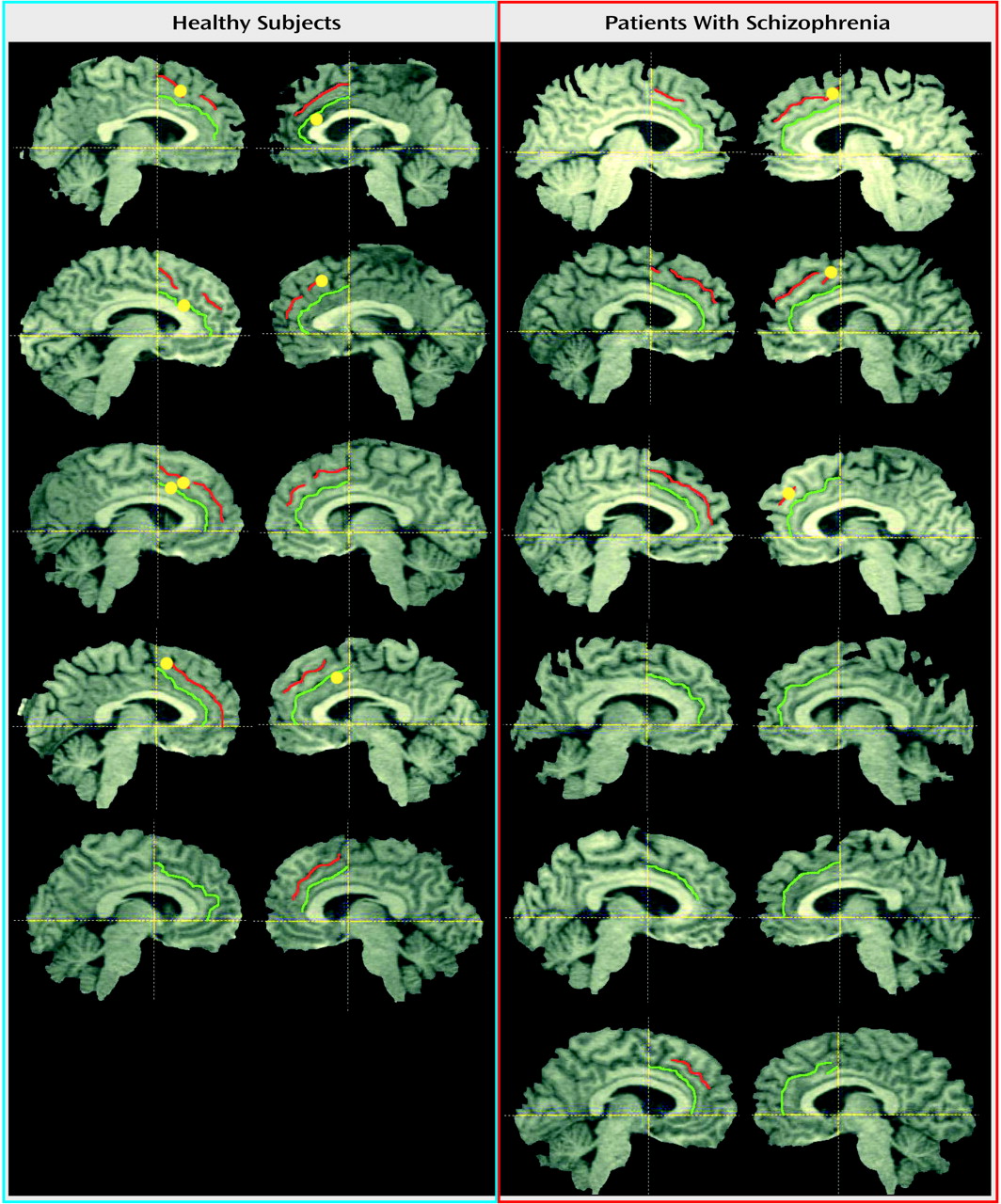
Examination of the underlying morphological characteristics of the activated regions (Figure 1) revealed that those patients who showed activation in the paralimbic-anterior cingulate also had a paracingulate sulcus in the activated hemisphere. In contrast, two of the three patients without activation of the anterior cingulate did not have a paracingulate sulcus in either hemisphere.

Discussion

Our finding that the limbic anterior cingulate was not activated in individual patients with schizophrenia during Stroop task performance is consistent with the group-averaged study by Carter et al. (2). Our findings also show that in some patients a separate, paralimbic region of the anterior cingulate is activated. Further studies should examine illness characteristics of patients whose paralimbic-anterior cingulate is activated compared with those with no activation in this region.
The paralimbic-anterior cingulate was activated only in patients with a paracingulate sulcus. Patients without a paracingulate sulcus displayed no activation across the entire anterior cingulate cortex. Furthermore, although the paralimbic anterior cingulate in the left hemisphere was activated in three of the four healthy subjects, in patients this region was activated only in the right hemisphere. These observations suggest a left-lateralized functional pathology in the paralimbic anterior cingulate of affected patients, consistent with our previous finding of left-lateralized anterior cingulate morphological anomalies (6). Together, our findings suggest that anterior cingulate sulcal/gyral variation has functional significance.
It is possible that the anterior cingulate morphological anomalies previously identified (6) and the functional abnormalities revealed in the current study reflect disturbed brain connectivity occurring early in life. Significant reductions in gray matter volume have been found in both the cingulate and paracingulate as well as in the prefrontal areas with which they connect (10). In addition, functional studies have found evidence of anterior cingulate-frontal disconnectivity (11). There is also evidence of discrete alterations of anterior cingulate intrinsic circuitry in affected patients (1). Such local and distal disruptions of anterior cingulate connectivity during neurodevelopment may affect its subsequent maturation and functional integrity and may be at the core of impaired attentional processing in schizophrenia (2).
Our finding that the supplementary motor area is consistently activated in patients, as it is in healthy subjects, suggests that the connectivity of this region is intact. Supplementary motor area activation presumably reflects the inhibitory component relevant to preparing the verbal response during Stroop task performance (5). Together, these findings are consistent with schizophrenia being a disorder of connectivity with specific involvement of the limbic anterior cingulate.
A number of methodological issues may limit these interpretations and warrant the replication and extension of our findings. These include the small number of patients and comparison subjects studied, the limited spatial resolution of PET, and the medication status of the patients.
TABLE 1
Figure 1. Location of Regional Cerebral Blood Flow Response During Performance of the Stroop Task in Five Healthy Comparison Subjects and Six Patients With Schizophreniaa
aParasagittal views of the left and right hemispheres show anterior cingulate morphological characteristics; the cingulate sulcus is highlighted in green, and the paracingulate sulcus is highlighted in red. Each yellow circle (where present) shows the single highest activation peak within the limbic cingulate sulcus and the paralimbic paracingulate sulcus for each individual. The images relate activation during positron emission tomography to the morphological characteristics in the medial surface. Functional activations were mapped 2–5 mm from the midline to emphasize anterior cingulate morphological features.

Footnote

Presented in part at the 8th International Congress on Schizophrenia Research, Whistler, British Colombia, Canada, April 30, 2001. Received Jan. 17, 2001; revisions received June 26 and Aug. 17, 2001; accepted Aug. 29, 2001. From the Cognitive Neuropsychiatry Research and Academic Unit, Department of Psychiatry, and the Howard Florey Institute, University of Melbourne, Victoria, Australia; Sunshine Hospital, Melbourne; the Applied Schizophrenia Division, Mental Health Research Institute, Parkville, Victoria; the School of Psychological Sciences, La Trobe University, Bundoora, Victoria; the PET Centre, Austin and Repatriation Medical Centre, Heidelberg, Melbourne; and the Brain Sciences Institute, Swinburne University of Technology, Hawthorn, Australia. Address reprint requests to Dr. Yücel, Cognitive Neuropsychiatry Unit, Mental Health Research Institute, Locked Bag 11, Parkville, Victoria, Australia 3052; [email protected] (e-mail). Supported by a Ph.D. scholarship from La Trobe University (Dr. Yücel), a National Alliance for Research on Schizophrenia and Depression Young Investigator Award (Dr. Stuart), National Health and Medical Research Council (NHMRC) grant 970599, the NHMRC Brain Research Network, the Woods Family, and the Ian Potter Foundation. The authors thank Drs. Phyllis Chua, James Olver, and Simon Collinson for their assistance with task and subject preparation and image acquisition. They also thank the PET Centre staff for their assistance and support throughout the study.

References

1.
Benes FM: Relationship of cingulate cortex to schizophrenia and other psychiatric disorders, in Neurobiology of Cingulate Cortex and Limbic Thalamus: A Comprehensive Handbook. Edited by Vogt BA, Gabriel MS. Boston, Birkhauser, 1993, pp 580-605
2.
Carter CS, Mintun M, Nichols T, Cohen JD: Anterior cingulate gyrus dysfunction and selective attention deficits in schizophrenia: [15O]H2O PET study during single-trial Stroop task performance. Am J Psychiatry 1997; 154:1670-1675
3.
Yücel M, Stuart GW, Maruff P, Velakoulis D, Crowe SF, Savage G, Pantelis C: Hemispheric and gender-related differences in the gross morphology of the anterior cingulate/paracingulate cortex in normal volunteers: an MRI morphometric study. Cereb Cortex 2001; 11:17-25
4.
Schlaug G, Knorr U, Seitz R: Inter-subject variability of cerebral activations in acquiring a motor skill: a study with positron emission tomography. Exp Brain Res 1994; 98:523-534
5.
Fink GR, Frackowiak RS, Pietrzyke U, Passingham RE: Multiple nonprimary motor areas in the human cortex. J Neurophysiol 1997; 77:2164-2174
6.
Pantelis C, Yücel M, Wood SJ, McGorry PD, Velakoulis D: The timing and functional consequences of structural brain abnormalities in schizophrenia. Neuroscience News 2001; 4:36-46
7.
Pardo JV, Pardo PJ, Janer KW, Raichle ME: The anterior cingulate cortex mediates processing selection in the Stroop attentional conflict paradigm. Proc Natl Acad Sci USA 1990; 87:256-259
8.
Braus DF, Ende G, Hubrich-Ungureanu P, Henn FA: Cortical response to motor stimulation in neuroleptic-naive first episode schizophrenics. Psychiatry Res 2000; 98:145-154
9.
Friston KJ, Holmes AP, Worsley KJ, Poline J-P, Frith CD, Frackowiak RS: Statistical parametric maps in functional imaging: a general linear approach. Hum Brain Mapp 1995; 2:189-210
10.
Goldstein JM, Goodman JM, Seidman LJ, Kennedy DN, Makris N, Lee H, Tourville J, Caviness VS Jr, Faraone SV, Tsuang MT: Cortical abnormalities in schizophrenia identified by structural magnetic resonance imaging. Arch Gen Psychiatry 1999; 56:537-547
11.
Spence SA, Liddle PF, Stefan MD, Hellewell JS, Sharma T, Friston KJ, Hirsch SR, Frith CD, Murray RM, Deakin JF, Grasby PM: Functional anatomy of verbal fluency in people with schizophrenia and those at genetic risk: focal dysfunction and distributed disconnectivity reappraised. Br J Psychiatry 2000; 176:52-60

Information & Authors

Information

Published In

Go to American Journal of Psychiatry
American Journal of Psychiatry
Pages: 251 - 254
PubMed: 11823267

History

Published online: 1 February 2002
Published in print: February 2002

Authors

Affiliations

Christos Pantelis, M.R.C.Psych., F.R.A.N.Z.C.P.
Geoffrey W. Stuart, Ph.D.
Stephen J. Wood, Ph.D.
Dennis Velakoulis, F.R.A.N.Z.C.P.
Henri J. Tochon-Danguy, Ph.D.
Gary F. Egan, Ph.D., M.B.A.

Metrics & Citations

Metrics

Citations

Export Citations

If you have the appropriate software installed, you can download article citation data to the citation manager of your choice. Simply select your manager software from the list below and click Download.

For more information or tips please see 'Downloading to a citation manager' in the Help menu.

Format
Citation style
Style
Copy to clipboard

There are no citations for this item

View Options

View options

PDF/ePub

View PDF/ePub

Get Access

Login options

Already a subscriber? Access your subscription through your login credentials or your institution for full access to this article.

Personal login Institutional Login Open Athens login
Purchase Options

Purchase this article to access the full text.

PPV Articles - American Journal of Psychiatry

PPV Articles - American Journal of Psychiatry

Not a subscriber?

Subscribe Now / Learn More

PsychiatryOnline subscription options offer access to the DSM-5-TR® library, books, journals, CME, and patient resources. This all-in-one virtual library provides psychiatrists and mental health professionals with key resources for diagnosis, treatment, research, and professional development.

Need more help? PsychiatryOnline Customer Service may be reached by emailing [email protected] or by calling 800-368-5777 (in the U.S.) or 703-907-7322 (outside the U.S.).

Media

Figures

Other

Tables

Share

Share

Share article link

Share