Full access
Editorial
Published Online: 1 August 2008

Neural Networks in Schizophrenia

As methods for high-resolution MRI continue to develop, our understanding of changes in brain structure in patients with psychiatric disorders improves. In patients with schizophrenia, decreases in the gray matter volume of a variety of cortical and subcortical brain regions have been repeatedly observed (1) . Two articles in this issue of the Journal add to this literature and go further by strengthening the hypothesis that schizophrenia involves abnormalities in specific cortical-subcortical networks. Ellison-Wright et al. (2) present a meta-analysis of MRI studies that reveals different patterns of structural changes in first-episode and chronic schizophrenia. Adapting a technique developed to combine data from functional MRI studies (3), the authors evaluated group differences in specific brain regions in reference to a three-dimensional coordinate system. The result of this adaptation, called anatomical likelihood estimation, adds to a growing literature on probabilistic approaches to neuroanatomical computation (4) . In patients with first-episode schizophrenia, Ellison-Wright et al. found evidence for decreased gray matter volume in the hippocampus, the caudate nucleus, the thalamus (mediodorsal nucleus), the insula, the anterior cingulate gyrus, the inferior frontal gyrus, and the cerebellum. Subcortical changes were similar in patients with chronic schizophrenia, but cortical changes were even more extensive.
As Ellison-Wright et al. note, this pattern of abnormality implicates a neural circuit originating with limbic input to the striatum, then the thalamus, and finally to the prefrontal and cingulate cortex ( Figure 1 ). Abnormalities of thalamocortical circuitry have previously been implicated in schizophrenia, particularly in reference to the pathophysiological concept of cognitive dysmetria (5) .
Figure 1. Gray Matter Deficits and Thalamocorticostriatal Circuit Dysfunction in Schizophrenia a
a The diagram on the left illustrates the circuit pathways. On the right is a diagrammatic representation of thalamocorticostriatal circuits superimposed on regions of gray matter showing signal decrease in both first-episode schizophrenia (yellow) and chronic schizophrenia (red). The thalamus (blue triangle) sends thalamocortical projections to the cortex (blue arrow). Cortical regions project (green arrow) to the caudate (pink circle) and then back to the thalamus (orange arrow). Components of these circuits show anatomical changes in schizophrenia, with the caudate head showing gray matter deficits in first-episode schizophrenia and more widespread cortical changes in chronic schizophrenia. The thalamus shows gray matter deficits in both first-episode and chronic schizophrenia.
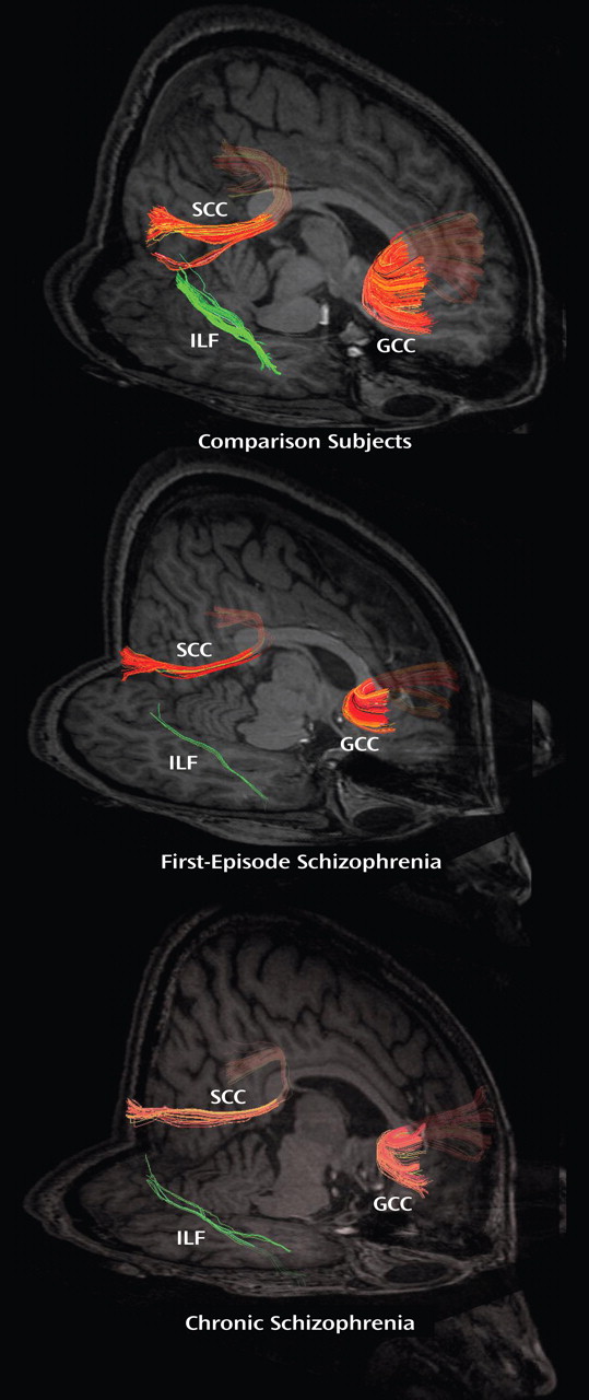
Also in this issue, Friedman et al. (6) report on their use of diffusion tensor imaging to examine the pattern of white matter changes in patients with first-episode and chronic schizophrenia. Using fractional anisotropy (FA) to assess the integrity of white matter connections (7), they found evidence in first-episode patients for decreased connectivity only in the inferior longitudinal fasciculus (at the trend level), while chronic patients showed a broader pattern of white matter disturbances ( Figure 2 ).
Figure 2. Regions of Fractional Anisotropy (FA) Reductions in Schizophrenia a
a Relative to healthy comparison subjects, patients with first-episode schizophrenia showed trend-level FA reductions in the left and right inferior longitudinal fasciculus. Patients with chronic schizophrenia showed significant FA reductions in the right forceps minor and the left inferior longitudinal fasciculus. ILF=inferior longitudinal fasciculus; GCC=genu of corpus callosum; SCC=splenium of corpus callosum.
The results of Friedman’s study, to date the largest to directly examine FA in first-episode and chronic schizophrenia patients, suggest a number of intriguing interpretations. As they note, one explanation may be that pathological white matter abnormalities may be present but too subtle to detect with current imaging methods in first-episode schizophrenia. Alternatively, white matter changes may develop as the disease process of schizophrenia progresses.
Taken together, the results of these two studies provide strong support for the hypothesis that schizophrenia involves abnormalities in networks of brain regions. They further suggest that such abnormalities may be progressive in at least some patients. If the “progression” hypothesis is confirmed in future studies, we must redouble our efforts at early and effective intervention for this potentially disabling disorder.

Footnotes

Address correspondence and reprint requests to Dr. Csernansky, Department of Psychiatry and Behavioral Sciences, Feinberg School of Medicine, Northwestern University, 446 E. Ontario, Suite 7-200, Chicago, IL 60611; [email protected] (e-mail). Editorial accepted for publication May 2008 (doi: 10.1176/appi.ajp.2008.08050700).
Dr. Csernansky has consulted for Eli Lilly, Sanofi-Aventis, and HoustonPharma. Dr. Freedman has reviewed this editorial and found no evidence of influence from these relationships. Dr. Cronenwett reports no competing interests.

References

1.
Shenton ME, Dickey CC, Frumin M, McCarley RW: A review of MRI findings in schizophrenia. Schizophr Res 2001; 49:1–52
2.
Ellison-Wright I, Glahn DC, Laird AR, Thelen SM, Bullmore E: The anatomy of first-episode and chronic schizophrenia: an anatomical likelihood estimation meta-analysis. Am J Psychiatry 2008; 165:1015–1023
3.
Turkeltaub PE, Eden GF, Jones KM, Zeffiro TA: Meta-analysis of the functional neuroanatomy of single-word reading: method and validation. Neuroimage 2002; 16:765–780
4.
Csernansky JG, Wang L, Joshi SC, Ratnanather JT, Miller MI: Computational anatomy and neuropsychiatric disease: probabilistic assessment of variation and statistical inference of group difference, hemispheric asymmetry, and time-dependent change. Neuroimage 2004; 23(suppl 1):S56–S68
5.
Andreasen NC, Nopoulos P, O’Leary DS, Miller DD, Wassink T, Flaum M: Defining the phenotype of schizophrenia: cognitive dysmetria and its neural mechanisms. Biol Psychiatry 1999; 46:908–920
6.
Friedman JI, Tang C, Carpenter D, Buchsbaum M, Schmeidler J, Flanagan L, Golembo S, Kanellopoulou I, Ng J, Hof PR, Harvey PD, Tsopelas ND, Stewart D, Davis KL: Diffusion tensor imaging findings in first-episode and chronic schizophrenia patients. Am J Psychiatry 2008; 165:1024–1032
7.
Taylor W, Hsu E, Krishnan R, MacFall J: Diffusion tensor imaging: background, potential, and utility in psychiatric research. Biol Psychiatry 2004; 55:201–207

Information & Authors

Information

Published In

Go to American Journal of Psychiatry
American Journal of Psychiatry
Pages: 937 - 939
PubMed: 18676594

History

Published online: 1 August 2008
Published in print: August, 2008

Authors

Affiliations

John G. Csernansky, M.D.
Will J. Cronenwett, M.D.

Metrics & Citations

Metrics

Citations

Export Citations

If you have the appropriate software installed, you can download article citation data to the citation manager of your choice. Simply select your manager software from the list below and click Download.

For more information or tips please see 'Downloading to a citation manager' in the Help menu.

Format
Citation style
Style
Copy to clipboard

There are no citations for this item

View Options

View options

PDF/ePub

View PDF/ePub

Get Access

Login options

Already a subscriber? Access your subscription through your login credentials or your institution for full access to this article.

Personal login Institutional Login Open Athens login
Purchase Options

Purchase this article to access the full text.

PPV Articles - American Journal of Psychiatry

PPV Articles - American Journal of Psychiatry

Not a subscriber?

Subscribe Now / Learn More

PsychiatryOnline subscription options offer access to the DSM-5-TR® library, books, journals, CME, and patient resources. This all-in-one virtual library provides psychiatrists and mental health professionals with key resources for diagnosis, treatment, research, and professional development.

Need more help? PsychiatryOnline Customer Service may be reached by emailing [email protected] or by calling 800-368-5777 (in the U.S.) or 703-907-7322 (outside the U.S.).

Media

Figures

Other

Tables

Share

Share

Share article link

Share