Full access
New Research
Published Online: 1 February 2011

Aberrant Brain Activation During a Working Memory Task in Psychotic Major Depression

Abstract

Objective:

The authors sought to better understand the neural circuitry associated with working memory deficits in psychotic major depression by examining brain function during an N-back task.

Method:

Study subjects were 16 patients with psychotic major depression, 15 patients with nonpsychotic major depression, and 19 healthy comparison subjects. Functional MRI data were collected while participants responded to letter stimuli that were repeated from the previous trial (1-back) or the one before that (2-back).

Results:

Relative to the healthy comparison group, both the psychotic and nonpsychotic major depression groups had significantly greater activation in the right parahippocampal gyrus during the 2-back task, and the psychotic major depression group showed this overactivation during the 1-back task as well. The nonpsychotic major depression group showed significantly lower activation than other groups in the right dorsolateral prefrontal cortex and greater activation than the healthy comparison group in the superior occipital cortex. The psychotic major depression group was unique in showing greater activation than both other groups in the right temporoparietal junction, a cluster that also demonstrated connectivity with activation in the left prefrontal cortex.

Conclusions:

The psychotic major depression group showed aberrant parahippocampal activation at a lower demand level than observed in nonpsychotic major depression. While the nonpsychotic major depression group showed abnormalities in frontal executive regions, the psychotic major depression group showed abnormalities in temporoparietal regions associated with orienting to unexpected stimuli. Considering the functional connectivity of this cluster with left dorsolateral prefrontal cortex regions, these findings may reflect neural compensation for sensory gating deficits in psychotic major depression.
An estimated 5%–20% of patients with major depression experience psychotic symptoms, including hallucinations and delusions (1, 2). Psychotic major depression is associated with more severe psychomotor symptoms and guilt (3, 4), significantly greater impairment, longer illness duration, and greater likelihood of recurrence compared with nonpsychotic major depression (1, 5, 6). However, the severity of a depressive episode does not necessarily determine whether psychotic features will be present (7). Unfortunately, psychotic major depression is frequently misdiagnosed (8), which can deter effective treatment.
A better understanding of the brain pathophysiology underlying psychotic major depression can lead to earlier detection and more effective treatments. Neuropsychological data from our lab have shown that patients with psychotic major depression have greater cognitive impairment than do patients with nonpsychotic major depression or healthy comparison subjects on tests of working memory, verbal memory, and psychomotor speed but not on tests of simple verbal attention (9), which implies abnormal brain circuits associated with memory encoding and executive function (10). However, the relationship of these cognitive impairments to neural function is not clear. For example, impairments in the function of temporal lobe memory systems, prefrontal lobe executive regions, or visual and attention networks could underlie working memory deficits in psychotic major depression.
Working memory tasks require the temporary maintenance and manipulation of information as well as sustained attention. The neurofunctional system subserving verbal working memory has been studied extensively in healthy comparison subjects using functional MRI (fMRI). Studies consistently show that the dorsolateral prefrontal cortex, typically in Brodmann's area 46, sustains activation during the delay periods in which individuals hold information in working memory (11) and suppresses activation to nonrelevant stimuli (12). The sustained attention and target detection component of a working memory task often activates a right hemisphere attention system, including regions of the temporoparietal cortex and inferior and middle frontal gyri. This network is consistently activated during detection of task-relevant stimuli, especially when they are infrequent (13). In the left hemisphere, the inferior parietal region is believed to be part of a storage buffer for verbal information (14). The hippocampus is also active during the delay period of a working memory task, but the parahippocampal gyrus, in contrast, is activated during the encoding and recognition parts of the working memory task and predicts successful long-term memory encoding (15).
Because of the widespread brain circuitry involved, patients with various psychiatric disorders have been found to show abnormal brain activation during working memory tasks. Patients with nonpsychotic major depression show abnormal prefrontal function during a verbal working memory task, which emphasizes the importance of comparing patients with psychotic and nonpsychotic major depression (1622). Because patients with schizophrenia also display deficits in working memory, several neuroimaging studies have found dorsolateral prefrontal cortex deficits in schizophrenia relative to comparison subjects (2325) and to patients with nonpsychotic major depression (24, 26), which suggests a common substrate for working memory deficits across diagnoses but with greater severity in schizophrenia. Given that psychotic major depression involves symptoms of both nonpsychotic major depression and schizophrenia, these patients may share common deficits in executive function. However, because high cortisol levels in psychotic major depression (9) are likely to affect hippocampal and parahippocampal systems as well, deficits in medial temporal lobe regions may differentiate psychotic major depression from nonpsychotic major depression.
In this study, we investigated the neural correlates of a verbal working memory task in patients with psychotic major depression in order to better understand the brain circuitry underlying working memory deficits in psychotic major depression. Patients with nonpsychotic major depression and healthy comparison subjects were included to differentiate between depressive subtypes as well as between these subtypes and unaffected individuals. We predicted that the psychotic major depression group would show a unique pattern of activation that reflects abnormalities in medial temporal lobe and parietal regions and similarities to the nonpsychotic major depression group in prefrontal regions.

Method

Participants

Participants were recruited from outpatient psychiatric clinics at Stanford University and through advertisements in the surrounding communities. Patients with psychotic and nonpsychotic major depression were diagnosed with the Structured Clinical Interview for DSM-IV (SCID) (27), and diagnoses were confirmed by the treating psychiatrist when available. Patients had to score at least 18 on the 21-item Hamilton Depression Rating Scale (HAM-D) (28) and at least 7 on the Thase Core Endogenomorphic Scale (29) to verify significant depressive and endogenous symptoms, respectively. Patients with psychotic major depression had to score at least 5 on the positive symptom subscale of the Brief Psychiatric Rating Scale (BPRS; 30). All patients met DSM-IV criteria for current unipolar major depressive episode. Patients with nonpsychotic major depression had no history of psychotic symptoms. Patients were excluded if they had active suicidality, obsessive-compulsive disorder, or bipolar disorder or had abused substances or received ECT within the past 6 months. All participants were allowed to continue their psychiatric medications but were required to maintain a stable medication regimen for at least 1 week before the study.
Healthy comparison subjects, recruited through advertisements, had to score less than 6 on the HAM-D, have no psychotic symptoms, and have no current or past axis I diagnoses according to SCID criteria.
Exclusion criteria for all participants included major medical illness, history of seizures or head injury, pregnancy or lactation, age <18 years, or use of estrogen supplements or birth control pills.
The study included 22 patients with psychotic major depression, 21 patients with nonpsychotic major depression, and 24 healthy comparison subjects. All participants gave written informed consent and received $250 for their participation. The study was approved by the Institutional Review Board of Stanford University.

The N-Back Task

Participants performed an N-back task during image acquisition. The task had three types of alternating blocks: four 1-back, four 2-back, and six control (“press for Z”) blocks. For all blocks, a succession of uniform-size single letters was presented, in both upper- and lowercase. Each letter was presented for 500 msec, with a 1,500 msec intertrial interval showing a fixation cross. Each block contained 12 letters and lasted for 24 seconds. Brief instructions were shown for 4 seconds at the beginning of the block (e.g., “Press for 1-back”). During the 1-back blocks, participants pressed the button when the current letter matched the one presented one trial back, e.g., a repeated letter such as A—A. During 2-back blocks, participants pressed the button when the current letter matched the one presented two trials back, e.g., in a “sandwich” pattern such as A—B—-A. During the “press for Z” blocks, participants pressed the button whenever the current letter was a Z. Participants pressed with the right index finger on a hand-held button box. Stimuli were projected onto a screen and viewed through a mirror attached to the head coil. All participants practiced the task before the scan. The scan lasted 7 minutes and 48 seconds, during which 232 frames were acquired. The E-Prime software program (www.pstnet.com) was used to present the task and collect responses.

fMRI Data Acquisition and Processing

Images were acquired on a 3-T General Electric (GE) Signa scanner using a standard GE whole-head coil (Lx platform, gradients 40 mT/m, 150 T/m/sec; GE Medical Systems, Milwaukee). A custom-built head restraint system and foam padding prevented head movement. A high-order shim (31) reduced blurring and signal loss from field inhomogeneities. Twenty-eight axial slices (4 mm thick, 0.5 mm skip) parallel to the anterior and posterior commissures and covering the whole brain were imaged using a T2*-weighted spiral pulse sequence (32) (repetition time=2,000 msec, echo time=30 msec, flip angle=80°, interleave=1, field of view=200 mm2, matrix=64×64, inplane spatial resolution=3.125 mm2).
fMRI data were processed using SPM5 (www.fil.ion.ucl.ac.uk/spm). Images were realigned to the third volume. Image distortion and spin history errors caused by abrupt motions were repaired by interpolation from the nearest unaffected volumes. These methods were implemented in the ArtRepair toolbox for SPM (http://cibsr.stanford.edu/tools/ArtRepair/ArtRepair.htm). Data were normalized to the Montreal Neurological Institute (MNI) echo-planar image template and resampled to a 2-mm3 matrix using sinc interpolation. Data were smoothed with a 4-mm full width at half maximum Gaussian filter and high-pass filtered at 120 seconds.
Individual statistics were computed using a fixed-effects model and a block design comparing 1-back with press for Z, and 2-back with press for Z. Group t tests used a random-effects model to examine activation within and between each group. A dual-significance corrected threshold of height at p=0.01 and cluster extent at p=0.01 was used within groups. To determine the location of significant clusters, coordinates were first converted to the Talairach template using the mni2tal function (http://imaging.mrc-cbu.cam.ac.uk/imaging/MniTalairach), and then brain regions were localized using the Talairach Daemon software (http://www.talairach.org/client.html) and also visually inspected by an experienced neuroimager (A.G.).
A multivariate analysis of variance was conducted in SPM5 using the independent factor group and the repeated factor task (1-back minus press for Z, and 2-back minus press for Z). Gender and response time were used as covariates (see below). A threshold of p=0.001, extent=10 defined significant activation that differed among the three groups across task conditions (main effect of group). Mean activation levels (t-scores) were extracted from significant clusters using MarsBar (http://marsbar.sourceforge.net/). Between-group t tests were then performed using SPSS (http://www.spss.com/) with a corrected significance threshold of p=0.0125 (or p=0.05/4 regions tested).
A connectivity analysis was conducted using the Psychophysiological Interaction module of SPM5. This post hoc analysis located brain regions functionally associated with the temporoparietal junction region in the psychotic major depression group. The temporoparietal junction cluster that was significantly increased in the psychotic major depression group was used as the seed region. For each participant, a 6-mm3 box was placed on the maximum voxel in the cluster. The average time course of the voxels that surpassed threshold (p=0.01) was extracted. A multiple regression identified voxels showing a significant interaction between that time course and the 2-back-press-for-Z contrast for each participant. A random-effects analysis combined individual results into a group result using a cluster-corrected threshold of dual height and extent at p=0.01.

Results

Data for five participants with psychotic major depression, two with nonpsychotic major depression, and three healthy comparison subjects were excluded because of response box failure resulting in N-back task accuracy below 50%. Scan data from one patient with psychotic major depression, four patients with nonpsychotic major depression, and two healthy comparison subjects were excluded because of movement artifacts during more than 20% of the scan. This left 16 participants in the psychotic major depression group, 15 in the nonpsychotic major depression group, and 19 in the healthy comparison group.
Table 1 summarizes participants' clinical and demographic characteristics. There were no group differences in age, handedness, or years of education, but the groups differed in gender distribution. The groups had similar IQ, as measured by full-scale estimates of premorbid intellectual functioning.
TABLE 1. Clinical, Demographic, and Task Performance Measures, by Group, in a Study of the Neural Circuitry Associated With Working Memory Deficits in Psychotic Major Depression
MeasurePsychotic Major Depression Group (N=16)Nonpsychotic Major Depression Group (N=15)Healthy Comparison Group (N=19)
 MeanSDMeanSDMeanSD
Age (years)34.1310.6839.8112.7434.8512.54
Education (years)16.25a3.7214.401.4015.952.15
IQb111.569.40104.0010.63111.289.35
Hamilton Depression Rating Scale (21-item) scorec27.883.5224.203.300.580.84
Brief Psychiatric Rating Scale scored28.755.3615.732.710.420.69
Brief Psychiatric Rating Scale positive symptom subscoree8.383.700.130.350.000.00
Length of current depressive episode (weeks)165.8f198.4155.5g217.1  
Task performance      
    1-Back      
        % Correct97.251.1396.411.2097.261.05
        Response time581.728.24650.029.96556.726.35
    2-Back      
        % Correct98.472.8499.030.3899.770.33
        Response timeh629.630.28683.932.13559.728.26
 N%N%N%
Femalei743.81066.7631.6
Left-handed2j15.4213.3210.5
Current medication      
    None212.5640.0  
    Anxiolytics425.000.0  
    Antidepressants850.0960.0  
    Antipsychotics1062.500.0  
    Mood stabilizer16.316.7  
Comorbid diagnoses      
    None531.3533.3  
    Anxiety disordersk318.8853.5  
    Bulimia nervosa16.300.0  
a
Based on 12 of 16 participants.
b
Wechsler Test of Reading estimate of premorbid full-scale IQ; means are based on N=9 patients with psychotic major depression, N=12 patients with nonpsychotic major depression, and N=18 healthy comparison subjects.
c
Psychotic major depression group > all others, nonpsychotic major depression group > healthy comparison group, p=0.0001.
d
Groups significantly different, p=0.0001.
e
Psychotic major depression group > all others, p<0.0001.
f
Based on 9 of 16 participants.
g
Based on 10 of 15 participants.
h
Nonpsychotic major depression group > healthy comparison group, p=0.008.
i
Groups significantly different, p=0.011.
j
Based on 13 of 16 participants.
k
Anxiety disorders in the psychotic major depression group included one patient with panic disorder, two with agoraphobia, two with gen-eralized anxiety disorder, one with posttraumatic stress disorder, and one with specific phobia. Anxiety disorders in the nonpsychotic major depression group included two patients with obsessive-compulsive disorder, three with specific phobia, three with panic disorder, four with social phobia, two with generalized anxiety disorder, two with agoraphobia, and two with posttraumatic stress disorder.
The patients for whom medication data were available had established antidepressant medication regimens with no changes; the psychotic major depression group (N=6) had an average of 20.3 weeks of antidepressant use (range=3–56, SD=19.9) and 9.8 weeks of antipsychotic use (range=5–26, SD=9.2). The nonpsychotic major depression group (N=9) had an average of 42.6 weeks of antidepressant use (range=4–156, SD=56.4).

N-Back Task Performance Results

All participants performed the task with a high degree of accuracy (Table 1). Response time for the 2-back task was different between groups, and further comparisons showed that this was due to significantly slower response time in the nonpsychotic major depression group compared to the healthy comparison group (p=0.002), even when controlling for group differences in gender (p=0.007). Therefore, all comparisons with the nonpsychotic major depression group during the 2-back task included response time as a covariate.

fMRI Results

Tables 2 and 3 list regions that were significantly activated during the 1-back and 2-back tasks within each group. All three groups activated regions typically associated with verbal working memory tasks, such as the left supramarginal gyrus, left inferior frontal gyrus, and left and right inferior parietal lobe.
TABLE 2. Significant Activation Within Each Group During the 1-Back Versus Press-for-Z Contrast
RegionBrodmann's AreapaCluster Size (Voxels)z ScoreTalairach Coordinates
xyz
Healthy comparison group (N=19)       
Right inferior frontal gyrus460.00013,5414.50573213
Right middle frontal gyrus46  4.29553215
 6  4.2336849
Left inferior parietal lobe400.0011,8174.47−42−4541
Left supramarginal gyrus40  4.19−42−4337
Right inferior parietal lobe400.00012,2004.1951−3652
Right superior parietal lobe7  4.0942−5849
Right postcentral gyrus40  3.9450−3251
Left middle frontal gyrus60.0031,3983.91−301455
 9  3.41−571730
Left superior frontal gyrus6  3.54−321451
Left inferior frontal gyrus9  3.51−571729
Psychotic major depression group (N=16)       
Right middle frontal gyrus60.00011,5304.0055642
 8  3.91551042
Right inferior frontal gyrus44  3.8653920
Left supramarginal gyrus400.00012,6104.26−42−4337
Left inferior parietal lobe40  4.11−42−4339
Right precuneus190.00012,0894.1032−6438
Right inferior parietal lobe39  3.6334−6238
 40  3.5944−4445
Left inferior frontal gyrus90.00013,4754.13−481723
Left middle frontal gyrus9  4.04−531331
 6  3.88−511444
Right medial frontal gyrus8  3.9282941
Nonpsychotic major depression group (N=15)       
Right inferior frontal gyrus460.00015,2785.84513911
Right middle frontal gyrus46  5.13573022
 6  4.9534048
Right inferior parietal lobe400.00014,3475.7440−5045
Left middle frontal gyrus60.00016,9835.26−36649
Left middle frontal gyrus46  4.37−442821
Right medial frontal gyrus8  5.00103141
Right superior frontal gyrus8  4.6883143
Left inferior frontal gyrus45  4.23−462621
Left inferior parietal lobe400.00013,5465.09−50−3941
Left supramarginal gyrus40  4.77−44−4137
Left lingual gyrus170.00011,7483.82−20−97−7
 18  3.71−20−99−5
Left inferior occipital gyrus17  3.80−22−97−7
Left inferior temporal gyrus20  3.50−57−49−14
Left fusiform gyrus19  3.48−50−69−12
 37  3.46−55−51−16
Left middle occipital gyrus19  3.40−50−67−10
Right inferior temporal gyrus200.00012,3863.8059−47−14
Right fusiform gyrus37  3.6455−49−16
Right middle temporal gyrus37  3.5059−49−9
a
Corrected cluster-wise.
TABLE 3. Significant Activation Within Each Group During the 2-Back Versus Press-for-Z Contrast
RegionBrodmann's AreapaCluster Size (Voxels)z ScoreTalairach Coordinates
xyz
Healthy comparison group (N=19)       
Left inferior parietal lobe400.000112,7507.06444341
Left supramarginal gyrus40  6.02424137
Right middle frontal gyrus60.000130,4536.1036646
Left middle frontal gyrus9  6.05511332
Right superior frontal gyrus8  5.7421851
Left superior frontal gyrus6  5.7321653
Right cuneus170.0055093.84147911
Right posterior cingulate30  3.2626647
Left inferior temporal gyrus200.0055323.34575114
Left fusiform gyrus37  3.23535116
Left middle occipital gyrus37  3.08556510
Psychotic major depression group (N=16)       
Right inferior frontal gyrus440.000111,5555.02571616
 45  4.78571320
Right middle frontal gyrus8  4.41281442
 6  4.41301244
Right insula13  4.3330174
Right angular gyrus390.000110,4134.59346036
Right precuneus39  4.45366238
Right inferior parietal lobe39  4.44366238
Left inferior parietal lobe40  4.43423944
Left precuneus19  4.32287441
Right subgyral angular gyrus39  4.27326036
Right medial frontal gyrus80.00016,3394.5862943
 6  4.2863537
Right superior frontal gyrus8  4.4263143
Left inferior frontal gyrus9  4.42501127
Nonpsychotic major depression group (N=15)       
Right inferior parietal lobe400.000110,0875.81484443
Right supramarginal gyrus40  5.26444537
Left caudate bodyNA0.000121,9105.7412716
Right caudate bodyNA  5.3012714
Right caudate headNA  5.1114125
a
Corrected cluster-wise.

Between-Group Analysis of Variance

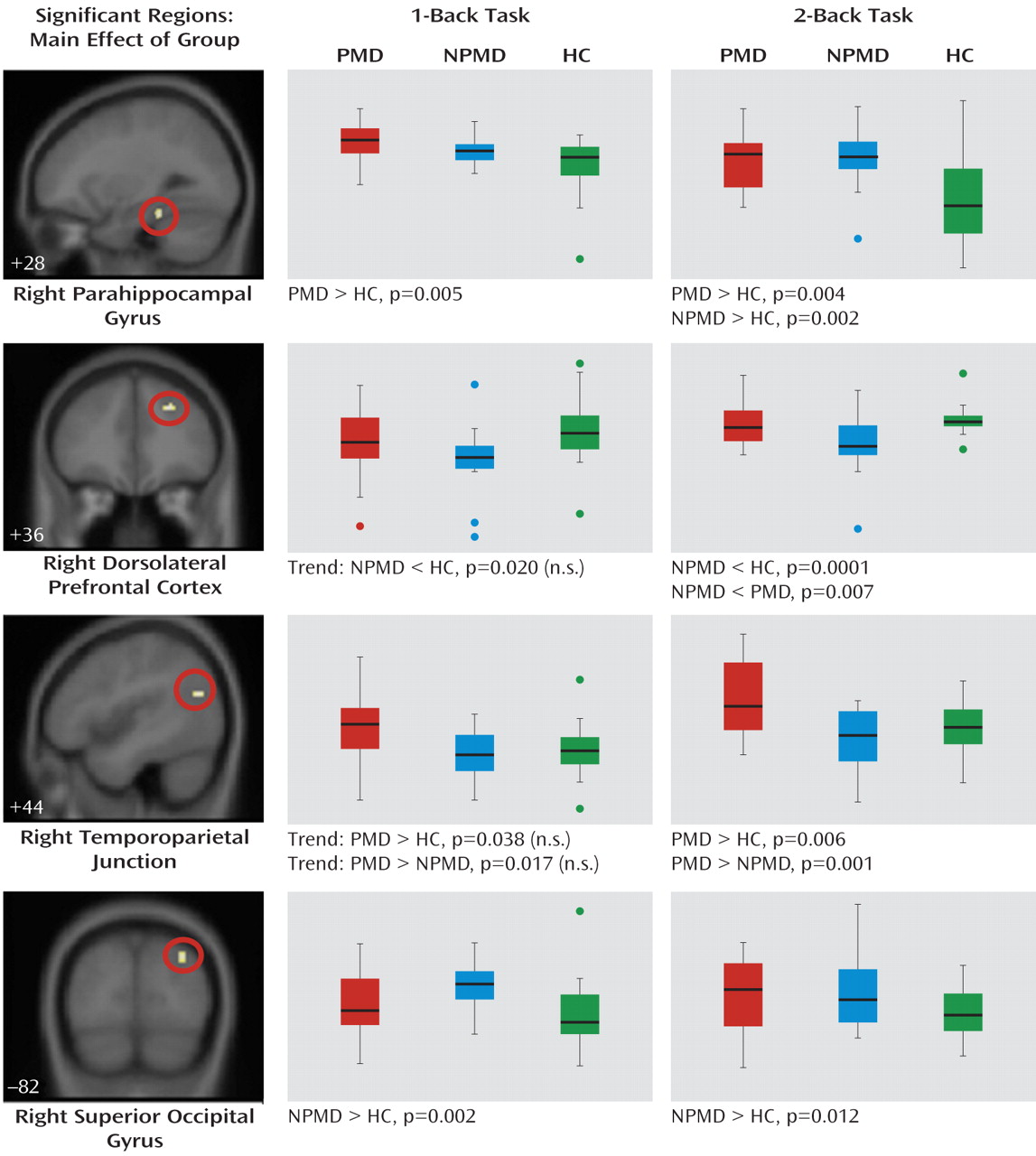
Four clusters of activation were significant (Figure 1). Activation in the right parahippocampal gyrus was significantly greater during both the 1-back and 2-back tasks in the psychotic major depression group relative to the healthy comparison group. The nonpsychotic major depression group showed greater parahippocampal activation during the 2-back task relative to the comparison group. The nonpsychotic major depression group showed significantly less activation in the right dorsolateral prefrontal cortex during the 2-back task relative to both groups and greater occipital cortex activation relative to the healthy comparison group during both tasks. The psychotic major depression group had significantly greater activation in the right temporoparietal junction during the 2-back task relative to both the nonpsychotic major depression and healthy comparison groups and nonsignificantly greater activation during the 1-back task.
FIGURE 1. Results of Three-Group Analysis of Variancea
a Boxplots show the distribution of mean activation within each group, including the minimum, median, and maximum values. Error bars show the standard deviation, and data points outside the box are outliers. The y-axis represents the mean T value of extracted activation, statistically corrected for gender and task performance, ranging from 0 to 2.5. Four clusters were significant, including the right parahippocampal gyrus (Brodmann's area 20/36; peak z=3.68; extent=26 voxels, Talairach coordinates (x, y, z) of peak=30, −36, −15); the right dorsolateral prefrontal cortex (Brodmann's area 9; peak z=3.68; extent=24 voxels; Talairach coordinates of peak=22, 37, 31); the right temporoparietal junction (Brodmann's area 39; peak z=3.60; extent=20 voxels; Talairach coordinates of peak=44, −67, 24); and the right superior occipital gyrus (Brodmann's area 19; peak z=3.52; extent=27 voxels; Talairach coordinates of peak=32, −78, 37). Results of between-group t tests comparing extracted clusters are shown under each graph (corrected significance threshold, p=0.0125). PMD=psychotic major depression group; NPMD=nonpsychotic major depression group; HC=healthy comparison group.
An important question is whether differences between the psychotic and nonpsychotic major depression groups can be attributed solely to differences in severity of depressive symptoms. However, group differences in brain activation did not change when covaried for HAM-D score. Also, we attempted to better understand the influence of medication on our results by examining brain activation in medicated compared with unmedicated participants. Only the nonpsychotic major depression group contained a sufficient number of unmedicated participants to perform this analysis. With the group divided into unmedicated (N=6) and medicated (N=9) subgroups, none of the regions were significantly different between subgroups.

Connectivity Results

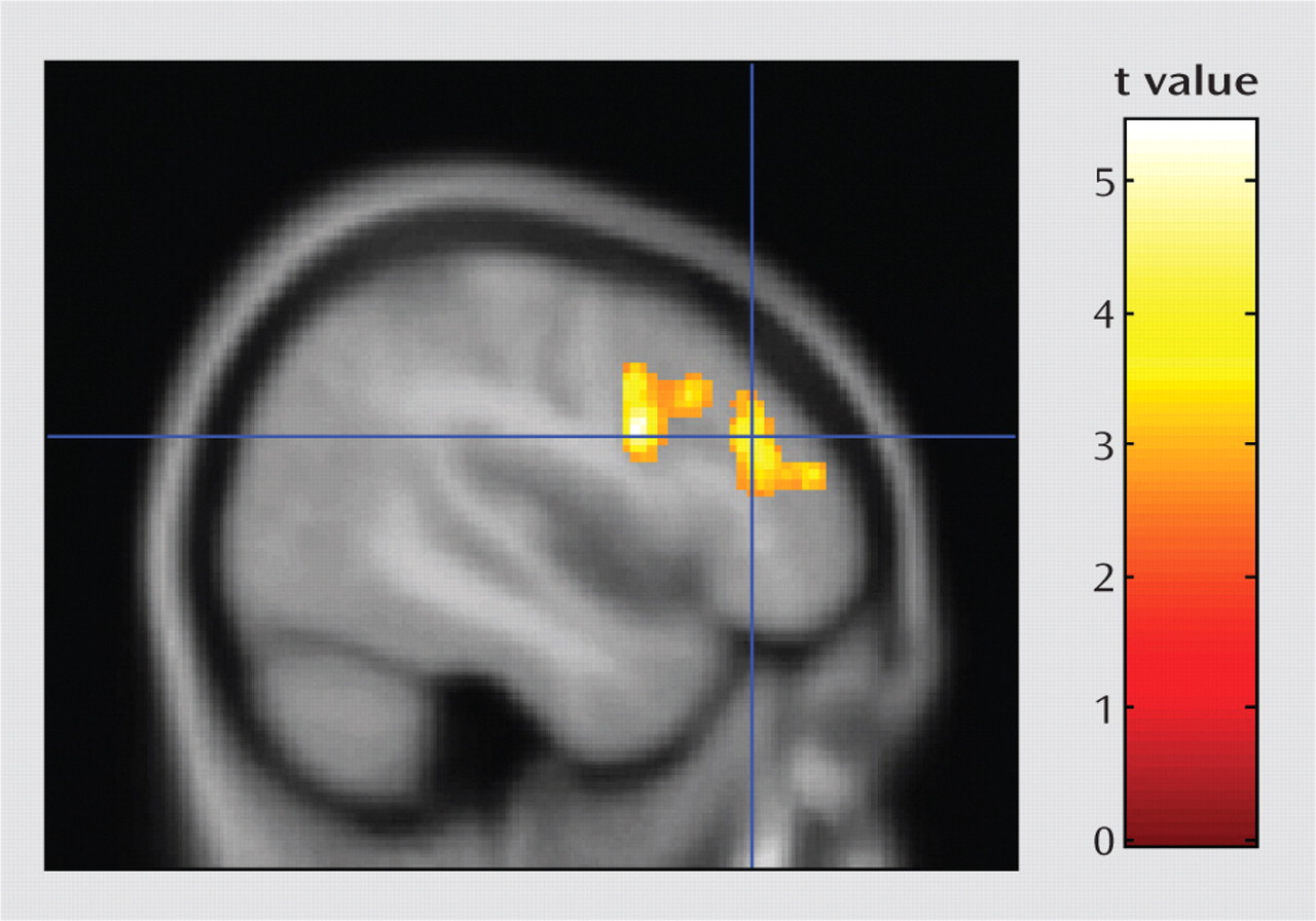
The psychophysiological interaction analysis was conducted to better understand functional circuits involving the temporoparietal junction during the 2-back task. Results showed that activation in a single large cluster in the left prefrontal cortex was significantly associated with activation in the right temporoparietal junction during the 2-back task (Figure 2). The left prefrontal cluster included three subpeaks: the dorsolateral prefrontal cortex, the middle frontal gyrus, and the inferior frontal gyrus.
FIGURE 2. Results of Functional Connectivity Analysis Within the Psychotic Major Depression Group Seeded With the Temporoparietal Junction Clustera
a A single significant cluster was found in the left prefrontal cortex (threshold: p=0.01 height and p=0.01 corrected cluster extent; cluster size=1,385 voxels; peak t=5.44; peak location, Talairach coordinates x, y, z=−46, 7, 24). The crosshairs shown in this figure arelocated at a subpeak in the dorsolateral prefrontal cortex (Brodmann'sarea 9/46). The cluster also includes the middle frontal gyrus (Brodmann's area 9/10) and the inferior frontal gyrus (Brodmann' area 44).

Discussion

We examined the neural correlates of working memory in patients with psychotic major depression compared to patients with nonpsychotic major depression and healthy comparison subjects. Given that several previous studies document working memory deficits in both psychotic and nonpsychotic major depression (910, 17, 20, 33), brain activation in these patient groups may reflect compensatory neural strategies as well as aberrant functional deficits. In the present study, both the psychotic and the nonpsychotic major depression groups showed increased parahippocampal activation relative to the healthy comparison group during the 2-back task, but the psychotic major depression group showed this difference during the less difficult 1-back task as well. These results suggest neural deficits common to both psychotic and nonpsychotic major depression in temporal lobe memory systems, albeit at a lower level of task demand in psychotic major depression. Only the nonpsychotic major depression group showed greater right occipital activation compared to the healthy comparison group, suggesting increased effort toward basic visual processing. The nonpsychotic major depression group also showed lower right dorsolateral prefrontal cortex activation compared to the psychotic major depression and healthy comparison groups during the 2-back task, although, contrary to predictions, no dorsolateral prefrontal cortex abnormalities were seen in the psychotic major depression group. The right temporoparietal junction was the only region where the psychotic major depression group showed significant overactivation compared to both the other groups during the 2-back task, with nonsignificantly greater activation during the 1-back task. In the psychotic major depression group, activation in the temporoparietal junction region showed psychophysiological connectivity with the left dorsolateral prefrontal cortex, which may reflect an executive function compensatory strategy. Controlling for group differences in severity of depressive symptoms did not eliminate observed differences between the psychotic and nonpsychotic major depression groups.
The temporoparietal junction region has been consistently shown in human and primate studies to be involved in reorienting attention to salient stimuli, especially when the stimuli are unexpected and important (13). The temporoparietal junction is part of the ventral frontoparietal system, as described by Corbetta and Shulman (13). Based on a large body of literature (34), these authors hypothesize that the ventral frontoparietal system functions as a “circuit breaker”; the system detects unexpected stimuli that both are behaviorally relevant and require a change in the current task set, such as reorienting attention to a fire alarm while working on your computer. This system is distinct from the dorsal attention system that prepares and applies goal-directed, top-down selection of task-relevant stimuli and responses. Also, the temporoparietal junction region is posterior and ventral to the inferior parietal lobe location (Brodmann's area 40/22) that is frequently activated bilaterally in neuroimaging studies of working memory, also called the “visuospatial scratch pad.”
As part of an automatic attention system, the temporoparietal junction could be important for sensory gating processes. Sensory gating has been suggested to be impaired in psychotic major depression, as data show that patients with this disorder have difficulties distinguishing relevant from irrelevant stimuli (35). In addition, we have observed abnormalities unique to patients with psychotic major depression during performance of the Stroop task (10), which requires sensory gating and filtering processes to resolve response conflict. A well-known measure of sensory gating is the prepulse inhibition test of the acoustic startle reflex (36). Although no studies of prepulse inhibition have focused on patients with psychotic major depression, it has been shown to be deficient in patients with schizophrenia but not in patients with nonpsychotic major depression (3739). Furthermore, a study of transgenic mice that overexpress corticotropin-releasing factor (proposed to be a model of psychotic depression) also show startle reflex gating deficits that are reversed by antipsychotic drugs (40). Because of psychotic symptoms and problems with filtering, the psychotic major depression group studied here may be expending significantly more neural resources for sensory gating and filtering, and this may be reflected in the increased activation of the temporoparietal junction region. The temporoparietal junction region may function to continually reorient attention to the task, perhaps because of internal and external distractions, including noise from the scanner, or possibly the intrusion of psychotic symptoms. Notably, the psychotic major depression group showed insula activation during the 2-back task, and reduced insula volume has been linked to psychotic symptoms in patients with psychotic major depression (J.D. Cohen et al., unpublished 2009 data). The post hoc analysis showing functional connectivity of the temporoparietal junction with the left prefrontal cortex suggests that top-down executive control over temporoparietal junction resources may compensate for sensory gating deficits, allowing accurate task performance.
The nonpsychotic major depression group showed increased activation in the parahippocampal cortex during the 2-back task, as did the psychotic major depression group during both the 1- and 2-back tasks. Traditionally, medial temporal lobe regions and the adjacent parahippocampal cortex have not been considered to be necessary for working memory tasks but to have an essential role in memory encoding and retrieval. However, a growing body of literature suggests that parahippocampal activation during working memory tasks is associated with encoding of the stimuli and predicts subsequent recall (15). Similar to the present study, a previous investigation (18) showed increased parahippocampal activation in patients with nonpsychotic major depression relative to healthy comparison subjects during a verbal working memory task. The present study goes further to find a similar deficit in psychotic major depression but at a lower level of task demand than in nonpsychotic major depression.
The dorsolateral prefrontal cortex is strongly associated with working memory tasks in healthy volunteers (11). Our study replicates previous reports of abnormal dorsolateral prefrontal cortex activation in nonpsychotic major depression. However, other studies show increased, rather than decreased, dorsolateral prefrontal cortex activation in nonpsychotic major depression during accurate task performance, usually in the left hemisphere (2022), but also in the right (19). The nonpsychotic major depression group also had lower right dorsolateral prefrontal cortex activation compared to the psychotic major depression group, and this difference remained after covarying for severity of depressive symptoms. This suggests that right hemisphere dorsolateral prefrontal cortex abnormalities contribute to working memory function in nonpsychotic major depression but not in psychotic major depression. However, the connectivity analysis suggests that the left dorsolateral prefrontal cortex may compensate for aberrant brain activation related to sensory gating in psychotic major depression.
These results support the hypothesis that psychotic major depression is distinct from nonpsychotic major depression on a neurofunctional basis. The study has several limitations, however, including the small sample size and medications taken by the clinical subjects, allowing us to detect only relatively large group differences. Although our analyses covaried for gender, imbalanced gender distributions across diagnoses may have contributed to our findings. The task was not difficult enough to demonstrate the previously shown deficits in working memory that have been documented for both patients with psychotic and nonpsychotic major depression. Greater group differences might have been detected by using a task that more rigorously challenged participants' working memory abilities, and an event-related analysis could separate trials on the basis of accuracy or response time for more detailed comparisons between groups.
It is possible that group differences in medication may have influenced brain activation. However, examining the distribution of medicated and unmedicated patients in each group did not reveal patterns suggesting a strong effect of medication. Moreover, we previously reported, using this sample of patients (9, 10), that medication status does not affect differences in cognitive test performance between symptomatic psychotic and nonpsychotic depressed groups. However, as psychiatric medications clearly affect brain function, we cannot rule out the possibility that medications influenced our results, which should thus be considered preliminary. Future studies would benefit from including only unmedicated subjects.
Footnotes

Received Dec. 2, 2009; revisions received May 7 and July 20, 2010; accepted Aug. 16, 2010
Dr. Schatzberg has served as a consultant to BrainCells, Eli Lilly, CNS Response, GlaxoSmithKline, Neuronetics, PharmaNeuroBoost, Sanofi, and Takeda and has equity in Corcept (cofounder), Merck, BrainCells, Xomaxon, CeNeRx, Forest, Pfizer, Neurocrine, Amylin, and PharmaNeuroBoost.

References

1.
Gaudiano BA, Dalrymple KL, Zimmerman M: Prevalence and clinical characteristics of psychotic versus nonpsychotic major depression in a general psychiatric outpatient clinic. Depress Anxiety 2009; 26:54–64
2.
Ohayon MM, Schatzberg AF: Prevalence of depressive episodes with psychotic features in the general population. Am J Psychiatry 2002; 159:1855–1861
3.
Keller J, Gomez RG, Kenna HA, Poesner J, DeBattista C, Flores B, Schatzberg AF: Detecting psychotic major depression using psychiatric rating scales. J Psychiatr Res 2006; 40:22–29
4.
Parker G, Hadzi-Pavlovic D, Hickie I, Boyce P, Mitchell P, Wilhelm K, Brodaty H: Distinguishing psychotic and non-psychotic melancholia. J Affect Disord 1991; 22:135–148
5.
Gaudiano BA, Young D, Chelminski I, Zimmerman M: Depressive symptom profiles and severity patterns in outpatients with psychotic vs nonpsychotic major depression. Compr Psychiatry 2008; 49:421–429
6.
Keller J, Schatzberg AF, Maj M: Current issues in the classification of psychotic major depression. Schizophr Bull 2007; 33:877–885
7.
Forty L, Jones L, Jones I, Cooper C, Russell E, Farmer A, McGuffin P, Craddock N: Is depression severity the sole cause of psychotic symp-toms during an episode of unipolar major depression? a study both between and within subjects. J Affect Disord 2009; 114:103–109
8.
Rothschild AJ, Winer J, Flint AJ, Mulsant BH, Whyte EM, Heo M, Fratoni S, Gabriele M, Kasapinovic S, Meyers BS; Study of Pharmacotherapy of Psychotic Depression (STOP-PD) Collaborative Study Group: Missed diagnosis of psychotic depression at 4 academic medical cen-ters. J Clin Psychiatry 2008; 69:1293–1296
9.
Gomez RG, Fleming SH, Keller J, Flores B, Kenna H, DeBattista C, Solvason B, Schatzberg AF: The neuropsychological profile of psychotic major depression and its relation to cortisol. Biol Psychiatry 2006; 60:472–478
10.
Schatzberg AF, Posener JA, DeBattista C, Kalehzan BM, Rothschild AJ, Shear PK: Neuropsychological deficits in psychotic versus nonpsy-chotic major depression and no mental illness. Am J Psychiatry 2000; 157:1095–1100
11.
D'Esposito M: From cognitive to neural models of working memory. Philos Trans R Soc Lond B Biol Sci 2007; 362:761–772
12.
Yoon JH, Curtis CE, D'Esposito M: Differential effects of distraction during working memory on delay-period activity in the prefrontal cortex and the visual association cortex. Neuroimage 2006; 29:1117–1126
13.
Corbetta M, Shulman GL: Control of goal-directed and stimulus-driven attention in the brain. Nat Rev Neurosci 2002; 3:201–215
14.
Ravizza SM, Delgado MR, Chein JM, Becker JT, Fiez JA: Functional dissociations within the inferior parietal cortex in verbal working memory. Neuroimage 2004; 22:562–573
15.
Axmacher N, Schmitz DP, Weinreich I, Elger CE, Fell J: Interaction of working memory and long-term memory in the medial temporal lobe. Cereb Cortex 2008; 18:2868–2878
16.
Rose EJ, Ebmeier KP: Pattern of impaired working memory during major depression. J Affect Disord 2006; 90:149–161
17.
Marquand AF, Mourao-Miranda J, Brammer MJ, Cleare AJ, Fu CH: Neuroanatomy of verbal working memory as a diagnostic biomarker for depression. Neuroreport 2008; 19:1507–1511
18.
Walsh ND, Williams SC, Brammer MJ, Bullmore ET, Kim J, Suckling J, Mitterschiffthaler MT, Cleare AJ, Pich EM, Mehta MA, Fu CH: A longi-tudinal functional magnetic resonance imaging study of verbal working memory in depression after antidepressant therapy. Biol Psychiatry 2007; 62:1236–1243
19.
Fitzgerald PB, Srithiran A, Benitez J, Daskalakis ZZ, Oxley TJ, Kulkarni J, Egan GF: An fMRI study of prefrontal brain activation during multi-ple tasks in patients with major depressive disorder. Hum Brain Mapp 2008; 29:490–501
20.
Harvey PO, Fossati P, Pochon JB, Levy R, Lebastard G, Lehéricy S, Allilaire JF, Dubois B: Cognitive control and brain resources in major de-pression: an fMRI study using the n-back task. Neuroimage 2005; 26:860–869
21.
Walter H, Wolf RC, Spitzer M, Vasic N: Increased left prefrontal activation in patients with unipolar depression: an event-related, parametric, performance-controlled fMRI study. J Affect Disord 2007; 101:175–185
22.
Matsuo K, Glahn DC, Peluso MA, Hatch JP, Monkul ES, Najt P, Sanches M, Zamarripa F, Li J, Lancaster JL, Fox PT, Gao JH, Soares JC: Prefrontal hyperactivation during working memory task in untreated individuals with major depressive disorder. Mol Psychiatry 2007; 12:158–166
23.
Potkin SG, Turner JA, Brown GG, McCarthy G, Greve DN, Glover GH, Manoach DS, Belger A, Diaz M, Wible CG, Ford JM, Mathalon DH, Gollub R, Lauriello J, O'Leary D, van Erp TG, Toga AW, Preda A, Lim KO; FBIRN: Working memory and DLPFC inefficiency in schizophre-nia: the FBIRN study. Schizophr Bull 2009; 35:19–31
24.
Barch DM, Sheline YI, Csernansky JG, Snyder AZ: Working memory and prefrontal cortex dysfunction: specificity to schizophrenia compared with major depression. Biol Psychiatry 2003; 53:376–384
25.
Barch DM, Csernansky JG: Abnormal parietal cortex activation during working memory in schizophrenia: verbal phonological coding distur-bances versus domain-general executive dysfunction. Am J Psychiatry 2007; 164:1090–1098
26.
Walter H, Vasic N, Hose A, Spitzer M, Wolf RC: Working memory dysfunction in schizophrenia compared to healthy controls and patients with depression: evidence from event-related fMRI. Neuroimage 2007; 35:1551–1561
27.
First MB, Spitzer RL, Gibbon M, Williams JBW: Structured Clinical Interview for DSM-IV Axis I Disorders, Patient Edition (SCID-P), version 2. New York, New York State Psychiatric Institute, Biometrics Research, 1997
28.
Hamilton M: A rating scale for depression. J Neurol Neurosurg Psychiatry 1960; 23:56–62
29.
Thase ME, Hersen M, Bellack AS, Himmelhoch JM, Kupfer DJ: Validation of a Hamilton subscale for endogenomorphic depression. J Affect Disord 1983; 5:267–278
30.
Overall JE, Gorham DR: The Brief Psychiatric Rating Scale. Psychol Rep 1962; 10:799–812
31.
Kim DH, Adalsteinsson E, Glover G, Spielman S: SVD regularization algorithm for improved high-order shimming. Proceedings of the 8th Annual Meeting of the International Society for Magnetic Resonance in Medicine, Denver, 2000
32.
Glover GH, Lai S: Self-navigated spiral fMRI: interleaved versus single-shot. Magn Reson Med 1998; 39:361–368
33.
Gomez RG, Posener JA, Keller J, DeBattista C, Solvason B, Schatzberg AF: Effects of major depression diagnosis and cortisol levels on indices of neurocognitive function. Psychoneuroendocrinology 2009; 34:1012–1018
34.
Corbetta M, Kincade JM, Ollinger JM, McAvoy MP, Shulman GL: Voluntary orienting is dissociated from target detection in human posterior parietal cortex. Nat Neurosci 2000; 3:292–297
35.
Belanoff JK, Kalehzan M, Sund B, Ficek SKF, Schatzberg AF: Cortisol activity and cognitive changes in psychotic major depression. Am J Psychiatry 2001; 158:1612–1616
36.
Braff DL, Geyer MA, Swerdlow NR: Human studies of prepulse inhibition of startle: normal subjects, patient groups, and pharmacological studies. Psychopharmacology (Berl) 2001; 156:234–258
37.
Ludewig S, Ludewig K: No prepulse inhibition deficits in patients with unipolar depression. Depress Anxiety 2003; 17:224–225
38.
Perry W, Minassian A, Feifel D: Prepulse inhibition in patients with non-psychotic major depressive disorder. J Affect Disord 2004; 81:179–184
39.
Quednow BB, Westheide J, Kuhn KU, Werner P, Maier W, Hawellek B, Wagner M: Normal prepulse inhibition and habituation of acoustic star-tle response in suicidal depressive patients without psychotic symptoms. J Affect Disord 2006; 92:299–303
40.
Dirks A, Groenink L, Westphal KG, Olivier JD, Verdouw PM, van der Gugten J, Geyer MA, Olivier B: Reversal of startle gating deficits in transgenic mice overexpressing corticotropin-releasing factor by antipsychotic drugs. Neuropsychopharmacology 2003; 28:1790–1798

Information & Authors

Information

Published In

Go to American Journal of Psychiatry
American Journal of Psychiatry
Pages: 173 - 182
PubMed: 21078708

History

Received: 2 December 2009
Revision received: 7 May 2010
Revision received: 20 July 2010
Accepted: 16 August 2010
Published online: 1 February 2011
Published in print: February 2011

Authors

Details

Amy Garrett, Ph.D.
From the Center for Interdisciplinary Brain Sciences Research and the Department of Psychiatry and Behavioral Sciences, Stanford University School of Medicine.
Ryan Kelly, B.S.
From the Center for Interdisciplinary Brain Sciences Research and the Department of Psychiatry and Behavioral Sciences, Stanford University School of Medicine.
Rowena Gomez, Ph.D.
From the Center for Interdisciplinary Brain Sciences Research and the Department of Psychiatry and Behavioral Sciences, Stanford University School of Medicine.
Jennifer Keller, Ph.D.
From the Center for Interdisciplinary Brain Sciences Research and the Department of Psychiatry and Behavioral Sciences, Stanford University School of Medicine.
Alan F. Schatzberg, M.D.
From the Center for Interdisciplinary Brain Sciences Research and the Department of Psychiatry and Behavioral Sciences, Stanford University School of Medicine.
Allan L. Reiss, M.D.
From the Center for Interdisciplinary Brain Sciences Research and the Department of Psychiatry and Behavioral Sciences, Stanford University School of Medicine.

Notes

Address correspondence and reprint requests to Dr. Garrett, Center for Interdisciplinary Brain Sciences Research, 401 Quarry Rd., Palo Alto, CA 94305-5719; [email protected] (e-mail).

Funding Information

All other authors report no financial relationships with commercial interests.

Metrics & Citations

Metrics

Citations

Export Citations

If you have the appropriate software installed, you can download article citation data to the citation manager of your choice. Simply select your manager software from the list below and click Download.

For more information or tips please see 'Downloading to a citation manager' in the Help menu.

Format
Citation style
Style
Copy to clipboard

View Options

View options

PDF/EPUB

View PDF/EPUB

Get Access

Login options

Already a subscriber? Access your subscription through your login credentials or your institution for full access to this article.

Personal login Institutional Login Open Athens login
Purchase Options

Purchase this article to access the full text.

PPV Articles - American Journal of Psychiatry

PPV Articles - American Journal of Psychiatry

Not a subscriber?

Subscribe Now / Learn More

PsychiatryOnline subscription options offer access to the DSM-5-TR® library, books, journals, CME, and patient resources. This all-in-one virtual library provides psychiatrists and mental health professionals with key resources for diagnosis, treatment, research, and professional development.

Need more help? PsychiatryOnline Customer Service may be reached by emailing [email protected] or by calling 800-368-5777 (in the U.S.) or 703-907-7322 (outside the U.S.).

Media

Figures

Other

Tables

Share

Share

Share article link

Share