Abstract

Objective:

A role for aberrant reward processing in the pathogenesis of depression has long been proposed. However, no review has yet examined its role in depression by integrating conceptual and quantitative findings across functional MRI (fMRI) and EEG methodologies. The authors quantified these effects, with an emphasis on development.

Method:

A total of 38 fMRI and 12 EEG studies were entered into fMRI and EEG meta-analyses. fMRI studies primarily examined reward anticipation and reward feedback. These were analyzed using the activation likelihood estimation method. EEG studies involved mainly the feedback-related negativity (FRN) event-related potential, and these studies were analyzed using random-effects meta-analysis of the association between FRN and depression.

Results:

Analysis of fMRI studies revealed significantly reduced striatal activation in depressed compared with healthy individuals during reward feedback. When region-of-interest analyses were included, reduced activation was also observed in reward anticipation, an effect that was stronger in individuals under age 18. FRN was also significantly reduced in depression, with pronounced effects in individuals under age 18. In longitudinal studies, reduced striatal activation in fMRI and blunted FRN in EEG were found to precede the onset of depression in adolescents.

Conclusions:

Taken together, the findings show consistent neural aberrations during reward processing in depression, namely, reduced striatal signal during feedback and blunted FRN. These aberrations may underlie the pathogenesis of depression and have important implications for development of new treatments.
Depression has a prevalence of 19% in the U.S. population (1), and over 300 million people suffer from the disorder worldwide (2). However, compared with many other medical conditions, we know little about its pathophysiology. In recent years, reward processing aberrations have been proposed as a candidate mechanism, which has implications for much-needed treatment breakthroughs (35). This quantitative review integrates the available evidence relating reward processing to depression.
Previous meta-analyses that included data on reward processing and depression have differed from this work in various aspects, including a focus on selected age groups (for example, excluding patients under 18) or on limited populations or only on patients with severe depression; analysis of region-of-interest-based studies; and use of lenient thresholds; some of these studies are also now outdated (610). Similarly, no previous quantitative review has pooled effects of electrophysiological studies exploring the association between reward processing and depression. While EEG’s spatial resolution is inferior to that of functional MRI (fMRI), its superior temporal resolution is particularly relevant to the study of reward processing dynamics. Moreover, feedback-related negativity (FRN; also termed reward positivity) has emerged as a powerful measure of reward processing (11, 12) implicated in depression (13), making it essential to include such studies. Notably, in this meta-analysis, we also focused on developmental effects, as both reward processes (14) and depression show developmental moderation (15).

Conceptual Links Between Depression and Reward Processing

Cardinal presentations of depression (16), most notably anhedonia, are thought to reflect alterations of the experience of reward (17, 18). The following paragraphs conceptually bridge clinical terminology with the burgeoning science of reward processing.
Rewards have been defined as stimuli that induce behaviors that help the animal organism obtain what is necessary for survival (19). In addition, rewards and punishers facilitate learning through positive or negative reinforcement: a reward (or lack of punishers) following a behavior will make the future occurrence of that behavior more likely, often eliciting feelings of pleasure; the opposite is true for behaviors followed by punishers (or lack of rewards). Reductions in reports of pleasure and approach-related behavior are a prominent feature of depression, and many suggest that they arise from aberrations in reward processing (3, 20).
In Table 1, we have adapted previous models (21, 22) to parse four sets of reward processing events and map their links to clinical phenomena. We term the first stage of reward processing prediction: it encompasses recognizing an object as potentially rewarding, a process that involves using existing knowledge about the value of objects. Anticipatory anhedonia, defined as a lack of interest in activities that used to be enjoyable, is the clinical depressive symptom that best maps onto this phase. In translational terms, this phase is typically captured by the reward or loss anticipation/prediction phase of an experiment, when a stimulus induces the subject to expect either a win or a loss. When attempting to engage prediction-related processes in translational work, the classic task is the monetary incentive delay paradigm (23).
TABLE 1. The Identified Phases of Reward Processing, Mapped Onto Their Associated Clinical and Translational Terminologies
Reward PhaseAssociated SymptomTranslational TermExample Experimental Task
PredictionAnticipatory anhedoniaReward/loss anticipationMonetary incentive delay task
DecisionImpaired decision makingChoiceIowa gambling task
ActionLow energyEffort expenditureEffort expenditure for rewards task
ExperienceConsummatory anhedoniaReward/loss feedbackMonetary incentive delay task
The second stage, decision, involves computing the cost associated with attaining a reward. Depressed patients often report decision-making problems (16, 24), sometimes seen by others as “lack of initiative.” These complaints best map onto this second stage of reward processing and in translational terms correspond to the decision part of an experiment, when a subject chooses between available options, for example, in a gambling task.
The third stage is action, during which effort is expended for a rewarding stimulus to be approached or a punisher to be avoided. Fatigue and low energy, commonly reported in patients with other depressive symptoms (16, 24), map onto this action component. In translational terms, this corresponds to a part of the experiment where a subject performs an action, such as a lever or button press, providing a quantification of task-related effort.
The final stage involves experience, which encompasses the consummation of a reward and the feelings that may be associated with it. This phase also entails the consolidation of this experience in memory, which may be accessed for future reward processing. Consummatory anhedonia, the lack of enjoyment from activities that used to be pleasant, best maps onto this phase. Translationally, this corresponds to a subject being faced with either a win or a loss outcome within a task, such as occurs in the monetary incentive delay task. In EEG studies, this is measured in terms of the FRN potential, or its reverse reward positivity (11), which occurs after feedback and is typically recorded at central to frontal-central regions of the scalp. FRN and reward positivity are the contrast of neural response to feedback of loss minus gain, and gain minus loss, respectively.
Reward processing involves many distinct components. One particularly key component of reward processing involves learning, whereby organisms update associated values attributed to objects and actions in their environment. Reward-related learning typically occurs through reward prediction errors, striatal dopamine-encoded signals that indicate the difference between anticipated and experienced reward (19). Such learning influences subsequent decision making and updates anticipation. In that sense, all the phases depicted in the model are part of reward-related learning.
Blunting of reward responses has been observed in major depression in adolescents, but it remains unclear whether the magnitude of this signal reduction varies across development. The sharp increase in depression incidence during adolescence (25) highlights the importance of examining this issue.

Method

Data Source and Search Strategy

We searched PubMed, Scopus, PsycINFO, and Web of Science for articles published in English from January 1, 2000, to February 1, 2017 (see Figure S1 in the online supplement), using the following terms and their derivatives: depression, anhedonia, reward, motivation, reinforcement, punishment and aversion, prediction error, decision making, and risk taking.

Inclusion and Exclusion Criteria

To be included, studies had to provide a measure of depression or anhedonia in people with major depressive disorder, in people at high risk of depression, or in healthy volunteers. We selected only studies that measured depression or depressive symptoms through questionnaires, structured interviews, or clinical diagnosis. In terms of reward paradigms employed, and following the classification described by Richards et al. (26), we included instrumental-reward tasks and decision-making tasks, which require participants to complete an action correctly in order to obtain a reward, as this action is linked to the reward value at a trial-by-trial level. Hence, reward paradigms in which rewards were presented passively were excluded. Either positive (e.g., winning money) or negative (e.g., losing money) reward manipulations were permitted. No age restrictions were applied. Exclusion criteria are detailed in the online supplement.
To be included in the analysis, fMRI studies had to have used a reward task and have reported on brain coordinates. Connectivity studies were excluded from the analysis.
Among EEG studies, we included studies that reported mean amplitude response to negative/loss and positive/gain feedback on a reward paradigm, either separately or in some combination of these, such as loss minus gain (FRN) or gain minus loss (reward positivity). The corresponding authors of 10 studies that met all but one of the inclusion criteria were contacted to inquire whether a compatible analysis had been conducted, such as mean amplitude extraction, rather than a peak approach. Where such analyses had been conducted, the means were requested for inclusion in the meta-analysis (as outlined in greater detail in the online supplement), which resulted in five of these studies being included.

Data Analysis

fMRI meta-analysis.

Of the 66 fMRI studies, 38 were included in the fMRI meta-analysis (see Tables 2 and 3 and the online supplement for further information), as they reported consistently the following contrasts: reward anticipation, reward feedback, and loss anticipation plus feedback (these phases were merged to reach a sufficient number of studies). For these contrasts of interest, 23 studies reported whole brain analyses, 15 reported region-of-interest analyses, and two reported both types of analysis.
TABLE 2. Summary of the Studies Included in the Meta-Analyses of fMRI and EEG Studies of Reward Processing in Depression
CharacteristicOverallSubjects Under Age 18Subjects Age 18 and Older
fMRI studies (N=38)   
Sample composition   
 Depressed subjects compared with healthy volunteers   
  Whole brain only15114
  Whole brain and region of interest24420
 Subjects at high risk of depression compared with healthy volunteers   
  Whole brain only624
  Whole brain and region of interest1055
 Depression on continuum   
  Whole brain only303
  Whole brain and region of interest835
 Overall   
  Whole brain onlya23320
  Region of interest only1578
Reward types   
 Monetary32923
 Primary211
 Affective3 3
 Accuracy2 2
Tasks used   
 Monetary incentive delay task13211
 Affective incentive delay task1 1
 Decision making1 1
 Wheel of fortune4 4
 Card guessing761
 Reward learning4 4
 Pavlovian prediction1 1
 Effort expenditure for rewards task1 1
 Modified reward task3 3
 Primary reward task11 
 Reward guessing task11 
 Gambling task1 1
EEG studies (N=12)   
Sample composition   
 Depressed subjects compared with healthy volunteers523
 Subjects with high risk of depression compared with healthy volunteers211
 Depression on continuum532
Reward types   
 Monetary1156
 Points11 
Tasks used   
 Doors guessing task743
 A gambling task3 3
 A reward guessing task22 
a
Significant difference between groups, p<0.001.
TABLE 3. Gender Distribution and Medication Status in the Samples Included in the Meta-Analyses of fMRI and EEG Studies of Reward Processing in Depressiona
 OverallSubjects Under Age 18Subjects Age 18 and Older
CharacteristicMeanSDRangeMeanSDRangeMeanSDRange
fMRI studies (N=38)         
Ageb (years)29.27.58–6513.51.68–1734.49.517–65
Percent femalec58.321.316–10077.717.250–10051.819.916–79
Percent medicated16.430.40–1000.81.50–4.521.733.50–100
EEG studies (N=12)         
Aged (years)19.97.28–6513.92.58–1726.04.418–65
Percent female70.232.248–10087.026.048–10070.817.455–100
Percent medicated18.716.70–4115.815.94.5–2720.620.50–41
a
Some studies did not report these characteristics.
b
Significant difference between groups in mean age, p<0.001.
c
Significant difference between groups in percent female, p<0.01.
d
Significant difference between groups in mean age, p<0.01.
To increase the power of our analyses, we compared the combined depression and high-risk groups to healthy volunteers, also including the studies that examined the effects of depressive symptoms on reward processing. This dimensional approach to depression is consistent with current nosological approaches to the disorder (27). However, in the online supplement, we describe analyses that include only studies comparing major depression and healthy volunteer groups.
Overall, we conducted 21 activation likelihood estimation (ALE) meta-analyses, a method proposed by Turkeltaub et al. (28) and Laird et al. (29). For our primary analyses, we included only the studies that examined whole brain activation and excluded region-of-interest and small-volume-correction studies; this is standard practice to avoid experimenter-imposed localization bias (6, 30). Hence, no studies with predefined region-of-interest masks were included. Instead, after whole brain analyses identified the caudate as the area that was significantly different between depressed and nondepressed subjects, region-of-interest studies of that region were added in follow-up analyses focusing on developmental effects. We did not impose specific requirements for the statistical thresholds or correction for multiple comparisons. To estimate the developmental influence of activation changes, studies were split between those with subjects under age 18 and those with subjects age 18 and older and analyzed separately. Then the two ALE images were contrasted to analyze the age-related differences.
The ALE analysis was implemented in GingerALE 2.1.1 (www.brainmap.org/ale). Except as otherwise indicated, all ALE images were family-wise error corrected for multiple comparisons at the whole brain level, using a cluster‐level inference correction to a p level of 0.05, with an uncorrected p level of 0.001 (see the online supplement for further details).

EEG meta-analysis.

Of the 32 EEG studies, 12 were included in the EEG meta-analyses (see Tables 2 and 3 and the online supplement for further information). To meta-analyze the EEG studies, all effects were coded to a direction consistent with loss minus gain feedback (i.e., FRN), where more negative values are indicative of a greater differentiation between the neural response to gain and loss feedback. To combine the effect sizes of the studies, correlation coefficients and mean differences were converted to standardized effect sizes (Cohen’s d). These were then subjected to a random-effects meta-analysis in Stata across all included studies. We report the variance of effect sizes attributable to heterogeneity using the I2 statistic, and between-study variance with tau-squared. All procedures of coefficient conversion and subsequent meta-analysis are described in more detail in the online supplement. Because of the small number of longitudinal studies meeting inclusion criteria, a separate meta-analysis on these could not be conducted.

Results

fMRI Meta-Analysis

Overall, the 38 fMRI studies (3168) examined 428 subjects with major depression, 225 subjects with high risk of depression, and 503 subjects from studies that correlated brain activity with continuous measures of depressive symptoms. (See Tables S2 and S3 in the online supplement for summaries of the study samples’ demographic and analytic characteristics.)

Studies examining whole brain activation.

Reward anticipation:
We found 12 whole-brain studies comprising 16 experiments, 84 foci, and 274 subjects. Meta-analysis showed no significant ALE clusters when correcting for multiple comparisons (see the online supplement for uncorrected results at p threshold of 0.001, focused in the caudate head).
Reward feedback:
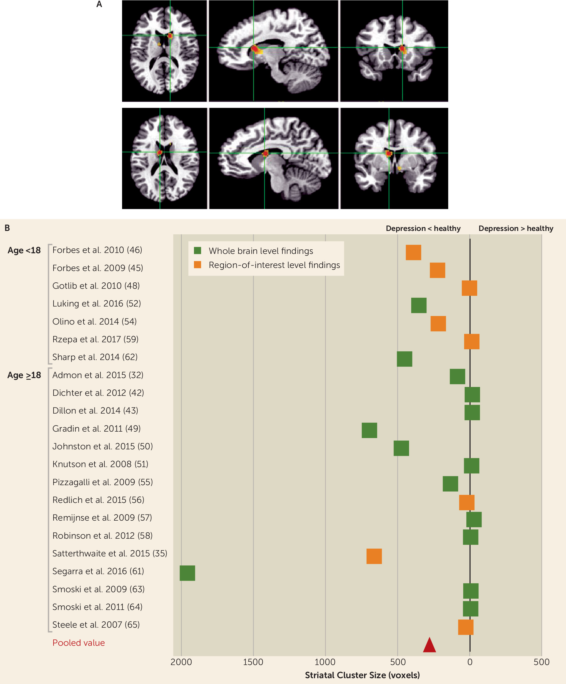
We found 14 studies comprising 17 experiments, 110 foci, and 306 subjects. Meta-analysis revealed a significant cluster in the right caudate body and head and the left caudate body (Figure 1A; 322 voxels, peak ALE value=0.016), showing a difference between depressed and healthy subjects. No other brain regions emerged as significant. Because ALE results only reflect a significant spatial overlap of reported coordinates, we also present a plot of the direction of effect of each individual study for the striatal findings (Figure 1B). As shown in the figure, 13 of the 14 studies (92.9%) reported decreased activity in depressed subjects. A single study (57) found a small cluster (5 voxels) of increased activation.
FIGURE 1. Alterations in Brain Activity During Reward Feedback, in Depressed Compared With Healthy Subjects: Meta-Analysis of fMRI Studiesa
a Panel A depicts results across whole brain studies, presented as activation likelihood estimation maps, showing significantly decreased activation in the right caudate head and body (x=+12, y=+14, z=+14). Panel B lists the studies included in the meta-analyses of reward feedback contrast, broken down by age and type, along with the striatal cluster extent and direction of effect (increased versus decreased in depression). (The cluster value in the Johnston et al. study [50] was reported as 10,871 voxels combining several regions, and this is not reflected in its position in the graph because of space concerns.)

Inclusion of region-of-interest studies.

We next included in the analyses the studies that reported region-of-interest findings. This larger study inclusion enabled us to compare results between subjects under age 18 and those 18 and older.
Reward anticipation:
We found 24 studies comprising 32 experiments, 119 foci, and 822 subjects. Meta-analysis revealed a significant cluster of decreased activity in depressed subjects, bilaterally at the caudate head as well as at the left putamen (see Table S4 and Figure S3 in online supplement).
When we divided these studies into over and under age 18, we found a stronger blunting of activity in the younger-age studies. (See Table S4 in the online supplement, which describes the cluster in the caudate when contrasting the ALE images of studies between those under age 18 compared with those 18 and older.)
Reward feedback:
We found 22 studies comprising 27 experiments, 135 foci, and 572 subjects. Meta-analysis showed a significant cluster of decreased activity in the caudate, the putamen, and the globus pallidus for depressed compared with healthy subjects (see Figure S6A in the online supplement). We found no significant difference between age groups (see Figure S6B–C in the online supplement).
Loss contrast meta-analysis showed no significant difference between depressed and healthy subjects (see the online supplement).
Sensitivity analyses are detailed in the online supplement.

EEG Meta-Analysis

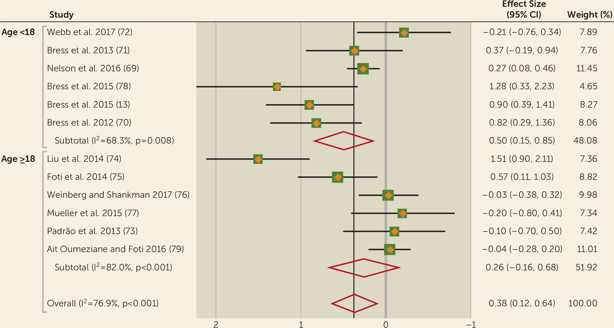
Random-effects meta-analysis across the 12 studies (13, 6979) yielded a statistically significant effect (z=2.82, p<0.01, two-tailed) with a pooled effect size (d) of 0.38 (95% CI=0.12, 0.64). There was high heterogeneity across studies (χ2=47.69, df=2, N=11, p<0.001; I2=76.9%). Between-study variance, as measured by tau-squared, was 0.15.
In a subsequent analysis, we tested age as a moderator of the relationship between FRN and depression. The analysis replicated the significant effect in studies with participants under age 18, with an effect size of 0.50 (95% CI=0.15, 0.85; z=2.78, p<0.01, two-tailed). Study heterogeneity in the younger group was moderate (χ2=15.76, df=2, N=5, p<0.05; I2=68.3%; tau-squared=0.12). However, in studies with samples over age 18, the association between FRN and depression was found to be nonsignificant (z=1.23, p=0.22, two-tailed). This result was based on a pooled effect size of 0.26 (95% CI=−0.16, 0.68), with a heterogeneity (χ2) of 27.77 (df=2, N=5, p<0.05; I2=82%; tau-squared=0.22). Despite this, the pooled weighted effect sizes within each age group were not significantly different from one another (z=0.62, p=0.54, two-tailed), as calculated according to Borenstein et al. (80). These results are summarized in Figure 2. See the online supplement for sensitivity analyses.
FIGURE 2. Effect Sizes for the Association Between Depression and Feedback-Related Negativity (FRN) in a Meta-Analysis of EEG Studiesa
a Effect sizes have been flipped for illustrative purposes, such that positive effect sizes, indicative of a blunting of FRN in depression, are located to the left of the null line. Weights are from random-effects analysis.

Longitudinal fMRI and EEG Studies

There was an insufficient number of longitudinal studies to conduct a separate meta-analysis (13, 3134, 6971, 8185). These findings are summarized in the online supplement.

Behavioral Findings

No statistics are presented here, as only seven (18%) fMRI studies showed a group difference (40, 57, 58, 60, 63, 65, 68), and only two EEG studies reported behavioral results (72, 73).

Discussion

This work links depression to aberrant reward processing. In particular, functional imaging and electrophysiological findings converge to show a blunted neural response to reward, and this effect may be more pronounced in individuals under age 18.
Our meta-analysis of fMRI studies found decreased striatal activity in subjects with depression compared with healthy volunteers during reward feedback. This finding is in keeping with the meta-analysis of Zhang et al. (6), although only 25% of studies included in our meta-analysis overlapped with those of Zhang et al., with the addition of several (N=15) new studies published since then. These findings cannot be attributed to localization bias, as they also occur in non-region-of-interest studies. We also found decreased anticipation activity in depressed subjects when we lowered the statistical threshold or added region-of-interest studies. Reward anticipation and feedback are distinguished conceptually; it has been suggested (86) that dopaminergic neurons are primarily associated with anticipation of reward (87). By contrast, opioid neurons are associated with consummation of reward and therefore the feedback phase. The fMRI measures do not allow distinctions at the neurotransmitter level, and macroscopic anatomical overlap should not be taken to imply mechanistic overlap. There were no significant results for fMRI contrasts of loss. Because of the small number of studies, we combined loss anticipation and feedback (88), and this may have diluted effects.
A previous meta-analysis (10) found no differences overall between healthy and depressed subjects, but that analysis focused on a broad range of emotional and learning-related responses, rather than on strictly defined reward processing, as our study did. Moreover, the authors excluded studies with participants under age 18, whereas our study used a developmental approach including all ages and comparing adolescence with adulthood. Furthermore, we found evidence from longitudinal studies that aberrations in reward processing were predictive of new-onset depression (33) and increased the risk for depression (81). Interestingly, a recent connectivity study (89) demonstrated that increased connectivity of the ventral striatum predicts depression, in keeping with striatal aberrations in this disorder.
Our fMRI findings fit with predictions from animal work (90) on the centrality of the striatum in reward processing. It is notable that the peak of activity difference between healthy volunteers and depressed subjects is in the caudate, rather than in the nucleus accumbens, a key part of the circuitry associated with reward processing (91). Indeed, there is substantial cytoarchitectural overlap between the accumbens and ventromedial parts of the caudate and putamen (92), and they are collectively designated as the ventral striatum (92, 93). The striatum receives rich input from various cortical areas, including the ventromedial prefrontal cortex, orbitofrontal cortex, and anterior cingulate cortex, as well as the amygdala and hippocampus (93, 94). This input is integrated and then translated into action via neighboring areas in the basal ganglia.
The overall association between FRN and depression yielded a significant effect size of 0.38 in the random-effects analysis. When we stratified our samples into subjects under age 18 and age 18 and older, significance was only found in youth depression but not adult depression, although the moderation statistic was not significant. We also noted evidence that blunted FRN is a predictor of future depression onset (71). Longitudinal effects were observed in adolescents only, as no studies examined this association in adults. Taken together, and in line with the fMRI studies, these results suggest a decreased brain sensitivity to anticipating and consuming rewards in depression. While fMRI studies suggest that this deficit involves the striatum, the source of the FRN is still debated; however, it may partially reflect striatal signals (95, 96) or the indirect influence of striatal signals on other neural regions (97, 98). It is worth speculating about the fact that there was a lower heterogeneity in the younger than the older subsamples. The younger subsamples were more likely to be community based, were narrower in age range, and had lower levels of medication, whereas the older sample was more diverse in terms of demographic variables. Medication was not consistently reported among the studies, and therefore we could not assess its effects on the outcomes. It should also be noted that the younger samples contained more females than did the older samples, which may have influenced the results.
The reward system is known to undergo transition during the adolescent period, with changes indexed by FRN (99, 100) and BOLD signal (14). More studies, particularly longitudinal studies of depressed individuals that span adolescence and adulthood, will be needed to understand the interaction between development and depression.
When considering these findings, several conceptual and empirical challenges need to be considered. First, postulating depression to be a generalized inability to anticipate or perceive pleasure (or avoid pain) may be overly simplistic. Depressed individuals can still crave rewards, as evidenced by the increased levels of drug and alcohol dependency in depression (101). Anhedonia is a core feature of depression closely linked to reward processing (102). Unfortunately, few studies have included measures of anhedonia to quantify the degree to which reward system dysfunction is moderated by anhedonia level. Moreover, depression studies are needed that combine the high temporal precision of EEG or magnetoencephalography with the spatial precision of fMRI.
Second, few studies have demonstrated aberrations in depression that span the three levels of explanation: brain circuitry, task behavior, and clinical symptoms. Indeed, many of the tasks addressing reward processing, notably the monetary incentive delay task, are less suited to capturing behavioral effects and reward experience (103). Developing tasks to overcome such shortcomings will be important. It will also be important to explore the interplay between reward and cognitions relevant to depression, such as executive control (104).
Third, future studies should go beyond typical case-control designs to include comparisons of reward processing between subjects with depression and other morbid groups. We found very few studies that directly compared reward processing in depression alongside other disorders. Two studies that compared reward processing across alcohol dependence, schizophrenia, depression, or bipolar disorder found that decreased striatal activity was correlated with depressive symptoms (35, 36).
Fourth, there were surprisingly few experimental studies embedded in treatment studies. Deep brain stimulation is the most direct way of testing this, although it is the most ethically challenging. Promising initial results of deep brain stimulation of the ventral striatum (105, 106) did not replicate in controlled studies (107). While some pharmacological (31, 108, 109) or psychological (110) interventions show promise in probing reward signal, they do not yet demonstrate that affecting reward modulates depressive symptoms.
Fifth, the extant studies in the literature allowed us to pool results for only two of the four postulated components of reward processing that we outlined above. Clearly, more research is needed to understand the functioning of the other component processes at the neural level in depression. Our review also could not address directly the important issue of reward learning (8), as there were not enough imaging or EEG studies of reward learning in depression that fit our criteria.
Sixth, we found no evidence of publication bias for the EEG studies but cannot exclude the possibility that the nonreporting of null results biased the fMRI findings.
Overall, these findings demonstrate consistent reward processing aberrations in depression, expressed as blunted striatal fMRI and FRN signals, during reward feedback. These aberrations, which potentially underlie the pathogenesis of depression, may have important implications for the development of new treatments.
Footnote

Supported in part by the NIMH Intramural Research Program (grant ZIA-MH002957-01).

Supplementary Material

File (appi.ajp.2018.17101124.ds001.pdf)

References

1.
Kessler RC, Bromet EJ: The epidemiology of depression across cultures. Annu Rev Public Health 2013; 34:119–138
2.
World Health Organization: Depression: A Global Crisis. Geneva, World Health Organization, 2012
3.
Treadway MT, Zald DH: Reconsidering anhedonia in depression: lessons from translational neuroscience. Neurosci Biobehav Rev 2011; 35:537–555
4.
Whitton AE, Treadway MT, Pizzagalli DA: Reward processing dysfunction in major depression, bipolar disorder, and schizophrenia. Curr Opin Psychiatry 2015; 28:7–12
5.
Insel T, Cuthbert B, Garvey M, et al: Research Domain Criteria (RDoC): toward a new classification framework for research on mental disorders. Am J Psychiatry 2010; 167:748–751
6.
Zhang WN, Chang SH, Guo LY, et al: The neural correlates of reward-related processing in major depressive disorder: a meta-analysis of functional magnetic resonance imaging studies. J Affect Disord 2013; 151:531–539
7.
Hamilton JP, Etkin A, Furman DJ, et al: Functional neuroimaging of major depressive disorder: a meta-analysis and new integration of baseline activation and neural response data. Am J Psychiatry 2012; 169:693–703
8.
Huys QJ, Pizzagalli DA, Bogdan R, et al: Mapping anhedonia onto reinforcement learning: a behavioural meta-analysis. Biol Mood Anxiety Disord 2013; 3:12
9.
Miller CH, Hamilton JP, Sacchet MD, et al: Meta-analysis of functional neuroimaging of major depressive disorder in youth. JAMA Psychiatry 2015; 72:1045–1053
10.
Müller VI, Cieslik EC, Serbanescu I, et al: Altered brain activity in unipolar depression revisited: meta-analyses of neuroimaging studies. JAMA Psychiatry 2017; 74:47–55
11.
Proudfit GH: The reward positivity: from basic research on reward to a biomarker for depression. Psychophysiology 2015; 52:449–459
12.
Hajcak G, Moser JS, Holroyd CB, et al: It’s worse than you thought: the feedback negativity and violations of reward prediction in gambling tasks. Psychophysiology 2007; 44:905–912
13.
Bress JN, Meyer A, Proudfit GH: The stability of the feedback negativity and its relationship with depression during childhood and adolescence. Dev Psychopathol 2015; 27:1285–1294
14.
Braams BR, van Duijvenvoorde AC, Peper JS, et al: Longitudinal changes in adolescent risk-taking: a comprehensive study of neural responses to rewards, pubertal development, and risk-taking behavior. J Neurosci 2015; 35:7226–7238
15.
Luking KR, Pagliaccio D, Luby JL, et al: Reward processing and risk for depression across development. Trends Cogn Sci 2016; 20:456–468
16.
Kendler KS: The phenomenology of major depression and the representativeness and nature of DSM criteria. Am J Psychiatry 2016; 173:771–780
17.
Berrios GE: The History of Mental Symptoms: Descriptive Psychopathology Since the Nineteenth Century. Cambridge, UK, Cambridge University Press, 1996
18.
Kessel EM, Klein DN: Depressivity and anhedonia, in The Dark Side of Personality: Science and Practice in Social, Personality, and Clinical Psychology. Edited by Ziegler-Hill V, Marcus DK. Washington, DC, American Psychological Association, 2016
19.
Schultz W: Dopamine reward prediction-error signalling: a two-component response. Nat Rev Neurosci 2016; 17:183–195
20.
Admon R, Pizzagalli DA: Dysfunctional reward processing in depression. Curr Opin Psychol 2015; 4:114–118
21.
Kring AM, Barch DM. The motivation and pleasure dimension of negative symptoms: neural substrates and behavioral outputs. European neuropsychopharmacology: the journal of the European College of Neuropsychopharmacology. 2014; 24:725–736
22.
Rizvi SJ, Pizzagalli DA, Sproule BA, et al: Assessing anhedonia in depression: potentials and pitfalls. Neurosci Biobehav Rev 2016; 65:21–35
23.
Knutson B, Westdorp A, Kaiser E, et al: fMRI visualization of brain activity during a monetary incentive delay task. Neuroimage 2000; 12:20–27
24.
Kendler KS: The genealogy of major depression: symptoms and signs of melancholia from 1880 to 1900. Mol Psychiatry 2017; 22:1539–1553
25.
Beesdo K, Höfler M, Leibenluft E, et al: Mood episodes and mood disorders: patterns of incidence and conversion in the first three decades of life. Bipolar Disord 2009; 11:637–649
26.
Richards JM, Plate RC, Ernst M: A systematic review of fMRI reward paradigms in adolescents versus adults: the impact of task design and implications for understanding neurodevelopment. Neurosci Biobehav Rev 2013; 37:976–991
27.
Corfield EC, Yang Y, Martin NG, et al: A continuum of genetic liability for minor and major depression. Transl Psychiatry 2017; 7:e1131
28.
Turkeltaub PE, Eden GF, Jones KM, et al: Meta-analysis of the functional neuroanatomy of single-word reading: method and validation. Neuroimage 2002; 16:765–780
29.
Laird AR, Fox PM, Price CJ, et al: ALE meta-analysis: controlling the false discovery rate and performing statistical contrasts. Hum Brain Mapp 2005; 25:155–164
30.
Bartra O, McGuire JT, Kable JW: The valuation system: a coordinate-based meta-analysis of BOLD fMRI experiments examining neural correlates of subjective value. Neuroimage 2013; 76:412–427
31.
Stoy M, Schlagenhauf F, Sterzer P, et al: Hyporeactivity of ventral striatum towards incentive stimuli in unmedicated depressed patients normalizes after treatment with escitalopram. J Psychopharmacol 2012; 26:677–688
32.
Admon R, Nickerson LD, Dillon DG, et al: Dissociable cortico-striatal connectivity abnormalities in major depression in response to monetary gains and penalties. Psychol Med 2015; 45:121–131
33.
Stringaris A, Vidal-Ribas Belil P, Artiges E, et al: The brain’s response to reward anticipation and depression in adolescence: dimensionality, specificity, and longitudinal predictions in a community-based sample. Am J Psychiatry 2015; 172:1215–1223
34.
Mori A, Okamoto Y, Okada G, et al: Behavioral activation can normalize neural hypoactivation in subthreshold depression during a monetary incentive delay task. J Affect Disord 2016; 189:254–262
35.
Satterthwaite TD, Kable JW, Vandekar L, et al: Common and dissociable dysfunction of the reward system in bipolar and unipolar depression. Neuropsychopharmacology 2015; 40:2258–2268
36.
Hägele C, Schlagenhauf F, Rapp M, et al: Dimensional psychiatry: reward dysfunction and depressive mood across psychiatric disorders. Psychopharmacology (Berl) 2015; 232:331–341
37.
Arrondo G, Segarra N, Metastasio A, et al: Reduction in ventral striatal activity when anticipating a reward in depression and schizophrenia: a replicated cross-diagnostic finding. Front Psychol 2015; 6:1280
38.
Casement MD, Keenan KE, Hipwell AE, et al: Neural reward processing mediates the relationship between insomnia symptoms and depression in adolescence. Sleep 2016; 39:439–447
39.
Chan RC, Li Z, Li K, et al: Distinct processing of social and monetary rewards in late adolescents with trait anhedonia. Neuropsychology 2016; 30:274–280
40.
Chandrasekhar Pammi VS, Pillai Geethabhavan Rajesh P, Kesavadas C, et al: Neural loss aversion differences between depression patients and healthy individuals: a functional MRI investigation. Neuroradiol J 2015; 28:97–105
41.
Chung YS, Barch D: Anhedonia is associated with reduced incentive cue related activation in the basal ganglia. Cogn Affect Behav Neurosci 2015; 15:749–767
42.
Dichter GS, Kozink RV, McClernon FJ, et al: Remitted major depression is characterized by reward network hyperactivation during reward anticipation and hypoactivation during reward outcomes. J Affect Disord 2012; 136:1126–1134
43.
Dillon DG, Dobbins IG, Pizzagalli DA: Weak reward source memory in depression reflects blunted activation of VTA/SN and parahippocampus. Soc Cogn Affect Neurosci 2014; 9:1576–1583
44.
Felder JN, Smoski MJ, Kozink RV, et al: Neural mechanisms of subclinical depressive symptoms in women: a pilot functional brain imaging study. BMC psychiatry 2012; 12:152
45.
Forbes E, Hariri A, Martin S, et al: Altered striatal activation predicting real-world positive affect in adolescent major depressive disorder. Am J Psychiatry 2009; 166:64–73
46.
Forbes EE, Ryan ND, Phillips ML, et al: Healthy adolescents’ neural response to reward: associations with puberty, positive affect, and depressive symptoms. J Am Acad Child Adolesc Psychiatry 2010; 49:162–172e5
47.
Gorka SM, Huggins AA, Fitzgerald DA, et al: Neural response to reward anticipation in those with depression with and without panic disorder. J Affect Disord 2014; 164:50–56
48.
Gotlib IH, Hamilton JP, Cooney RE, et al: Neural processing of reward and loss in girls at risk for major depression. Arch Gen Psychiatry 2010; 67:380–387
49.
Gradin VB, Kumar P, Waiter G, et al: Expected value and prediction error abnormalities in depression and schizophrenia. Brain 2011; 134:1751–1764
50.
Johnston BA, Tolomeo S, Gradin V, et al: Failure of hippocampal deactivation during loss events in treatment-resistant depression. Brain 2015; 138:2766–2776
51.
Knutson B, Bhanji JP, Cooney RE, et al: Neural responses to monetary incentives in major depression. Biol Psychiatry 2008; 63:686–692
52.
Luking KR, Pagliaccio D, Luby JL, et al: Depression risk predicts blunted neural responses to gains and enhanced responses to losses in healthy children. J Am Acad Child Adolesc Psychiatry 2016; 55:328–337
53.
Olino TM, McMakin DL, Dahl RE, et al: “I won, but I’m not getting my hopes up”: depression moderates the relationship of outcomes and reward anticipation. Psychiatry Res 2011; 194:393–395
54.
Olino TM, McMakin DL, Morgan JK, et al: Reduced reward anticipation in youth at high-risk for unipolar depression: a preliminary study. Dev Cogn Neurosci 2014; 8:55–64
55.
Pizzagalli DA, Holmes AJ, Dillon DG, et al: Reduced caudate and nucleus accumbens response to rewards in unmedicated individuals with major depressive disorder. Am J Psychiatry 2009; 166:702–710
56.
Redlich R, Dohm K, Grotegerd D, et al: Reward processing in unipolar and bipolar depression: a functional MRI study. Neuropsychopharmacology 2015; 40:2623–2631
57.
Remijnse PL, Nielen MM, van Balkom AJ, et al: Differential frontal-striatal and paralimbic activity during reversal learning in major depressive disorder and obsessive-compulsive disorder. Psychol Med 2009; 39:1503–1518
58.
Robinson OJ, Cools R, Carlisi CO, et al: Ventral striatum response during reward and punishment reversal learning in unmedicated major depressive disorder. Am J Psychiatry 2012; 169:152–159
59.
Rzepa E, Fisk J, McCabe C: Blunted neural response to anticipation, effort, and consummation of reward and aversion in adolescents with depression symptomatology. J Psychopharmacol 2017; 31:303–311
60.
Schiller CE, Minkel J, Smoski MJ, et al: Remitted major depression is characterized by reduced prefrontal cortex reactivity to reward loss. J Affect Disord 2013; 151:756–762
61.
Segarra N, Metastasio A, Ziauddeen H, et al: Abnormal frontostriatal activity during unexpected reward receipt in depression and schizophrenia: relationship to anhedonia. Neuropsychopharmacology 2016; 41:2001–2010
62.
Sharp C, Kim S, Herman L, et al: Major depression in mothers predicts reduced ventral striatum activation in adolescent female offspring with and without depression. J Abnorm Psychol 2014; 123:298–309
63.
Smoski MJ, Felder J, Bizzell J, et al: fMRI of alterations in reward selection, anticipation, and feedback in major depressive disorder. J Affect Disord 2009; 118:69–78
64.
Smoski MJ, Rittenberg A, Dichter GS: Major depressive disorder is characterized by greater reward network activation to monetary than pleasant image rewards. Psychiatry Res 2011; 194:263–270
65.
Steele JD, Kumar P, Ebmeier KP: Blunted response to feedback information in depressive illness. Brain 2007; 130:2367–2374
66.
Ubl B, Kuehner C, Kirsch P, et al: Altered neural reward and loss processing and prediction error signalling in depression. Soc Cogn Affect Neurosci 2015; 10:1102–1112
67.
Ubl B, Kuehner C, Kirsch P, et al: Neural reward processing in individuals remitted from major depression. Psychol Med 2015; 45:3549–3558
68.
Yang XH, Huang J, Lan Y, et al: Diminished caudate and superior temporal gyrus responses to effort-based decision making in patients with first-episode major depressive disorder. Prog Neuropsychopharmacol Biol Psychiatry 2016; 64:52–59
69.
Nelson BD, Perlman G, Klein DN, et al: Blunted neural response to rewards as a prospective predictor of the development of depression in adolescent girls. Am J Psychiatry 2016; 173:1223–1230
70.
Bress JN, Smith E, Foti D, et al: Neural response to reward and depressive symptoms in late childhood to early adolescence. Biol Psychol 2012; 89:156–162
71.
Bress JN, Foti D, Kotov R, et al: Blunted neural response to rewards prospectively predicts depression in adolescent girls. Psychophysiology 2013; 50:74–81
72.
Webb CA, Auerbach RP, Bondy E, et al: Abnormal neural responses to feedback in depressed adolescents. J Abnorm Psychol 2017; 126:19–31
73.
Padrão G, Mallorquí A, Cucurell D, et al: Neurophysiological differences in reward processing in anhedonics. Cogn Affect Behav Neurosci 2013; 13:102–115
74.
Liu WH, Wang LZ, Shang HR, et al: The influence of anhedonia on feedback negativity in major depressive disorder. Neuropsychologia 2014; 53:213–220
75.
Foti D, Carlson JM, Sauder CL, et al: Reward dysfunction in major depression: multimodal neuroimaging evidence for refining the melancholic phenotype. Neuroimage 2014; 101:50–58
76.
Weinberg A, Shankman SA: Blunted reward processing in remitted melancholic depression. Clin Psychol Sci 2017; 5:14–25
77.
Mueller EM, Panitz C, Pizzagalli DA, et al: Midline theta dissociates agentic extraversion and anhedonic depression. Pers Individ Diff 2015; 79:172–177
78.
Bress JN, Meyer A, Hajcak G: Differentiating anxiety and depression in children and adolescents: evidence from event-related brain potentials. J Clin Child Adolesc Psychol 2015; 44:238–249
79.
Ait Oumeziane B, Foti D: Reward-related neural dysfunction across depression and impulsivity: a dimensional approach. Psychophysiology 2016; 53:1174–1184
80.
Borenstein M, Hedges LV, Higgins JPT, et al: Introduction to Meta-Analysis. Chichester, UK, John Wiley and Sons, 2009
81.
Morgan JK, Olino TM, McMakin DL, et al: Neural response to reward as a predictor of increases in depressive symptoms in adolescence. Neurobiol Dis 2013; 52:66–74
82.
Telzer EH, Fuligni AJ, Lieberman MD, et al: Neural sensitivity to eudaimonic and hedonic rewards differentially predict adolescent depressive symptoms over time. Proc Natl Acad Sci USA 2014; 111:6600–6605
83.
Morgan JK, Shaw DS, Olino TM, et al: History of depression and frontostriatal connectivity during reward processing in late adolescent boys. J Clin Child Adolescent Psychol 2016; 45:59–68
84.
Carl H, Walsh E, Eisenlohr-Moul T, et al: Sustained anterior cingulate cortex activation during reward processing predicts response to psychotherapy in major depressive disorder. J Affect Disord 2016; 203:204–212
85.
Walsh E, Carl H, Eisenlohr-Moul T, et al: Attenuation of frontostriatal connectivity during reward processing predicts response to psychotherapy in major depressive disorder. Neuropsychopharmacology 2017; 42:831–843
86.
Di Chiara G, North RA: Neurobiology of opiate abuse. Trends Pharmacol Sci 1992; 13:185–193
87.
Bressan RA, Crippa JA: The role of dopamine in reward and pleasure behaviour: review of data from preclinical research. Acta Psychiatr Scand Suppl 2005; (427):14–21
88.
Eickhoff SB, Nichols TE, Laird AR, et al: Behavior, sensitivity, and power of activation likelihood estimation characterized by massive empirical simulation. Neuroimage 2016; 137:70–85
89.
Pan PM, Sato JR, Salum GA, et al: Ventral striatum functional connectivity as a predictor of adolescent depressive disorder in a longitudinal community-based. Am J Psychiatry 2017; 174:1112–1119
90.
Schultz W, Dayan P, Montague PR: A neural substrate of prediction and reward. Science 1997; 275:1593–1599
91.
Robbins TW, Everitt BJ: Neurobehavioural mechanisms of reward and motivation. Curr Opin Neurobiol 1996; 6:228–236
92.
Heimer L, de Olmos JS, Alheid GF, et al: The human basal forebrain, part II, in Handbook of Chemical Neuroanatomy, The Primate Nervous System, Part III. Edited by Bloom FE, Björklund A, Hökfelt T. Amsterdam, Elsevier, 1999;15:57–226
93.
Haber SN: Neuroanatomy of reward: a view from the ventral striatum, in Neurobiology of sensation and reward. Edited by Gottfried JA. Boca Raton, Fla, CRC Press, 2011, pp 235–262
94.
Rolls ET: The orbitofrontal cortex and emotion in health and disease, including depression. Neuropsychologia (Epub ahead of print, Sept 24, 2017)
95.
Carlson JM, Foti D, Mujica-Parodi LR, et al: Ventral striatal and medial prefrontal BOLD activation is correlated with reward-related electrocortical activity: a combined ERP and fMRI study. Neuroimage 2011; 57:1608–1616
96.
Foti D, Weinberg A, Dien J, et al: Event-related potential activity in the basal ganglia differentiates rewards from nonrewards: temporospatial principal components analysis and source localization of the feedback negativity. Hum Brain Mapp 2011; 32:2207–2216
97.
Holroyd CB, Coles MGH: The neural basis of human error processing: reinforcement learning, dopamine, and the error-related negativity. Psychol Rev 2002; 109:679–709
98.
Luu P, Tucker DM, Derryberry D, et al: Electrophysiological responses to errors and feedback in the process of action regulation. Psychol Sci 2003; 14:47–53
99.
Crowley MJ, Wu J, Hommer RE, et al: A developmental study of the feedback-related negativity from 10–17 years: age and sex effects for reward versus non-reward. Dev Neuropsychol 2013; 38:595–612
100.
Zottoli TM, Grose-Fifer J: The feedback-related negativity (FRN) in adolescents. Psychophysiology 2012; 49:413–420
101.
Conway KP, Compton W, Stinson FS, et al: Lifetime comorbidity of DSM-IV mood and anxiety disorders and specific drug use disorders: results from the National Epidemiologic Survey on Alcohol and Related Conditions. J Clin Psychiatry 2006; 67:247–257
102.
Zhang B, Lin P, Shi H, et al: Mapping anhedonia-specific dysfunction in a transdiagnostic approach: an ALE meta-analysis. Brain Imaging Behav 2016; 10:920–939
103.
National Advisory Mental Health Council Workgroup on Tasks and Measures for Research Domain Criteria (RDoC, National Institutes of Health): Behavioral Assessment Methods for RDoC Constructs. Bethesda, Md, National Institute of Mental Health, August 2016 (https://www.nimh.nih.gov/about/advisory-boards-and-groups/namhc/reports/rdoc_council_workgroup_report_153440.pdf)
104.
Han G, Klimes-Dougan B, Jepsen S, et al: Selective neurocognitive impairments in adolescents with major depressive disorder. J Adolesc 2012; 35:11–20
105.
Malone DA Jr, Dougherty DD, Rezai AR, et al: Deep brain stimulation of the ventral capsule/ventral striatum for treatment-resistant depression. Biol Psychiatry 2009; 65:267–275
106.
Schlaepfer TE, Cohen MX, Frick C, et al: Deep brain stimulation to reward circuitry alleviates anhedonia in refractory major depression. Neuropsychopharmacology 2008; 33:368–377
107.
Dougherty DD, Rezai AR, Carpenter LL, et al: A randomized sham-controlled trial of deep brain stimulation of the ventral capsule/ventral striatum for chronic treatment-resistant depression. Biol Psychiatry 2015; 78:240–248
108.
Admon R, Kaiser RH, Dillon DG, et al: Dopaminergic enhancement of striatal response to reward in major depression. Am J Psychiatry 2017; 174:378–386
109.
Lally N, Nugent AC, Luckenbaugh DA, et al: Neural correlates of change in major depressive disorder anhedonia following open-label ketamine. J Psychopharmacol 2015; 29:596–607
110.
Rice F, Rawal A, Riglin L, et al: Examining reward-seeking, negative self-beliefs, and over-general autobiographical memory as mechanisms of change in classroom prevention programs for adolescent depression. J Affect Disord 2015; 186:320–327

Information & Authors

Information

Published In

Go to American Journal of Psychiatry
American Journal of Psychiatry
Pages: 1111 - 1120
PubMed: 29921146

History

Received: 18 October 2017
Revision received: 7 February 2018
Revision received: 19 March 2018
Accepted: 26 March 2018
Published online: 20 June 2018
Published in print: November 01, 2018

Keywords

  1. Depression
  2. Reward
  3. fMRI
  4. EEG
  5. Meta-analysis

Authors

Details

Hanna Keren, Ph.D. [email protected]
From the Mood, Brain, and Development Unit, the Section on Mood Dysregulation and Neuroscience, and the Section on Development and Affective Neuroscience, Emotion and Development Branch, NIMH, Bethesda, Md.; the Department of Human Development and Quantitative Methodology, University of Maryland, College Park; the Department of Psychiatry, Laboratório Interdisciplinar de Neurociências Clínicas, Universidade Federal de São Paulo, São Paulo, Brazil; and the Department of Child and Adolescent Psychiatry, Institute of Psychiatry, Psychology, and Neuroscience, King’s College London.
Georgia O’Callaghan, Ph.D.
From the Mood, Brain, and Development Unit, the Section on Mood Dysregulation and Neuroscience, and the Section on Development and Affective Neuroscience, Emotion and Development Branch, NIMH, Bethesda, Md.; the Department of Human Development and Quantitative Methodology, University of Maryland, College Park; the Department of Psychiatry, Laboratório Interdisciplinar de Neurociências Clínicas, Universidade Federal de São Paulo, São Paulo, Brazil; and the Department of Child and Adolescent Psychiatry, Institute of Psychiatry, Psychology, and Neuroscience, King’s College London.
Pablo Vidal-Ribas, M.Sc.
From the Mood, Brain, and Development Unit, the Section on Mood Dysregulation and Neuroscience, and the Section on Development and Affective Neuroscience, Emotion and Development Branch, NIMH, Bethesda, Md.; the Department of Human Development and Quantitative Methodology, University of Maryland, College Park; the Department of Psychiatry, Laboratório Interdisciplinar de Neurociências Clínicas, Universidade Federal de São Paulo, São Paulo, Brazil; and the Department of Child and Adolescent Psychiatry, Institute of Psychiatry, Psychology, and Neuroscience, King’s College London.
George A. Buzzell, Ph.D.
From the Mood, Brain, and Development Unit, the Section on Mood Dysregulation and Neuroscience, and the Section on Development and Affective Neuroscience, Emotion and Development Branch, NIMH, Bethesda, Md.; the Department of Human Development and Quantitative Methodology, University of Maryland, College Park; the Department of Psychiatry, Laboratório Interdisciplinar de Neurociências Clínicas, Universidade Federal de São Paulo, São Paulo, Brazil; and the Department of Child and Adolescent Psychiatry, Institute of Psychiatry, Psychology, and Neuroscience, King’s College London.
Melissa A. Brotman, Ph.D.
From the Mood, Brain, and Development Unit, the Section on Mood Dysregulation and Neuroscience, and the Section on Development and Affective Neuroscience, Emotion and Development Branch, NIMH, Bethesda, Md.; the Department of Human Development and Quantitative Methodology, University of Maryland, College Park; the Department of Psychiatry, Laboratório Interdisciplinar de Neurociências Clínicas, Universidade Federal de São Paulo, São Paulo, Brazil; and the Department of Child and Adolescent Psychiatry, Institute of Psychiatry, Psychology, and Neuroscience, King’s College London.
Ellen Leibenluft, M.D.
From the Mood, Brain, and Development Unit, the Section on Mood Dysregulation and Neuroscience, and the Section on Development and Affective Neuroscience, Emotion and Development Branch, NIMH, Bethesda, Md.; the Department of Human Development and Quantitative Methodology, University of Maryland, College Park; the Department of Psychiatry, Laboratório Interdisciplinar de Neurociências Clínicas, Universidade Federal de São Paulo, São Paulo, Brazil; and the Department of Child and Adolescent Psychiatry, Institute of Psychiatry, Psychology, and Neuroscience, King’s College London.
Pedro M. Pan, M.D., Ph.D
From the Mood, Brain, and Development Unit, the Section on Mood Dysregulation and Neuroscience, and the Section on Development and Affective Neuroscience, Emotion and Development Branch, NIMH, Bethesda, Md.; the Department of Human Development and Quantitative Methodology, University of Maryland, College Park; the Department of Psychiatry, Laboratório Interdisciplinar de Neurociências Clínicas, Universidade Federal de São Paulo, São Paulo, Brazil; and the Department of Child and Adolescent Psychiatry, Institute of Psychiatry, Psychology, and Neuroscience, King’s College London.
Liana Meffert, B.Sc.
From the Mood, Brain, and Development Unit, the Section on Mood Dysregulation and Neuroscience, and the Section on Development and Affective Neuroscience, Emotion and Development Branch, NIMH, Bethesda, Md.; the Department of Human Development and Quantitative Methodology, University of Maryland, College Park; the Department of Psychiatry, Laboratório Interdisciplinar de Neurociências Clínicas, Universidade Federal de São Paulo, São Paulo, Brazil; and the Department of Child and Adolescent Psychiatry, Institute of Psychiatry, Psychology, and Neuroscience, King’s College London.
Ariela Kaiser, B.A.
From the Mood, Brain, and Development Unit, the Section on Mood Dysregulation and Neuroscience, and the Section on Development and Affective Neuroscience, Emotion and Development Branch, NIMH, Bethesda, Md.; the Department of Human Development and Quantitative Methodology, University of Maryland, College Park; the Department of Psychiatry, Laboratório Interdisciplinar de Neurociências Clínicas, Universidade Federal de São Paulo, São Paulo, Brazil; and the Department of Child and Adolescent Psychiatry, Institute of Psychiatry, Psychology, and Neuroscience, King’s College London.
Selina Wolke, M.Sc.
From the Mood, Brain, and Development Unit, the Section on Mood Dysregulation and Neuroscience, and the Section on Development and Affective Neuroscience, Emotion and Development Branch, NIMH, Bethesda, Md.; the Department of Human Development and Quantitative Methodology, University of Maryland, College Park; the Department of Psychiatry, Laboratório Interdisciplinar de Neurociências Clínicas, Universidade Federal de São Paulo, São Paulo, Brazil; and the Department of Child and Adolescent Psychiatry, Institute of Psychiatry, Psychology, and Neuroscience, King’s College London.
Daniel S. Pine, M.D.
From the Mood, Brain, and Development Unit, the Section on Mood Dysregulation and Neuroscience, and the Section on Development and Affective Neuroscience, Emotion and Development Branch, NIMH, Bethesda, Md.; the Department of Human Development and Quantitative Methodology, University of Maryland, College Park; the Department of Psychiatry, Laboratório Interdisciplinar de Neurociências Clínicas, Universidade Federal de São Paulo, São Paulo, Brazil; and the Department of Child and Adolescent Psychiatry, Institute of Psychiatry, Psychology, and Neuroscience, King’s College London.
Argyris Stringaris, M.D., Ph.D.
From the Mood, Brain, and Development Unit, the Section on Mood Dysregulation and Neuroscience, and the Section on Development and Affective Neuroscience, Emotion and Development Branch, NIMH, Bethesda, Md.; the Department of Human Development and Quantitative Methodology, University of Maryland, College Park; the Department of Psychiatry, Laboratório Interdisciplinar de Neurociências Clínicas, Universidade Federal de São Paulo, São Paulo, Brazil; and the Department of Child and Adolescent Psychiatry, Institute of Psychiatry, Psychology, and Neuroscience, King’s College London.

Notes

Address correspondence to Dr. Keren ([email protected]).

Author Contributions

The first two authors contributed equally to this article.

Competing Interests

The authors report no financial relationships with commercial interests.

Metrics & Citations

Metrics

Citations

Export Citations

If you have the appropriate software installed, you can download article citation data to the citation manager of your choice. Simply select your manager software from the list below and click Download.

For more information or tips please see 'Downloading to a citation manager' in the Help menu.

Format
Citation style
Style
Copy to clipboard

View Options

View options

PDF/EPUB

View PDF/EPUB

Login options

Already a subscriber? Access your subscription through your login credentials or your institution for full access to this article.

Personal login Institutional Login Open Athens login
Purchase Options

Purchase this article to access the full text.

PPV Articles - American Journal of Psychiatry

PPV Articles - American Journal of Psychiatry

Not a subscriber?

Subscribe Now / Learn More

PsychiatryOnline subscription options offer access to the DSM-5-TR® library, books, journals, CME, and patient resources. This all-in-one virtual library provides psychiatrists and mental health professionals with key resources for diagnosis, treatment, research, and professional development.

Need more help? PsychiatryOnline Customer Service may be reached by emailing [email protected] or by calling 800-368-5777 (in the U.S.) or 703-907-7322 (outside the U.S.).

Media

Figures

Other

Tables

Share

Share

Share article link

Share