Abstract

Objective:

Childhood maltreatment is a major risk factor for psychopathology associated with interpersonal problems in adulthood, but the etiological pathways involved are still unclear. The authors propose that childhood maltreatment confers risk for dysfunctional behavior in social interactions by altering interpersonal distance preference and the processing of social touch.

Methods:

Ninety-two medication-free adults (64 of them female) with low, medium, and high levels of childhood maltreatment were tested with an interpersonal distance paradigm and subsequently underwent a social touch functional MRI task during which they rated the perceived comfort of slow touch (C-tactile [CT] optimal speed; 5 cm/s) and fast touch (non-CT-optimal speed; 20 cm/s).

Results:

Participants with high childhood maltreatment levels preferred a larger interpersonal distance and experienced fast touch as less comforting compared with participants with no or moderate childhood maltreatment experiences. On the neural level, participants with severe childhood maltreatment exhibited exaggerated responses to fast touch in the right somatosensory and posterior insular cortex, which correlated with lower comfort ratings. Severe childhood maltreatment was associated with decreased activation in the right hippocampus in response to slow touch. This response pattern was not moderated or mediated by childhood maltreatment–associated region-specific reductions in gray matter volume.

Conclusions:

The study findings suggest that higher childhood maltreatment levels are associated with hypersensitivity characterized by a preference for larger interpersonal distance and discomfort of fast touch. These dysregulations were manifested in a sensory cortical hyperreactivity and limbic CT-related hypoactivation. These results may shed light on why individuals with severe childhood maltreatment exhibit an increased susceptibility to interpersonal dysfunctions and psychiatric disorders in adulthood.
Childhood maltreatment dramatically increases the risk for psychopathology in adulthood. This association appears to be particularly pronounced for psychiatric disorders associated with social avoidance, such as posttraumatic stress disorder (PTSD) and mood disorders (1). Accumulating evidence indicates that childhood maltreatment negatively affects adult social functioning, which in turn confers vulnerability to future psychiatric morbidity (2). Specifically, childhood maltreatment often manifests in dysfunctional interpersonal behavior, including not only social withdrawal and isolation (3) but also aggressive and antisocial tendencies (4).
A large body of research points toward the biological embedding of early psychosocial adversity as an etiological link between childhood maltreatment and psychopathology (5). Imaging studies have found that childhood maltreatment is associated with hyperresponsiveness to threatening faces in the amygdala and insula (6) and hypoactivation to reward signals in the hippocampus and insula (7). Moreover, morphological studies have consistently linked early psychosocial adversities to anatomical alterations in sensory systems and large-scale networks associated with social threat detection and stress response (5), including in the hippocampus and insula (8). Surprisingly, however, it is still unknown whether childhood maltreatment and the associated neuroplasticity have detrimental effects on key processes of social interactions such as interpersonal distance and social touch.
Interpersonal distance is automatically regulated during social interactions, and intrusions into the personal space induce a sense of threat and discomfort (9). Converging evidence suggests that the regulation of interpersonal distance is amygdala dependent (10) and functions as a control mechanism to regulate arousal and stimulation intensity during social interactions (11). Previous research found a preference for larger interpersonal distance in physically abused children (12) and adults with PTSD (13), an association that may be explained by higher trait-like levels of sensory sensitivity (14) in individuals with traumatic experiences.
Social touch is a crucial source of sensory experience that shapes trajectories of brain development, promotes a sense of body ownership, and serves as a homeostatic regulator of acute stress (15). The perception of interpersonal touch acts on both sensory and emotional systems via different low-threshold mechanoreceptor afferents in hairy skin. Aβ-fibers conduct high-speed impulses (50 m/s) that relay discriminative tactile information, whereas C-tactile (CT) fibers primarily convey affective properties of touch at slow stimulation velocities (1–10 cm/s) (15). While both tactile stimulation velocities elicit activations in the primary somatosensory cortex and the insula, fast speed tactile stimulation is more likely to activate the primary somatosensory and slow speed tactile stimulation is more likely to activate the posterior insula (16), the key hub for interoceptive processing (17). Altered touch sensitivity has been observed in a variety of psychiatric disorders, including anorexia nervosa (18) and autism spectrum disorder (19).
The rationale of the present study was to examine interpersonal distance preferences and social touch processing as core metrics of social interactions in individuals with a history of childhood maltreatment. Participants with varying degrees of childhood maltreatment first participated in an established interpersonal distance task and subsequently underwent a social touch paradigm with concomitant functional MRI (fMRI), during which participants rated the comfort of affective (i.e., slow) and discriminative (i.e., fast) touch. Additionally, we used voxel-based morphometry (VBM) to study childhood maltreatment–associated region-specific differences in gray matter volume. We hypothesized that participants with severe childhood maltreatment experiences would prefer larger interpersonal distances and perceive social touch as less comfortable compared with participants with no or marginal childhood maltreatment experiences. We expected that these effects would be accompanied by diminished neural responses to slow touch and heightened responses to fast touch in the primary somatosensory cortex, insula, hippocampus, and amygdala. Furthermore, we explored whether structural changes or childhood maltreatment–associated symptoms such as depression and PTSD scores moderated or mediated the effects of childhood maltreatment.

Methods

Study Subjects

We tested a total of 92 medication-free adults (64 of them female; mean age, 27.8 years [SD=8.50]) with varying levels of childhood maltreatment. The study was approved by the local ethics committee of the Medical Faculty of the University of Bonn, and all participants provided written informed consent (for more detail, see the online supplement). Participants were recruited from the local population through online advertisements and public postings between November 2015 and July 2017. All postings specified the objective of the study: to examine the effects of childhood maltreatment on sensory processing. Individuals selected for study eligibility assessment had varying degrees of childhood maltreatment, including individuals with no reported history of maltreatment. All potential participants were thoroughly screened for lifetime history of psychiatric disorders using the German version of the Structured Clinical Interview for DSM-IV (20). The Clinician-Administered PTSD Scale (CAPS) was used for diagnosing and measuring the severity of current PTSD (21). The lifetime prevalence distribution of DSM-IV disorders within the sample is presented in Table S1 in the online supplement. Exclusion criteria were psychotic disorders, neurological abnormalities, history of head trauma, use of psychotropic medication, and MRI contraindications. The 25-item retrospective Childhood Trauma Questionnaire (CTQ) was administered to assess history of abuse and neglect. The CTQ measures five types of maltreatment: emotional, physical, and sexual abuse and emotional and physical neglect (22). Prevalence rates for each subtype of maltreatment within the study sample are reported in Table S2 in the online supplement. The reliability of the CTQ was high in the present sample (Cronbach’s alpha=0.94). Depressive symptoms within the previous 2 weeks and subjective stress in the past month were assessed with the Beck Depression Inventory–II (23) and the Perceived Stress Scale (24). The general attitude toward touch was evaluated using a Social Touch Questionnaire (25). Seven participants had to be excluded from the fMRI analysis because of technical malfunctions or excessive head motion (>3 mm/degree) during scanning, leaving 85 participants for the final analyses (see Figure S1 in the online supplement).

Interpersonal Distance Paradigm

We applied an adapted version of an established stop-distance paradigm (10). Participants were positioned at one end of the testing room with their toes on a line marked on the floor and were instructed to move toward an unfamiliar experimenter from a start distance of 2 m. In the first two of four consecutive trials, participants were asked to stop at their ideal distance, and in the last two trials, participants were instructed to stop at a distance at which they felt slightly uncomfortable. The final chin-to-chin distance was measured with a digital laser measurer (error=±0.003 m).

Social Touch Paradigm

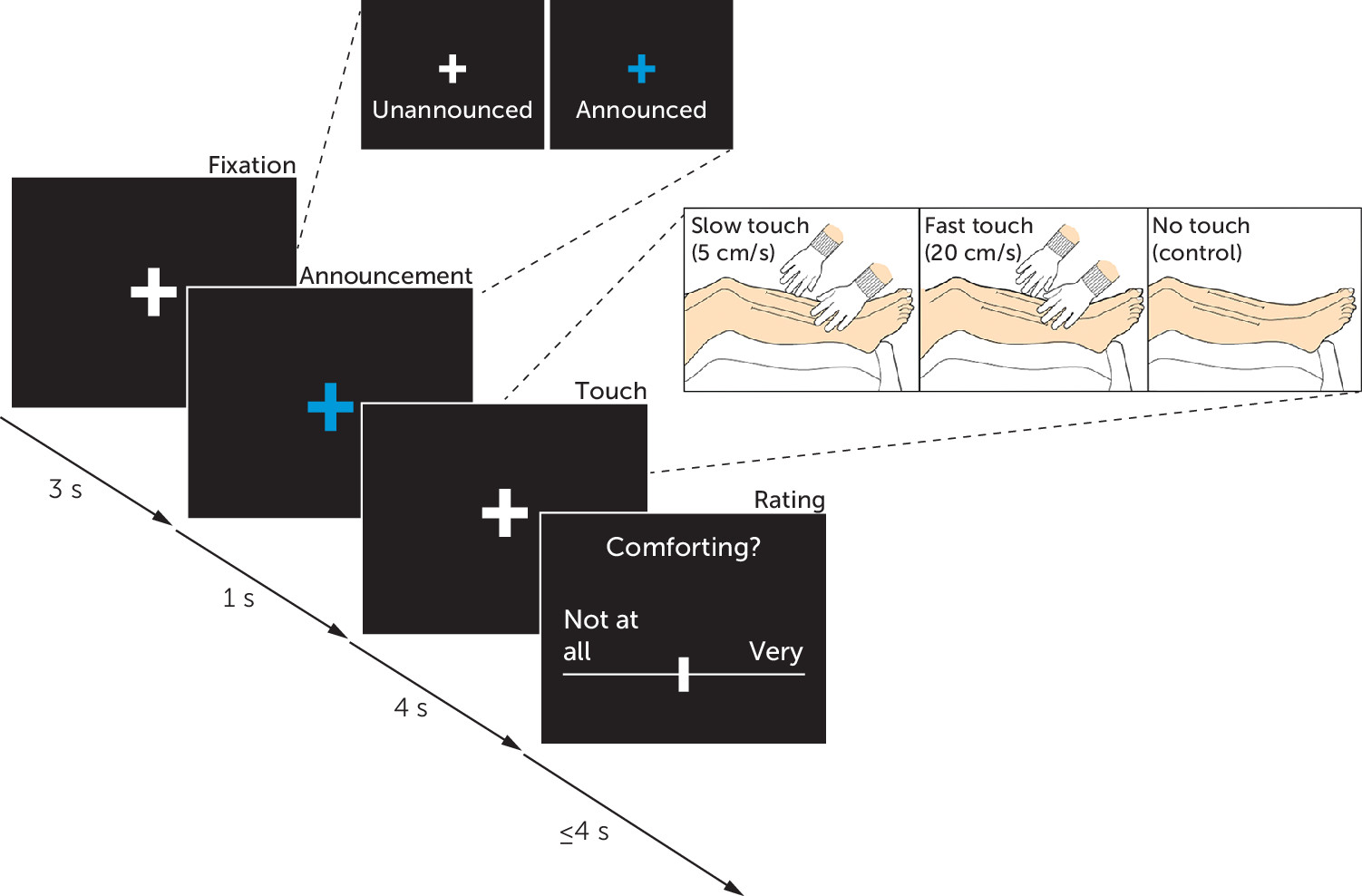
For the fMRI scan, we employed an adapted version of a previous social touch paradigm (26). Participants were asked to rate the perceived comfort of gentle, dynamic social tactile stimulations that were manually administered across 20 cm of their shins using speeds of ∼5 cm/s (slow touch, CT-optimal speed) and ∼20 cm/s (fast touch, non-CT-optimal speed), as well as a no-touch control condition (Figure 1). In 50% of the trials, imminent tactile stimulations were announced by the color of the fixation cross (see the online supplement). For the duration of the fMRI experiment, the entire opening of the scanner was covered with fabric so that the participants could not see their legs or the experimenter. The experimenter was trained in the delivery of the tactile stimuli with constant pressure at both speed levels and was guided by audio cues during the experiment to ensure constant stroking velocity. Participants were unaware of which experimenter administered the tactile stimulations.
FIGURE 1. Experimental design of the social touch taska
a During functional MRI, participants were asked to rate the perceived comfort of gentle, dynamic social tactile stimulations that were manually administered across 20 cm of their shins using speeds of ∼5 cm/s (slow touch, C-tactile [CT] optimal speed) and ∼20 cm/s (fast touch, non-CT-optimal speed), as well as a no-touch control condition. In each trial, tactile stimulation was administered over the course of 4 s, after which participants rated the comfort of the tactile stimulus on a 100-point visual analogue scale ranging from not at all (0) to very (100).The next trial started immediately after the response was recorded or after a maximum of 4 s. In 50% of the trials, imminent tactile stimulations were visually announced by a blue fixation cross appearing on the screen for the last second of the 4 s interstimulus interval. Each condition (slow touch announced, slow touch unannounced, fast touch announced, fast touch unannounced, no touch) was presented 20 times in random order, resulting in 100 trials over a period of about 20 minutes. All tactile stimulations were delivered to both shins simultaneously in proximo-distal orientation by a trained experimenter wearing cotton gloves.

Image Acquisition

T2*-weighted echoplanar images and high-resolution anatomical images were collected on a 3-T MRI system (Siemens, Erlangen, Germany). fMRI data were analyzed using SPM12 (http://www.fil.ion.ucl.ac.uk/spm) embedded in MATLAB, release R2010b (MathWorks, Natick, Mass.). Structural images were preprocessed with the CAT12 toolbox (Computational Anatomy Toolbox 12, Structural Brain Mapping group, Jena University Hospital, Jena, Germany) (for details, see the online supplement).

Data Analysis

Data analysis was performed using childhood maltreatment as a continuous (see the online supplement) and as a categorical measure. For the categorical approach, participants were stratified into low (mean CTQ score, 26.61 [SD=0.28; N=33]), medium (mean CTQ score, 35.53 [SD=0.67; N=30]) and high (mean CTQ score, 63.35 [SD=2.61; N=29]) levels of childhood maltreatment exposure (low CM, medium CM, and high CM) by means of a tertile split of CTQ sum scores. Sociodemographic and psychometric data for the three childhood maltreatment groups are summarized in Table S3 in the online supplement. Given that both operationalizations of childhood maltreatment yielded a consistent pattern of results (see the online supplement), we present the results of the categorical approach in order to illustrate the impact of severe relative to low and medium levels of childhood maltreatment on social touch processing.
For the fMRI statistical analysis, we used a two-level random-effects approach based on the general linear model as implemented in SPM12. On the first level, onsets and durations of the experimental conditions were modeled by a stick function convolved with a hemodynamic response function. Cardiac and respiratory noise correction was conducted using the PhysIO toolbox (27). We then included the movement parameters and physiological noise regressors as nuisance regressors in the design matrix. On the second level, group-specific response patterns to social touch were assessed by computing the main contrasts of interest (low CMSlow>Fast > high CMSlow>Fast and high CMSlow>Fast > low CMSlow>Fast) using a flexible factorial design. To investigate differences in region-specific gray matter volume between the three childhood maltreatment level groups, we employed a full factorial design, controlling for total intracranial volume, age, and sex, with age and sex orthogonalized in relation to total intracranial volume. Results were assessed by the main contrast of interest (low CM > high CM). The main fMRI and VBM analysis focused on a set of a priori bilateral regions of interest consisting of the amygdala, hippocampus, insula, and primary somatosensory cortex, which were anatomically defined according to the Wake Forest University PickAtlas, version 3.0. The family-wise error rate was used to correct p values for multiple comparisons, and p<0.05 was considered significant.
Statistical analyses were conducted with SPSS, version 24 (IBM, Armonk, N.Y.). Childhood maltreatment groups were compared with analyses of variance (ANOVAs). Post hoc t tests were Bonferroni corrected to account for multiple comparisons. We also assessed childhood maltreatment as a continuous variable by performing supplemental regression analyses (see the online supplement). Mediation and moderation analyses were carried out to examine the effects of potential confounders, using the PROCESS macro for SPSS, version 3.1 (28). We tested potential mediator and moderator effects of the covariates gray matter volume, age, sex, total education time (years), depressive symptoms, Perceived Stress Scale score, and CAPS score on the relationship between childhood maltreatment and behavioral ratings and parameter estimates extracted from significant clusters of the blood-oxygen-level-dependent response analysis (see the online supplement).

Results

Behavioral Results

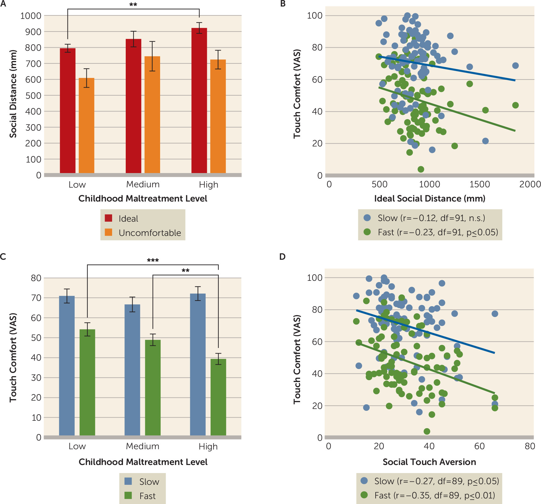
An ANOVA with the ideal distance for a social interaction as dependent variable showed a difference between low, medium, and high childhood maltreatment groups, which fell short of significance (F=2.65, df=2, 88, p=0.076; ηp2=0.06) (Figure 2A). Post hoc t tests revealed that participants with high levels of childhood maltreatment preferred a larger interpersonal distance compared with participants with low childhood maltreatment levels (t=2.88, df=60, Bonferroni-corrected p=0.008; d=0.74), but there was no significant difference between medium and low or medium and high childhood maltreatment levels (all p values >0.24). Childhood maltreatment groups did not differ significantly in the perception of slightly uncomfortable interpersonal distance (all p values >0.19). Moreover, a preference for larger ideal interpersonal distances was associated with lower comfort ratings of fast touch in the fMRI paradigm (r=−0.23, df=91, p=0.02) (Figure 2B), suggesting that a common denominator such as sensory sensitivity may affect both social metrics.
FIGURE 2. Interpersonal distance preferences and social touch preferences among adults with low, medium, and high levels of childhood maltreatmenta
a Participants with high levels of childhood maltreatment preferred larger ideal (comfortable) distances in the stop-distance paradigm compared with participants with low levels of childhood maltreatment (panel A). The preferred ideal interpersonal distance in the stop-distance paradigm correlated negatively with comfort rating of fast touch (panel B). Slow touch was rated as significantly more comforting than fast touch, and participants with high levels of childhood maltreatment perceived fast touch as significantly less comforting compared with participants with low and medium levels of childhood maltreatment (panel C). Comfort ratings of slow and fast tactile stimulations correlated negatively with general social touch aversion (panel D). Indicated p values are Bonferroni corrected. Error bars indicate standard error of the mean. VAS=visual analogue scale.
*p≤0.05. **p≤0.01.
A 2×2×3 mixed ANOVA with the within-subject factors announcement (announced touch, unannounced touch) and touch velocity (slow, fast) and the between-subject factor childhood maltreatment group (low, medium, high) revealed main effects of announcement (F=4.09, df=1, 89, p=0.046; ηp2=0.04) and touch velocity (F=61.3, df=1, 89, p<0.001; ηp2=0.41) and a touch velocity–by–childhood maltreatment group interaction (F=3.02, df=2, 89, p=0.045; ηp2=0.07) (Figure 2C). Fast touch and unannounced touch were rated as less comfortable, but the announcement did not alter the effect of childhood maltreatment (all p values >0.44). Post hoc t tests revealed that participants with high levels of childhood maltreatment rated fast touch as less comforting compared with participants with medium levels of childhood maltreatment (t=−2.36, df=57, Bonferroni-corrected p=0.03; d=0.63) and those with low levels of childhood maltreatment (t=−3.36, df=60, Bonferroni-corrected p=0.002; d=0.86); there was no significant difference for slow touch (all p values >0.29).
Further analyses showed that lower comfort ratings of slow (r=−0.27, df=89, p=0.01) and fast touch (r=−0.35, df=89, p=0.001) (Figure 2D) were associated with higher everyday social touch aversion. Together, these findings suggest that higher childhood maltreatment levels bias individuals toward a more cautious and negative perception of interpersonal contact.

fMRI Results

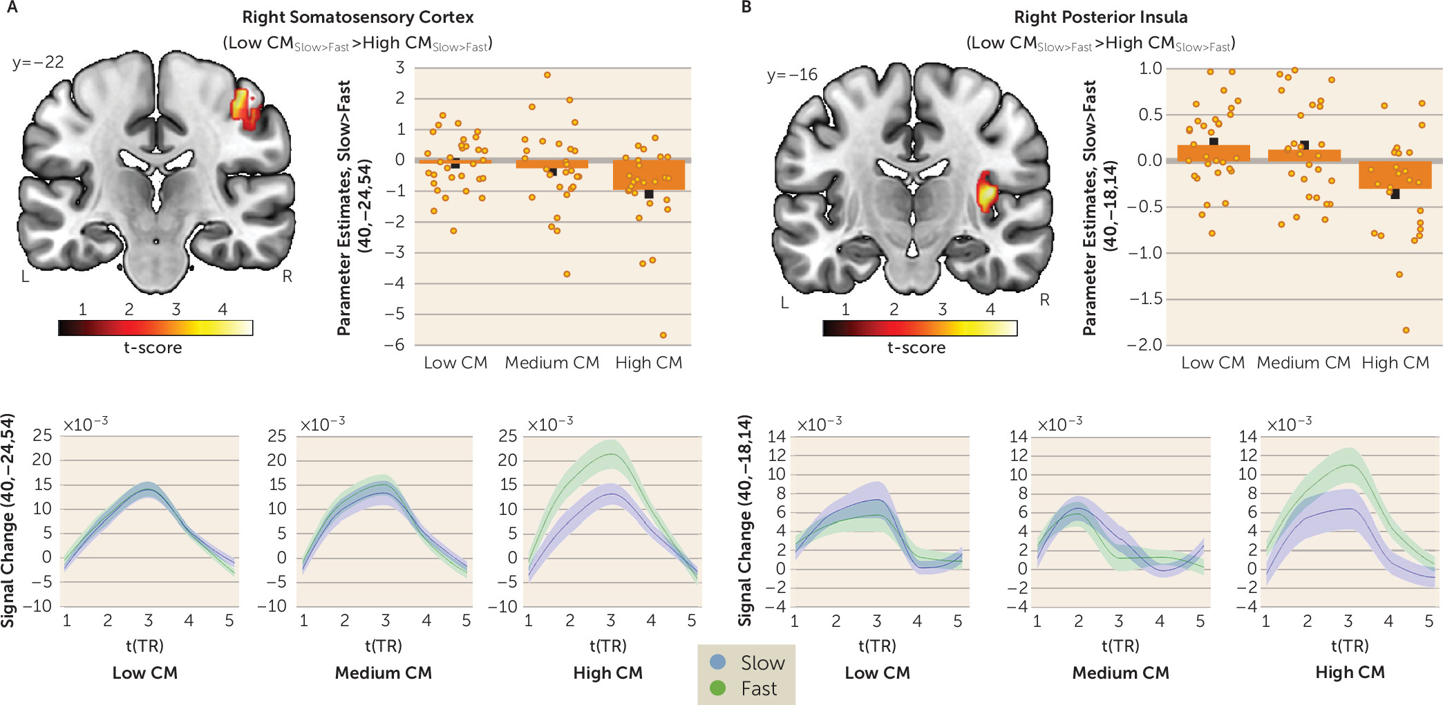
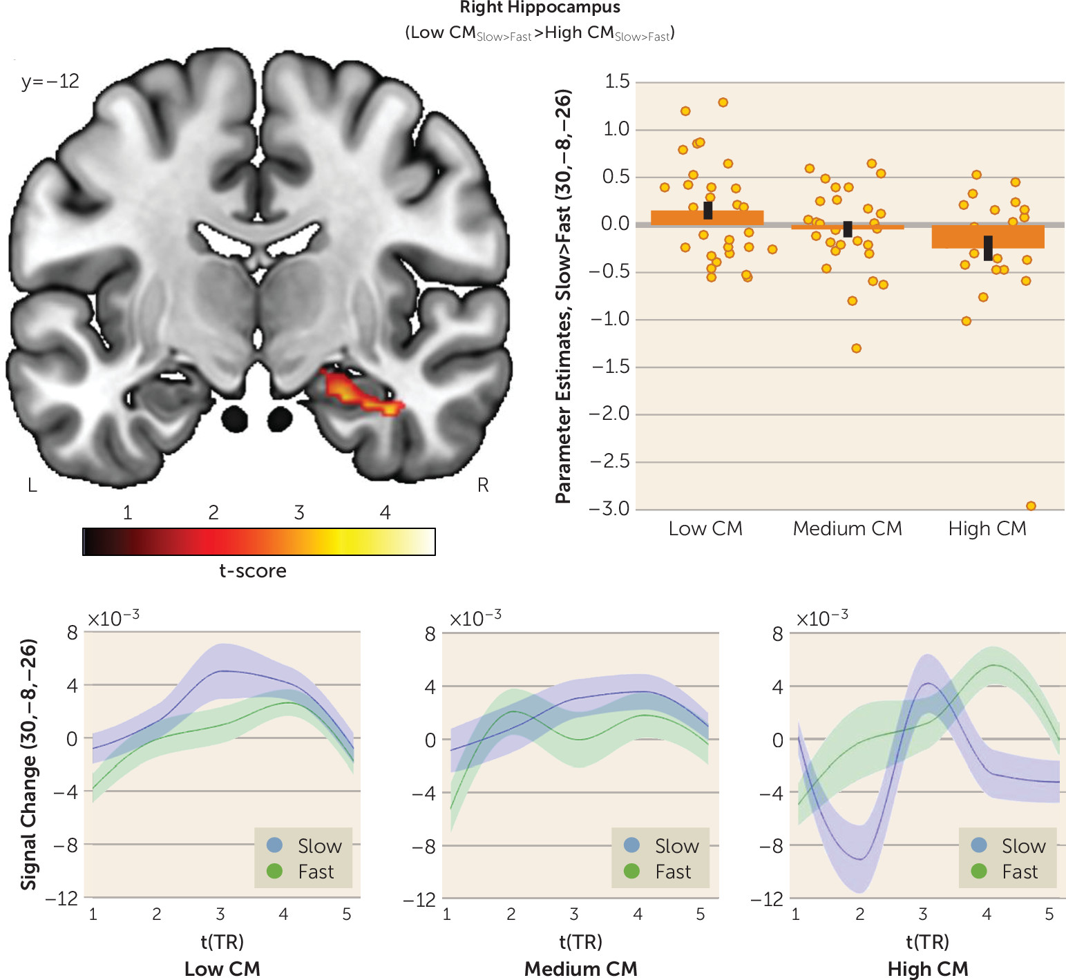
In participants with low levels of childhood maltreatment, slow relative to fast touch produced widespread activations at the whole-brain level, including the hippocampus and the insula (see Table S4 in the online supplement). Notably, participants with high levels of childhood maltreatment exhibited increased cortical reactivity to fast touch in the right primary somatosensory cortex (peak Montreal Neurological Institute coordinates (x, y, z): 40, −24, 54; t=4.56, df=246, family-wise error corrected p [pFWE]=0.004; d=0.75) (Figure 3A) and the right posterior insula (40, −18, −14; t=4.24, df=246, pFWE=0.007; d=0.96) (Figure 3B) compared with participants with low levels of childhood maltreatment. Furthermore, participants with high childhood maltreatment levels showed decreased limbic responsiveness to slow touch in the right hippocampus (30, −8, −26; t=4.13, df=246, pFWE=0.006; d=−0.67) (Figure 4) compared with participants with low levels of childhood maltreatment; they also showed a decrease in response to slow touch in the right amygdala, although this difference fell short of significance (26, −2, −22; t=2.95, df=246, pFWE=0.065; d=−0.47). Hyperreactivity of both the right primary somatosensory cortex (r=−0.51, df=85, p<0.001) and the right posterior insula (r=−0.23, df=85, p=0.035) in response to fast touch was associated with lower perceived comfort of fast touch. We also found a positive association between hippocampal response to slow touch and the perceived comfort of slow touch, although the difference did not reach significance (r=0.21, df=85, p=0.055). We did not detect significant higher-order interactions of announcement. Regression analyses confirmed the observed response pattern (see the online supplement).
FIGURE 3. Social touch functional MRI task results for adults with low, medium, and high levels of childhood maltreatment: right somatosensory cortex and posterior insulaa
a Participants with high levels of childhood maltreatment exhibited heightened neural responses to fast touch in the right somatosensory cortex (panel A) and the right posterior insula (panel B) compared with participants with low levels of childhood maltreatment levels. Error bars (upper graphs) and shaded areas (lower graphs) indicate standard error of the mean. CM=childhood maltreatment; TR=repetition time.
FIGURE 4. Social touch functional MRI task results for adults with low, medium, and high levels of childhood maltreatment: right hippocampusa
a Participants with high levels of childhood maltreatment exhibited diminished responses to slow touch (C-tactile [CT] optimal speed) in the right hippocampus compared with participants with low levels of childhood maltreatment. Error bars (upper graph) and shaded areas (lower graphs) indicate standard error of the mean. CM=childhood maltreatment; TR=repetition time.

Voxel-Based Morphometry Results

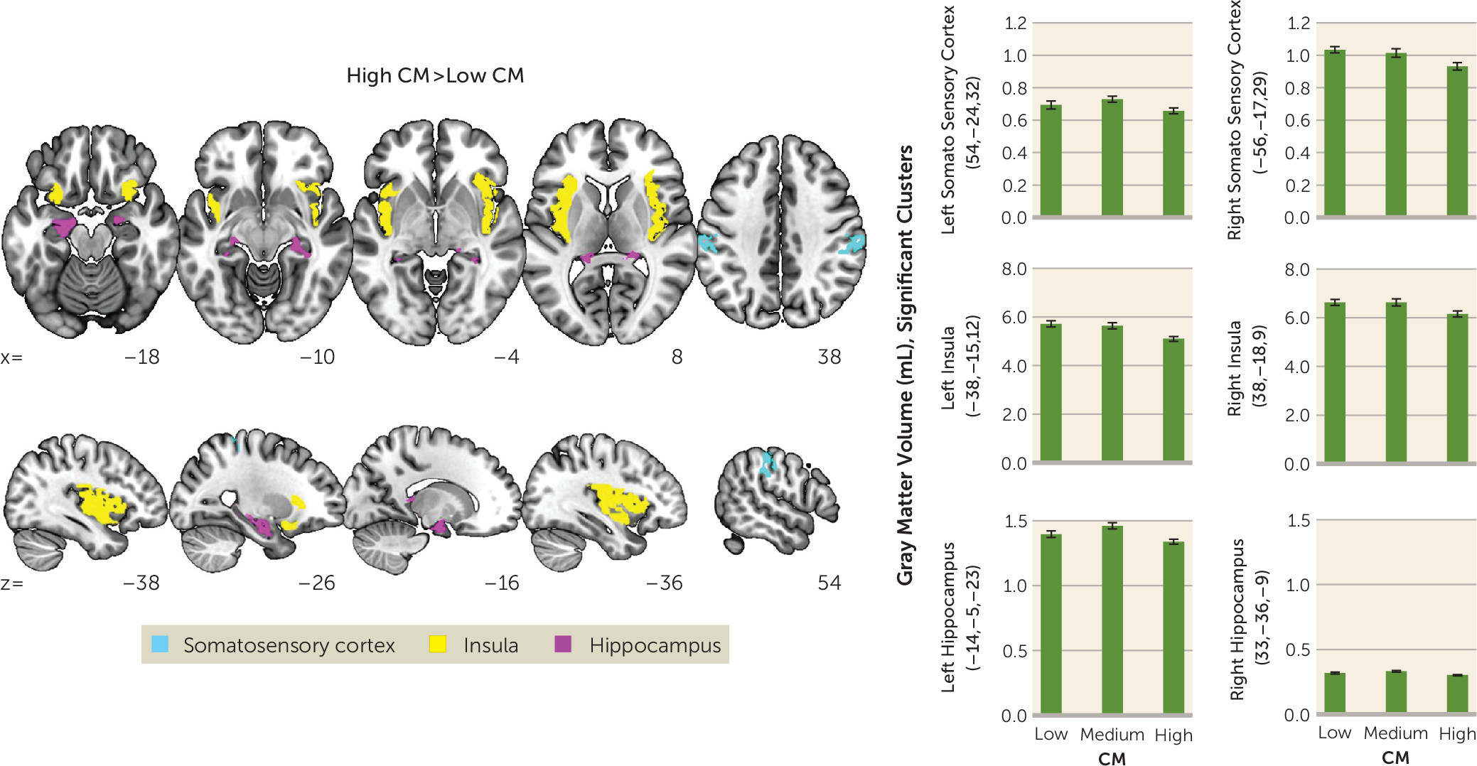
As expected, high compared with low levels of childhood maltreatment were associated with significant reductions of gray matter volume in the left and right hippocampus (coordinates: 33, −36, −9; t=3.71, df=76, pFWE=0.013; −14, −5, −23; t=3.18, df=76, pFWE=0.048), the left and right primary somatosensory cortex (54, −24, 32; t=4.91, df=76, pFWE=0.001; −56, −17, 29; t=4.24, df=76, pFWE=0.009), the left and right posterior insula (33, −18, 9; t=5.51, df=76, pFWE<0.001; −38, −15, 12; t=5.77, df=76, pFWE<0.001) and left amygdala (−20, 3, −20; t=3.75, df=76, pFWE=0.003) (Figure 5). We observed a similar pattern of results in the comparison between participants with medium and high levels of childhood maltreatment (see the online supplement). However, there were no significant differences in gray matter volume between participants with low and medium childhood maltreatment (all p values >0.05). The whole-brain results of the group contrasts are listed in Table S5 in the online supplement.
FIGURE 5. Voxel-based morphometry results for adults with low, medium, and high levels of childhood maltreatmenta
a Region-based morphometry analysis revealed significantly reduced gray matter volume in the somatosensory cortex, insula, and hippocampus in participants with severe childhood maltreatment compared with participants with a low childhood maltreatment severity. Error bars indicate standard error of the mean. CM=childhood maltreatment.

Mediation and Moderation Effects

General social touch aversion mediated the effect of childhood maltreatment on the perceived comfort of slow touch (β=−0.16, SE=0.08, 95% CI=−0.33, −0.03) and fast touch (β=−0.09, SE=0.05, 95% CI=−0.19, −0.01), indicating that childhood maltreatment may affect the experience of social touch irrespective of the specific task context. Of note, we observed an indirect effect of social touch aversion on the relationship between childhood maltreatment and subjective stress levels (β=0.04, SE=0.02, 95% CI=0.05, 0.08), showing that the detrimental effects of childhood maltreatment on subjective stress are mediated by social touch aversion. Moderation analysis revealed that CAPS score moderated the effect of childhood maltreatment on fast touch ratings (t=2.90, df=81, p=0.005). The Johnson-Neyman technique showed that the relationship between childhood maltreatment and fast touch ratings was significant when the CAPS sum score was less than 19.45 (β=−0.203, SE=0.102, p=0.05), but not significant with higher symptom loads. Given a strong positive correlation between CAPS score and childhood maltreatment (r=0.52, df=89, p<0.001), this moderation suggests a ceiling effect, such that the negative impact of traumatic experiences on social touch may reach a plateau. No significant mediation or moderation effects were observed for other covariates. Thus, the behavioral and neural effects of childhood maltreatment were not confounded by sociodemographic factors, PTSD symptom loads, depression and stress levels, or childhood maltreatment–associated region-specific reductions in gray matter volume (all 95% confidence intervals overlapped with zero).

Effect of Trauma Type

All five CTQ subscales were significantly intercorrelated, because a large percentage of the study sample (42.4%) had experienced multiple types of maltreatment during childhood. Furthermore, applying a Fisher’s r-to-z transformation revealed that the correlation coefficients did not significantly differ between CTQ subscales, suggesting that trauma-induced behavioral and neural changes cannot be attributed to a single trauma type in the present sample (see the online supplement).

Discussion

In this study, we investigated whether childhood maltreatment is associated with altered interpersonal distance preferences and social touch processing later in life. As hypothesized, participants with a high level of childhood maltreatment preferred larger interpersonal distance in a stop-distance paradigm. Participants with severe childhood maltreatment also expressed discomfort of discriminative (i.e., fast) touch in an fMRI social touch paradigm, which was paralleled by increased responses to fast touch in the right primary somatosensory cortex and posterior insula and diminished responses to affective (i.e., slow) touch in the right hippocampus. Our findings add to the burgeoning evidence indicating that childhood maltreatment has long-term detrimental effects on sensory processing of social information (5, 29).
Previous research demonstrated that a higher perceived risk of physical threat is associated with larger interpersonal distance, which may facilitate escape during potentially violent interactions (30). It is well established that individuals with a history of childhood maltreatment exhibit increased sensitivity to threat (29), and therefore they may use the self-protective and arousal regulation functions of larger interpersonal distance (11) to mitigate emotional and physical threats during interactions. Thus, a need for a larger personal space may constitute another dimension of the phenotypic hypervigilance to threat signals following childhood maltreatment (29). Furthermore, our finding of larger interpersonal distance preferences in individuals with high childhood maltreatment levels were linked to both general touch aversion and perceived discomfort of discriminative touch, indicating overall avoidance of interpersonal sensory stimulation to reduce emotional distress.
On the neural level, childhood maltreatment–associated responses to social touch suggest a cortical sensory hyperreactivity in the primary somatosensory cortex and posterior insula in response to discriminative touch alongside a CT-specific hypoactivation in the hippocampus. The primary somatosensory cortex has a functional role for motor control (15), and exaggerated primary somatosensory cortex responses have been associated with tactile defensiveness (31). Thus, increased primary somatosensory cortex activation to discriminative touch may reflect a motor preparation to initiate a flight response. The insula encodes interoceptive signals from the body and integrates these signals in a posterior-to-anterior fashion, ultimately serving emotional and body awareness (17). Moreover, hyperreactivity of the posterior insula has been observed in trauma-exposed adolescents in response to conflicting visual stimuli (32), which may indicate increased salience detection of external signals, consistent with the proposed function of the posterior insula as a multisensory magnitude detector in the context of pain perception (33). In the present study, increased posterior insula activation was associated with diminished comfort of fast touch, suggesting that this potentiated sensory signal may contribute to an interoceptive integration and awareness that make individuals with high childhood maltreatment levels feel tense and uneasy. In addition to these sensory effects, we observed changes in limbic touch responsiveness. The hippocampus plays a crucial role in the consolidation and retrieval of episodic memory (34) and is highly susceptible to early stress (5). Furthermore, a population of reward-associated cells has been identified in the hippocampus (35), and activation in this region is altered in the context of rewarding stimuli (35, 36). Thus, childhood maltreatment–associated hippocampal hypoactivation may reflect decreased encoding of affective touch as a result of the retrieval of past experiences of abuse and neglect when affective touch was initially associated with low reward salience. Moreover, we found a decrease in amygdala responses to affective touch, although it fell short of significance. Recent neuroimaging studies suggest that the amygdala may extract the emotional value of somatosensory inputs (37) and show diminished amygdala response to positive facial stimuli in individuals with childhood maltreatment (8). Reduced amygdala responses to CT-optimal affective touch may therefore reflect diminished emotional salience of positive social signals.
Our findings of childhood maltreatment–associated structural deficits mirror previous reports of childhood maltreatment–associated reduction of gray matter volume in the hippocampus, primary somatosensory cortex, insula (8), and amygdala (38). Both childhood maltreatment–associated hippocampal and amygdala volume reductions have been linked to behavioral disturbances later in life (38). Interestingly, modifications of sensory systems may convey the specific forms of early adverse experiences (5). Thus, childhood maltreatment–associated structural deficits in the primary somatosensory cortex and insula may reflect neuroplastic adaptations as a function of early physical abuse and/or touch deprivation to promote avoidance and diminish approach responses toward traumatic reminders (5). Notably, structural changes neither moderated nor mediated the observed response pattern to social touch, underscoring the hypothesis that structural modifications may keep functional adaptation pathways intact to allow for rapid threat responses (5).
Furthermore, significant detrimental effects on interpersonal distance and social touch were evident in participants with severe childhood maltreatment, whereas a medium level of childhood maltreatment did not produce significant changes. This response pattern corroborates previous findings of a dose-dependent effect of childhood maltreatment on psychosocial functioning and comorbidity (39). Moreover, avoidance tendencies toward everyday social touch appear to hinder individuals with a history of childhood maltreatment from experiencing the stress-buffering effects of physical contact. It is noteworthy that the announcement of the subsequent touch did not significantly reduce childhood maltreatment–associated changes in response to social touch. Thus, the childhood maltreatment effect on social touch and physical proximity cannot be explained only as a consequence of biased cognition (29) but should also be considered a product of fundamentally altered sensory experiences.
Our results may have important implications for the understanding and effective treatment of childhood maltreatment and associated psychopathology. Adults with a history of childhood maltreatment may exhibit exaggerated responses to interpersonal physical contact, which in turn may affect their psychosocial functioning in daily life. Hypersensitivity and sensory avoidance have been linked to social withdrawal and an inability to initiate relationships (40). Moreover, hyperresponsiveness to touch was found to fully mediate social impairments in autism spectrum disorder (19), suggesting that sensory dysregulations in the tactile domain may confer vulnerability to interpersonal difficulties. Recent findings demonstrated that sensory profiles mediate the relationship between childhood maltreatment and health-related quality of life in patients with affective disorders (41). Furthermore, neural hyperresponsiveness to aversive facial stimuli has been shown to predict poor treatment response in PTSD (42). Thus, randomized clinical trials are warranted to evaluate treatment-induced changes and the predictive validity of social touch and physical proximity. Current cognitive-behavioral therapeutic approaches do not directly address these aberrant autonomic and somatic responses. Previous research has, however, demonstrated the effectiveness of massage interventions in the treatment of patients with sexual abuse (43) and PTSD (44). Along these lines, the use of complementary bottom-up, body-based interventions could help individuals with a history of childhood maltreatment to facilitate their participation in social interactions by learning to tolerate and enjoy the comforts of social touch in a safe environment (45).
Limitations of this study include the co-occurrence and interrelatedness of multiple childhood maltreatment types in the sample, which hindered us from disentangling the effects of specific types of maltreatment. However, childhood family adversities are often clustered, rendering multitype maltreatment highly prevalent in the population (39). Another limitation is the retrospective and self-report nature of childhood maltreatment assessment, which may be subject to negative recall bias due to current elevated levels of depression and psychological distress (46). Although we thoroughly controlled for current depressive symptoms and subjective stress levels, a recall-related underreporting of childhood maltreatment may have influenced our results.
In conclusion, we provide first evidence that severe childhood maltreatment is associated with larger interpersonal distance preferences and adverse responses to social touch. We propose changes in early sensory processing as the underlying mechanism of these associations. This sensory dysregulation may explain why individuals with severe childhood maltreatment often suffer from difficulties in establishing and maintaining close social bonds later in life.
Acknowledgments

Dr. Hurlemann and Dr. Scheele are supported by an Else-Kröner-Fresenius-Stiftung grant (2017_A35). The authors thank Paul Jung for programming assistance and Alexandra Patin for proofreading the manuscript.

Supplementary Material

File (appi.ajp.2019.19020212.ds001.pdf)

References

1.
Scott KM, Smith DR, Ellis PM: Prospectively ascertained child maltreatment and its association with DSM-IV mental disorders in young adults. Arch Gen Psychiatry 2010; 67:712–719
2.
Braithwaite EC, O’Connor RM, Degli-Esposti M, et al: Modifiable predictors of depression following childhood maltreatment: a systematic review and meta-analysis. Transl Psychiatry 2017; 7:e1162
3.
Copeland WE, Shanahan L, Hinesley J, et al: Association of childhood trauma exposure with adult psychiatric disorders and functional outcomes. JAMA Netw Open 2018; 1:e184493
4.
Alink LR, Cicchetti D, Kim J, et al: Longitudinal associations among child maltreatment, social functioning, and cortisol regulation. Dev Psychol 2012; 48:224–236
5.
Teicher MH, Samson JA, Anderson CM, et al: The effects of childhood maltreatment on brain structure, function, and connectivity. Nat Rev Neurosci 2016; 17:652–666
6.
McCrory EJ, De Brito SA, Sebastian CL, et al: Heightened neural reactivity to threat in child victims of family violence. Curr Biol 2011; 21:R947–R948
7.
Gerin MI, Puetz VB, Blair RJR, et al: A neurocomputational investigation of reinforcement-based decision making as a candidate latent vulnerability mechanism in maltreated children. Dev Psychopathol 2017; 29:1689–1705
8.
Dannlowski U, Stuhrmann A, Beutelmann V, et al: Limbic scars: long-term consequences of childhood maltreatment revealed by functional and structural magnetic resonance imaging. Biol Psychiatry 2012; 71:286–293
9.
Hayduk LA: Personal space: where we now stand. Psychol Bull 1983; 94:293–335
10.
Kennedy DP, Gläscher J, Tyszka JM, et al: Personal space regulation by the human amygdala. Nat Neurosci 2009; 12:1226–1227
11.
Uzzell D, Horne N: The influence of biological sex, sexuality, and gender role on interpersonal distance. Br J Soc Psychol 2006; 45:579–597
12.
Vranić A: Personal space in physically abused children. Environ Behav 2003; 35:550–565
13.
Bogović A, Ivezić E, Filipčić I: Personal space of war veterans with PTSD: some characteristics and comparison with healthy individuals. Psychiatr Danub 2016; 28:77–81
14.
Perry A, Nichiporuk N, Knight RT: Where does one stand: a biological account of preferred interpersonal distance. Soc Cogn Affect Neurosci 2016; 11:317–326
15.
McGlone F, Wessberg J, Olausson H: Discriminative and affective touch: sensing and feeling. Neuron 2014; 82:737–755
16.
Morrison I: ALE meta-analysis reveals dissociable networks for affective and discriminative aspects of touch. Hum Brain Mapp 2016; 37:1308–1320
17.
Craig AD: How do you feel—now? The anterior insula and human awareness. Nat Rev Neurosci 2009; 10:59–70
18.
Crucianelli L, Cardi V, Treasure J, et al: The perception of affective touch in anorexia nervosa. Psychiatry Res 2016; 239:72–78
19.
Lundqvist LO: Hyper-responsiveness to touch mediates social dysfunction in adults with autism spectrum disorders. Res Autism Spectr Disord 2015; 9:13–20
20.
Wittchen HU, Wunderlich U, Gruschwitz S, et al: Strukturiertes Klinisches Interview für DSM-IV. Goettingen, Hogrefe, 1997
21.
Weathers FW, Bovin MJ, Lee DJ, et al: The Clinician-Administered PTSD Scale for DSM-5 (CAPS-5): development and initial psychometric evaluation in military veterans. Psychol Assess 2018; 30:383–395
22.
Bernstein DP, Fink L, Handelsman L, et al: Initial reliability and validity of a new retrospective measure of child abuse and neglect. Am J Psychiatry 1994; 151:1132–1136
23.
Beck AT, Steer RA, Brown GK: Manual for the Beck Depression Inventory–II. San Antonio, Tex, Psychological Corporation, 1996
24.
Cohen S, Kamarck T, Mermelstein R: A global measure of perceived stress. J Health Soc Behav 1983; 24:385–396
25.
Wilhelm FH, Kochar AS, Roth WT, et al: Social anxiety and response to touch: incongruence between self-evaluative and physiological reactions. Biol Psychol 2001; 58:181–202
26.
McGlone F, Olausson H, Boyle JA, et al: Touching and feeling: differences in pleasant touch processing between glabrous and hairy skin in humans. Eur J Neurosci 2012; 35:1782–1788
27.
Kasper L, Bollmann S, Diaconescu AO, et al: The PhysIO toolbox for modeling physiological noise in fMRI data. J Neurosci Methods 2017; 276:56–72
28.
Hayes AF: Introduction to Mediation, Moderation, and Conditional Process Analysis: A Regression Based Approach. New York, Guilford, 2013
29.
Jaffee SR: Child maltreatment and risk for psychopathology in childhood and adulthood. Annu Rev Clin Psychol 2017; 13:525–551
30.
Edney JJ, Walker EC, Jordan NL: Is there reactance in personal space? J Soc Psychol 1976; 100:207–217
31.
Pissiota A, Frans O, Fernandez M, et al: Neurofunctional correlates of posttraumatic stress disorder: a PET symptom provocation study. Eur Arch Psychiatry Clin Neurosci 2002; 252:68–75
32.
Marusak HA, Etkin A, Thomason ME: Disrupted insula-based neural circuit organization and conflict interference in trauma-exposed youth. Neuroimage Clin 2015; 8:516–525
33.
Baliki MN, Geha PY, Apkarian AV: Parsing pain perception between nociceptive representation and magnitude estimation. J Neurophysiol 2009; 101:875–887
34.
Tulving E: Episodic memory: from mind to brain. Annu Rev Psychol 2002; 53:1–25
35.
Tryon VL, Penner MR, Heide SW, et al: Hippocampal neural activity reflects the economy of choices during goal-directed navigation. Hippocampus 2017; 27:743–758
36.
Gauthier JL, Tank DW: A dedicated population for reward coding in the hippocampus. Neuron 2018; 99:179–193.e7
37.
Lucas MV, Anderson LC, Bolling DZ, et al: Dissociating the neural correlates of experiencing and imagining affective touch. Cereb Cortex 2015; 25:2623–2630
38.
Hanson JL, Nacewicz BM, Sutterer MJ, et al: Behavioral problems after early life stress: contributions of the hippocampus and amygdala. Biol Psychiatry 2015; 77:314–323
39.
Green JG, McLaughlin KA, Berglund PA, et al: Childhood adversities and adult psychiatric disorders in the National Comorbidity Survey Replication, I: associations with first onset of DSM-IV disorders. Arch Gen Psychiatry 2010; 67:113–123
40.
Miller LJ, Anzalone ME, Lane SJ, et al: Concept evolution in sensory integration: a proposed nosology for diagnosis. Am J Occup Ther 2007; 61:135–140
41.
Serafini G, Gonda X, Pompili M, et al: The relationship between sensory processing patterns, alexithymia, traumatic childhood experiences, and quality of life among patients with unipolar and bipolar disorders. Child Abuse Negl 2016; 62:39–50
42.
van Rooij SJ, Kennis M, Vink M, et al: Predicting treatment outcome in PTSD: a longitudinal functional MRI study on trauma-unrelated emotional processing. Neuropsychopharmacology 2016; 41:1156–1165
43.
Price C: Body-oriented therapy in recovery from child sexual abuse: an efficacy study. Altern Ther Health Med 2005; 11:46–57
44.
Jain S, McMahon GF, Hasen P, et al: Healing touch with guided imagery for PTSD in returning active duty military: a randomized controlled trial. Mil Med 2012; 177:1015–1021
45.
van der Kolk B: The Body Keeps the Score: Brain, Mind, and Body in the Healing of Trauma. New York, Viking, 2014
46.
Colman I, Kingsbury M, Garad Y, et al: Consistency in adult reporting of adverse childhood experiences. Psychol Med 2016; 46:543–549

Information & Authors

Information

Published In

Go to American Journal of Psychiatry
American Journal of Psychiatry
Pages: 37 - 46
PubMed: 31416339

History

Received: 25 February 2019
Revision received: 29 May 2019
Accepted: 2 July 2019
Published online: 16 August 2019
Published in print: January 01, 2020

Keywords

  1. Brain Imaging Techniques
  2. Cognitive Neuroscience

Authors

Details

Ayline Maier, M.Sc. [email protected]
Division of Medical Psychology (Maier, Gieling, Heinen-Ludwig, Stefan, Hurlemann, Scheele), Center for Economics and Neuroscience (Schultz), and Department of Psychiatry (Hurlemann), University of Bonn, Bonn, Germany; Department of Psychology, Laboratory for Biological Psychology, Ruhr-University of Bochum, Bochum, Germany (Güntürkün); Clinical Hospital of Chengdu Brain Science Institute and Key Laboratory for Neuroinformation, University of Electronic Science and Technology of China, Chengdu (Becker); and Department of Psychiatry, University of Oldenburg Medical Campus, Bad Zwischenahn, Germany (Hurlemann).
Caroline Gieling
Division of Medical Psychology (Maier, Gieling, Heinen-Ludwig, Stefan, Hurlemann, Scheele), Center for Economics and Neuroscience (Schultz), and Department of Psychiatry (Hurlemann), University of Bonn, Bonn, Germany; Department of Psychology, Laboratory for Biological Psychology, Ruhr-University of Bochum, Bochum, Germany (Güntürkün); Clinical Hospital of Chengdu Brain Science Institute and Key Laboratory for Neuroinformation, University of Electronic Science and Technology of China, Chengdu (Becker); and Department of Psychiatry, University of Oldenburg Medical Campus, Bad Zwischenahn, Germany (Hurlemann).
Luca Heinen-Ludwig
Division of Medical Psychology (Maier, Gieling, Heinen-Ludwig, Stefan, Hurlemann, Scheele), Center for Economics and Neuroscience (Schultz), and Department of Psychiatry (Hurlemann), University of Bonn, Bonn, Germany; Department of Psychology, Laboratory for Biological Psychology, Ruhr-University of Bochum, Bochum, Germany (Güntürkün); Clinical Hospital of Chengdu Brain Science Institute and Key Laboratory for Neuroinformation, University of Electronic Science and Technology of China, Chengdu (Becker); and Department of Psychiatry, University of Oldenburg Medical Campus, Bad Zwischenahn, Germany (Hurlemann).
Vlad Stefan
Division of Medical Psychology (Maier, Gieling, Heinen-Ludwig, Stefan, Hurlemann, Scheele), Center for Economics and Neuroscience (Schultz), and Department of Psychiatry (Hurlemann), University of Bonn, Bonn, Germany; Department of Psychology, Laboratory for Biological Psychology, Ruhr-University of Bochum, Bochum, Germany (Güntürkün); Clinical Hospital of Chengdu Brain Science Institute and Key Laboratory for Neuroinformation, University of Electronic Science and Technology of China, Chengdu (Becker); and Department of Psychiatry, University of Oldenburg Medical Campus, Bad Zwischenahn, Germany (Hurlemann).
Johannes Schultz, M.D., Ph.D.
Division of Medical Psychology (Maier, Gieling, Heinen-Ludwig, Stefan, Hurlemann, Scheele), Center for Economics and Neuroscience (Schultz), and Department of Psychiatry (Hurlemann), University of Bonn, Bonn, Germany; Department of Psychology, Laboratory for Biological Psychology, Ruhr-University of Bochum, Bochum, Germany (Güntürkün); Clinical Hospital of Chengdu Brain Science Institute and Key Laboratory for Neuroinformation, University of Electronic Science and Technology of China, Chengdu (Becker); and Department of Psychiatry, University of Oldenburg Medical Campus, Bad Zwischenahn, Germany (Hurlemann).
Onur Güntürkün, M.Sc., Ph.D.
Division of Medical Psychology (Maier, Gieling, Heinen-Ludwig, Stefan, Hurlemann, Scheele), Center for Economics and Neuroscience (Schultz), and Department of Psychiatry (Hurlemann), University of Bonn, Bonn, Germany; Department of Psychology, Laboratory for Biological Psychology, Ruhr-University of Bochum, Bochum, Germany (Güntürkün); Clinical Hospital of Chengdu Brain Science Institute and Key Laboratory for Neuroinformation, University of Electronic Science and Technology of China, Chengdu (Becker); and Department of Psychiatry, University of Oldenburg Medical Campus, Bad Zwischenahn, Germany (Hurlemann).
Benjamin Becker, M.Sc., Ph.D.
Division of Medical Psychology (Maier, Gieling, Heinen-Ludwig, Stefan, Hurlemann, Scheele), Center for Economics and Neuroscience (Schultz), and Department of Psychiatry (Hurlemann), University of Bonn, Bonn, Germany; Department of Psychology, Laboratory for Biological Psychology, Ruhr-University of Bochum, Bochum, Germany (Güntürkün); Clinical Hospital of Chengdu Brain Science Institute and Key Laboratory for Neuroinformation, University of Electronic Science and Technology of China, Chengdu (Becker); and Department of Psychiatry, University of Oldenburg Medical Campus, Bad Zwischenahn, Germany (Hurlemann).
René Hurlemann, M.D., Ph.D.
Division of Medical Psychology (Maier, Gieling, Heinen-Ludwig, Stefan, Hurlemann, Scheele), Center for Economics and Neuroscience (Schultz), and Department of Psychiatry (Hurlemann), University of Bonn, Bonn, Germany; Department of Psychology, Laboratory for Biological Psychology, Ruhr-University of Bochum, Bochum, Germany (Güntürkün); Clinical Hospital of Chengdu Brain Science Institute and Key Laboratory for Neuroinformation, University of Electronic Science and Technology of China, Chengdu (Becker); and Department of Psychiatry, University of Oldenburg Medical Campus, Bad Zwischenahn, Germany (Hurlemann).
Dirk Scheele, M.Sc., Ph.D. [email protected]
Division of Medical Psychology (Maier, Gieling, Heinen-Ludwig, Stefan, Hurlemann, Scheele), Center for Economics and Neuroscience (Schultz), and Department of Psychiatry (Hurlemann), University of Bonn, Bonn, Germany; Department of Psychology, Laboratory for Biological Psychology, Ruhr-University of Bochum, Bochum, Germany (Güntürkün); Clinical Hospital of Chengdu Brain Science Institute and Key Laboratory for Neuroinformation, University of Electronic Science and Technology of China, Chengdu (Becker); and Department of Psychiatry, University of Oldenburg Medical Campus, Bad Zwischenahn, Germany (Hurlemann).

Notes

Send correspondence to Ms. Maier ([email protected]) and Dr. Scheele ([email protected]).

Funding Information

Else-Kröner-Fresenius-Stiftung (EKFS) grant: 2017_A35
The authors report no financial relationships with commercial interests.

Metrics & Citations

Metrics

Citations

Export Citations

If you have the appropriate software installed, you can download article citation data to the citation manager of your choice. Simply select your manager software from the list below and click Download.

For more information or tips please see 'Downloading to a citation manager' in the Help menu.

Format
Citation style
Style
Copy to clipboard

View Options

View options

PDF/EPUB

View PDF/EPUB

Login options

Already a subscriber? Access your subscription through your login credentials or your institution for full access to this article.

Personal login Institutional Login Open Athens login
Purchase Options

Purchase this article to access the full text.

PPV Articles - American Journal of Psychiatry

PPV Articles - American Journal of Psychiatry

Not a subscriber?

Subscribe Now / Learn More

PsychiatryOnline subscription options offer access to the DSM-5-TR® library, books, journals, CME, and patient resources. This all-in-one virtual library provides psychiatrists and mental health professionals with key resources for diagnosis, treatment, research, and professional development.

Need more help? PsychiatryOnline Customer Service may be reached by emailing [email protected] or by calling 800-368-5777 (in the U.S.) or 703-907-7322 (outside the U.S.).

Media

Figures

Other

Tables

Share

Share

Share article link

Share