Abstract

Objective:

Thalamus models of psychosis implicate association nuclei in the pathogenesis of psychosis and mechanisms of cognitive impairment. Studies to date have provided conflicting findings for structural deficits specific to these nuclei. The authors sought to characterize thalamic structural abnormalities in psychosis and a neurodevelopmental cohort, and to determine whether nuclear volumes were associated with cognitive function.

Methods:

Thalamic nuclei volumes were tested in a cross-sectional sample of 472 adults (293 with psychosis) and the Philadelphia Neurodevelopmental Cohort (PNC), consisting of 1,393 youths (398 with psychosis spectrum symptoms and 609 with other psychopathologies), using a recently developed, validated method for segmenting thalamic nuclei and complementary voxel-based morphometry. Cognitive function was measured with the Screen for Cognitive Impairment in Psychiatry in the psychosis cohort and the Penn Computerized Neurocognitive Battery in the PNC.

Results:

The psychosis group had smaller pulvinar, mediodorsal, and, to a lesser extent, ventrolateral nuclei volumes compared with the healthy control group. Youths with psychosis spectrum symptoms also had smaller pulvinar volumes, compared with both typically developing youths and youths with other psychopathologies. Pulvinar volumes were positively correlated with general cognitive function.

Conclusions:

The study findings demonstrate that smaller thalamic association nuclei represent a neurodevelopmental abnormality associated with psychosis, risk for psychosis in youths, and cognitive impairment. Identifying specific thalamic nuclei abnormalities in psychosis has implications for early detection of psychosis risk and treatment of cognitive impairment in psychosis.
Multiple lines of evidence implicate the thalamus in psychotic disorders, including smaller thalamus volume, decreased activation during task performance, abnormal functional and anatomical connectivity with the cortex, reduced expression of biochemical markers of neuronal integrity, abnormal sleep spindles, and lower cell numbers in some thalamic nuclei (113). These findings contributed to the development of several models of psychosis, many of which propose a neurodevelopmental basis for thalamus pathology and emphasize thalamic abnormalities in the mechanisms of cognitive impairment (1418).
Several models further propose selective dysfunction of specific thalamic nuclei, particularly thalamic association nuclei, including the mediodorsal and pulvinar nuclei (1418). There is abundant evidence that connectivity of some association nuclei (e.g., the mediodorsal nucleus) are abnormal (3, 4); however, evidence of selective anatomical abnormalities is sparse and inconsistent. For instance, while postmortem studies consistently find smaller volume and lower cell numbers in the pulvinar (1921), mediodorsal nucleus findings are mixed (22, 23), and small sample sizes raise broader concerns about replicability and generalization from postmortem studies (10, 23). Similarly, the handful of neuroimaging studies examining specific thalamic nuclei report both smaller and normal mediodorsal and pulvinar volumes (2427). Inconsistent neuroimaging results are likely due to a combination of factors, including modest sample sizes and use of idiosyncratic methods for quantifying thalamic nuclei that have not been widely adopted by the neuroimaging community.
The recent development of a novel method for segmenting the thalamus has created a new opportunity to investigate the thalamus in psychosis. Specifically, adapting an approach developed to segment hippocampal subfields (28), Iglesias and colleagues (29) built a probabilistic atlas from ex vivo MRI and histological data that can be applied to standard T1-weighted in vivo MRI to segment thalamic nuclei using Bayesian inference. Critically, their method is able to recover the three-dimensional structure of histological data from ex vivo MRI; yields volumes that are in good agreement with other histological atlases of the thalamus; has excellent test-retest reliability for most thalamic nuclei (interclass correlation coefficients ranging from 0.86 to 0.99); is robust against changes in MRI contrast; and demonstrates good correspondence between in vivo MRI and established neuropathology in neurological disorders (i.e., Alzheimer’s disease). Moreover, their method is included in FreeSurfer, one of the most widely used software packages for quantifying brain anatomy, ensuring dissemination to the broader neuroimaging community (29).
We applied the method described above to a large cohort of individuals with psychotic disorders (N>450) and the Philadelphia Neurodevelopmental Cohort (PNC) (N=1,601), which includes youths with psychosis spectrum symptoms, in order to clarify the anatomical specificity, neurodevelopmental basis, and cognitive correlates of thalamic pathology in psychosis. Our investigation had three aims. First, we sought to characterize thalamic nuclei volumes in psychosis; we hypothesized that thalamic mediodorsal and pulvinar nuclei would be smaller in psychosis. Second, we sought to determine whether thalamic abnormalities extend to youths with psychosis spectrum symptoms and to establish whether thalamic volume abnormalities are specific to psychosis or are related to psychopathology more broadly. Consistent with a neurodevelopmental basis for thalamic pathology in psychosis, we hypothesized that youths with psychosis spectrum symptoms, but not youths with other psychopathologies, would demonstrate a pattern of volume abnormality similar to that of individuals with psychotic disorders. And third, we sought to establish the cognitive correlates of thalamic nuclei volumes in individuals with a psychotic disorder and youths with psychosis spectrum symptoms. In keeping with models proposing thalamic dysfunction in the mechanisms of cognitive impairment in psychosis, as well as abundant evidence demonstrating the importance of the mediodorsal and pulvinar nuclei in higher cognitive abilities (3032), we hypothesized that the volume of the mediodorsal and pulvinar nuclei would correlate with cognitive function across typically developing individuals, individuals with psychosis, and youths with psychosis spectrum symptoms.

Methods

Study Participants

Psychosis cohort.

Our psychosis cohort was drawn from a repository study comprising 593 individuals who participated in one of three neuroimaging projects (grant numbers CT00762866, 1R01MH070560, 1R01MH102266). Detailed study procedures and clinical characterizations are provided in the online supplement. After excluding 121 individuals who did not meet study criteria, including neuroimaging quality assurance, our final sample included 179 healthy individuals and 293 individuals with psychotic disorders (schizophrenia spectrum symptoms, N=199; psychotic bipolar disorder, N=94). The participants’ demographic characteristics are presented in Table 1. Neuropsychological functioning was assessed with the Screen for Cognitive Impairment in Psychiatry (33); a composite z-score was created by averaging accuracy scores across this instrument’s five subtests: working memory, processing speed, verbal fluency, and immediate and delayed word list recall.
TABLE 1. Demographic and clinical characteristics of study participants in the psychosis cohorta
CharacteristicHealthy Group (N=179)Psychosis Group (N=293)Analysis
 N%N%dfχ2p
Gender    10.110.741
 Female7340.811539.2   
 Male10659.217860.8   
Race    20.030.985
 White12469.320570.0   
 African American4625.77425.3   
 Other95.0144.8   
 MeanSDMeanSDdft or Fp
Age (years)29.210.230.111.7470–0.860.388
Education (years)15.42.113.62.24418.96<0.001
Parental education (years)14.72.414.52.64330.680.496
SCIP composite z-score0.120.65–0.920.9645612.81<0.001
Estimated premorbid IQ111.111.2101.915.24586.85<0.001
Duration of illness (years)  8.210.6   
PANSS       
 Positive score  16.78.6   
 Negative score  13.86.4   
 General score  29.48.5   
YMRS score  6.19.4   
Antipsychotic dosage (mg/day, chlorpromazine equivalents)  325.4216.9   
a
PANSS=Positive and Negative Syndrome Scale; SCIP=Screen for Cognitive Impairment in Psychiatry; YMRS=Young Mania Rating Scale.

Philadelphia Neurodevelopmental Cohort (PNC).

The PNC was obtained from the database of Genotypes and Phenotypes (dbGaP). We used the most recent version of the PNC available on dbGaP (Study Accession phs000607.v3.p2), which consists of 9,498 children and youths 8–21 years of age and includes the complete baseline neuroimaging sample (N=1,601). Of these 1,601 participants, 1,393 were included in the present study after we excluded participants who did not meet our inclusion criteria, including neuroimaging quality assurance. Using procedures similar to those of previous PNC studies (34), we classified individuals as typically developing (N=386), those with psychosis spectrum symptoms (N=398), and those with other psychopathology (N=609). The participants’ demographic characteristics are summarized in Table 2. Youths with other psychopathologies were defined as those who had suprathreshold psychopathology symptoms but did not meet psychosis spectrum criteria. Details on participant selection and clinical characterization are provided in the online supplement.
TABLE 2. Demographic characteristics of study participants in the Philadelphia Neurodevelopmental Cohort
CharacteristicTypically Developing Group (N=386)Psychosis Spectrum Group (N=398)Other Psychopathology Group (N=609)Analysis
 N%N%N%dfχ2p 
Gender       2.970.227 
 Female18949.021153.033254.5    
 Male19751.018747.027745.5    
Race       59.39<0.001 
 White21355.212130.429348.1    
 African American12833.222857.324840.7    
 Other4311.14812.16410.5    
 MeanSDMeanSDMeanSD t or F Contrasts
Age (years)14.13.715.93.114.73.62, 139026.01<0.001PS>OP>TD
Education (years)7.13.68.22.77.73.52, 139011.30<0.001PS>OP>TD
Parental education (years)14.52.513.52.214.12.22, 138220.07<0.001TD>OP>PS
WRAT standard score105.615.898.516.9102.515.92, 138719.20<0.001TD>OP>PS
 MeanSEMeanSEMeanSE    
CNB composite z-scoreb0.070.02–0.020.020.050.022, 13794.630.010TD>OP>PS
a
CNB=Computerized Neurocognitive Battery; OP=other psychopathology group; PS=psychosis spectrum group; TD=typically developing group; WRAT=Wide Range Achievement Test.
b
Adjusted for age, sex, and parental education.
Cognition was assessed with the Penn Computerized Neurocognitive Battery (35), which consists of 14 tests covering five main cognitive domains: executive function, episodic memory, social cognition, complex cognition, and sensorimotor ability. This instrument was scored as previously described (35), and a composite score was created by averaging accuracy scores across the four nonmotor cognitive domains.

Neuroimaging

Image data storage and processing took place on the Vanderbilt University Institute of Imaging Science Center for Computational Imaging XNAT (36, 37). The processing pipelines were containerized using Singularity and were built at SingularityHub (38) (https://singularity-hub.org).

MRI data acquisition and thalamic nuclei segmentation.

MRI acquisition parameters, thalamic segmentation details, quality control measures, and FreeSurfer segmentation examples in selected participants are presented in the online supplement. Briefly, T1-weighted images were processed using the FreeSurfer (release 6) image analysis suite with standard parameters (39, 40) and the thalamus segmentation module to quantify thalamic nuclei volumes (29). We studied five nuclei groups—the mediodorsal, pulvinar, ventrolateral, ventral anterior, and ventral posterolateral nuclei—because of 1) their large size relative to neuroimaging data resolution (i.e., 1 mm3); 2) reliable segmentation (29); and 3) putative involvement in psychosis (e.g., mediodorsal, pulvinar). Several nuclei, including some relevant to psychosis, were not examined because of their small size and/or lack of contrast with surrounding white matter (e.g., lateral and medial geniculate nuclei).

Voxel-based morphometry analysis of the thalamus.

To further localize thalamic structural abnormalities and enhance the scientific rigor of our study by examining the consistency of results across methods, we complemented the FreeSurfer-based segmentation approach described above with a voxel-based morphometry (VBM) analysis using the Computational Anatomy Toolbox 12 (CAT12, version 12.5) in SPM12 (version 7487). See the online supplement for a detailed description of the VBM analysis, including quality control measures. Group analyses were restricted to only voxels within the thalamus using a whole thalamus mask. The resulting statistical parametric maps were cluster-level corrected at p<0.05 (family-wise error corrected), for a voxel-wise threshold of p<0.001. Additionally, to determine whether volume loss was more pronounced in specific nuclei, the statistical parametric maps were overlaid on the Krauth atlas of thalamic nuclei (41) and the percentage overlap was calculated for each nucleus across statistical thresholds ranging from p values of 10−3 to 10−5.

Statistical Analysis

Group analyses.

FreeSurfer thalamic nuclei volumes were analyzed using mixed analysis of covariance (ANCOVA) models in SPSS, version 26 (IBM, Armonk, N.Y.), with the five thalamic nuclei as within-subject variables and group as the between-subject variable. Nuclei were combined across the left and right hemispheres. Significant nuclei-by-group interactions were followed up with separate ANCOVAs for each nucleus. The critical alpha was Bonferroni-corrected for the five nuclei tested, resulting in an alpha of 0.01. For all statistical analyses, age, sex, intracranial volume, and project for the psychosis cohort were included as covariates of no interest.

Associations with cognition.

A priori planned associations between the mediodorsal and pulvinar volumes and overall cognitive function (i.e., composite scores on the Screen for Cognitive Impairment in Psychiatry and composite z-scores on the Penn Computerized Neurocognitive Battery) were calculated separately for the psychosis cohort and the PNC. Associations between cognition and mediodorsal and pulvinar nuclei volumes in each cohort were tested with linear regression models predicting cognition from the volume of each nucleus. In each model, group was included as a fixed factor to account for mean group differences, and age, sex, intracranial volume, and project for the psychosis cohort were included as covariates of no interest. The critical alpha was Bonferroni-corrected for two nuclei tested, resulting in an alpha of 0.025.

Results

Thalamic Nuclei Volumes in Psychosis

Group differences.

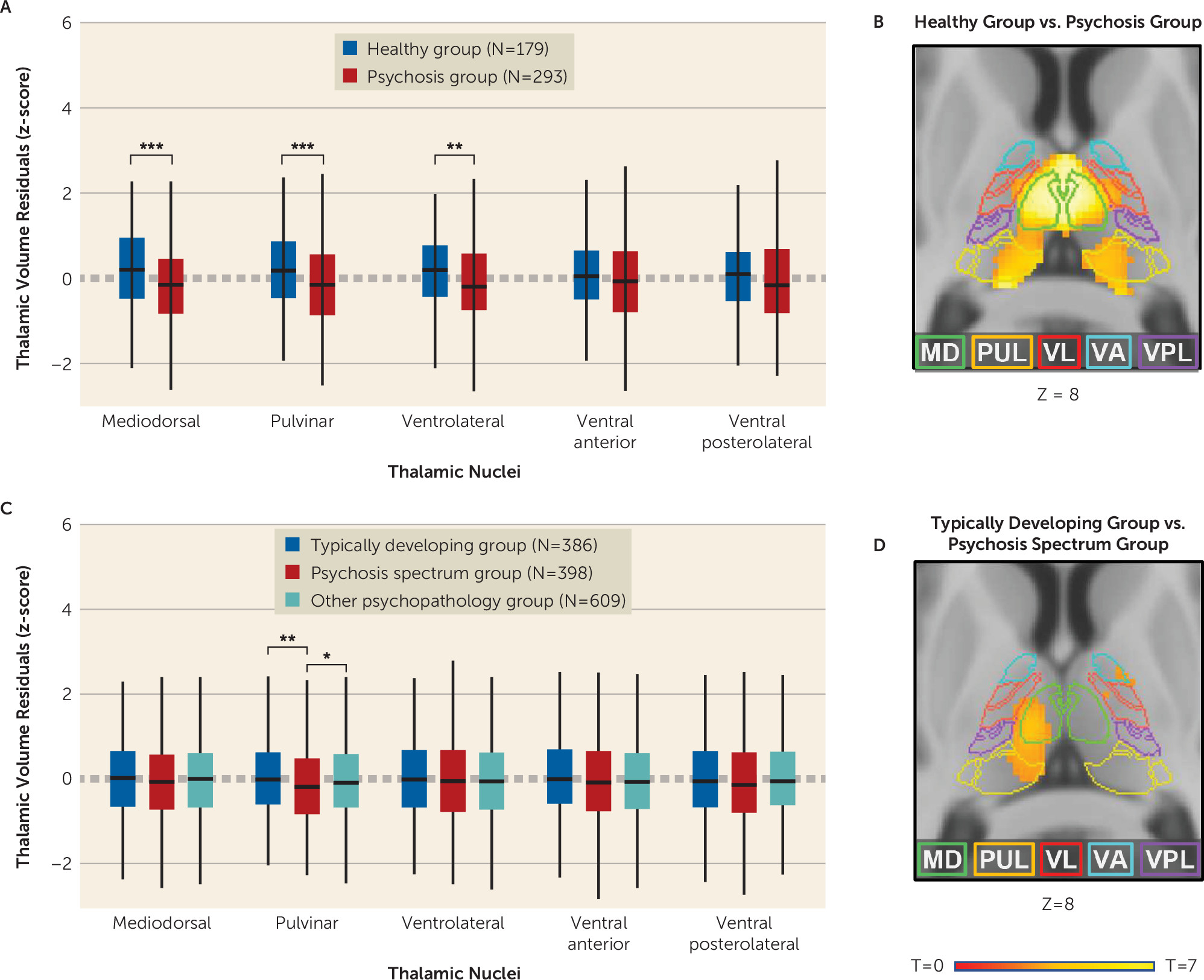
There was a significant nuclei-by-group interaction (F=6.894, df=4, 1860, p<0.001) and a main effect of group (F=14.573, df=1, 465, p<0.001), indicating differential volume reduction across the five thalamic nuclei superimposed on a general decrease in thalamic volumes in psychosis. As shown in Figure 1A and Table 3, mediodorsal, pulvinar, and ventrolateral nuclei volumes were smaller in the psychosis group (2.60% [p<0.001], 2.74% [p<0.001], and 1.94% [p=0.006] smaller, respectively). See the online supplement for complete statistical results.
FIGURE 1. Thalamic nuclei volumes in the psychosis cohort and Philadelphia Neurodevelopmental Cohorta
a In panel A, individuals with psychosis show significantly smaller thalamus volumes in the mediodorsal (MD), pulvinar (PUL), and ventrolateral (VL) nuclei, but not the ventral anterior (VA) or ventral posterolateral (VPL) nuclei, as observed in standardized nuclei volumes from FreeSurfer. In panel B, the finding of smaller thalamus volumes in individuals with psychosis is supported by a voxel-based morphometry (VBM) analysis. In panel C, youths with psychosis spectrum symptoms show smaller pulvinar volumes compared with both typically developing youths and youths with other psychopathology in standardized thalamic volumes from FreeSurfer. In panel D, VBM analyses show smaller thalamic volume in youths with psychosis spectrum symptoms compared with typically developing youths. This cluster was less extensive than that observed in the psychosis cohort. VBM analyses are presented at a cluster-level corrected p threshold of 0.05 (family-wise error corrected), for a voxel-wise threshold of 0.001.
***p<0.001. **p<0.01. *p<0.05.
TABLE 3. Mean volumes of thalamic nuclei in the psychosis cohort and the Philadelphia Neurodevelopmental Cohort
 VolumeAnalysis
Cohort and StructureMean (mm3)SEMean (mm3)SEMean (mm3)SECohen’s fReduction (%)Reduction (%)p
Psychosis cohort
 Healthy group Psychosis group    In psychosis group  
Mediodorsal2,04110.71,9888.3  0.1822.60 <0.001
Pulvinar3,39118.73,29814.6  0.1822.74 <0.001
Ventrolateral2,89215.92,83612.4  0.1281.94 0.006
Ventral anterior8595.48474.2  0.0841.40 0.071
Ventral posterolateral1,6219.21,5997.2  0.0901.36 0.059
Philadelphia Neurodevelopment Cohort
 Typically developing group Psychosis spectrum group Other psychopathology group  In psychosis spectrum groupIn other psychopathology group 
Mediodorsal2,2289.52,2109.42,2187.50.0320.080.450.429
Pulvinar3,51817.43,44217.33,49813.70.0842.160.570.006
Ventrolateral3,04011.53,02711.43,0309.10.0320.430.330.681
Ventral anterior8943.78883.78882.90.0320.670.670.448
Ventral posterolateral1,7698.81,7538.71,7676.90.0450.900.110.343
Supplemental analyses examining the effects of psychotic disorder diagnosis (i.e., schizophrenia spectrum symptoms, bipolar disorder with psychotic features), hemisphere, age, and sex, as well as group-by-age and group-by-sex interactions, were performed and are reported in the online supplement. Briefly, the results were similar across psychotic disorders, and there was no evidence of group-by-hemisphere, group-by-age, or group-by-sex interactions. While no associations were hypothesized, we also examined the correlations between thalamus nuclei volumes and clinical symptoms of psychosis, including positive and negative symptoms. Briefly, none of the thalamus volumes correlated with clinical symptoms. See the online supplement for complete statistical results.

Voxel-based morphometry analysis.

As shown in Figure 1B, and consistent with the FreeSurfer-based results presented above, VBM analyses revealed lower gray matter volume in psychosis in a cluster encompassing the left and right mediodorsal and pulvinar and the left ventrolateral nuclei. Overlaying the results on the Krauth thalamus atlas indicated that the cluster covered large portions of the left and right mediodorsal (left, 84.5%; right, 92.9%), pulvinar (left, 67.8%; right, 60.6%), but much less of the ventrolateral (left, 16.2%; right, 11.7%) nuclei at a standard p=0.001 threshold. With increasingly stringent thresholds, the cluster continued to cover a portion of the mediodorsal nucleus and pulvinar, but not the ventrolateral nucleus. Detailed results from the VBM analysis are presented in the online supplement.

Thalamic Nuclei Volumes in Youths With Psychosis Spectrum Symptoms

Group differences.

There was a significant nuclei-by-group interaction (F=3.216, df=8, 5548, p=0.001), although not a significant main effect of group (F=2.646, df=2, 1387, p=0.07). As shown in Figure 1C and Table 3, post hoc tests indicated that only the pulvinar exhibited a main effect of group (F=5.206, df=2, 1387, p=0.006). Pulvinar volume was significantly smaller in youths with psychosis spectrum symptoms compared with both typically developing youths (2.16% smaller; p=0.002) and youths with other psychopathologies (1.60% smaller; p=0.01) but did not differ between typically developing youths and youths with other psychopathologies (0.57% smaller in youths with other psychopathologies; p=0.38). See the online supplement for complete statistical results.
Supplemental analyses examining the effects of hemisphere, age, and sex, as well as group-by-age and group-by-sex interactions, were performed and are presented in the online supplement. Briefly, there was no evidence for group-by-hemisphere, group-by-age, or group-by-sex interactions in any nucleus.

Sensitivity analysis.

To ensure that smaller pulvinar volumes in youths with psychosis spectrum symptoms were not driven by group differences in demographic variables (i.e., age, sex, race), we performed a sensitivity analysis by creating subsamples of the psychosis spectrum and typically developing youth groups matched for age, sex, and race. Matching was conducted using the MatchIt package, version 3.0.2 (42) in R, version 3.6.1 (R Core Team, 2019), and resulted in a final sample of 150 youths with psychosis spectrum symptoms and 133 typically developing youths. The main effect of group remained significant for the pulvinar, reflecting smaller pulvinar volume in the psychosis spectrum group (2.56% smaller, p=0.03). No other nucleus showed significant group effects. See the online supplement for complete details of the sensitivity analysis.

Voxel-based morphometry analysis.

As shown in Figure 1D, VBM analysis revealed significantly lower gray matter volume in youths with psychosis spectrum symptoms in a cluster in the left thalamus encompassing mediodorsal, pulvinar, and anterior nuclei and a cluster in the right thalamus with a peak in the anterior thalamus. Overlaying the results on the Krauth thalamus atlas indicated that lower thalamus volume in the psychosis spectrum group covered the left mediodorsal (34.3%), pulvinar (10.7%), and ventrolateral nuclei (22.6%). Detailed results of the VBM analysis are presented in the online supplement.

Thalamic Nuclei Volumes and Cognition in Psychotic Disorders and in Youths With Psychosis Spectrum Symptoms

Psychosis cohort.

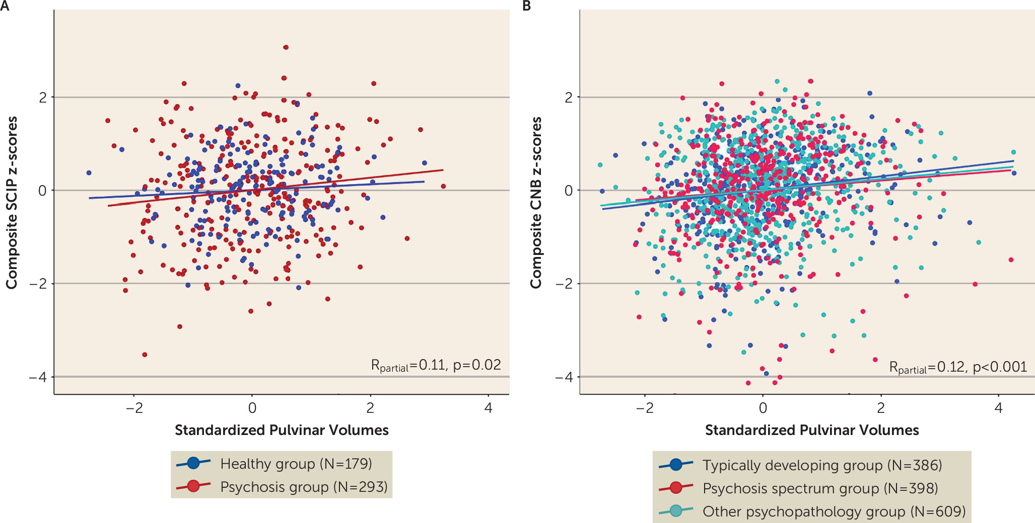
As shown in Figure 2A, overall cognitive function (i.e., Screen for Cognitive Impairment in Psychiatry composite z-score) correlated with pulvinar (Rpartial=0.111, p=0.02) but not mediodorsal nuclei volumes (Rpartial=0.087, p=0.06) after correction for multiple comparisons.
FIGURE 2. Correlation between pulvinar volumes and cognitive function in the psychosis cohort and Philadelphia Neurodevelopmental Cohorta
a Pulvinar volumes were significantly associated with overall cognitive function as measured with the Screen for Cognitive Impairment in Psychiatry (SCIP) composite z-score in the psychosis cohort (panel A) and the Computerized Neurocognitive Battery (CNB) composite z-score in the Philadelphia Neurodevelopmental Cohort (panel B).

PNC.

As shown in Figure 2B, overall neuropsychological functioning (i.e., Penn Computerized Neurocognitive Battery composite z-score) was positively correlated with pulvinar volumes (Rpartial=0.121, p<0.001) at the corrected statistical threshold, but not mediodorsal nuclei volumes (Rpartial=0.035, p=0.193).

Discussion

We examined thalamic nuclei volumes in a large cohort of individuals with psychotic disorders and a community-ascertained neurodevelopmental cohort, the PNC. We used a recently developed, validated method included in the FreeSurfer software package for segmenting thalamic nuclei on conventional T1-weighted MRI and complemented this approach with a VBM analysis.
The study findings contribute to our understanding of thalamic pathology in psychosis in several ways. First, they clarify the anatomical specificity of thalamic structural abnormalities in psychosis. Using complementary segmentation and voxel-based approaches, we found that lower thalamic volume was most pronounced in the mediodorsal and pulvinar nuclei. As touched upon earlier, findings from postmortem (10) and neuroimaging studies (2427) are mixed, likely because of a combination of several factors, including small sample sizes and, in the case of neuroimaging studies, variable methods used to measure specific thalamic nuclei. Our results support thalamic models of psychotic disorders that emphasize dysfunction among association nuclei.
Second, our finding that youths with psychosis spectrum symptoms also demonstrate smaller thalamic volumes is consistent with a neurodevelopmental basis for thalamic abnormalities in psychosis (14, 15). Disruption of the thalamus during development may affect cortical development in regions involved in the pathogenesis of psychosis (e.g., the prefrontal cortex). For instance, animal studies show that disrupted thalamus development is associated with lower cell density and volume in neuroanatomically connected cortical regions (43, 44). In our neurodevelopmental cohort, smaller pulvinar volume was specific to the psychosis spectrum group. Smaller pulvinar volumes were constant across age in both samples, suggesting that structural abnormalities in the pulvinar are present prior to the onset of psychosis. VBM analysis indicated that smaller volume in the psychosis spectrum group also encompassed a portion of the mediodorsal nucleus. Indeed, qualitative comparison of VBM results indicates that there is significant overlap in thalamic volume loss in the psychosis sample and in the psychosis spectrum youth sample, although, not surprisingly, volume loss is less extensive in the psychosis spectrum group.
While previous studies have identified smaller thalamic volume in youths exhibiting symptoms of psychosis, including individuals at clinical high risk (45, 46), the present results extend these findings in critical ways. First, we included a large cohort of individuals with psychotic disorders to compare concordance across the psychosis spectrum. Second, we examined volumes of specific thalamic nuclei, in contrast to most previous studies, which examined whole thalamus volumes only. Third, our sample size is considerably larger than those of many previous studies, including a recent study that used an earlier release of the PNC data that did not include the complete neuroimaging sample (N=997, compared with N=1,601 in the present study) and separated youths with psychosis spectrum symptoms only from those with psychosis and bipolar spectrum symptoms (47). Fourth, in contrast to all previous studies (47, 48), we established that smaller thalamic volume is specific to youths with psychosis spectrum symptoms by including a large sample of youths with other psychopathology. Finally, we examined associations between thalamus volume and cognitive function.
One major implication of smaller thalamic nuclei volumes in both psychosis and in youths with psychosis spectrum symptoms is that this pathological abnormality contributes to the cognitive impairment considered to be a core feature of psychotic disorders (49). This is supported in our data, as thalamic nuclei volumes, and of the pulvinar specifically, are associated with general cognitive function. Moreover, the association, while modest, was strikingly similar across the psychosis and youth cohorts. Previous studies have found that whole thalamus volume and pulvinar-cortex covariance predict cognition in schizophrenia (50, 51). The pulvinar is intimately involved in cognitive functions, particularly as it relates to flexible, goal-oriented direction of attention (30). The pulvinar also has a role in synchronizing cortical activity during attention (52, 53), and lesions of the pulvinar reduce attention-related signals in the cortex (53), suggesting that it may influence cognition through thalamo-cortical interactions. Inappropriate modulation of attention through cortical networks that include the thalamus (e.g., the salience network) is hypothesized as a core deficit in schizophrenia that impairs the integrity of sensory information and context processing to control goal-directed behavior (54). Furthermore, positron emission tomography studies in schizophrenia have demonstrated reduced dopamine D2 receptor binding in the pulvinar and mediodorsal nucleus specifically, with lower binding being associated with more severe psychotic symptoms (55, 56).
Our study had several strengths, including large sample sizes, complementary methods for measuring thalamic volumes, and convergent findings across both methods and cohorts. Nevertheless, there are several limitations. While our use of an automated segmentation technique allowed us to obtain a well-validated segmentation of larger thalamic nuclei, we did not include several nuclei because of their small size and poor contrast in standard 1-mm3 T1 imaging, some of which may be relevant to psychosis, such as the lateral and medial geniculate nuclei. Another limitation was our use of cross-sectional samples, which limits our ability to investigate changes in the volumes of thalamic nuclei over time in individuals with psychotic disorders and youths with psychosis spectrum symptoms.

Conclusions

Thalamic association nuclei, including the pulvinar and mediodorsal nuclei, are smaller in individuals with psychotic disorders. Our findings also demonstrate that in a large cohort of youths with psychosis risk, as compared with youths with other psychopathology and taking into account specific thalamic nuclei, smaller thalamic association nuclei volumes represent a neurodevelopmental abnormality associated with cognitive impairment and higher risk for developing a psychotic disorder. Identification of specific thalamic nuclei affected in psychosis provides potential targets in the treatment of psychosis and cognitive impairment in psychosis with new neuromodulation technology, such as focused ultrasound therapy (57).

Acknowledgments

This work was conducted in part using the resources of the Center for Computational Imaging at the Vanderbilt University Institute of Imaging Sciences and the Advanced Computing Center for Research and Education at Vanderbilt University.
The authors thank the individuals who participated in the study, as well as Kristan Armstrong, Erin Brosey, Molly Boyce, Victoria Fox, Yasmeen Iqbal, Margo Menkes, Austin Woolard, Katherine Seldin, and Margee Quinn for their assistance in recruiting and screening study participants.
Supplementary Material

File (appi.ajp.2020.19101099.ds001.pdf)

References

1.
Brugger S, Davis JM, Leucht S, et al: Proton magnetic resonance spectroscopy and illness stage in schizophrenia: a systematic review and meta-analysis. Biol Psychiatry 2011; 69:495–503
2.
Giraldo-Chica M, Rogers BP, Damon SM, et al: Prefrontal-thalamic anatomical connectivity and executive cognitive function in schizophrenia. Biol Psychiatry 2018; 83:509–517
3.
Giraldo-Chica M, Woodward ND: Review of thalamocortical resting-state fMRI studies in schizophrenia. Schizophr Res 2017; 180:58–63
4.
Ramsay IS: An activation likelihood estimate meta-analysis of thalamocortical dysconnectivity in psychosis. Biol Psychiatry Cogn Neurosci Neuroimaging 2019; 4:859–869
5.
Minzenberg MJ, Laird AR, Thelen S, et al: Meta-analysis of 41 functional neuroimaging studies of executive function in schizophrenia. Arch Gen Psychiatry 2009; 66:811–822
6.
Huang AS, Rogers BP, Woodward ND: Disrupted modulation of thalamus activation and thalamocortical connectivity during dual task performance in schizophrenia. Schizophr Res 2019; 210:270–277
7.
Adriano F, Spoletini I, Caltagirone C, et al: Updated meta-analyses reveal thalamus volume reduction in patients with first-episode and chronic schizophrenia. Schizophr Res 2010; 123:1–14
8.
van Erp TGM, Hibar DP, Rasmussen JM, et al: Subcortical brain volume abnormalities in 2028 individuals with schizophrenia and 2540 healthy controls via the ENIGMA consortium. Mol Psychiatry 2016; 21:547–553
9.
Hibar DP, Westlye LT, van Erp TGM, et al: Subcortical volumetric abnormalities in bipolar disorder. Mol Psychiatry 2016; 21:1710–1716
10.
Dorph-Petersen K-A, Lewis DA: Postmortem structural studies of the thalamus in schizophrenia. Schizophr Res 2017; 180:28–35
11.
Parnaudeau S, O’Neill P-K, Bolkan SS, et al: Inhibition of mediodorsal thalamus disrupts thalamofrontal connectivity and cognition. Neuron 2013; 77:1151–1162
12.
Buchmann A, Dentico D, Peterson MJ, et al: Reduced mediodorsal thalamic volume and prefrontal cortical spindle activity in schizophrenia. Neuroimage 2014; 102:540–547
13.
Baran B, Karahanoğlu FI, Mylonas D, et al: Increased thalamocortical connectivity in schizophrenia correlates with sleep spindle deficits: evidence for a common pathophysiology. Biol Psychiatry Cogn Neurosci Neuroimaging 2019; 4:706–714
14.
Andreasen NC: The role of the thalamus in schizophrenia. Can J Psychiatry 1997; 42:27–33
15.
Jones EG: Cortical development and thalamic pathology in schizophrenia. Schizophr Bull 1997; 23:483–501
16.
Sim K, Cullen T, Ongur D, et al: Testing models of thalamic dysfunction in schizophrenia using neuroimaging. J Neural Transm (Vienna) 2006; 113:907–928
17.
Pergola G, Selvaggi P, Trizio S, et al: The role of the thalamus in schizophrenia from a neuroimaging perspective. Neurosci Biobehav Rev 2015; 54:57–75
18.
Steullet P: Thalamus-related anomalies as candidate mechanism-based biomarkers for psychosis. Schizophr Res (Online ahead of print, May 27, 2019)
19.
Byne W, Buchsbaum MS, Mattiace LA, et al: Postmortem assessment of thalamic nuclear volumes in subjects with schizophrenia. Am J Psychiatry 2002; 159:59–65
20.
Highley JR, Walker MA, Crow TJ, et al: Low medial and lateral right pulvinar volumes in schizophrenia: a postmortem study. Am J Psychiatry 2003; 160:1177–1179
21.
Byne W, Fernandes J, Haroutunian V, et al: Reduction of right medial pulvinar volume and neuron number in schizophrenia. Schizophr Res 2007; 90:71–75
22.
Pakkenberg B: Pronounced reduction of total neuron number in mediodorsal thalamic nucleus and nucleus accumbens in schizophrenics. Arch Gen Psychiatry 1990; 47:1023–1028
23.
Young KA, Holcomb LA, Yazdani U, et al: Elevated neuron number in the limbic thalamus in major depression. Am J Psychiatry 2004; 161:1270–1277
24.
Gilbert AR, Rosenberg DR, Harenski K, et al: Thalamic volumes in patients with first-episode schizophrenia. Am J Psychiatry 2001; 158:618–624
25.
Kemether EM, Buchsbaum MS, Byne W, et al: Magnetic resonance imaging of mediodorsal, pulvinar, and centromedian nuclei of the thalamus in patients with schizophrenia. Arch Gen Psychiatry 2003; 60:983–991
26.
Horga G, Bernacer J, Dusi N, et al: Correlations between ventricular enlargement and gray and white matter volumes of cortex, thalamus, striatum, and internal capsule in schizophrenia. Eur Arch Psychiatry Clin Neurosci 2011; 261:467–476
27.
Pergola G, Trizio S, Di Carlo P, et al: Grey matter volume patterns in thalamic nuclei are associated with familial risk for schizophrenia. Schizophr Res 2017; 180:13–20
28.
Iglesias JE, Augustinack JC, Nguyen K, et al: A computational atlas of the hippocampal formation using ex vivo, ultra-high resolution MRI: application to adaptive segmentation of in vivo MRI. Neuroimage 2015; 115:117–137
29.
Iglesias JE, Insausti R, Lerma-Usabiaga G, et al: A probabilistic atlas of the human thalamic nuclei combining ex vivo MRI and histology. Neuroimage 2018; 183:314–326
30.
Saalmann YB, Kastner S: Cognitive and perceptual functions of the visual thalamus. Neuron 2011; 71:209–223
31.
Rikhye RV, Wimmer RD, Halassa MM: Toward an integrative theory of thalamic function. Annu Rev Neurosci 2018; 41:163–183
32.
Ouhaz Z, Fleming H, Mitchell AS: Cognitive functions and neurodevelopmental disorders involving the prefrontal cortex and mediodorsal thalamus. Front Neurosci 2018; 12:33
33.
Purdon SE: The Screen for Cognitive Impairment in Psychiatry (SCIP): Instructions and Three Alternate Forms. Edmonton, Alberta, PNL Inc, 2005
34.
Roalf DR, Quarmley M, Calkins ME, et al: Temporal lobe volume decrements in psychosis spectrum youths. Schizophr Bull 2017; 43:601–610
35.
Gur RC, Richard J, Calkins ME, et al: Age group and sex differences in performance on a computerized neurocognitive battery in children age 8–21. Neuropsychology 2012; 26:251–265
36.
Harrigan RL, Yvernault BC, Boyd BD, et al: Vanderbilt University Institute of Imaging Science Center for Computational Imaging XNAT: a multimodal data archive and processing environment. Neuroimage 2016; 124(Pt B):1097–1101
37.
Huo Y, Blaber J, Damon SM, et al: Towards portable large-scale image processing with high-performance computing. J Digit Imaging 2018; 31:304–314
38.
Sochat VV, Prybol CJ, Kurtzer GM: Enhancing reproducibility in scientific computing: metrics and registry for Singularity containers. PLoS One 2017; 12:e0188511
39.
Dale AM, Fischl B, Sereno MI: Cortical surface-based analysis, I: segmentation and surface reconstruction. Neuroimage 1999; 9:179–194
40.
Fischl B, Salat DH, Busa E, et al: Whole brain segmentation: automated labeling of neuroanatomical structures in the human brain. Neuron 2002; 33:341–355
41.
Krauth A, Blanc R, Poveda A, et al: A mean three-dimensional atlas of the human thalamus: generation from multiple histological data. Neuroimage 2010; 49:2053–2062
42.
Ho DE, Imai K, King G, et al: MatchIt: nonparametric preprocessing for parametric causal inference. Journal of Statistical Software 2011; 42:1–28 (https://r.iq.harvard.edu/docs/matchit/2.4-18/matchit.pdf)
43.
Ouhaz Z, Ba-M’hamed S, Mitchell AS, et al: Behavioral and cognitive changes after early postnatal lesions of the rat mediodorsal thalamus. Behav Brain Res 2015; 292:219–232
44.
Selemon LD, Wang L, Nebel MB, et al: Direct and indirect effects of fetal irradiation on cortical gray and white matter volume in the macaque. Biol Psychiatry 2005; 57:83–90
45.
Lunsford-Avery JR, Orr JM, Gupta T, et al: Sleep dysfunction and thalamic abnormalities in adolescents at ultra high-risk for psychosis. Schizophr Res 2013; 151:148–153
46.
Harrisberger F, Buechler R, Smieskova R, et al: Alterations in the hippocampus and thalamus in individuals at high risk for psychosis. NPJ Schizophr 2016; 2:16033
47.
Jalbrzikowski M, Freedman D, Hegarty CE, et al: Structural brain alterations in youth with psychosis and bipolar spectrum symptoms. J Am Acad Child Adolesc Psychiatry 2019; 58:1079–1091
48.
Jacobs GR, Ameis SH, Ji JL, et al: Developmentally divergent sexual dimorphism in the cortico-striatal-thalamic-cortical psychosis risk pathway. Neuropsychopharmacology 2019; 44:1649–1658
49.
Elvevag B, Goldberg TE: Cognitive impairment in schizophrenia is the core of the disorder. Crit Rev Neurobiol 2000; 14:1–21
50.
Ramsay IS, Fryer S, Boos A, et al: Response to targeted cognitive training correlates with change in thalamic volume in a randomized trial for early schizophrenia. Neuropsychopharmacology 2018; 43:590–597
51.
Mitelman SA, Byne W, Kemether EM, et al: Correlations between volumes of the pulvinar, centromedian, and mediodorsal nuclei and cortical Brodmann’s areas in schizophrenia. Neurosci Lett 2006; 392:16–21
52.
Saalmann YB, Pinsk MA, Wang L, et al: The pulvinar regulates information transmission between cortical areas based on attention demands. Science 2012; 337:753–756
53.
Zhou H, Schafer RJ, Desimone R: Pulvinar-cortex interactions in vision and attention. Neuron 2016; 89:209–220
54.
Palaniyappan L, Simmonite M, White TP, et al: Neural primacy of the salience processing system in schizophrenia. Neuron 2013; 79:814–828
55.
Yasuno F, Suhara T, Okubo Y, et al: Low dopamine d(2) receptor binding in subregions of the thalamus in schizophrenia. Am J Psychiatry 2004; 161:1016–1022
56.
Kessler RM, Woodward ND, Riccardi P, et al: Dopamine D2 receptor levels in striatum, thalamus, substantia nigra, limbic regions, and cortex in schizophrenic subjects. Biol Psychiatry 2009; 65:1024–1031
57.
Legon W, Ai L, Bansal P, et al: Neuromodulation with single-element transcranial focused ultrasound in human thalamus. Hum Brain Mapp 2018; 39:1995–2006

Information & Authors

Information

Published In

Go to American Journal of Psychiatry
American Journal of Psychiatry
Pages: 1159 - 1167
PubMed: 32911995

History

Received: 28 October 2019
Revision received: 7 February 2020
Revision received: 27 April 2020
Accepted: 11 May 2020
Published online: 11 September 2020
Published in print: December 01, 2020

Keywords

  1. Schizophrenia Spectrum and Other Psychotic Disorders
  2. Neuroanatomy
  3. Bipolar and Related Disorders
  4. Bipolar II Disorder
  5. Cognition/Learning/Memory

Authors

Details

Anna S. Huang, Ph.D [email protected]
Department of Psychiatry and Behavioral Sciences, Vanderbilt University Medical Center, Nashville (Huang, Sheffield, Blackford, Heckers, Woodward); Vanderbilt University Institute of Imaging Sciences, Nashville (Rogers); Department of Psychiatry, University of Pittsburgh, Pittsburgh (Jalbrzikowski); Department of Psychiatry, Yale University School of Medicine, New Haven, Conn. (Anticevic); Research and Development, Department of Veterans Affairs Medical Center, Nashville (Blackford).
Baxter P. Rogers, Ph.D
Department of Psychiatry and Behavioral Sciences, Vanderbilt University Medical Center, Nashville (Huang, Sheffield, Blackford, Heckers, Woodward); Vanderbilt University Institute of Imaging Sciences, Nashville (Rogers); Department of Psychiatry, University of Pittsburgh, Pittsburgh (Jalbrzikowski); Department of Psychiatry, Yale University School of Medicine, New Haven, Conn. (Anticevic); Research and Development, Department of Veterans Affairs Medical Center, Nashville (Blackford).
Julia M. Sheffield, Ph.D
Department of Psychiatry and Behavioral Sciences, Vanderbilt University Medical Center, Nashville (Huang, Sheffield, Blackford, Heckers, Woodward); Vanderbilt University Institute of Imaging Sciences, Nashville (Rogers); Department of Psychiatry, University of Pittsburgh, Pittsburgh (Jalbrzikowski); Department of Psychiatry, Yale University School of Medicine, New Haven, Conn. (Anticevic); Research and Development, Department of Veterans Affairs Medical Center, Nashville (Blackford).
Maria E. Jalbrzikowski, Ph.D
Department of Psychiatry and Behavioral Sciences, Vanderbilt University Medical Center, Nashville (Huang, Sheffield, Blackford, Heckers, Woodward); Vanderbilt University Institute of Imaging Sciences, Nashville (Rogers); Department of Psychiatry, University of Pittsburgh, Pittsburgh (Jalbrzikowski); Department of Psychiatry, Yale University School of Medicine, New Haven, Conn. (Anticevic); Research and Development, Department of Veterans Affairs Medical Center, Nashville (Blackford).
Alan Anticevic, Ph.D
Department of Psychiatry and Behavioral Sciences, Vanderbilt University Medical Center, Nashville (Huang, Sheffield, Blackford, Heckers, Woodward); Vanderbilt University Institute of Imaging Sciences, Nashville (Rogers); Department of Psychiatry, University of Pittsburgh, Pittsburgh (Jalbrzikowski); Department of Psychiatry, Yale University School of Medicine, New Haven, Conn. (Anticevic); Research and Development, Department of Veterans Affairs Medical Center, Nashville (Blackford).
Jennifer Urbano Blackford, Ph.D
Department of Psychiatry and Behavioral Sciences, Vanderbilt University Medical Center, Nashville (Huang, Sheffield, Blackford, Heckers, Woodward); Vanderbilt University Institute of Imaging Sciences, Nashville (Rogers); Department of Psychiatry, University of Pittsburgh, Pittsburgh (Jalbrzikowski); Department of Psychiatry, Yale University School of Medicine, New Haven, Conn. (Anticevic); Research and Development, Department of Veterans Affairs Medical Center, Nashville (Blackford).
Stephan Heckers, M.D.
Department of Psychiatry and Behavioral Sciences, Vanderbilt University Medical Center, Nashville (Huang, Sheffield, Blackford, Heckers, Woodward); Vanderbilt University Institute of Imaging Sciences, Nashville (Rogers); Department of Psychiatry, University of Pittsburgh, Pittsburgh (Jalbrzikowski); Department of Psychiatry, Yale University School of Medicine, New Haven, Conn. (Anticevic); Research and Development, Department of Veterans Affairs Medical Center, Nashville (Blackford).
Neil D. Woodward, Ph.D
Department of Psychiatry and Behavioral Sciences, Vanderbilt University Medical Center, Nashville (Huang, Sheffield, Blackford, Heckers, Woodward); Vanderbilt University Institute of Imaging Sciences, Nashville (Rogers); Department of Psychiatry, University of Pittsburgh, Pittsburgh (Jalbrzikowski); Department of Psychiatry, Yale University School of Medicine, New Haven, Conn. (Anticevic); Research and Development, Department of Veterans Affairs Medical Center, Nashville (Blackford).

Notes

Send correspondence to Dr. Huang ([email protected]).

Competing Interests

Dr. Anticevic has served as a consultant and as a scientific board member for, and holds equity in, BlackThorn Therapeutics. The other authors report no financial relationships with commercial interests.

Funding Information

National Center for Research Resourceshttp://dx.doi.org/10.13039/100000097: 1-UL-1-TR000445
National Institute of Mental Healthhttp://dx.doi.org/10.13039/100000025: R01 MH070560, R01 MH102266, R01 MH115000
The Jack Martin, MD, Research Professorship in Psychopharmacology:
Charlotte and Donald Test Fund:
Supported by NIMH grants R01 MH102266 (to Dr. Woodward), R01 MH115000 (to Dr. Woodward and Dr. Anticevic), and R01 MH070560 (to Dr. Heckers); the Charlotte and Donald Test Fund; the Jack Martin, M.D., Research Professorship in Psychopharmacology (to Dr. Blackford); and the Vanderbilt Institute for Clinical and Translational Research (through grant 1-UL-1-TR000445 from the National Center for Research Resources/NIH).

Metrics & Citations

Metrics

Citations

Export Citations

If you have the appropriate software installed, you can download article citation data to the citation manager of your choice. Simply select your manager software from the list below and click Download.

For more information or tips please see 'Downloading to a citation manager' in the Help menu.

Format
Citation style
Style
Copy to clipboard

View Options

View options

PDF/EPUB

View PDF/EPUB

Login options

Already a subscriber? Access your subscription through your login credentials or your institution for full access to this article.

Personal login Institutional Login Open Athens login
Purchase Options

Purchase this article to access the full text.

PPV Articles - American Journal of Psychiatry

PPV Articles - American Journal of Psychiatry

Not a subscriber?

Subscribe Now / Learn More

PsychiatryOnline subscription options offer access to the DSM-5-TR® library, books, journals, CME, and patient resources. This all-in-one virtual library provides psychiatrists and mental health professionals with key resources for diagnosis, treatment, research, and professional development.

Need more help? PsychiatryOnline Customer Service may be reached by emailing [email protected] or by calling 800-368-5777 (in the U.S.) or 703-907-7322 (outside the U.S.).

Media

Figures

Other

Tables

Share

Share

Share article link

Share