Abstract

Objective:

Compulsive behaviors are a core feature of obsessive-compulsive spectrum disorders but appear across a broad spectrum of psychological conditions. It is thought that compulsions reflect a failure to override habitual behaviors “stamped in” through repeated practice and short-term distress reduction. Animal models suggest a possible causal role of the orbitofrontal cortex (OFC) in compulsive behaviors, but human studies have largely been limited by correlational designs. The goal of this study was to establish the first experimental evidence in humans for a mechanistic model in order to inform further experimental work and the eventual development of novel mechanistic treatments involving synergistic biological-behavioral pairings.

Methods:

After a baseline assessment, 69 individuals with compulsive behavior disorders were randomly assigned, in a double-blind, between-subjects design, to receive a single session of one of two active stimulation conditions targeting the left OFC: intermittent theta burst stimulation (iTBS), expected to increase OFC activity, or continuous TBS (cTBS), expected to decrease activity (both conditions, 600 pulses at 110% of target resting motor threshold). In both conditions, brain modulation was paired with a subsequent computer task providing practice in overriding a clinically relevant habit (an overlearned shock avoidance behavior), delivered during the expected window of OFC increase or decrease. Pre- and post-TBS functional MRI assessments were conducted of target engagement and compulsive behaviors performed in response to an idiographically designed stressful laboratory probe.

Results:

cTBS and iTBS modulated OFC activation in the expected directions. cTBS, relative to iTBS, exhibited a beneficial impact on acute laboratory assessments of compulsive behaviors 90 minutes after TBS. These acute behavioral effects persisted 1 week after cTBS.

Conclusions:

Experimental modulation of the OFC, within the behavioral context of habit override training, affected short-term markers of compulsive behavior vulnerability. The findings help delineate a causal translational model, serving as an initial precursor to mechanistic intervention development.
Compulsive behaviors are prominent across a wide range of psychiatric disorders, notably in compulsive behavior spectrum disorders (e.g., obsessive-compulsive disorder [OCD]), and are associated with marked impairment and staggering societal burden (1). In all their diverse manifestations, compulsive behaviors can be characterized as a failure of flexible goal-directed behavior to override habitual behaviors (2). While these behaviors may serve a function initially (e.g., short-term reduction of distress, perception of greater certainty), they typically become highly aversive and are recognized as contrary to life goals.
“Dual-system” theories of instrumental action (3) posit that the decision to perform an action is determined by the balance between the habitual system, which supports inflexible stimulus-response associations that are “stamped in” through repetition, and the goal-directed system, which supports flexible application of knowledge and motivational contingencies. Neuropsychological executive deficits in patients with OCD (e.g., deficits in set shifting, reversal learning, and response inhibition) are potentially consistent with a core imbalance in the ability to flexibly implement goal-directed behavior and override habit (4, 5). Recent studies have more specifically suggested that, in the context of habit formation, goal-directed behavior is impaired in patients with compulsive behaviors (e.g., 6, 7).
Both animal and human findings converge on alterations within cortico-striato-thalamo-cortical (CSTC) circuits as a key factor in compulsive behaviors and highlight one key structure in particular—the orbitofrontal cortex (OFC). However, ambiguity persists regarding the OFC’s role as either a driver or an overrider of compulsive behaviors. The dominant neurocognitive account of imaging findings in OCD suggests that greater activation of a “direct,” excitatory, feed-forward CSTC pathway relative to an “indirect,” inhibitory pathway results in overall hyperactivation of CSTC circuits (8, 9). This includes hyperactivity in the OFC at rest and during symptom provocation, one of the best-replicated neuroimaging patterns in OCD patients (10). However, decreased activation of the OFC is observed in OCD during some cognitive tasks (10), consistent with the OFC’s critical role in promoting goal-directed behavior. Furthermore, a large meta-analytic effort failed to identify consistent cortical thickness abnormalities in the OFC (11). Such inconsistencies likely reflect the inherent imprecision of correlational methodologies, functional MRI (fMRI) susceptibility artifacts within the OFC, heterogeneity among patients, and the OFC’s diverse and nonstatic role within CSTC loops, supporting a broad array of behavioral responses via pathways within and beyond CSTC circuits.
The correlational nature of both neural and neuropsychological findings in patients hinders the differentiation of neural patterns that are causal in producing compulsive behaviors from those that are effects or “epiphenomena.” In contrast, animal research techniques afford exquisite experimental precision in modulating neural circuitry relevant to compulsive behavior and measuring downstream behavioral effects. Such studies have demonstrated a causal role for the OFC in modulating compulsive behaviors and habit, yet the direction of effect remains ambiguous. Optogenetic studies in rodents (12) have established that brief but repeated activation of medial OFC-to-ventral striatum glutamatergic synapses induces excessive grooming behavior in mice (10, 13). Optogenetic disruption of activity in a nearby ventromedial prefrontal cortex (vmPFC) region blocked habit formation and expression in rats (14, 15), further suggesting that the OFC helps drive habit and compulsive behaviors. However, the OFC plays an equally critical role in promoting goal-directed behavior. Consequently, chemogenetic inhibition of the OFC can disrupt goal-directed behavior in mice, while optogenetic OFC activation increases goal-directed behavior (16) and inhibitory control over a habitual grooming response (17).
The direct relevance of these opposing mechanisms to humans who engage in problematic compulsive behaviors, and the ability to harness these insights in the development of novel treatments, remains unclear without a direct parallel test of causality in humans to explicate the OFC’s directional role. This is particularly critical given that cross-species comparisons suggest important discrepancies between findings with regard to the OFC’s functions and functional organization that make rodent homologues an imperfect approximation of humans (18). Theta burst stimulation (TBS) (19), a form of transcranial magnetic stimulation (TMS), has been used for noninvasive causal manipulations to acutely, experimentally manipulate brain function in two opposing directions. TBS may create a 50- to 60-minute window of increased (with intermittent “bursts” of stimulation; iTBS) or decreased (with continuous pulses; cTBS) neural activity using a potent, very brief (40–120 seconds total) approach. Although the majority of TBS studies have targeted the motor cortex, data supporting the safety, tolerability, and target engagement of iTBS and cTBS targeting the OFC and frontopolar cortex (FPC) have recently begun to accumulate in healthy volunteers (20) and clinical populations (21).
However, relative to animal models, limitations exist in the spatial and circuit-level precision of currently available methods for noninvasive stimulation in humans, which complicates hypothesis tests, given substantial functional heterogeneity within the OFC and neighboring FPC regions (22, 23). To overcome this, we used immediate post-TBS completion of a “habit override” task, thereby isolating the specific OFC functional mechanism that we aimed to augment, effectively aiming to “exercise” clinically relevant habit override abilities directly after stimulation.
We used a single session of iTBS and cTBS to test competing hypotheses relevant to the development of a comprehensive pathophysiological model of compulsive behaviors in patients—namely, that increased or decreased OFC activation might be beneficial, enhancing patients’ capacity to override compulsive behaviors. Given spatial diffusion of the TMS signal, with the goal of modulating the OFC at large, we selected a left OFC/FPC target (left Brodmann’s area [BA] 10, at its orbital junction with BA 11). This target was selected both in order to mimic the methods of previous TBS studies that provided crucial validation for the safety, tolerability, and effectiveness (i.e., target engagement) of modulating orbital areas using a standard TMS coil (24, 25) and for consistency with a previous TMS study in OCD patients that also targeted the left OFC/FPC and showed clinical benefit (26). To focus the effects of the stimulation and thereby enhance both the theoretical and the clinical implications of the findings, TBS was followed immediately by behavioral practice in a habit override task. This provided a behavioral context for the OFC/FPC manipulation and allowed us to ask the more specific question of what role the OFC plays in promoting—or interfering with—the acquisition of a highly clinically relevant skill (overriding avoidance habits). The primary study outcomes were cerebral blood flow in the OFC (target engagement) and participants’ acute capacity to override compulsive behaviors during an idiographic stressful laboratory probe, measured on the same day (90 minutes after TBS) and, in exploratory analyses, at 1-week follow-up. The study’s goal was to establish the first experimental evidence in humans for a mechanistic model in order to inform further experimental work and, ideally, the eventual development of novel mechanistic treatments involving synergistic biological-behavioral pairings (27).

Methods

Study Design

The study design (Figure 1) included parallel-arm randomization and patient and assessor blinding. Because our primary comparisons were for iTBS versus cTBS, all participants were allocated to one of these two arms, using a between-group design to eliminate crossover and practice effects. However, we used a baseline single-blind sham TBS lead-in to create parallel procedures at the baseline assessment. As detailed further below and in Figure 1, each of the two principal visits (sham-controlled baseline and active TBS) included pre-TBS habit acquisition, TBS (sham or active), habit override training, fMRI assessment of target engagement, a 45-minute break to allow acute TBS effects to dissipate, and a postacute (90 minutes after TBS) laboratory probe of compulsive behaviors. A 1-week follow-up assessment included the laboratory probe of compulsive behaviors as well as a repeat clinical interview. Clinical assessors were successfully blinded to treatment allocation (see the online supplement).
FIGURE 1. Flowchart illustrating the design of a theta burst stimulation (TBS) study of compulsive behaviorsa
a Based on TBS meta-analyses in the motor cortex (49), acute increases (with iTBS) and decreases (with cTBS) were expected to persist for approximately 50–60 minutes after the active TBS session, with large effect sizes peaking 10–15 minutes after TBS (i.e., during the habit override training task). Preliminary findings further suggested that effects of similar TBS protocols might extend to dorsolateral and medial PFC/OFC regions (20, 24, 5052). BOLD=blood-oxygen-level-dependent; CB probe=compulsive behavior probe; fMRI=functional MRI; MB-ME=multiband/multiecho; MPRAGE= magnetization-prepared rapid acquisition with gradient echo; OFC=orbitofrontal cortex; PFC=prefrontal cortex; pcASL=pseudo-continuous arterial spin labeling; TBS=theta burst stimulation; iTBS=intermittent theta burst stimulation; cTBS=continuous theta burst stimulation; Y-BOCS=Yale-Brown Obsessive Compulsive Scale, 2nd edition.

Participants

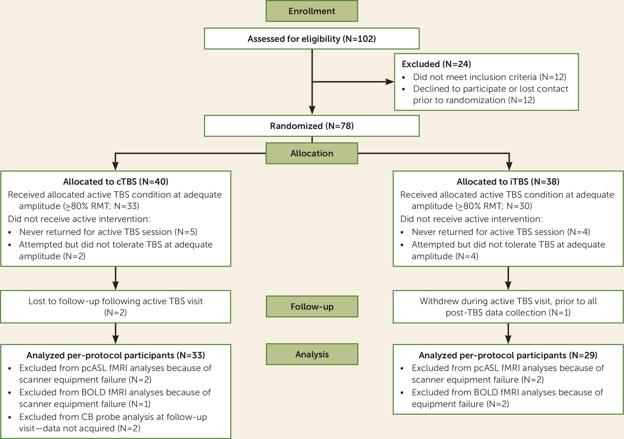
Seventy-eight adults (ages 18–55) underwent randomized assignment and completed all baseline assessment procedures, including the single-blind sham TBS session; of these, 69 participants returned to attempt active TBS (Figure 2). Two clinical inclusion criteria (see the online supplement for full details of inclusion and exclusion criteria) were used to identify clinically meaningful forms of compulsive behavior (i.e., both DSM-5 obsessive-compulsive and related disorders and “diagnostic orphans” with similar problematic compulsive behaviors): a score greater than one standard deviation above the published mean of healthy control subjects on at least one self-report scale of compulsive behaviors, and clinically significant compulsive behaviors per clinician rating on the compulsions subscale of the Yale-Brown Obsessive Compulsive Scale, 2nd edition (28). According to baseline diagnostic interviews, these transdiagnostic inclusion criteria yielded a sample with an average of 2.22 DSM-5 obsessive-compulsive and related disorders (see Table 1 for sample characteristics).
FIGURE 2. CONSORT flow diagram for a theta burst stimulation (TBS) study of compulsive behaviorsa
a As indicated by unpaired t tests (continuous variables) and chi-square tests (categorical variables), the participants excluded from all analyses (N=16) did not differ at baseline from the analyzed sample (N=62) on any neural or behavioral (compulsive behavior probe) outcome measure (p values >0.30) or on any demographic or clinical features reported in Table 1 (p values >0.24). No follow-up data were acquired for any participant who dropped out midway through the study, as we were unable to reach these participants for reassessment. BOLD=blood-oxygen-level-dependent; CB probe=compulsive behavior probe; cTBS=continuous TBS; fMRI=functional MRI; iTBS=intermittent TBS; pcASL=pseudo-continuous arterial spin labeling; RMT=resting motor threshold.
TABLE 1. Demographic and clinical characteristics of the sample included in the main analyses in a theta burst stimulation (TBS) study of compulsive behaviorsa
Characteristic or MeasureFull Analyzed Sample (N=62)cTBS Sample (N=33)iTBS Sample (N=29)
MeanSDMeanSDMeanSD
Age (years)29.859.1829.39.030.49.5
N%N%N%
Caucasian539027822690
Female467426792069
Taking psychotropic medication254013391240
Principal diagnosis
 Obsessive-compulsive disorder (OCD)325216491655
 Excoriation disorder19311133828
 Trichotillomania71139414
 Body dysmorphic disorder473913
OCD diagnosis (principal or secondary)609732972897
Comorbid depressive disorder233711331241
Generalized anxiety disorder19311030931
Eating disorder5726310
MeanSDMeanSDMeanSD
Y-BOCS
 Obsessions subscale score9.134.089.54.18.74.1
 Compulsions subscale scoreb12.793.0713.02.712.63.5
 Total score21.926.0830.35.829.76.4
a
No significant difference between the cTBS and iTBS groups on any variable (p values >0.43) or between the randomized (intent-to-treat) sample and the analyzed (per-protocol) sample (p values >0.24). cTBS=continuous TBS; iTBS=intermittent TBS; Y-BOCS=Yale-Brown Obsessive Compulsive Scale, 2nd edition.
b
Although the full Y-BOCS was administered for the purposes of clinical characterization, only the compulsions subscale, which captures transdiagnostic compulsive behavior severity, was utilized as a study inclusion criterion.

TBS

After determining resting motor threshold (RMT) through standard procedures (29), the TMS coil (a figure-8 MCF-B65; MagVenture, Denmark) was positioned stereotaxically over a navigational-system-identified left OFC/FPC anatomical target (see the online supplement for details on neuronavigation).
We followed clinical TMS conventions and adapted published protocols (25) to increase tolerability and comfort and achieved high compliance with TBS procedures, as observed in other clinical populations (21). This included a stimulation ramp-up block (600 pulses beginning at 0% and ramping up gradually over the entire block, such that few pulses were delivered at an efficacious amplitude; see the online supplement), followed by the active block (600 pulses), which was delivered at the target amplitude of 110% of RMT (as in previous OFC/FPC cTBS studies [24]) or at the maximum amplitude tolerated by the participant during ramp-up. Based on findings from TBS clinical studies targeting the prefrontal cortex (PFC) (30, 31) and electric field simulations (see the online supplement), a minimum tolerated amplitude of 85% of RMT was considered adequate for efficacious stimulation, and thus participants who completed the active TBS block at ≥85% of RMT (N=59 at 110%; N=4 at 86%−104%) were included in all analyses. For these participants, the TBS pulse amplitude as a percentage of the maximum stimulator output was 55.8% (SD=6.6; range=38−68) for cTBS and 52.6% (SD=5.9; range=41−65) for iTBS.
In all conditions, pulses were applied in a theta burst pattern (bursts of three stimuli at 50 Hz repeated at 5 Hz frequency) using a MagPro X100 stimulator (MagVenture). iTBS consisted of 20 trains, each lasting 2 seconds with intertrain intervals of 8 seconds, for a total of 192 seconds. cTBS consisted of one continuous train of 40 seconds. Sham TBS (delivered to all participants at the baseline session) utilized the participant’s allocated active TBS protocol over the same target, run at 20% of maximum stimulator output to provide a consistent absolute level of scalp stimulation and therefore sensation (32%−57% of RMT, below the threshold expected to induce reliable cortical effects). See Figure S2 in the online supplement for a representative computer simulation of the distribution of the electric field induced by TBS in the cortex.

Habit Override Training

During the expected window of TBS modulation (on the active TBS day, and during the same window on the sham TBS day), a habit override training task was administered, modeled after previous OCD research (6) (see Figure S1 in the online supplement). In brief, in habit acquisition (delivered prior to TBS), participants were instructed that their goal was to avoid receiving shocks to the left and right foot by pressing appropriate buttons whenever a conditioned cue appeared. Across 480 trials (3 seconds per trial, 24 minutes total), participants overlearned these avoidance behaviors. In the subsequent habit override task (240 trials, 3 seconds per trial, 12 minutes total), which was administered 15–30 minutes after TBS, the same pairs of cues were presented, but one of the two overlearned habits was “devalued.” A written and verbal instruction was provided informing participants which one of the two electrodes had been disconnected and stating that they should attempt to resist the relevant “devalued” avoidance response (i.e., override that habit), while continuing the remaining “valued” habit. As detailed in the online supplement, performance patterns conformed to expectations and suggested that overriding the shock-avoidance habit was challenging for participants.

fMRI Assessments

Scanning was performed in a Siemens 3-T Prisma Scanner, beginning approximately 25 minutes after TBS (sham or active TBS), immediately following completion of the habit override task, allowing all fMRI data collection to occur within the anticipated window of acute TBS modulation. During a 4-minute resting-state block, collected both at the baseline (post–sham TBS) and post–active TBS visits, two-dimensional pseudo-continuous arterial spin labeling (pcASL) was acquired (TR=4.9 seconds; TE=16 ms; 25 4-mm slices with 1-mm interslice gap; 3.28×3.28 mm voxels; labeling duration=1800 ms; postlabeling delay=1800 ms; 25 pairs of labeled/control acquisitions) and preprocessed as recommended using SPM’s ASL perfusion MRI signal processing toolbox (ASLtbx; details are provided in the online supplement). pcASL provides stable, interpretable quantification of absolute brain activation (cerebral blood flow) (32), with adequate test-retest reliability and convergent validity with positron emission tomography (33, 34). A broad prefrontal/anterior mask was defined by all voxels within the Montreal Neurological Institute template with y≥22. A voxel-wise search was then performed within this mask for clusters differentially modulated by TBS condition (voxel-wise p=0.005, map-wise p<0.05).
During an additional 7-minute resting-state block, blood-oxygen-level-dependent (BOLD) data were acquired with a multiband/multiecho (MB-ME) sequence optimized to capture BOLD signal in ventral/OFC regions (TR=1 second; TE=13.2, 38.76, and 64.32 ms; multiband factor=5; flip angle=49°; matrix=68×68; resolution=3-mm isotropic voxels; 40 oblique slices; 480 volumes), which has been found to substantially reduce susceptibility artifact and increases BOLD signal-to-noise ratio by up to 80% in the OFC (3537). Given budgetary and scanner time constraints, the MB-ME sequence was acquired only at the second MRI visit, after active iTBS or cTBS. At the first session (following sham TBS), a high-resolution structural scan (using MPRAGE [magnetization-prepared rapid acquisition with gradient echo]) was obtained instead, for input to the TMS neuronavigation system. We observed post hoc that this customized MB-ME sequence was indeed less susceptible to signal dropout in orbital areas relative to pcASL; thus, this secondary measure of left OFC target engagement was used strictly to confirm whether differential modulation extended to anatomically defined left BA 47 and 11, where pcASL signal dropout was observed.
After standard preprocessing in AFNI (details are provided in the online supplement), to quantify the intensity of spontaneous brain activity during the resting state (38), AFNI’s 3dRSFC tool was applied to quantify fractional amplitude of low-frequency (0.01–0.1 Hz) fluctuations (fALFF), a putative index of the absolute level of resting activation following TBS. Mean fALFF within a priori target regions (left BA 47 and 11, defined within AFNI’s TT_Daemon atlas) was extracted for each individual and compared across the two TBS conditions with an unpaired t test.

Laboratory-Triggered Compulsive Behavior Assessment

A laboratory probe of compulsive behavior vulnerability was completed at the baseline sham visit and the active TBS visit (90 minutes after TBS) and, as an exploratory probe of the durability of TBS’s impact, repeated at the 1-week follow-up visit. We idiographically designed, collaboratively with the participant, a laboratory task involving a triggering object, image, or scenario that corresponded to compulsive behaviors typical to the participant. The idiographic trigger was presented to the participant alone in a controlled laboratory environment, along with the means to engage in typical compulsive behaviors (e.g., hand sanitizer/sink; mirror for skin picking; necessary media for reassurance seeking/checking). To assess ability to voluntarily override compulsive behaviors, participants were instructed to confront the trigger and then resist or decrease compulsive behaviors to the extent possible, and were then left alone for 5 minutes. Posttask ratings of urges (0–100), effort (0–100), and success (i.e., total estimated duration of time spent engaged in compulsive behaviors) were collected. The ratings were correlated with scores on the compulsions subscale of the gold-standard Yale-Brown Obsessive Compulsive Scale (see Table S1 in the online supplement), suggesting that we captured clinically relevant symptoms, but on an immediate time scale suitable for sensitively capturing an acute clinical impact.
See the online supplement for details on our analytic strategy, including the rationale for the per-protocol analyses.

Results

Modulation of the OFC/FPC Target

Tolerability.

The two conditions were equally well tolerated; 94% (N=33/35) of participants tolerated cTBS stimulation at ≥85% of RMT, and 88% (N=30/34) tolerated iTBS (Fisher’s exact test, p=0.43) (further details are provided in the online supplement). No serious or unanticipated adverse events occurred.

pcASL.

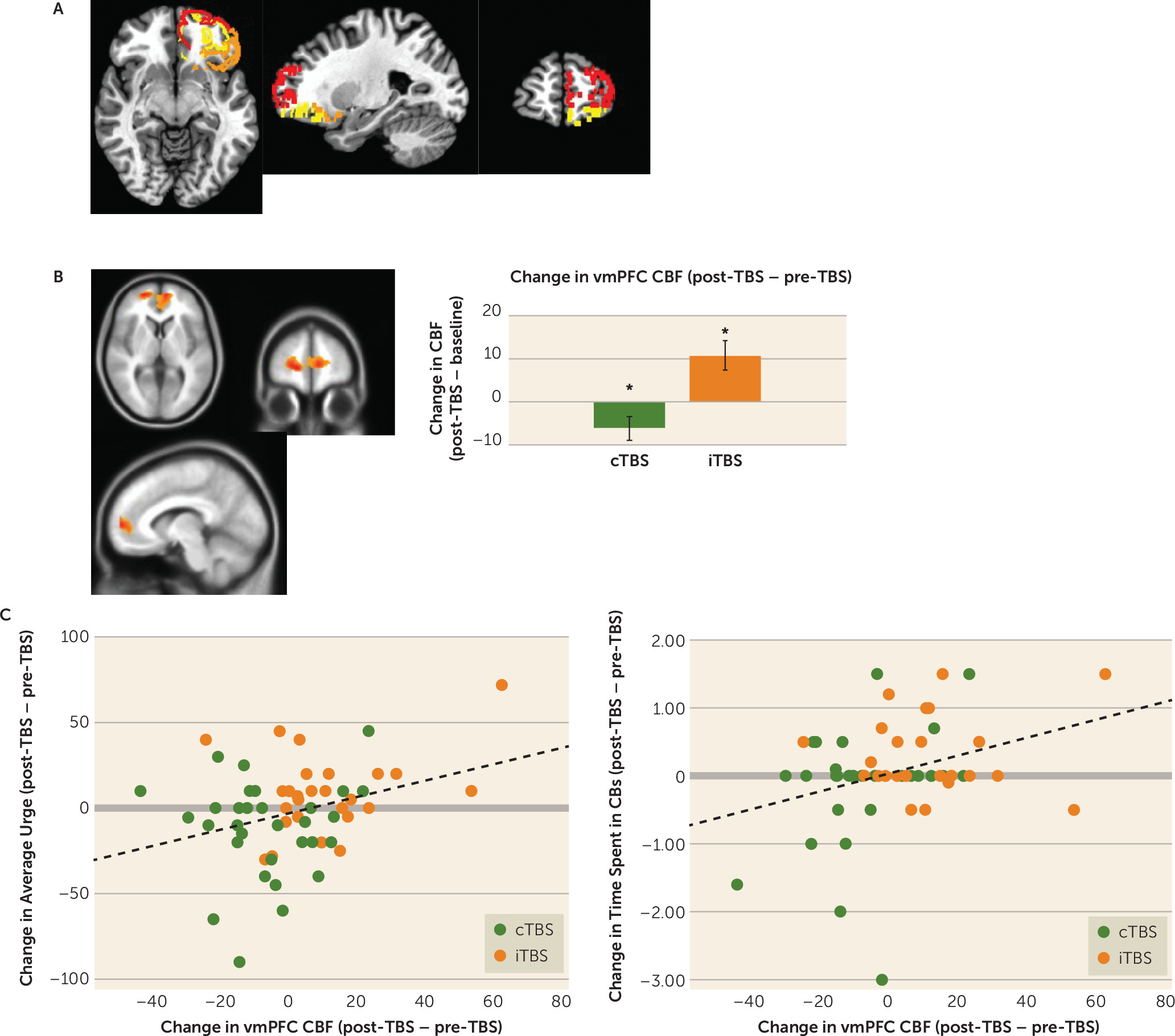
The pcASL sequence acquired both before (at the baseline/sham TBS visit) and after active cTBS/iTBS showed that TBS condition moderated pre- to post-TBS changes in cerebral blood flow in the target area of BA 10 and a surrounding vmPFC area (voxel-wise uncorrected p<0.005, N=736 voxels within PFC mask; map-wise corrected p<0.01; partial eta-squared=0.21) (Figure 3B), with the expected direction of modulation observed for both cTBS (decrease: post hoc paired t=−2.17, df=30, p=0.038; d=0.39) and iTBS (increase: paired t=3.14, df=26, p=0.004; d=0.68).
FIGURE 3. Theta burst stimulation (TBS) target and target engagement analyses in a TBS study of compulsive behaviorsa
a Panel A depicts the a priori anatomical stimulation target area (left orbitofrontal cortex/frontopolar cortex) and anatomical subregions. For participant-level neuronavigation on the active stimulation day, the target location reflecting a focal point of stimulation within the left Brodmann’s area [BA] 10 was verified on the basis of each participant’s individual structural MPRAGE (magnetization-prepared rapid acquisition with gradient echo) data acquired at the baseline scan (details are provided in the online supplement). BA 10 is shown in red, BA 11 in yellow, and BA 47 in orange. In panel B, a voxel-wise search, performed within a broad prefrontal cortex brain mask, identified a cluster in which the pre- to post-TBS change in cerebral blood flow (CBF), captured at rest with a pseudo-continuous arterial spin labeling (pcASL) sequence, was modulated by TBS condition (voxel-wise p<0.005, within-mask map-wise corrected p<0.01, N=736 voxels). The cluster spanned orbitofrontal/frontopolar (BA 10) and neighboring ventromedial prefrontal cortex (vmPFC) areas, with local maxima in both the left BA 10 (x=−8, y=56, z=4) and the right BA 10 (x=16, y=56, z=0). Within this cluster, according to post hoc paired t tests comparing the baseline scan to the post-TBS scan, continuous TBS (cTBS) significantly reduced cerebral blood flow (t=−2.17, df=30, p=0.038; d=0.39), and intermittent TBS (iTBS) significantly increased cerebral blood flow (t=3.14, df=26, p=0.004; d=0.68). Mean values are shown, and error bars indicate SEM. In panel C, scatterplots depict correlations between change scores (post-TBS at active TBS visit – baseline visit) in ventromedial prefrontal cortex (vmPFC) CBF (cluster depicted in panel B) and behavioral outcomes (urge strength, time spent in compulsive behaviors [CBs]) across all participants. Across individuals, larger reductions in CBF correlated with larger reductions in urge strength (r=0.32, uncorrected p=0.015, false discovery rate corrected [FDR] p=0.045) and time spent in compulsive behaviors (r=0.32, uncorrected p=0.015, pFDR=0.023).

fALFF.

Lower fALFF was observed following cTBS, relative to following iTBS, in the left BA 47 (t=−2.02, df=57, uncorrected p=0.048; Cohen’s d=0.53) but not the left BA 11 (t=0.27, df=57, uncorrected p=0.79; d=0.07).

Acute Effect of OFC/FPC Modulation on Compulsive Behaviors

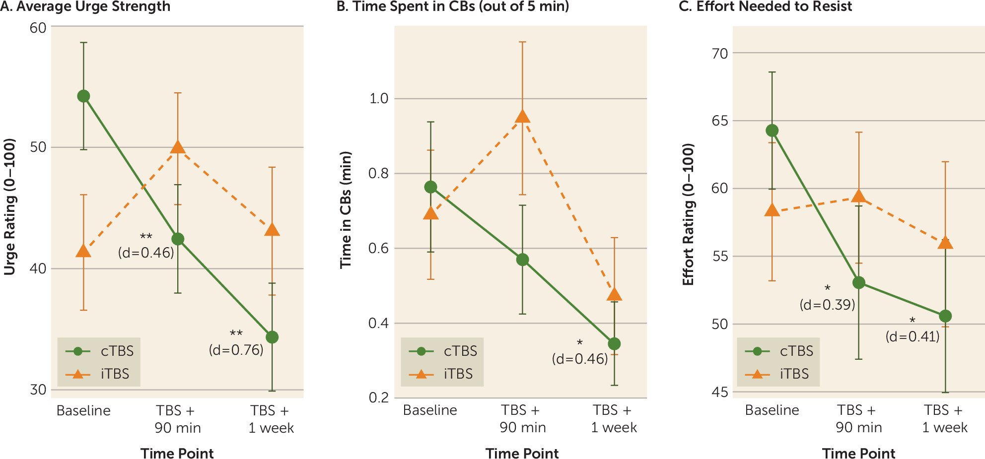
In linear mixed models focusing on the two primary time points (baseline and active TBS + 90 minutes), group-by-time interactions were observed in laboratory measures of compulsive behavior urge strength (β=20.32; t=3.28, df=60, uncorrected p=0.002, false discovery rate corrected [FDR] p=0.006; effect size r=0.39) and time spent engaged in idiographic compulsive behaviors (β=0.45; t=2.45, df=60, uncorrected p=0.017, pFDR=0.026; effect size r=0.30). For effort needed to resist compulsive behaviors, the interaction was nonsignificant (β=12.25; t=1.71, df=60, uncorrected p=0.093, pFDR=0.093; effect size r=0.22), although a significant pre- to post-TBS reduction was observed in the cTBS group (p=0.032; d=−0.39) but not the iTBS group (p=0.85; d=−0.04). Across all three indices (Figure 4), the pattern of change suggested that cTBS produced pre- to post-TBS improvements, while indices were unchanged or (nonsignificantly) worsened after iTBS.
FIGURE 4. Behavioral effects of theta burst stimulation (TBS) condition on laboratory probe measures in a TBS study of compulsive behaviorsa
a Among participants who received continuous TBS (cTBS) (N=33), relative to the baseline sham-TBS visit, active cTBS followed by habit override training led to decreased urges to complete compulsive behaviors (panel A), time spent performing compulsive behaviors (panel B), and effort needed to resist compulsive behaviors during a 5-minute laboratory probe (panel C), with significant effects persisting at a 1-week follow-up. Participants who received intermittent TBS (iTBS) (N=29) exhibited either nonsignificant (p values >0.07) increases (panels A, B) or no changes (panel C) from baseline at 90 minutes post-TBS, with no significant effects observed at the 1-week follow-up. Mean values are shown, and error bars indicate SEM. The variables in panels A–C did not differ significantly at baseline across the cTBS and iTBS samples (omnibus multivariate analysis of variance, F=1.3, df=3, 59, p=0.29; uncorrected p values for group difference on individual variables, 0.074–0.71). Asterisks indicate p values for pairwise comparisons within group, comparing each post-TBS time point to baseline value. Cohen’s d effect sizes, noted for all significant within-group changes, are calculated with Cohen’s classical formula (i.e., unadjusted for repeated measurement), which provides uniform, conservative quantification of standardized effect sizes independent of the study design.
*p<0.05. **p<0.01.
Across individuals, larger cerebral blood flow reductions in the vmPFC functional region of interest (Figure 3C) from pre- to post–active TBS correlated with larger reductions in urge strength (r=0.32, uncorrected p=0.015, pFDR=0.045) and time spent in compulsive behaviors (r=0.32, uncorrected p=0.015, pFDR=0.023), but not effort (r=−0.02, uncorrected p=0.88, pFDR=0.88).
In exploratory within-group analyses probing the durability of change at the 1-week follow-up, in the cTBS group, significant decreases from baseline were observed in urge strength (paired t=−3.39, df=30, uncorrected p=0.002, pFDR=0.006; d=0.76), time spent in compulsive behaviors (paired t=−2.51, df=30, uncorrected p=0.018, pFDR=0.027; d=0.46), and effort needed to resist (paired t=−2.28, df=30, uncorrected p=0.030, pFDR=0.03; d=0.51) (Figure 4), while no significant enduring effects were found for iTBS (uncorrected p values >0.31; d values, 0.07–0.25).

Discussion

In this study, we found that a single session of intermittent or continuous TBS was successful in 1) increasing and decreasing (respectively) cerebral blood flow within the left OFC/FPC (BA 10) and surrounding vmPFC areas (Figure 3), and 2) modulating the left ventrolateral OFC (BA47), according to a secondary index of spontaneous brain activity (fALFF). Furthermore, the single 40-second session of cTBS, paired with the context of behavioral training in habit override, reduced acute markers of clinically relevant compulsive behaviors measured in the laboratory, relative to iTBS (Figure 4). Acute in-laboratory behavioral improvements in the cTBS condition endured at 1-week follow-up, with medium to large effect sizes. Although this suggested possible durability that exceeded what would typically be expected following a single session of rTMS delivered in isolation (i.e., without a paired behavioral manipulation), effects of the brief, single-session manipulation did not extend to clinical symptom scales that measure more generalized and enduring symptomatic patterns (see the online supplement).
Our findings add to a growing body of published studies suggesting that brief, noninvasive TBS procedures can have an impact on resting or baseline activation in the OFC/FPC and nearby ventral medial PFC areas—even when using a standard figure-8 TMS coil (e.g., 24, 25). Notably, consistent with expected spatial diffusion of TMS signal, evidence of target engagement extended to a broader medial PFC area, suggesting modulation of a wider PFC network with the potential to influence a broad range of functions.
Neuroanatomical models of OCD, which center broadly on CSTC circuits, have provided important groundwork, but progress has been hindered by the inherent limitations of correlational (e.g., cross-sectional group comparison) designs in human studies. Here we circumvented that limitation by performing causal TBS studies, which led to findings consistent with animal experiments (e.g., optogenetic stimulation of the OFC produced excessive grooming in mice [10, 13]). By decreasing OFC/FPC activity in compulsive behavior patients via cTBS, we posit that we may have pulled a chronically hyperactive OFC state down from the “ceiling,” opening a window of opportunity for more reflexive OFC activation and adaptive, goal-directed learning to occur. Within this window of opportunity, behavioral training may provide critical opportunities to make clinical effects of neuromodulation more efficient, stronger, and/or more enduring. Similarly, a multisite randomized controlled trial of “deep TMS” targeting the medial PFC/anterior cingulate (39) suggested that active stimulation outperformed sham stimulation in reducing OCD symptoms, when both active and sham TMS were paired with the context of a prestimulation behavioral manipulation (brief exposure to a trigger). This led the U.S. Food and Drug Administration to approve marketing of deep rTMS to OCD patients, provided the behavioral exposure context protocol is specifically followed, under the assumption that this behavioral framing may be an influential factor. “State-dependent” neuromodulation (4042) may thus play a key role within emerging theory-driven, synergistic biological-behavioral treatments (27).
This study had several limitations. It was powered only to detect medium effect sizes or larger; yet several such effects were detected. Per-protocol analyses, which measure the effect of using (i.e., tolerating) a given treatment, have less real-world clinical relevance and greater possibility of selection bias than intent-to-treat analyses, which measure the effect of merely being assigned/randomized to a treatment; well-powered studies should confirm these initial findings using an intent-to-treat principle. Emerging motor physiology work suggests that the directionality of TBS effects is affected by a complex interaction between RMT, cortical depth, and number of pulses (e.g., 43), suggesting that future work would be optimized by a more idiographic and nuanced approach to dosimetry (e.g., 44). Furthermore, neural target engagement was quantified as single-point averages reflecting spontaneous/baseline activation in a priori regions, whereas TBS effects on network-level activation and functional connectivity may be broad and dynamic (45). Definitive experimental studies should include 1) both sham and cTBS treatment arms, with and without habit override training, to thoroughly assess key unanswered questions about necessary and sufficient intervention components; 2) larger samples permitting diagnosis-specific analyses, particularly in an effort to better delineate effects on relatively “compulsive” versus relatively “impulsive” behaviors (4648); and 3) healthy control subjects, to assess whether procedures “normalize” neural and/or behavioral patterns. Such rigorous experimental studies will serve as essential precursors to facilitate optimized, mechanistically targeted treatment development.

Conclusions

We posit that by decreasing drive toward habitual and compulsive behaviors, down-regulation of the OFC/FPC (via cTBS) may provide a window of opportunity in which to enhance goal-directed behavior, after first “muting” OFC-mediated habit system interference. Our novel, fully automated behavioral training in habit override may have leveraged this acute brain state, creating a short-term benefit observed specifically on idiographic compulsive behaviors, which persisted for 1 week. Our study results advance an integrative picture of both neural and behavioral pathophysiological processes that contribute to compulsive behaviors. Future studies would benefit from inclusion of crucial experimental controls (e.g., sham TBS, cTBS without habit override) to clarify the role that behavioral context may play in leveraging an acute OFC/FPC state manipulation. With further extension of these initial experimental findings, this line of inquiry could represent a first step toward developing and clinically optimizing neuromodulation therapies (e.g., multisession synergistic biobehavioral treatments) that are well grounded in basic experimental science and leverage acute neuroplasticity to maximize clinical benefit.

Acknowledgments

The authors gratefully acknowledge Daniel M. Blumberger, M.D., Sabine Wilhelm, Ph.D., the study participants, and all members of the Clinical Application of Neuroscience lab for their contributions to this work.

Footnote

ClinicalTrials.gov identifier: NCT03265015.

Supplementary Material

File (appi.ajp.2020.20060821.ds001.pdf)

References

1.
Eaton WW, Martins SS, Nestadt G, et al : The burden of mental disorders. Epidemiol Rev 2008 ; 30 : 1 – 14
2.
Gillan CM, Robbins TW, Sahakian BJ, et al : The role of habit in compulsivity. Eur Neuropsychopharmacol 2016 ; 26:828–840
3.
de Wit S, Dickinson A : Associative theories of goal-directed behaviour: a case for animal-human translational models. Psychol Res 2009 ; 73 : 463 – 476
4.
Abramovitch A, Abramowitz JS, Mittelman A : The neuropsychology of adult obsessive-compulsive disorder: a meta-analysis. Clin Psychol Rev 2013 ; 33 : 1163 – 1171
5.
Shin NY, Lee TY, Kim E, et al : Cognitive functioning in obsessive-compulsive disorder: a meta-analysis. Psychol Med 2014 ; 44 : 1121 – 1130
6.
Gillan CM, Apergis-Schoute AM, Morein-Zamir S, et al : Functional neuroimaging of avoidance habits in obsessive-compulsive disorder. Am J Psychiatry 2015 ; 172 : 284 – 293
7.
Gillan CM, Kosinski M, Whelan R, et al : Characterizing a psychiatric symptom dimension related to deficits in goal-directed control. Elife 2016 ; 5 : e11305
8.
Saxena S, Rauch SL : Functional neuroimaging and the neuroanatomy of obsessive-compulsive disorder. Psychiatr Clin North Am 2000 ; 23 : 563 – 586
9.
Pauls DL, Abramovitch A, Rauch SL, et al : Obsessive-compulsive disorder: an integrative genetic and neurobiological perspective. Nat Rev Neurosci 2014 ; 15 : 410 – 424
10.
Ahmari SE : Using mice to model obsessive compulsive disorder: from genes to circuits. Neuroscience 2016 ; 321 : 121 – 137
11.
Boedhoe PSW, Schmaal L, Abe Y, et al : Cortical abnormalities associated with pediatric and adult obsessive-compulsive disorder: findings from the ENIGMA Obsessive-Compulsive Disorder Working Group. Am J Psychiatry 2018 ; 175 : 453 – 462
12.
Deisseroth K : Optogenetics: 10 years of microbial opsins in neuroscience. Nat Neurosci 2015 ; 18 : 1213 – 1225
13.
Ahmari SE, Spellman T, Douglass NL, et al : Repeated cortico-striatal stimulation generates persistent OCD-like behavior. Science 2013 ; 340 : 1234 – 1239
14.
Smith KS, Virkud A, Deisseroth K, et al : Reversible online control of habitual behavior by optogenetic perturbation of medial prefrontal cortex. Proc Natl Acad Sci USA 2012 ; 109 : 18932 – 18937
15.
Smith KS, Graybiel AM : A dual operator view of habitual behavior reflecting cortical and striatal dynamics. Neuron 2013 ; 79 : 361 – 374
16.
Gremel CM, Costa RM : Orbitofrontal and striatal circuits dynamically encode the shift between goal-directed and habitual actions. Nat Commun 2013 ; 4 : 2264
17.
Burguière E, Monteiro P, Feng G, et al : Optogenetic stimulation of lateral orbitofronto-striatal pathway suppresses compulsive behaviors. Science 2013 ; 340 : 1243 – 1246
18.
Wallis JD : Cross-species studies of orbitofrontal cortex and value-based decision-making. Nat Neurosci 2011 ; 15 : 13 – 19
19.
Huang Y-Z, Edwards MJ, Rounis E, et al : Theta burst stimulation of the human motor cortex. Neuron 2005 ; 45 : 201 – 206
20.
Grossheinrich N, Rau A, Pogarell O, et al : Theta burst stimulation of the prefrontal cortex: safety and impact on cognition, mood, and resting electroencephalogram. Biol Psychiatry 2009 ; 65 : 778 – 784
21.
Hanlon CA, Philip NS, Price RB, et al : A case for the frontal pole as an empirically derived neuromodulation treatment target. Biol Psychiatry 2019 ; 85 : e13 – e14
22.
Ongür D, Price JL : The organization of networks within the orbital and medial prefrontal cortex of rats, monkeys, and humans. Cereb Cortex 2000 ; 10 : 206 – 219
23.
Kahnt T, Chang LJ, Park SQ, et al : Connectivity-based parcellation of the human orbitofrontal cortex. J Neurosci 2012 ; 32 : 6240 – 6250
24.
Hanlon CA, Dowdle LT, Austelle CW, et al : What goes up, can come down: novel brain stimulation paradigms may attenuate craving and craving-related neural circuitry in substance dependent individuals. Brain Res 2015 ; 1628 ( Pt A ): 199 – 209
25.
Hanlon CA, Dowdle LT, Correia B, et al : Left frontal pole theta burst stimulation decreases orbitofrontal and insula activity in cocaine users and alcohol users. Drug Alcohol Depend 2017 ; 178 : 310 – 317
26.
Ruffini C, Locatelli M, Lucca A, et al : Augmentation effect of repetitive transcranial magnetic stimulation over the orbitofrontal cortex in drug-resistant obsessive-compulsive disorder patients: a controlled investigation. Prim Care Companion J Clin Psychiatry 2009 ; 11 : 226 – 230
27.
Wilkinson ST, Holtzheimer PE, Gao S, et al : Leveraging neuroplasticity to enhance adaptive learning: the potential for synergistic somatic-behavioral treatment combinations to improve clinical outcomes in depression. Biol Psychiatry 2019 ; 85 : 454 – 465
28.
Storch EA, Rasmussen SA, Price LH, et al : Development and psychometric evaluation of the Yale-Brown Obsessive Compulsive Scale, Second Edition. Psychol Assess 2010 ; 22 : 223 – 232
29.
Rossini PM, Burke D, Chen R, et al : Non-invasive electrical and magnetic stimulation of the brain, spinal cord, roots, and peripheral nerves: basic principles and procedures for routine clinical and research application: an updated report from an IFCN Committee. Clin Neurophysiol 2015 ; 126 : 1071 – 1107
30.
Philip NS, Barredo J, Aiken E, et al : Theta-burst transcranial magnetic stimulation for posttraumatic stress disorder. Am J Psychiatry 2019 ; 176 : 939 – 948
31.
Li CT, Chen MH, Juan CH, et al : Efficacy of prefrontal theta-burst stimulation in refractory depression: a randomized sham-controlled study. Brain 2014 ; 137 : 2088 – 2098
32.
Chen JJ, Jann K, Wang DJJ : Characterizing resting-state brain function using arterial spin labeling. Brain Connect 2015 ; 5 : 527 – 542
33.
Heijtel DFR, Mutsaerts HJMM, Bakker E, et al : Accuracy and precision of pseudo-continuous arterial spin labeling perfusion during baseline and hypercapnia: a head-to-head comparison with 15O H2O positron emission tomography. Neuroimage 2014 ; 92 : 182 – 192
34.
Kilroy E, Apostolova L, Liu C, et al : Reliability of two-dimensional and three-dimensional pseudo-continuous arterial spin labeling perfusion MRI in elderly populations: comparison with 15O-water positron emission tomography. J Magn Reson Imaging 2014 ; 39 : 931 – 939
35.
Poser BA, Versluis MJ, Hoogduin JM, et al : BOLD contrast sensitivity enhancement and artifact reduction with multiecho EPI: parallel-acquired inhomogeneity-desensitized fMRI. Magn Reson Med 2006 ; 55 : 1227 – 1235
36.
Kundu P, Brenowitz ND, Voon V, et al : Integrated strategy for improving functional connectivity mapping using multiecho fMRI. Proc Natl Acad Sci USA 2013 ; 110 : 16187 – 16192
37.
Kim T, Zhao T, Bae KT : Enhancement of functional MRI signal at high-susceptibility regions of brain using simultaneous multiecho multithin-slice summation imaging technique. J Magn Reson Imaging 2016 ; 44 : 478 – 485
38.
Zou QH, Zhu CZ, Yang Y, et al : An improved approach to detection of amplitude of low-frequency fluctuation (ALFF) for resting-state fMRI: fractional ALFF. J Neurosci Methods 2008 ; 172 : 137 – 141
39.
Carmi L, Tendler A, Bystritsky A, et al : Efficacy and safety of deep transcranial magnetic stimulation for obsessive-compulsive disorder: a prospective multicenter randomized double-blind placebo-controlled trial. Am J Psychiatry 2019 ; 176:931–938
40.
Silvanto J, Pascual-Leone A : State-dependency of transcranial magnetic stimulation. Brain Topogr 2008 ; 21 : 1 – 10
41.
Silvanto J, Muggleton N, Walsh V : State-dependency in brain stimulation studies of perception and cognition. Trends Cogn Sci 2008 ; 12 : 447 – 454
42.
Micoulaud-Franchi JA, Fond G, Dumas G : Cyborg psychiatry to ensure agency and autonomy in mental disorders: a proposal for neuromodulation therapeutics. Front Hum Neurosci 2013 ; 7 : 463
43.
Chung SW, Hill AT, Rogasch NC, et al : Use of theta-burst stimulation in changing excitability of motor cortex: a systematic review and meta-analysis. Neurosci Biobehav Rev 2016 ; 63 : 43 – 64
44.
Beynel L, Davis SW, Crowell CA, et al : Site-specific effects of online rTMS during a working memory task in healthy older adults. Brain Sci 2020 ; 10 : E255
45.
Singh A, Erwin-Grabner T, Sutcliffe G, et al : Default mode network alterations after intermittent theta burst stimulation in healthy subjects. Transl Psychiatry 2020 ; 10 : 75
46.
Robbins TW, Vaghi MM, Banca P : Obsessive-compulsive disorder: puzzles and prospects. Neuron 2019 ; 102 : 27 – 47
47.
Fineberg NA, Potenza MN, Chamberlain SR, et al : Probing compulsive and impulsive behaviors, from animal models to endophenotypes: a narrative review. Neuropsychopharmacology 2010 ; 35 : 591 – 604
48.
Figee M, Pattij T, Willuhn I, et al : Compulsivity in obsessive-compulsive disorder and addictions. Eur Neuropsychopharmacol 2016 ; 26 : 856 – 868
49.
Wischnewski M, Schutter DJLG : Efficacy and time course of theta burst stimulation in healthy humans. Brain Stimul 2015 ; 8 : 685 – 692
50.
Prause N, Siegle GJ, Deblieck C, et al : EEG to primary rewards: predictive utility and malleability by brain stimulation. PLoS One 2016 ; 11 : e0165646
51.
Chistyakov AV, Rubicsek O, Kaplan B, et al : Safety, tolerability, and preliminary evidence for antidepressant efficacy of theta-burst transcranial magnetic stimulation in patients with major depression. Int J Neuropsychopharmacol 2010 ; 13 : 387 – 393
52.
Mastropasqua C, Bozzali M, Ponzo V, et al : Network based statistical analysis detects changes induced by continuous theta-burst stimulation on brain activity at rest. Front Psychiatry 2014 ; 5 : 97

Information & Authors

Information

Published In

Go to American Journal of Psychiatry
American Journal of Psychiatry
Pages: 459 - 468
PubMed: 33726523

History

Received: 9 June 2020
Revision received: 11 September 2020
Revision received: 19 October 2020
Accepted: 25 November 2020
Published online: 17 March 2021
Published in print: May 01, 2021

Keywords

  1. Theta Burst Stimulation
  2. Transcranial Magnetic Stimulation
  3. Compulsive Behaviors
  4. Obsessive-Compulsive Disorder
  5. Orbitofrontal Cortex
  6. Goal-Directed Behavior

Authors

Details

Rebecca B. Price, Ph.D. [email protected]
Department of Psychiatry, University of Pittsburgh, Pittsburgh (Price, Ferrarelli, Kim, Karim, Renard, Kaskie, Degutis, Wears, Brown, Siegle, Wallace, Ahmari); Department of Psychology, Trinity College Dublin, Dublin (Gillan); Department of Cancer Biology, Wake Forest University, Winston-Salem N.C. (Hanlon); Department of Psychiatry and Behavioral Sciences, Duke University, Durham N.C. (Vienneau, Peterchev).
Claire M. Gillan, Ph.D.
Department of Psychiatry, University of Pittsburgh, Pittsburgh (Price, Ferrarelli, Kim, Karim, Renard, Kaskie, Degutis, Wears, Brown, Siegle, Wallace, Ahmari); Department of Psychology, Trinity College Dublin, Dublin (Gillan); Department of Cancer Biology, Wake Forest University, Winston-Salem N.C. (Hanlon); Department of Psychiatry and Behavioral Sciences, Duke University, Durham N.C. (Vienneau, Peterchev).
Colleen Hanlon, Ph.D.
Department of Psychiatry, University of Pittsburgh, Pittsburgh (Price, Ferrarelli, Kim, Karim, Renard, Kaskie, Degutis, Wears, Brown, Siegle, Wallace, Ahmari); Department of Psychology, Trinity College Dublin, Dublin (Gillan); Department of Cancer Biology, Wake Forest University, Winston-Salem N.C. (Hanlon); Department of Psychiatry and Behavioral Sciences, Duke University, Durham N.C. (Vienneau, Peterchev).
Fabio Ferrarelli, M.D., Ph.D.
Department of Psychiatry, University of Pittsburgh, Pittsburgh (Price, Ferrarelli, Kim, Karim, Renard, Kaskie, Degutis, Wears, Brown, Siegle, Wallace, Ahmari); Department of Psychology, Trinity College Dublin, Dublin (Gillan); Department of Cancer Biology, Wake Forest University, Winston-Salem N.C. (Hanlon); Department of Psychiatry and Behavioral Sciences, Duke University, Durham N.C. (Vienneau, Peterchev).
Tae Kim, Ph.D.
Department of Psychiatry, University of Pittsburgh, Pittsburgh (Price, Ferrarelli, Kim, Karim, Renard, Kaskie, Degutis, Wears, Brown, Siegle, Wallace, Ahmari); Department of Psychology, Trinity College Dublin, Dublin (Gillan); Department of Cancer Biology, Wake Forest University, Winston-Salem N.C. (Hanlon); Department of Psychiatry and Behavioral Sciences, Duke University, Durham N.C. (Vienneau, Peterchev).
Helmet T. Karim, Ph.D.
Department of Psychiatry, University of Pittsburgh, Pittsburgh (Price, Ferrarelli, Kim, Karim, Renard, Kaskie, Degutis, Wears, Brown, Siegle, Wallace, Ahmari); Department of Psychology, Trinity College Dublin, Dublin (Gillan); Department of Cancer Biology, Wake Forest University, Winston-Salem N.C. (Hanlon); Department of Psychiatry and Behavioral Sciences, Duke University, Durham N.C. (Vienneau, Peterchev).
Marlee Renard, B.A.
Department of Psychiatry, University of Pittsburgh, Pittsburgh (Price, Ferrarelli, Kim, Karim, Renard, Kaskie, Degutis, Wears, Brown, Siegle, Wallace, Ahmari); Department of Psychology, Trinity College Dublin, Dublin (Gillan); Department of Cancer Biology, Wake Forest University, Winston-Salem N.C. (Hanlon); Department of Psychiatry and Behavioral Sciences, Duke University, Durham N.C. (Vienneau, Peterchev).
Rachel Kaskie, B.S.
Department of Psychiatry, University of Pittsburgh, Pittsburgh (Price, Ferrarelli, Kim, Karim, Renard, Kaskie, Degutis, Wears, Brown, Siegle, Wallace, Ahmari); Department of Psychology, Trinity College Dublin, Dublin (Gillan); Department of Cancer Biology, Wake Forest University, Winston-Salem N.C. (Hanlon); Department of Psychiatry and Behavioral Sciences, Duke University, Durham N.C. (Vienneau, Peterchev).
Michelle Degutis, B.S.
Department of Psychiatry, University of Pittsburgh, Pittsburgh (Price, Ferrarelli, Kim, Karim, Renard, Kaskie, Degutis, Wears, Brown, Siegle, Wallace, Ahmari); Department of Psychology, Trinity College Dublin, Dublin (Gillan); Department of Cancer Biology, Wake Forest University, Winston-Salem N.C. (Hanlon); Department of Psychiatry and Behavioral Sciences, Duke University, Durham N.C. (Vienneau, Peterchev).
Anna Wears, B.A.
Department of Psychiatry, University of Pittsburgh, Pittsburgh (Price, Ferrarelli, Kim, Karim, Renard, Kaskie, Degutis, Wears, Brown, Siegle, Wallace, Ahmari); Department of Psychology, Trinity College Dublin, Dublin (Gillan); Department of Cancer Biology, Wake Forest University, Winston-Salem N.C. (Hanlon); Department of Psychiatry and Behavioral Sciences, Duke University, Durham N.C. (Vienneau, Peterchev).
Emelina P. Vienneau, B.S.E.
Department of Psychiatry, University of Pittsburgh, Pittsburgh (Price, Ferrarelli, Kim, Karim, Renard, Kaskie, Degutis, Wears, Brown, Siegle, Wallace, Ahmari); Department of Psychology, Trinity College Dublin, Dublin (Gillan); Department of Cancer Biology, Wake Forest University, Winston-Salem N.C. (Hanlon); Department of Psychiatry and Behavioral Sciences, Duke University, Durham N.C. (Vienneau, Peterchev).
Angel V. Peterchev, Ph.D.
Department of Psychiatry, University of Pittsburgh, Pittsburgh (Price, Ferrarelli, Kim, Karim, Renard, Kaskie, Degutis, Wears, Brown, Siegle, Wallace, Ahmari); Department of Psychology, Trinity College Dublin, Dublin (Gillan); Department of Cancer Biology, Wake Forest University, Winston-Salem N.C. (Hanlon); Department of Psychiatry and Behavioral Sciences, Duke University, Durham N.C. (Vienneau, Peterchev).
Vanessa Brown, Ph.D.
Department of Psychiatry, University of Pittsburgh, Pittsburgh (Price, Ferrarelli, Kim, Karim, Renard, Kaskie, Degutis, Wears, Brown, Siegle, Wallace, Ahmari); Department of Psychology, Trinity College Dublin, Dublin (Gillan); Department of Cancer Biology, Wake Forest University, Winston-Salem N.C. (Hanlon); Department of Psychiatry and Behavioral Sciences, Duke University, Durham N.C. (Vienneau, Peterchev).
Greg J. Siegle, Ph.D.
Department of Psychiatry, University of Pittsburgh, Pittsburgh (Price, Ferrarelli, Kim, Karim, Renard, Kaskie, Degutis, Wears, Brown, Siegle, Wallace, Ahmari); Department of Psychology, Trinity College Dublin, Dublin (Gillan); Department of Cancer Biology, Wake Forest University, Winston-Salem N.C. (Hanlon); Department of Psychiatry and Behavioral Sciences, Duke University, Durham N.C. (Vienneau, Peterchev).
Meredith L. Wallace, Ph.D.
Department of Psychiatry, University of Pittsburgh, Pittsburgh (Price, Ferrarelli, Kim, Karim, Renard, Kaskie, Degutis, Wears, Brown, Siegle, Wallace, Ahmari); Department of Psychology, Trinity College Dublin, Dublin (Gillan); Department of Cancer Biology, Wake Forest University, Winston-Salem N.C. (Hanlon); Department of Psychiatry and Behavioral Sciences, Duke University, Durham N.C. (Vienneau, Peterchev).
Susanne E. Ahmari, M.D., Ph.D.
Department of Psychiatry, University of Pittsburgh, Pittsburgh (Price, Ferrarelli, Kim, Karim, Renard, Kaskie, Degutis, Wears, Brown, Siegle, Wallace, Ahmari); Department of Psychology, Trinity College Dublin, Dublin (Gillan); Department of Cancer Biology, Wake Forest University, Winston-Salem N.C. (Hanlon); Department of Psychiatry and Behavioral Sciences, Duke University, Durham N.C. (Vienneau, Peterchev).

Notes

Send correspondence to Dr. Price ([email protected]).

Funding Information

Supported by NIMH grant R21MH112770 and by the Clinical and Translational Sciences Institute at the University of Pittsburgh (UL1-TR-001857).Dr. Peterchev is an inventor on patents and patent applications related to TMS, and he has received travel funds as well as patent royalties from Rogue Research; he has received research grants, travel funds, consulting fees, and equipment donation from Tal Medical/Neurex, research grant, hardware donations, and patent application support from Magstim, equipment loans and hardware donations from MagVenture, and consulting fees from Neuronetics and Advise Connect Inspire. Dr. Wallace has served as a statistical consultant for Noctem. The other authors report no financial relationships with commercial interests.

Metrics & Citations

Metrics

Citations

Export Citations

If you have the appropriate software installed, you can download article citation data to the citation manager of your choice. Simply select your manager software from the list below and click Download.

For more information or tips please see 'Downloading to a citation manager' in the Help menu.

Format
Citation style
Style
Copy to clipboard

View Options

View options

PDF/EPUB

View PDF/EPUB

Login options

Already a subscriber? Access your subscription through your login credentials or your institution for full access to this article.

Personal login Institutional Login Open Athens login
Purchase Options

Purchase this article to access the full text.

PPV Articles - American Journal of Psychiatry

PPV Articles - American Journal of Psychiatry

Not a subscriber?

Subscribe Now / Learn More

PsychiatryOnline subscription options offer access to the DSM-5-TR® library, books, journals, CME, and patient resources. This all-in-one virtual library provides psychiatrists and mental health professionals with key resources for diagnosis, treatment, research, and professional development.

Need more help? PsychiatryOnline Customer Service may be reached by emailing [email protected] or by calling 800-368-5777 (in the U.S.) or 703-907-7322 (outside the U.S.).

Media

Figures

Other

Tables

Share

Share

Share article link

Share