Abstract

Objective:

Maternal psychological stress during pregnancy is a common risk factor for psychiatric disorders in offspring, but little is known about how heterogeneity of stress trajectories during pregnancy affect brain systems and behavioral phenotypes in infancy. This study was designed to address this gap in knowledge.

Methods:

Maternal anxiety, stress, and depression were assessed at multiple time points during pregnancy in two independent low-risk mother-infant cohorts (N=115 and N=2,156). Trajectories in maternal stress levels in relation to infant negative affect were examined in both cohorts. Neonatal amygdala resting-state functional connectivity MRI was examined in a subset of one cohort (N=60) to explore the potential relationship between maternal stress trajectories and brain systems in infants relevant to negative affect.

Results:

Four distinct trajectory clusters, characterized by changing patterns of stress over time, and two magnitude clusters, characterized by severity of stress, were identified in the original mother-infant cohort (N=115). The magnitude clusters were not associated with infant outcomes. The trajectory characterized by increasing stress in late pregnancy was associated with blunted development of infant negative affect. This relationship was replicated in the second, larger cohort (N=2,156). In addition, the trajectories that included increasing or peak maternal stress in late pregnancy were related to stronger neonatal amygdala functional connectivity to the anterior insula and the ventromedial prefrontal cortex in the exploratory analysis.

Conclusions:

The trajectory of maternal stress appears to be important for offspring brain and behavioral development. Understanding heterogeneity in trajectories of maternal stress and their influence on infant brain and behavioral development is critical to developing targeted interventions.
Maternal psychological stress during pregnancy, such as symptoms of depression and anxiety and perceived stress, is a common and potentially modifiable risk factor for offspring psychiatric and other health disorders. Maternal psychological stress during pregnancy is considered primarily in terms of its severity (13). However, several lines of evidence highlight how the dynamic nature of psychosocial stress over the course of pregnancy might be an equally important indicator of the long-term health of offspring (4, 5). Fetal neurodevelopment is a dynamic process that is differentially sensitive to environmental influences during distinct phases (3, 69). Thus, it is likely that the effect of any insult, such as maternal stress, may depend just as much on the timing and rate of change of the stressor as it does on the severity of the stressor. Despite such evidence, there is a limited understanding of how heterogeneity in maternal psychological stress during pregnancy relates to offspring neurodevelopment.
The fetus receives cues about the extrauterine environment through stress-sensitive aspects of maternal-placental-fetal biology (4, 10), potentially influencing brain systems sensitive to stress and commonly implicated in neuropsychiatric disorders. Psychological and biological mediators of maternal stress experienced during pregnancy have been associated with altered limbic system development in infants (1113). Because the limbic system plays an important role in the expression and regulation of stress and emotions, these stress-induced alterations may increase the risk for emotional dysregulation (14, 15), heightened negative affect and stress reactivity (16, 17), and subsequent psychopathology (18) in offspring. Offspring development of negative emotionality is of particular interest as a transdiagnostic endophenotype of susceptibility for subsequent psychiatric disorders (19, 20).
In the present study, we used a data-driven approach to characterize individual longitudinal trajectories of maternal stress during pregnancy. We further examined the relevance of heterogeneity in maternal stress trajectories for neonatal amygdala connectivity and affect development in the offspring. Finally, replication analyses with a large independent data set provided an opportunity to examine the generalizability of these findings, with potential implications for intervention and prevention efforts during the perinatal period.

Methods

Participants

We examined maternal stress trajectories and offspring outcomes with data sets from two cohorts. The first cohort included mother-infant dyads enrolled as part of a prospective longitudinal cohort study conducted at the University of California, Irvine (UCI) from February 2011 to November 2018 (21). Mothers with singleton pregnancies without cord, placental, or uterine abnormalities were recruited through prenatal clinics during early pregnancy. Exclusion criteria included maternal psychotropic medication, corticosteroid, or substance use; infant prematurity (<34 weeks’ gestation), or evidence of infant genetic, congenital, or neurological disorder. A subset of mothers (N=115) was selected based on the completion of maternal stress measures in early pregnancy and at 1 month postpartum. For a flow diagram illustrating each step of the study, see Figure S1 in the online supplement. All procedures were approved by the institutional review board at UCI. For detailed demographic data, see Table S1 in the online supplement.
The second, replication cohort of mother-infant dyads was part of a prospective longitudinal cohort study conducted at the University of Turku as part of the FinnBrain Birth Cohort Study from December 2011 to April 2015 (22). Mothers were recruited during their first trimester of pregnancy. A subset of mother-infant dyads (N=2,156) was selected based on completion of maternal prenatal depression and anxiety measures and at least one infant negative affect measure (at 6, 12, or 24 months). Study protocols were approved by the ethics committee of the Hospital District of Southwest Finland. For detailed demographic data, see Table S2 in the online supplement. All participants provided written consent after receiving a complete description of the study.

Maternal Psychological Stress Measures

Participants in the UCI cohort completed the Center for Epidemiologic Studies Depression Scale (CES-D) (23), Perceived Stress Scale (PSS) (24), and State-Trait Anxiety Inventory (STAI) (25) in early (mean=12.84 weeks, SD=1.83), middle (mean=20.50 weeks, SD=1.44), and late (mean=30.48 weeks, SD=1.39) pregnancy and at 1, 3, 6, 9, 12, and 24 months postpartum. Composite stress scores were created for early, middle, and late pregnancy and 1 month postpartum (for details, see the Methods section in the online supplement). We will refer to maternal stress from pregnancy through 1 month postpartum as perinatal stress.
Participants in the FinnBrain cohort completed the Edinburgh Postnatal Depression Scale (26) and the Symptom Checklist–90 (SCL-90) (27) in early (mean=15.2 weeks, SD=2.7), middle (mean=25.3 weeks, SD=1.5), and late (mean=35.1 weeks, SD=1.8) pregnancy and at 6, 12, and 24 months postpartum. Data from the anxiety subscale of the SCL-90 were evaluated to parallel the anxiety measure in the UCI cohort. Composite stress scores were created for early, middle, and late pregnancy (for details, see the Methods section in the online supplement).

Infant Negative Affect

In the UCI cohort, infant negative affect was assessed with the Infant Behavior Questionnaire–Revised (IBQ-R) (28), completed by mothers at infant ages 3, 6, 9, and 12 months, and the Early Childhood Behavior Questionnaire, short form (ECBQ) (29), completed at infant age 24 months. A latent growth model (using Mplus 8 [30]) was used to define infant negative affect development from 3 to 24 months of age (for details, see the Methods section in the online supplement).
In the FinnBrain cohort, infant negative affect was assessed with the IBQ-R (28), completed by mothers at infant ages of 6 and 12 months, and the ECBQ (29), completed at infant age 24 months (N=1,062 mothers with complete data from all time points).

Resting-State Functional Connectivity MRI

Brain imaging was available only for a subset of the UCI cohort (N=60) and thus considered exploratory (31). Data were acquired when infants were approximately 1 month old (mean=28.42 days, SD=13.31), during natural sleep. Data acquisition and preprocessing procedures have been described previously (see the Methods section in the online supplement) (11, 21, 32). We previously identified patterns of increased neonatal amygdala connectivity with both the ventromedial prefrontal cortex (vmPFC) and the anterior insula that predicted infant negative affect (32, 33). We focused on these predefined connections because previous work indicated vulnerability of the amygdala to early-life stress, beginning in the prenatal period (11, 12, 15, 32, 33).

Amygdala Connections

Automated amygdala segmentation was performed using a multitemplate, multimodality-based method that combines T1- and T2-weighted high-resolution images (34). Following anterior-posterior realignment, amygdala segmentations were manually corrected using the ITK-SNAP tool (35). For resting-state functional connectivity MRI analyses, amygdalae were transformed to atlas space based on previously computed atlas transformations (11).
Anterior insula and vmPFC regions were also defined in previous reports (32, 33). Predefined individualized amygdala regions of interest were used as seed regions for resting-state functional connectivity MRI to generate whole-brain voxel-wise connectivity maps. A search algorithm from the 4dfp suite of image processing programs (https://github.com/robbisg/4dfp_tools) was used to identify regions of interest surrounding peak voxels within the whole-brain thresholded results map from our previous studies (11, 32, 33). We focused on these two hypothesized circuits of interest, amygdala-to-vmPFC and amygdala-to-anterior insula connectivity, to reduce multiple comparisons. The time course of the signal was averaged across voxels within each region of interest and then correlated with the time course for the amygdala signal. The resulting z-transformed correlation coefficients (r), representing the strength of the connections, were extracted and used for post hoc analyses.

Analytical Approach

The functional random forest is a novel approach designed to capture unknown heterogeneity in samples (36, 37), which we extended here to characterize heterogeneity of maternal perinatal stress trajectories. The approach integrates three validated techniques: functional data analysis, random forest, and community detection (using Infomap). Community detection is applied in two ways to identify “model-based clusters” and “correlation-based clusters” (for more detail on these approaches, see the Methods section and Figure S4 in the online supplement). Both approaches capture longitudinal symptom heterogeneity in a flexible and data-driven manner. Mothers from the UCI cohort were included in the model if they provided data from three of the four assessments, including the first and the last time points.
Post hoc analyses (see Tables S4–S11 and Figures S2 and S3 in the online supplement) were conducted to examine the validity of identified clusters and further characterize differences in clusters. Modularity permutation tests (see Tables S15–S18 in the online supplement) showed that all identified clusters had significantly greater modularity than clusters comprising randomly assigned participants, suggesting that the identified clusters represented stable subgroups.
A simple multiple regression approach was used to examine relationships between maternal stress clusters and both infant amygdala connections and negative affect development. Gestational age at birth and infant age at scan were included as covariates in all analyses to account for neonatal brain maturity at the time of MRI acquisition. Additional covariates, including infant sex, maternal annual income, and maternal obstetric risk factors, were tested to ensure that model results remained consistent. Both magnitude clusters and trajectory clusters were included in the same model to evaluate their independent contributions to infant outcomes. Maternal postnatal composite stress scores were included as a time-varying covariate at each time point when negative affect was assessed to account for its role as a possible contributing factor to infant negative affect development.
For the replication study with the FinnBrain cohort, we extracted trajectory model parameters for clusters from the correlation-based cluster analysis using multinomial logistic regression on the UCI maternal composite stress scores. These parameters were then mapped to the FinnBrain data (for additional details, see the Methods section in the online supplement). Classification of FinnBrain participants was based on the fit of the FinnBrain stress scores from early, middle, and late pregnancy with the maternal stress trajectories identified in the UCI cohort.
Repeated-measures analysis of covariance (ANCOVA) with data from the FinnBrain cohort was used to examine infant negative affect development, accounting for infant sex, gestational age, and maternal postnatal depression. ANCOVA was used to model negative affect development because there were not enough time points in the FinnBrain cohort to include a quadratic term in the latent growth model, which we have found necessary to capture the trajectory of infant negative affect development over the first 2 years of life (38). Infant negative affect scores were residualized for postnatal depression at each time point to more closely match the time-varying covariate approach used in the latent growth model, as described in the online supplement.

Results

Maternal Trajectories of Perinatal Stress

Model-based clustering approach and magnitude of maternal perinatal stress.
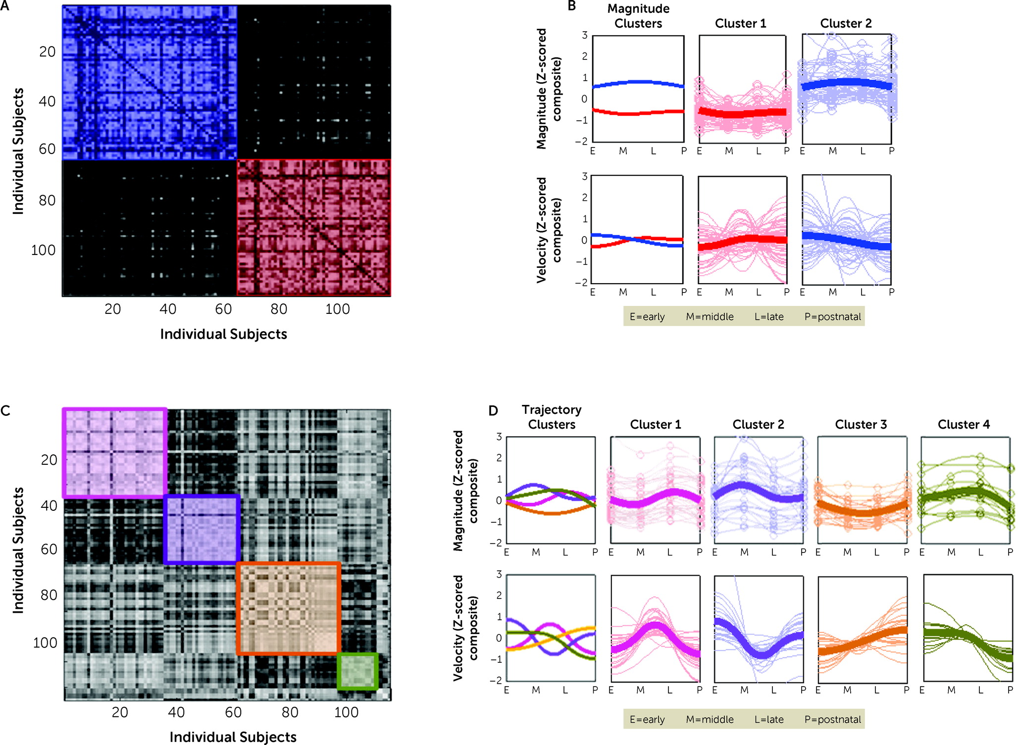
This study used data from mother-infant dyads that included maternal stress measures from early pregnancy to 1 month postpartum and infant behavior assessment of negative affect up to 24 months of age (UCI cohort; N=115) (21). Maternal stress scores from the UCI cohort were entered into the functional random forest algorithm. The model-based approach identified two clusters containing the same number of participants (N=57), and a third cluster, containing a single participant, was excluded from the analysis because this cluster was considered unreliable. The two remaining clusters had significantly greater modularity than clusters comprising randomly assigned participants (Q=0.729, p<0.001) (see Table S15 in the online supplement), and they captured magnitude differences in maternal perinatal stress (t=113.47, df=1998, p<0.001) (Figure 1A). These clusters differed significantly from each other at every time point (early, middle, and late pregnancy and early postpartum) in a two-way ANOVA (all p<0.001), reflecting mothers with high and low mean stress composite scores (higher scores reflect greater stress). We refer to these clusters as “magnitude clusters.” We plotted the associated path of the magnitude clusters, which clearly reflected the difference between high and low stress scores (Figure 1B). We also plotted the scores for each individual stress measure (i.e., CES-D, PSS, and STAI scores) by magnitude cluster assignment and found that the overall trends were the same for each contributing measure (see Figure S2 in the online supplement).
FIGURE 1. Identification of distinct clusters of patterns of perinatal stress in the primary cohort with functional random foresta
aMaternal perinatal stress measures were completed during early, middle, and late pregnancy and 1-month postnatal time points. Panel A shows the sorted proximity matrix resulting from the model-based approach. Two distinct clusters were identified. Panel B shows two distinct trajectories reflecting high and low maternal perinatal stress, resulting from a model-based approach. The far left column shows the central tendency of both magnitude clusters. The top row shows trajectories based on the magnitude of individual stress levels, with the central tendency of each cluster presented as a bold line. Magnitude cluster 1 (red) had lower maternal perinatal stress scores. Magnitude cluster 2 (blue) had higher scores and greater variability. The bottom row shows the velocity of perinatal stress (rate of change in maternal stress scores) with the central tendency of each cluster presented as a bold line. The clusters did not show differing patterns in terms of velocity. Panel C shows the sorted proximity matrix resulting from the correlation-based approach. Four clusters were identified. Panel D shows four distinct trajectories reflecting differences in peak stress and velocity of stress over time, resulting from a correlation-based approach. The far left column shows the central tendency of all trajectory clusters. The top row shows trajectories based on the magnitude of individual stress levels, with the central tendency of each cluster presented as a bold line. The bottom row shows the velocity of perinatal stress (rate of change in maternal stress scores) with the central tendency of each cluster presented as a bold line. The colors in panels A and C correspond to the colors used in panels B and D.

Correlation-based clustering approach and shape and velocity of maternal perinatal stress.

The second arm of the functional random forest uses a correlation-based approach to model longitudinal heterogeneity. This analysis used data from the same participants and identified four correlation-based clusters (Q=0.811, p<0.001) (Figure 1C), with distinct trajectories defined by the shape and velocity of change in maternal perinatal stress (Figure 1D). We refer to these clusters as “trajectory clusters.” Trajectory 1 (N=35) showed a peak in stress in late pregnancy, trajectory 2 (N=27) had a peak in middle pregnancy, trajectory 3 (N=26) showed an increase in stress late in pregnancy, and trajectory 4 (N=14) showed postnatal decline in stress. Clusters with fewer than 10 participants were not considered reliable and were excluded from subsequent analyses. The final trajectory clusters accounted for 102 mothers. The 13 mother-infant dyads that belonged to clusters excluded from the analyses did not differ significantly from dyads included in the analyses (see the online supplement). Similar to the first approach, we plotted scores for individual measures by trajectory cluster assignment. Trajectories 1 and 3 were consistent across all individual measures; trajectory 2 showed differences in magnitude of the measure of perceived stress, with high severity and minimal change; and trajectory 4 showed differences in anxiety and depression, with a pattern of decreasing, increasing, then decreasing stress (see Figure S3 in the online supplement).

Replication in FinnBrain Cohort

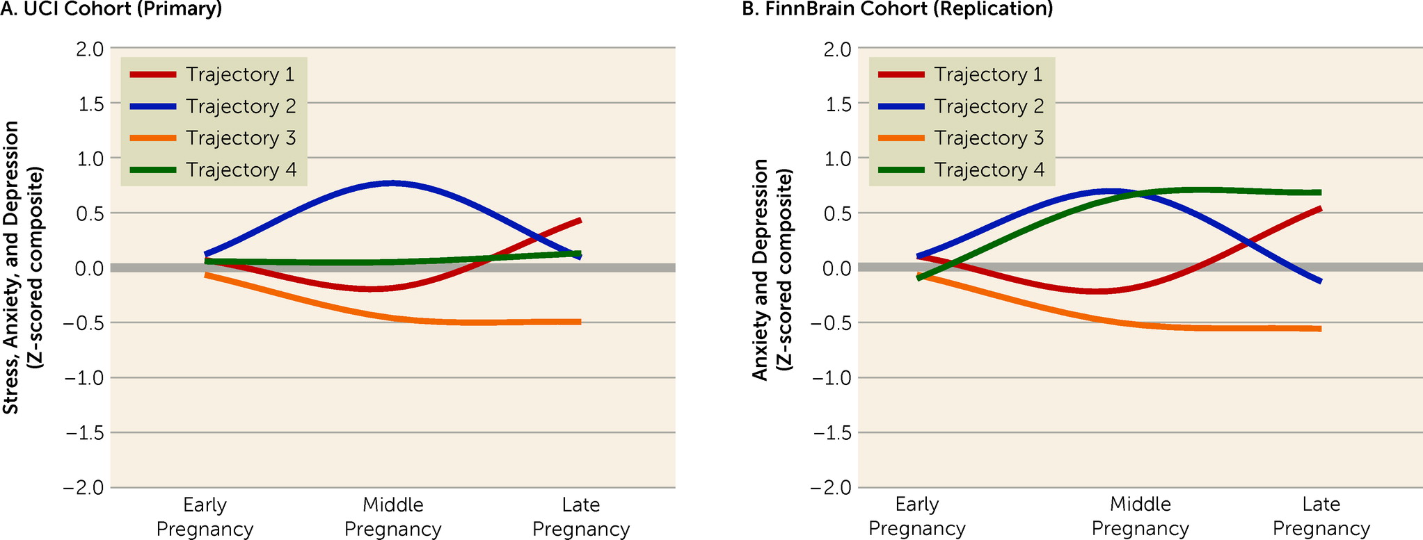
To examine whether the maternal clusters could be replicated in an independent sample, we applied the parameters for the UCI trajectory clusters to maternal antenatal stress principal component scores from a separate independent cohort (FinnBrain cohort; N=2,156) (25). FinnBrain trajectory 1 showed the same pattern of decreasing then increasing levels of stress found in UCI trajectory 1. FinnBrain trajectory 2 similarly mimicked the increasing then decreasing levels of stress observed in UCI trajectory 2. FinnBrain trajectory 3 showed an overall decline in stress followed by a flattening, a pattern that was similar to UCI trajectory 3. In contrast, FinnBrain trajectory 4 diverged from UCI trajectory 4 and showed increasing levels of stress followed by a flattening in levels. Trajectory 4 was composed of the fewest participants in the UCI cohort, which potentially contributed to the observed differences. In Figure 2, the first three time points of the UCI trajectories (primary cohort) are plotted beside the FinnBrain trajectories (replication cohort).
FIGURE 2. Similar stress trajectories in the primary and replication cohorts resulting from a correlation-based approacha
aStress measures were completed during early, middle, and late pregnancy in both cohorts. Panel A shows four distinct trajectories, reflecting differences in antenatal stress, resulting from the correlation-based approach in the University of California, Irvine (UCI) cohort (primary data set). Panel B shows four similar trajectories reflecting differences in antenatal stress that were identified in the FinnBrain cohort (replication data set) using the model parameters derived from the UCI cohort analysis.

Maternal Perinatal Stress Clusters and Infant Negative Affect Development

Using the clusters identified in the primary cohort, we first examined the association between maternal clusters and infant negative affect development by adding dummy-coded magnitude and trajectory clusters as predictors, along with relevant covariates, to the latent growth model of negative affect from 3 to 24 months of age. The magnitude and trajectory clusters were included in the same model to evaluate their independent contributions to infant negative affect development.

Magnitude clusters.

The magnitude clusters predicted only the intercept term (B=0.339, p=0.008), suggesting that higher maternal perinatal stress was related to elevated infant negative affect at 3 months of age (see Table S20 in the online supplement). When maternal postnatal stress scores were introduced as time-varying covariates at each time point that the IBQ was completed, the magnitude clusters no longer predicted the intercept (B=0.179, p=0.217), suggesting that postnatal maternal stress explains the association between the magnitude of maternal perinatal stress and infant negative affect at 3 months.

Trajectory clusters.

Maternal trajectory clusters did not predict the intercept or the quadratic term. However, trajectory 3 (increased stress in late pregnancy) predicted less linear development (B=−0.918, p=0.032) of infant negative affect (Figure 3A; see also Table S20 in the online supplement). When maternal postnatal stress scores were adjusted for, trajectory 3 continued to significantly predict the linear term of infant negative affect development (B=−0.921, p=0.031). The plot of infant negative affect development (Figure 3A) suggests that infants of mothers in trajectory 3 (increased stress in late pregnancy) do not follow the typical inverted U-shaped pattern of negative affect development, beginning at approximately 12 months of age. Infant negative affect differed significantly between trajectory 3 and the other clusters at 12 months (t=−2.841, df=69, p=0.01).
FIGURE 3. Negative affect of infants over time in the primary and replication cohortsa
aPanel A shows negative affect development in the University of California, Irvine (UCI) cohort (primary data set). The quadratic-shaped function resulting from this model reflects an increase in negative affect mean scores through 12 months of age and a subsequent decrease in scores through 24 months of age. Panel B shows negative affect development in the FinnBrain cohort (replication data set). Mean scores show negative affect increasing through 12 months of age then decreasing through 24 months of age. Panel C shows four trajectories of the different clusters. Trajectory 3 independently predicts the slope of infant negative affect development in the UCI cohort. As indicated by an asterisk, trajectory 3 differs significantly from the other clusters at 12 months of age (t=−2.841, df=69, p=0.01). Panel D shows that the trajectories of the different clusters were associated with infant negative affect development in the FinnBrain cohort. Trajectory clusters significantly predicted infant negative affect scores at different time points (F=3.04, df=3, 1058, p=0.028). As indicated by asterisks, trajectory 3 differed significantly from all other clusters, and it was associated with lower negative affect mean scores than the other clusters.
We also examined the association between composite stress scores at each time point during pregnancy and infant negative affect development (see Tables S26–S29 in the online supplement). Stress during middle and late pregnancy was associated with an increasing slope and negative quadratic term in the model of infant negative affect when postnatal maternal stress was adjusted for.

Replication of Association Between Maternal Antenatal Stress and Infant Negative Affect

Using the maternal antenatal stress clusters, we modeled infant negative affect development over the first 2 years of life. Consistent with findings from the UCI cohort, repeated-measures ANOVA suggested a statistically significant change in negative affect between 6 and 24 months (F=111.319, df=3, 1058, p<0.001) (for additional details, see the Methods section in the online supplement).
We then examined the association between FinnBrain maternal trajectory clusters and infant negative affect development with repeated-measures ANCOVA and relevant covariates. Consistent with the results from the UCI cohort, maternal trajectory clusters showed a significant main effect of group on infant negative affect over time (F=3.04, df=3, p=0.028). This effect remained significant after adjusting for infant sex, length of gestation, and postnatal depression (F=3.23, df=3, p=0.022). Post hoc analyses examining the contribution of specific trajectories also demonstrated a pattern similar to that identified in the UCI cohort. Specifically, there was a significant difference between trajectory 3 and all other trajectories of infant negative affect at 6 months (F=4.423, df=3, 1058, p=0.004), 12 months (F=6.674, df=3, 1058, p<0.001), and 24 months (F=6.011, df=3, 1058, p<0.001). The difference between trajectories 3 and 4 remained significant when maternal postnatal depression, infant sex, and gestational age were controlled for (12 months: B=−0.202, p=0.020; 24 months: B=−0.151, p=0.018). This suggests that, as in the UCI cohort, infants of mothers in the trajectory 3 cluster showed a blunted divergent pattern of negative affect development.

Maternal Perinatal Stress Clusters and Infant Functional Connectivity
To examine the effects of the maternal magnitude and trajectory clusters on the infant brain, we evaluated neonatal functional connectivity of offspring in the UCI cohort. Only a subset of the cohort had brain imaging data available (N=60), and thus, the analysis is considered exploratory (31). Maternal trajectory clusters were dummy coded and included as predictors in all analyses (see the Methods section in the online supplement). The magnitude and trajectory clusters were included in the same model to evaluate their independent contributions to infant functional connectivity.
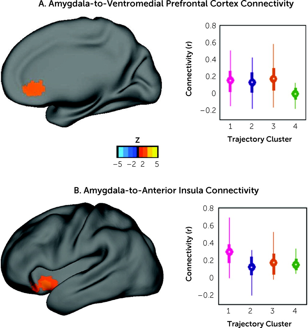
The magnitude clusters, capturing symptom severity, were not associated with neonatal amygdala functional connectivity to the vmPFC (B=0.015, p=0.746) or the anterior insula (B=0.008, p=0.841). However, the trajectory clusters showed significant associations with amygdala connectivity. Trajectory 1 (peak stress in late pregnancy) was significantly associated with stronger amygdala connectivity to the vmPFC (B=0.173, p=0.011) and the anterior insula (B=0.166, p=0.006) (Figure 4). Trajectory 3 (increase in stress late in pregnancy) also significantly predicted stronger amygdala connectivity to the vmPFC (B=0.172, p=0.011) (Figure 4A). Trajectories 2 (peak stress in middle pregnancy) and 4 (postnatal decline in stress) were not associated with alterations in amygdala connectivity. To understand the contribution of data from individual time points, we also examined the associations between stress scores at each time point during pregnancy and neonatal amygdala connectivity (see Table S25 in the online supplement).
FIGURE 4. Maternal perinatal stress trajectory clusters are associated with amygdala connectivity in neonatal infantsa
aLeft amygdala connectivity to two regions of interest identified in a previous study (32) are displayed here for the University of California, Irvine (UCI) cohort. Gestational age at birth and infant age at scan were included as covariates to account for neonatal brain maturity at the time of MRI acquisition. Correlation values (r) are reported for amygdala functional connectivity. Circles represent cluster means. Bolded color bars represent interquartile range and thin lines represent data from the 2.5th to the 97.5th percentile. Panel A shows that maternal trajectory clusters 1 and 3 were associated with stronger infant amygdala connectivity to the ventromedial prefrontal cortex. This connectivity among offspring of mothers included in cluster 2 appeared to be stronger than that in cluster 4, but this difference did not reach statistical significance (B=0.127, p=0.069). Panel B shows that maternal trajectory cluster 1 was associated with increased infant amygdala connectivity to the anterior insula bilaterally.

Discussion

We found that trajectories of maternal perinatal stress were related to infant brain phenotypes and negative affect development over the first 2 years of life. Offspring of mothers with increasing or peak stress late in pregnancy showed increased amygdala functional connectivity at 1 month of age and a blunted pattern of negative affect development over the first 2 years of life. Interestingly, the overall magnitude of stress was not associated with brain or behavioral development of the offspring when considered in the same models as the trajectory of stress. The relationship between the trajectory of maternal stress during pregnancy and infant negative affect development was replicated in an independent cohort comprising more than 2,000 mother-infant dyads. The shapes of the stress trajectories were mostly replicated in this larger cohort, although participants in the trajectory 4 cluster showed lower stress during late pregnancy in the replication cohort compared with the primary cohort. Overall, the data highlight that the trajectory of maternal stress may contribute to both brain and affect development of offspring in a manner that is independent of stress magnitude.
The finding that maternal psychological stress during pregnancy is related to infant emotional development, especially negative emotionality, is not surprising, given the literature on maternal stress and infant outcomes. However, a large majority of earlier studies considered only the magnitude of stress. Here, we showed that heterogeneity exists in maternal stress trajectories during pregnancy, with regard to timing and rate of change, and this variance in trajectories is an important characteristic affecting brain and behavioral outcomes of offspring. When women were grouped by either the overall magnitude of stress or the timing and rate of change in stress (trajectory), the trajectory appeared to be more important for brain and behavioral development of the offspring.

Maternal Stress in Middle or Late Pregnancy and Infant Negative Affect Development

Two ways to consider our overall findings in the context of the literature are that the entire trajectory is an important parameter for offspring development that needs to be considered, and that in the absence of the trajectory (e.g., when studies sample only at one time point), the time when that sample is taken is critically important. To highlight this point, we followed up our trajectory analyses by examining each perinatal time point separately. Maternal stress during pregnancy, examined at single time points, did not predict neonatal amygdala functional connectivity, suggesting that the entire trajectory is an important parameter with regard to the observed brain outcomes. Maternal stress during middle and late pregnancy was associated with blunted development of infant negative affect over the first 2 years of life, indicating that sampling in middle or late pregnancy may be more likely to reveal associations between antenatal stress and infant negative affect development.

Maternal Stress Trajectories and Neonatal Amygdala Connectivity

In the UCI cohort, trajectories with either peaks or increases in maternal stress in late pregnancy, such as trajectories 1 and 3, were related strongly to amygdala connectivity and negative affect development in the offspring, even when the overall magnitude of maternal stress is accounted for. The distinct effects of maternal stress during late pregnancy on the limbic system of offspring is in line with previous studies examining maternal anxiety and infant hippocampal development, suggesting that the timing of stress may play a role in infant developmental outcomes (39, 40).
Trajectory 3 was associated with distinct patterns of stronger amygdala connectivity in offspring and a pattern of infant negative affect development that diverged from the other trajectories and previous findings. The most similar trajectory in the FinnBrain cohort was also associated with a divergent pattern of infant negative affect development, indicating the robustness of this finding.
Although neonates of mothers in trajectory 1 also had stronger amygdala-to-vmPFC connectivity, these infants did not demonstrate decreased negative affect development. Trajectory 1 might be associated with activation of an unexplored compensatory circuit that compensates and renders affect development more similar to that observed for trajectories 2 or 4. Interestingly, trajectory 1 was also associated with stronger connectivity between the amygdala and the anterior insula. Stronger connectivity between the amygdala and the anterior insula, regions involved in the salience network, has been shown to predict higher levels of fear in infants, internalizing symptoms in early infancy, and anxiety in children (32, 33, 41, 42). The more typical trajectory of negative affect development for infants of mothers in trajectory 1 may relate to the contribution of stronger amygdala-to-insula connectivity to higher levels of fear. Unfortunately, the analyses were not powered to test relationships between these neonatal functional connections and infant negative affect development within this cohort. Considering our recent work highlighting the generally small effect sizes for association studies in brain imaging (31), larger longitudinal studies are needed to validate and test the full pathway from heterogeneity of maternal psychological stress trajectories to offspring affect development via alterations in early brain development.

Maternal Stress Trajectories and Differential Infant Negative Affect Development

Consistent with our previous work and the literature, infant negative affect increases over the first year of life and then decreases through 24 months of age, resulting in an inverted U-shaped trajectory (33, 38, 4345). An increase in negative affect over the first year, as infants explore their environment and encounter strangers, is expected and normative. The increase in negative emotionality and the resulting need for internal and external forms of emotional regulation likely supports the development of regulatory and coping skills.
In the UCI cohort, trajectory 3 was associated with blunted infant negative affect development at 12 months of age, representing a deviation from an expected pattern. The much larger replication sample confirmed this particular finding, but it also suggested a similar pattern of divergence at the other time points, with lower levels of negative affect at 6, 12, and 24 months of age. It is likely that the UCI cohort was underpowered to reveal this broader effect across the time span, and highlights in very clear terms the importance of replication of similar types of work in large, well-powered samples, as was done here. The blunted development of infant negative affect may have been associated with an overall pattern of lower maternal stress during pregnancy because the increase in maternal stress was observed in late pregnancy into the early postpartum period. However, because all models were adjusted for magnitude of stress, the association appears to be unique to the trajectory of stress during pregnancy and the trajectory of infant negative affect. Longitudinal studies in high-risk samples with greater variability in maternal stress and infant negative affect are needed to ascertain whether this divergent pattern of negative affect development is adaptive or maladaptive.

Maternal-Placental-Fetal Biology, Maternal Psychological Stress, and Brain Outcomes of Offspring

There is growing evidence to suggest that the fetus may be especially sensitive to extrauterine cues transmitted via stress-sensitive aspects of maternal-placental-fetal biology during late pregnancy. Cortisol bioavailability, which is altered during increased maternal psychological stress, plays a key role in fetal maturation and is associated with risk of psychopathology in offspring (46). Cortisol is involved in fetal brain neurogenesis, synaptogenesis, and axonal growth through glucocorticoid receptors (46). Although maternal cortisol levels vary during pregnancy, there is a surge during late pregnancy (47). Several studies have shown that maternal cortisol during late pregnancy is associated with infant reactivity and negative emotionality (48), a finding potentially explained by the high concentration of glucocorticoid receptors present in the amygdala (46). Increased psychological stress is also associated with increasing levels of inflammatory markers such as interleukin-6 (IL-6) and other cytokines that activate inflammatory processes in the fetal brain through both direct and indirect pathways (11). Like cortisol, IL-6 and other cytokines are important for fetal brain development but can disrupt cellular survival and proliferation and, if the levels are too high, disrupt synaptogenesis. Spann et al. (49) found that higher levels of inflammation during the third trimester were associated with the strength of neonatal functional connectivity within the salience network, including the medial prefrontal cortex. Similarly, Rudolph et al. (50) showed that elevated maternal IL-6 concentrations during the third trimester were most strongly associated with reduced infant working memory at 2 years of age.

Importance of the Third Trimester for Brain Development

The third trimester is a critical time for fetal brain development and is characterized by increasing connectivity. Long-distance corticocortical connections are established during this time frame and functional connections are strengthened (51, 52). The amygdala and anterior insula demonstrate rapid development during the third trimester of pregnancy (53). There is also evidence of strong coactivation of the anterior insula and amygdala during this time (53). Early functional specialization of the insula and subcortical synchronization of the amygdala suggest that these regions are particularly important for survival during early infancy (5355). During the final few weeks of pregnancy, white matter myelination, synaptic connectivity, and cortical growth increase rapidly (5659). The period right before birth is characterized by a rapid and dramatic increase in the total number of physical connections between neurons of the cerebral cortex and is therefore considered a critical period for development of the cortical connectome (59). Rapid cortical maturation, circuit formation, and increased neuronal connectivity are hallmarks of third-trimester neurodevelopment and may explain why this period is particularly sensitive to increases in maternal stress (6, 7, 60).

Limitations and Additional Considerations

There are several limitations of this study to consider. First, maternal stress was characterized using self-report measures. Although multimethod assessment is preferable, the use of self-report measures to characterize stress is clinically relevant given that this is how depression and anxiety are typically monitored during routine antenatal care. Similarly, infant behavior was based on maternal report measures. Infant emotions are difficult to assess and could reflect maternal mood, expectations, or recall. We addressed this limitation by including maternal stress at each infant behavior time point as a covariate. We are likely constraining variance with this approach, given the high correlation between prenatal and postnatal stress measures; however, this is unavoidable in an observational study. Second, the measures of anxiety and depression used in the UCI cohort differed from those used in the FinnBrain cohort. While we attempted to account for postnatal maternal stress in our models, some of the observed effects may be postnatal in origin. Importantly, the replication of findings across two independent cohorts increases our level of confidence in the capacity of these measures to detect meaningful variation in the constructs of interest. Additionally, the sample size used to initially characterize the trajectories was small (N=115). Functional random forest should be used to examine larger and more diverse cohorts to see if additional groups emerge and if there are different interactions between magnitude and trajectory.
The neuroimaging component of this study focused on the resting-state functional connectivity between the amygdala and two regions of interest. The processing, interpretation, and expression of negative affect involves multiple brain regions and networks beyond those explored in the present study. Additional work is required to explore the full extent of how maternal perinatal stress trajectories relate to the structure and function of the neonatal brain. Studies are needed to explore the role of maternal stress in the postnatal environment. These exploratory statistical analyses were not powered to test the role of functional connectivity as a mediator in our model of maternal perinatal stress on the development of infant negative affect (e.g., see [31]). However, based on the literature, it is likely that alterations in the limbic system associated with stress exposure during pregnancy explain some of the differences observed here in the development of infant negative affect over the first 2 years of life.

Conclusions

The identified trajectories provide information on the timing and variability of stress during pregnancy. Importantly, the trajectory clusters identified by the functional random forest differed in terms of the timing of peak stress and rate of changes in stress during pregnancy. Given the sequential process of neurodevelopment, alterations in psychological stress are likely to have a differential impact on brain development depending on the timing of the peak stress or the timing of the change. Consistent with this conceptualization, we found that maternal perinatal stress trajectories were related to both infant neurodevelopment and psychosocial development in a manner independent of the overall magnitude of stress exposure. This study highlights the importance of considering variability in maternal perinatal stress over time, as opposed to focusing predominantly on stress severity. Advancing understanding of heterogeneity in perinatal psychological stress represents an important step toward understanding contributing factors to the experience of stress and the implications for offspring development. Finally, gaining a more nuanced understanding of perinatal stress and its influence on infant neurobiological and psychosocial development is critical for refining and improving preventive interventions designed to support maternal and infant mental health.

Supplementary Material

File (appi.ajp.21111176.ds001.pdf)

REFERENCES

1.
Weinstock M: The long-term behavioural consequences of prenatal stress. Neurosci Biobehav Rev 2008; 32:1073–1086
2.
Van den Bergh BR, van den Heuvel MI, Lahti M, et al: Prenatal developmental origins of behavior and mental health: the influence of maternal stress in pregnancy. Neurosci Biobehav Rev 2020; 117:26–64
3.
Graignic-Philippe R, Dayan J, Chokron S, et al: Effects of prenatal stress on fetal and child development: a critical literature review. Neurosci Biobehav Rev 2014; 43:137–162
4.
Heim CM, Entringer S, Buss C: Translating basic research knowledge on the biological embedding of early-life stress into novel approaches for the developmental programming of lifelong health. Psychoneuroendocrinology 2019; 105:123–137
5.
Mora PA, Bennett IM, Elo IT, et al: Distinct trajectories of perinatal depressive symptomatology: evidence from growth mixture modeling. Am J Epidemiol 2009; 169:24–32
6.
Tau GZ, Peterson BS: Normal development of brain circuits. Neuropsychopharmacology 2010; 35:147–168
7.
Andescavage NN, du Plessis A, McCarter R, et al: Complex trajectories of brain development in the healthy human fetus. Cereb Cortex 2017; 27:5274–5283
8.
Vasung L, Abaci Turk E, Ferradal SL, et al: Exploring early human brain development with structural and physiological neuroimaging. NeuroImage 2019; 187:226–254
9.
Gao W, Alcauter S, Smith JK, et al: Development of human brain cortical network architecture during infancy. Brain Struct Funct 2015; 220:1173–1186
10.
Buss C, Entringer S, Moog NK, et al: Intergenerational transmission of maternal childhood maltreatment exposure: implications for fetal brain development. J Am Acad Child Adolesc Psychiatry 2017; 56:373–382
11.
Graham AM, Rasmussen JM, Rudolph MD, et al: Maternal systemic interleukin-6 during pregnancy is associated with newborn amygdala phenotypes and subsequent behavior at 2 years of age. Biol Psychiatry 2018; 83:109–119
12.
Qiu A, Anh TT, Li Y, et al: Prenatal maternal depression alters amygdala functional connectivity in 6-month-old infants. Transl Psychiatry 2015; 5:e508
13.
Rifkin-Graboi A, Meaney MJ, Chen H, et al: Antenatal maternal anxiety predicts variations in neural structures implicated in anxiety disorders in newborns. J Am Acad Child Adolesc Psychiatry 2015; 54:313–321
14.
Burghy CA, Stodola DE, Ruttle PL, et al: Developmental pathways to amygdala-prefrontal function and internalizing symptoms in adolescence. Nat Neurosci 2012; 15:1736–1741
15.
Buss C, Davis EP, Shahbaba B, et al: Maternal cortisol over the course of pregnancy and subsequent child amygdala and hippocampus volumes and affective problems. Proc Natl Acad Sci USA 2012; 109:E1312–E1319
16.
Davis EP, Glynn LM, Waffarn F, et al: Prenatal maternal stress programs infant stress regulation. J Child Psychol Psychiatry 2011; 52:119–129
17.
Yong Ping E, Laplante DP, Elgbeili G, et al: Prenatal maternal stress predicts stress reactivity at 2½ years of age: the Iowa Flood Study. Psychoneuroendocrinology 2015; 56:62–78
18.
Etkin A, Wager TD: Functional neuroimaging of anxiety: a meta-analysis of emotional processing in PTSD, social anxiety disorder, and specific phobia. Am J Psychiatry 2007; 164:1476–1488
19.
Insel T, Cuthbert B, Garvey M, et al: Research Domain Criteria (RDoC): toward a new classification framework for research on mental disorders. Am J Psychiatry 2010; 167:748–751
20.
Damme KSF, Norton ES, Briggs-Gowan MJ, et al: Developmental patterning of irritability enhances prediction of psychopathology in pre-adolescence: improving RDoC with developmental science. J Psychopathol Clin Sci 2022; 131:556–566
21.
Moog NK, Entringer S, Rasmussen JM, et al: Intergenerational effect of maternal exposure to childhood maltreatment on newborn brain anatomy. Biol Psychiatry 2018; 83:120–127
22.
Karlsson L, Tolvanen M, Scheinin NM, et al: Cohort profile: the FinnBrain Birth Cohort Study (FinnBrain). Int J Epidemiol 2018; 47:15–16j
23.
Radloff LS: The CES-D scale: a self-report depression scale for research in the general population. Appl Psychol Meas 1977; 1:385–401
24.
Cohen S, Kamarck T, Mermelstein R: A global measure of perceived stress. J Health Soc Behav 1983; 24:385–396
25.
Spielberger CD, Gorsuch RL, Lushene RE: Manual for the State-Trait Anxiety Inventory. Palo Alto, Calif, Consulting Psychologists Press, 1970
26.
Cox JL, Holden JM, Sagovsky R: Detection of postnatal depression: development of the 10-item Edinburgh Postnatal Depression Scale. Br J Psychiatry 1987; 150:782–786
27.
Derogatis LR, Lipman RS, Covi L: SCL-90: an outpatient psychiatric rating scale—preliminary report. Psychopharmacol Bull 1973; 9:13–28
28.
Parade SH, Leerkes EM: The reliability and validity of the Infant Behavior Questionnaire-Revised. Infant Behav Dev 2008; 31:637–646
29.
Putnam SP, Gartstein MA, Rothbart MK: Measurement of fine-grained aspects of toddler temperament: the Early Childhood Behavior Questionnaire. Infant Behav Dev 2006; 29:386–401
30.
Muthén LK, Muthén BO: Mplus User’s Guide. Eighth edition. Los Angeles, Muthén & Muthén, 2017. www.StatModel.com
31.
Marek S, Tervo-Clemmens B, Calabro FJ, et al: Reproducible brain-wide association studies require thousands of individuals. Nature 2022; 603:654–660
32.
Graham AM, Buss C, Rasmussen JM, et al: Implications of newborn amygdala connectivity for fear and cognitive development at 6-months-of-age. Dev Cogn Neurosci 2016; 18:12–25
33.
Thomas E, Buss C, Rasmussen JM, et al: Newborn amygdala connectivity and early emerging fear. Dev Cogn Neurosci 2019; 37:100604
34.
Wang J, Vachet C, Rumple A, et al: Multi-atlas segmentation of subcortical brain structures via the AutoSeg software pipeline. Front Neuroinform 2014; 8:7
35.
Yushkevich PA, Piven J, Hazlett HC, et al: User-guided 3D active contour segmentation of anatomical structures: significantly improved efficiency and reliability. Neuroimage 2006; 31:1116–1128
36.
Feczko E, Balba NM, Miranda-Dominguez O, et al: Subtyping cognitive profiles in autism spectrum disorder using a functional random forest algorithm. Neuroimage 2018; 172:674–688
37.
Feczko E, Miranda-Dominguez O, Marr M, et al: The heterogeneity problem: approaches to identify psychiatric subtypes. Trends Cogn Sci 2019; 23:584–601
38.
Partridge T, Lerner JV: A latent growth-curve approach to difficult temperament. Infant Child Dev 2007; 16:255–265
39.
Qiu A, Rifkin-Graboi A, Chen H, et al: Maternal anxiety and infants’ hippocampal development: timing matters. Transl Psychiatry 2013; 3:e306
40.
Rifkin-Graboi A, Quan J, Richmond J, et al: Greater caregiving risk, better infant memory performance? Hippocampus 2018; 28:497–511
41.
Qin S, Young CB, Duan X, et al: Amygdala subregional structure and intrinsic functional connectivity predicts individual differences in anxiety during early childhood. Biol Psychiatry 2014; 75:892–900
42.
Rogers CE, Sylvester CM, Mintz C, et al: Neonatal amygdala functional connectivity at rest in healthy and preterm infants and early internalizing symptoms. J Am Acad Child Adolesc Psychiatry 2017; 56:157–166
43.
Garstein M, Rothbart MK: Studying infant temperament via the Revised Infant Behavior Questionnaire. Infant Behav Dev 2003; 26:64–86
44.
Braungart-Rieker JM, Hill-Soderlund AL, Karrass J: Fear and anger reactivity trajectories from 4 to 16 months: the roles of temperament, regulation, and maternal sensitivity. Dev Psychol 2010; 46:791–804
45.
Brooker RJ, Buss KA, Lemery-Chalfant K, et al: The development of stranger fear in infancy and toddlerhood: normative development, individual differences, antecedents, and outcomes. Dev Sci 2013; 16:864–878
46.
Graham AM, Rasmussen JM, Entringer S, et al: Maternal cortisol concentrations during pregnancy and sex-specific associations with neonatal amygdala connectivity and emerging internalizing behaviors. Biol Psychiatry 2019; 85:172–181
47.
Stoye DQ, Andrew R, Grobman WA, et al: Maternal glucocorticoid metabolism across pregnancy: a potential mechanism underlying fetal glucocorticoid exposure. J Clin Endocrinol Metab 2020; 105:e782–e790
48.
Braithwaite EC, Pickles A, Sharp H, et al: Maternal prenatal cortisol predicts infant negative emotionality in a sex-dependent manner. Physiol Behav 2017; 175:31–36
49.
Spann MN, Monk C, Scheinost D, et al: Maternal immune activation during the third trimester is associated with neonatal functional connectivity of the salience network and fetal to toddler behavior. J Neurosci 2018; 38:2877–2886
50.
Rudolph MD, Graham AM, Feczko E, et al: Maternal IL-6 during pregnancy can be estimated from newborn brain connectivity and predicts future working memory in offspring. Nat Neurosci 2018; 21:765–772
51.
Ball G, Aljabar P, Zebari S, et al: Rich-club organization of the newborn human brain. Proc Natl Acad Sci USA 2014; 111:7456–7461
52.
Jakab A, Schwartz E, Kasprian G, et al: Fetal functional imaging portrays heterogeneous development of emerging human brain networks. Front Hum Neurosci 2014; 8:852
53.
Scheinost D, Chang J, Lacadie C, et al: Hypoconnectivity between anterior insula and amygdala associates with future vulnerabilities in social development in a neurodiverse sample of neonates. Scientific Rep 2022; 12:16230
54.
Alcauter S, Lin W, Smith JK, et al: Consistent anterior–posterior segregation of the insula during the first 2 years of life. Cereb Cortex 2015; 25:1176–1187
55.
Salzwedel AP, Stephens RL, Goldman BD, et al: Development of amygdala functional connectivity during infancy and its relationship with 4-year behavioral outcomes. Biol Psychiatry Cogn Neurosci Neuroimaging 2019; 4:62–71
56.
Hüppi PS, Warfield S, Kikinis R, et al: Quantitative magnetic resonance imaging of brain development in premature and mature newborns. Ann Neurol 1998; 43:224–235
57.
Garcia KE, Robinson EC, Alexopoulos D, et al: Dynamic patterns of cortical expansion during folding of the preterm human brain. Proc Natl Acad Sci USA 2018; 115:3156–3161
58.
Collin G, Sporns O, Mandl RCW, van den Heuvel MP: Structural and functional aspects relating to cost and benefit of rich club organization in the human cerebral cortex. Cereb Cortex 2014; 24:2258–2267
59.
Van den Heuvel MP, Kersbergen KJ, de Reus MA, et al: The neonatal connectome during preterm brain development. Cereb Cortex 2015; 25:3000–3013
60.
Thomason ME, Grove LE, Lozon TA, Jr., et al: Age-related increases in long-range connectivity in fetal functional neural connectivity networks in utero. Dev Cogn Neurosci 2015; 11:96–104

Information & Authors

Information

Published In

Go to American Journal of Psychiatry
American Journal of Psychiatry
Pages: 766 - 777
PubMed: 37670606

History

Received: 28 November 2021
Revision received: 5 September 2022
Revision received: 11 April 2023
Revision received: 17 May 2023
Revision received: 26 May 2023
Accepted: 31 May 2023
Published online: 6 September 2023
Published in print: October 1, 2023

Keywords

  1. Development
  2. Emotion
  3. Functional Random Forest
  4. Neuroimaging
  5. Pre/Peri/Postnatal Issues
  6. Stress

Authors

Details

Mollie C. Marr, M.D., Ph.D.
Department of Behavioral Neuroscience (Marr, Graham, Sturgeon, Schifsky, Fair) and Department of Psychiatry (Graham, Fair), Oregon Health and Science University School of Medicine, Portland; Department of Psychiatry, Massachusetts General Hospital, Boston (Marr); Department of Psychiatry, McLean Hospital, Belmont, Mass. (Marr); Masonic Institute for the Developing Brain, Institute of Child Development (Fair), and Department of Pediatrics (Feczko, Fair), University of Minnesota, Minneapolis; Department of Psychology and Speech–Language Pathology, University of Turku, Turku, Finland (Nolvi, Korja); Institute of Medical Psychology, Charité–Universitätsmedizin Berlin, corporate member of Freie Universität Berlin and Humboldt–Universität zu Berlin, Berlin (Nolvi, Entringer, Buss); Department of Neuroscience, Earlham College, Richmond, Ind. (Thomas); Development, Health, and Disease Research Program and Departments of Pediatrics, Psychiatry and Human Behavior, Obstetrics and Gynecology, and Epidemiology, University of California, Irvine, School of Medicine, Irvine (Rasmussen, Entringer, Wadhwa, Buss); Department of Pediatrics, University of California, Irvine, School of Medicine, Orange (Rasmussen, Entringer, Wadhwa, Buss); Departments of Psychiatry and Human Behavior (Entringer, Wadhwa), Obstetrics and Gynecology (Wadhwa), and Epidemiology (Wadhwa), University of California, Irvine, School of Medicine, Orange; FinnBrain Birth Cohort Study, Turku Brain and Mind Center, Department of Clinical Medicine, University of Turku (Korja, H. Karlsson, L. Karlsson); Centre for Population Health Research, University of Turku and Turku University Hospital (Korja, H. Karlsson, L. Karlsson); Department of Paediatrics and Adolescent Medicine (L. Karlsson) and Department of Psychiatry (H. Karlsson), Department of Clinical Medicine, Turku University Hospital and University of Turku; Department of Psychiatry, University of North Carolina at Chapel Hill School of Medicine, Chapel Hill (Gilmore); Department of Psychiatry, University of North Carolina at Chapel Hill, Chapel Hill (Styner).
Alice M. Graham, Ph.D.
Department of Behavioral Neuroscience (Marr, Graham, Sturgeon, Schifsky, Fair) and Department of Psychiatry (Graham, Fair), Oregon Health and Science University School of Medicine, Portland; Department of Psychiatry, Massachusetts General Hospital, Boston (Marr); Department of Psychiatry, McLean Hospital, Belmont, Mass. (Marr); Masonic Institute for the Developing Brain, Institute of Child Development (Fair), and Department of Pediatrics (Feczko, Fair), University of Minnesota, Minneapolis; Department of Psychology and Speech–Language Pathology, University of Turku, Turku, Finland (Nolvi, Korja); Institute of Medical Psychology, Charité–Universitätsmedizin Berlin, corporate member of Freie Universität Berlin and Humboldt–Universität zu Berlin, Berlin (Nolvi, Entringer, Buss); Department of Neuroscience, Earlham College, Richmond, Ind. (Thomas); Development, Health, and Disease Research Program and Departments of Pediatrics, Psychiatry and Human Behavior, Obstetrics and Gynecology, and Epidemiology, University of California, Irvine, School of Medicine, Irvine (Rasmussen, Entringer, Wadhwa, Buss); Department of Pediatrics, University of California, Irvine, School of Medicine, Orange (Rasmussen, Entringer, Wadhwa, Buss); Departments of Psychiatry and Human Behavior (Entringer, Wadhwa), Obstetrics and Gynecology (Wadhwa), and Epidemiology (Wadhwa), University of California, Irvine, School of Medicine, Orange; FinnBrain Birth Cohort Study, Turku Brain and Mind Center, Department of Clinical Medicine, University of Turku (Korja, H. Karlsson, L. Karlsson); Centre for Population Health Research, University of Turku and Turku University Hospital (Korja, H. Karlsson, L. Karlsson); Department of Paediatrics and Adolescent Medicine (L. Karlsson) and Department of Psychiatry (H. Karlsson), Department of Clinical Medicine, Turku University Hospital and University of Turku; Department of Psychiatry, University of North Carolina at Chapel Hill School of Medicine, Chapel Hill (Gilmore); Department of Psychiatry, University of North Carolina at Chapel Hill, Chapel Hill (Styner).
Eric Feczko, Ph.D.
Department of Behavioral Neuroscience (Marr, Graham, Sturgeon, Schifsky, Fair) and Department of Psychiatry (Graham, Fair), Oregon Health and Science University School of Medicine, Portland; Department of Psychiatry, Massachusetts General Hospital, Boston (Marr); Department of Psychiatry, McLean Hospital, Belmont, Mass. (Marr); Masonic Institute for the Developing Brain, Institute of Child Development (Fair), and Department of Pediatrics (Feczko, Fair), University of Minnesota, Minneapolis; Department of Psychology and Speech–Language Pathology, University of Turku, Turku, Finland (Nolvi, Korja); Institute of Medical Psychology, Charité–Universitätsmedizin Berlin, corporate member of Freie Universität Berlin and Humboldt–Universität zu Berlin, Berlin (Nolvi, Entringer, Buss); Department of Neuroscience, Earlham College, Richmond, Ind. (Thomas); Development, Health, and Disease Research Program and Departments of Pediatrics, Psychiatry and Human Behavior, Obstetrics and Gynecology, and Epidemiology, University of California, Irvine, School of Medicine, Irvine (Rasmussen, Entringer, Wadhwa, Buss); Department of Pediatrics, University of California, Irvine, School of Medicine, Orange (Rasmussen, Entringer, Wadhwa, Buss); Departments of Psychiatry and Human Behavior (Entringer, Wadhwa), Obstetrics and Gynecology (Wadhwa), and Epidemiology (Wadhwa), University of California, Irvine, School of Medicine, Orange; FinnBrain Birth Cohort Study, Turku Brain and Mind Center, Department of Clinical Medicine, University of Turku (Korja, H. Karlsson, L. Karlsson); Centre for Population Health Research, University of Turku and Turku University Hospital (Korja, H. Karlsson, L. Karlsson); Department of Paediatrics and Adolescent Medicine (L. Karlsson) and Department of Psychiatry (H. Karlsson), Department of Clinical Medicine, Turku University Hospital and University of Turku; Department of Psychiatry, University of North Carolina at Chapel Hill School of Medicine, Chapel Hill (Gilmore); Department of Psychiatry, University of North Carolina at Chapel Hill, Chapel Hill (Styner).
Saara Nolvi, Ph.D.
Department of Behavioral Neuroscience (Marr, Graham, Sturgeon, Schifsky, Fair) and Department of Psychiatry (Graham, Fair), Oregon Health and Science University School of Medicine, Portland; Department of Psychiatry, Massachusetts General Hospital, Boston (Marr); Department of Psychiatry, McLean Hospital, Belmont, Mass. (Marr); Masonic Institute for the Developing Brain, Institute of Child Development (Fair), and Department of Pediatrics (Feczko, Fair), University of Minnesota, Minneapolis; Department of Psychology and Speech–Language Pathology, University of Turku, Turku, Finland (Nolvi, Korja); Institute of Medical Psychology, Charité–Universitätsmedizin Berlin, corporate member of Freie Universität Berlin and Humboldt–Universität zu Berlin, Berlin (Nolvi, Entringer, Buss); Department of Neuroscience, Earlham College, Richmond, Ind. (Thomas); Development, Health, and Disease Research Program and Departments of Pediatrics, Psychiatry and Human Behavior, Obstetrics and Gynecology, and Epidemiology, University of California, Irvine, School of Medicine, Irvine (Rasmussen, Entringer, Wadhwa, Buss); Department of Pediatrics, University of California, Irvine, School of Medicine, Orange (Rasmussen, Entringer, Wadhwa, Buss); Departments of Psychiatry and Human Behavior (Entringer, Wadhwa), Obstetrics and Gynecology (Wadhwa), and Epidemiology (Wadhwa), University of California, Irvine, School of Medicine, Orange; FinnBrain Birth Cohort Study, Turku Brain and Mind Center, Department of Clinical Medicine, University of Turku (Korja, H. Karlsson, L. Karlsson); Centre for Population Health Research, University of Turku and Turku University Hospital (Korja, H. Karlsson, L. Karlsson); Department of Paediatrics and Adolescent Medicine (L. Karlsson) and Department of Psychiatry (H. Karlsson), Department of Clinical Medicine, Turku University Hospital and University of Turku; Department of Psychiatry, University of North Carolina at Chapel Hill School of Medicine, Chapel Hill (Gilmore); Department of Psychiatry, University of North Carolina at Chapel Hill, Chapel Hill (Styner).
Elina Thomas, Ph.D.
Department of Behavioral Neuroscience (Marr, Graham, Sturgeon, Schifsky, Fair) and Department of Psychiatry (Graham, Fair), Oregon Health and Science University School of Medicine, Portland; Department of Psychiatry, Massachusetts General Hospital, Boston (Marr); Department of Psychiatry, McLean Hospital, Belmont, Mass. (Marr); Masonic Institute for the Developing Brain, Institute of Child Development (Fair), and Department of Pediatrics (Feczko, Fair), University of Minnesota, Minneapolis; Department of Psychology and Speech–Language Pathology, University of Turku, Turku, Finland (Nolvi, Korja); Institute of Medical Psychology, Charité–Universitätsmedizin Berlin, corporate member of Freie Universität Berlin and Humboldt–Universität zu Berlin, Berlin (Nolvi, Entringer, Buss); Department of Neuroscience, Earlham College, Richmond, Ind. (Thomas); Development, Health, and Disease Research Program and Departments of Pediatrics, Psychiatry and Human Behavior, Obstetrics and Gynecology, and Epidemiology, University of California, Irvine, School of Medicine, Irvine (Rasmussen, Entringer, Wadhwa, Buss); Department of Pediatrics, University of California, Irvine, School of Medicine, Orange (Rasmussen, Entringer, Wadhwa, Buss); Departments of Psychiatry and Human Behavior (Entringer, Wadhwa), Obstetrics and Gynecology (Wadhwa), and Epidemiology (Wadhwa), University of California, Irvine, School of Medicine, Orange; FinnBrain Birth Cohort Study, Turku Brain and Mind Center, Department of Clinical Medicine, University of Turku (Korja, H. Karlsson, L. Karlsson); Centre for Population Health Research, University of Turku and Turku University Hospital (Korja, H. Karlsson, L. Karlsson); Department of Paediatrics and Adolescent Medicine (L. Karlsson) and Department of Psychiatry (H. Karlsson), Department of Clinical Medicine, Turku University Hospital and University of Turku; Department of Psychiatry, University of North Carolina at Chapel Hill School of Medicine, Chapel Hill (Gilmore); Department of Psychiatry, University of North Carolina at Chapel Hill, Chapel Hill (Styner).
Darrick Sturgeon, B.S.
Department of Behavioral Neuroscience (Marr, Graham, Sturgeon, Schifsky, Fair) and Department of Psychiatry (Graham, Fair), Oregon Health and Science University School of Medicine, Portland; Department of Psychiatry, Massachusetts General Hospital, Boston (Marr); Department of Psychiatry, McLean Hospital, Belmont, Mass. (Marr); Masonic Institute for the Developing Brain, Institute of Child Development (Fair), and Department of Pediatrics (Feczko, Fair), University of Minnesota, Minneapolis; Department of Psychology and Speech–Language Pathology, University of Turku, Turku, Finland (Nolvi, Korja); Institute of Medical Psychology, Charité–Universitätsmedizin Berlin, corporate member of Freie Universität Berlin and Humboldt–Universität zu Berlin, Berlin (Nolvi, Entringer, Buss); Department of Neuroscience, Earlham College, Richmond, Ind. (Thomas); Development, Health, and Disease Research Program and Departments of Pediatrics, Psychiatry and Human Behavior, Obstetrics and Gynecology, and Epidemiology, University of California, Irvine, School of Medicine, Irvine (Rasmussen, Entringer, Wadhwa, Buss); Department of Pediatrics, University of California, Irvine, School of Medicine, Orange (Rasmussen, Entringer, Wadhwa, Buss); Departments of Psychiatry and Human Behavior (Entringer, Wadhwa), Obstetrics and Gynecology (Wadhwa), and Epidemiology (Wadhwa), University of California, Irvine, School of Medicine, Orange; FinnBrain Birth Cohort Study, Turku Brain and Mind Center, Department of Clinical Medicine, University of Turku (Korja, H. Karlsson, L. Karlsson); Centre for Population Health Research, University of Turku and Turku University Hospital (Korja, H. Karlsson, L. Karlsson); Department of Paediatrics and Adolescent Medicine (L. Karlsson) and Department of Psychiatry (H. Karlsson), Department of Clinical Medicine, Turku University Hospital and University of Turku; Department of Psychiatry, University of North Carolina at Chapel Hill School of Medicine, Chapel Hill (Gilmore); Department of Psychiatry, University of North Carolina at Chapel Hill, Chapel Hill (Styner).
Emma Schifsky, B.A.
Department of Behavioral Neuroscience (Marr, Graham, Sturgeon, Schifsky, Fair) and Department of Psychiatry (Graham, Fair), Oregon Health and Science University School of Medicine, Portland; Department of Psychiatry, Massachusetts General Hospital, Boston (Marr); Department of Psychiatry, McLean Hospital, Belmont, Mass. (Marr); Masonic Institute for the Developing Brain, Institute of Child Development (Fair), and Department of Pediatrics (Feczko, Fair), University of Minnesota, Minneapolis; Department of Psychology and Speech–Language Pathology, University of Turku, Turku, Finland (Nolvi, Korja); Institute of Medical Psychology, Charité–Universitätsmedizin Berlin, corporate member of Freie Universität Berlin and Humboldt–Universität zu Berlin, Berlin (Nolvi, Entringer, Buss); Department of Neuroscience, Earlham College, Richmond, Ind. (Thomas); Development, Health, and Disease Research Program and Departments of Pediatrics, Psychiatry and Human Behavior, Obstetrics and Gynecology, and Epidemiology, University of California, Irvine, School of Medicine, Irvine (Rasmussen, Entringer, Wadhwa, Buss); Department of Pediatrics, University of California, Irvine, School of Medicine, Orange (Rasmussen, Entringer, Wadhwa, Buss); Departments of Psychiatry and Human Behavior (Entringer, Wadhwa), Obstetrics and Gynecology (Wadhwa), and Epidemiology (Wadhwa), University of California, Irvine, School of Medicine, Orange; FinnBrain Birth Cohort Study, Turku Brain and Mind Center, Department of Clinical Medicine, University of Turku (Korja, H. Karlsson, L. Karlsson); Centre for Population Health Research, University of Turku and Turku University Hospital (Korja, H. Karlsson, L. Karlsson); Department of Paediatrics and Adolescent Medicine (L. Karlsson) and Department of Psychiatry (H. Karlsson), Department of Clinical Medicine, Turku University Hospital and University of Turku; Department of Psychiatry, University of North Carolina at Chapel Hill School of Medicine, Chapel Hill (Gilmore); Department of Psychiatry, University of North Carolina at Chapel Hill, Chapel Hill (Styner).
Jerod M. Rasmussen, Ph.D.
Department of Behavioral Neuroscience (Marr, Graham, Sturgeon, Schifsky, Fair) and Department of Psychiatry (Graham, Fair), Oregon Health and Science University School of Medicine, Portland; Department of Psychiatry, Massachusetts General Hospital, Boston (Marr); Department of Psychiatry, McLean Hospital, Belmont, Mass. (Marr); Masonic Institute for the Developing Brain, Institute of Child Development (Fair), and Department of Pediatrics (Feczko, Fair), University of Minnesota, Minneapolis; Department of Psychology and Speech–Language Pathology, University of Turku, Turku, Finland (Nolvi, Korja); Institute of Medical Psychology, Charité–Universitätsmedizin Berlin, corporate member of Freie Universität Berlin and Humboldt–Universität zu Berlin, Berlin (Nolvi, Entringer, Buss); Department of Neuroscience, Earlham College, Richmond, Ind. (Thomas); Development, Health, and Disease Research Program and Departments of Pediatrics, Psychiatry and Human Behavior, Obstetrics and Gynecology, and Epidemiology, University of California, Irvine, School of Medicine, Irvine (Rasmussen, Entringer, Wadhwa, Buss); Department of Pediatrics, University of California, Irvine, School of Medicine, Orange (Rasmussen, Entringer, Wadhwa, Buss); Departments of Psychiatry and Human Behavior (Entringer, Wadhwa), Obstetrics and Gynecology (Wadhwa), and Epidemiology (Wadhwa), University of California, Irvine, School of Medicine, Orange; FinnBrain Birth Cohort Study, Turku Brain and Mind Center, Department of Clinical Medicine, University of Turku (Korja, H. Karlsson, L. Karlsson); Centre for Population Health Research, University of Turku and Turku University Hospital (Korja, H. Karlsson, L. Karlsson); Department of Paediatrics and Adolescent Medicine (L. Karlsson) and Department of Psychiatry (H. Karlsson), Department of Clinical Medicine, Turku University Hospital and University of Turku; Department of Psychiatry, University of North Carolina at Chapel Hill School of Medicine, Chapel Hill (Gilmore); Department of Psychiatry, University of North Carolina at Chapel Hill, Chapel Hill (Styner).
John H. Gilmore, M.D.
Department of Behavioral Neuroscience (Marr, Graham, Sturgeon, Schifsky, Fair) and Department of Psychiatry (Graham, Fair), Oregon Health and Science University School of Medicine, Portland; Department of Psychiatry, Massachusetts General Hospital, Boston (Marr); Department of Psychiatry, McLean Hospital, Belmont, Mass. (Marr); Masonic Institute for the Developing Brain, Institute of Child Development (Fair), and Department of Pediatrics (Feczko, Fair), University of Minnesota, Minneapolis; Department of Psychology and Speech–Language Pathology, University of Turku, Turku, Finland (Nolvi, Korja); Institute of Medical Psychology, Charité–Universitätsmedizin Berlin, corporate member of Freie Universität Berlin and Humboldt–Universität zu Berlin, Berlin (Nolvi, Entringer, Buss); Department of Neuroscience, Earlham College, Richmond, Ind. (Thomas); Development, Health, and Disease Research Program and Departments of Pediatrics, Psychiatry and Human Behavior, Obstetrics and Gynecology, and Epidemiology, University of California, Irvine, School of Medicine, Irvine (Rasmussen, Entringer, Wadhwa, Buss); Department of Pediatrics, University of California, Irvine, School of Medicine, Orange (Rasmussen, Entringer, Wadhwa, Buss); Departments of Psychiatry and Human Behavior (Entringer, Wadhwa), Obstetrics and Gynecology (Wadhwa), and Epidemiology (Wadhwa), University of California, Irvine, School of Medicine, Orange; FinnBrain Birth Cohort Study, Turku Brain and Mind Center, Department of Clinical Medicine, University of Turku (Korja, H. Karlsson, L. Karlsson); Centre for Population Health Research, University of Turku and Turku University Hospital (Korja, H. Karlsson, L. Karlsson); Department of Paediatrics and Adolescent Medicine (L. Karlsson) and Department of Psychiatry (H. Karlsson), Department of Clinical Medicine, Turku University Hospital and University of Turku; Department of Psychiatry, University of North Carolina at Chapel Hill School of Medicine, Chapel Hill (Gilmore); Department of Psychiatry, University of North Carolina at Chapel Hill, Chapel Hill (Styner).
Martin Styner, Ph.D.
Department of Behavioral Neuroscience (Marr, Graham, Sturgeon, Schifsky, Fair) and Department of Psychiatry (Graham, Fair), Oregon Health and Science University School of Medicine, Portland; Department of Psychiatry, Massachusetts General Hospital, Boston (Marr); Department of Psychiatry, McLean Hospital, Belmont, Mass. (Marr); Masonic Institute for the Developing Brain, Institute of Child Development (Fair), and Department of Pediatrics (Feczko, Fair), University of Minnesota, Minneapolis; Department of Psychology and Speech–Language Pathology, University of Turku, Turku, Finland (Nolvi, Korja); Institute of Medical Psychology, Charité–Universitätsmedizin Berlin, corporate member of Freie Universität Berlin and Humboldt–Universität zu Berlin, Berlin (Nolvi, Entringer, Buss); Department of Neuroscience, Earlham College, Richmond, Ind. (Thomas); Development, Health, and Disease Research Program and Departments of Pediatrics, Psychiatry and Human Behavior, Obstetrics and Gynecology, and Epidemiology, University of California, Irvine, School of Medicine, Irvine (Rasmussen, Entringer, Wadhwa, Buss); Department of Pediatrics, University of California, Irvine, School of Medicine, Orange (Rasmussen, Entringer, Wadhwa, Buss); Departments of Psychiatry and Human Behavior (Entringer, Wadhwa), Obstetrics and Gynecology (Wadhwa), and Epidemiology (Wadhwa), University of California, Irvine, School of Medicine, Orange; FinnBrain Birth Cohort Study, Turku Brain and Mind Center, Department of Clinical Medicine, University of Turku (Korja, H. Karlsson, L. Karlsson); Centre for Population Health Research, University of Turku and Turku University Hospital (Korja, H. Karlsson, L. Karlsson); Department of Paediatrics and Adolescent Medicine (L. Karlsson) and Department of Psychiatry (H. Karlsson), Department of Clinical Medicine, Turku University Hospital and University of Turku; Department of Psychiatry, University of North Carolina at Chapel Hill School of Medicine, Chapel Hill (Gilmore); Department of Psychiatry, University of North Carolina at Chapel Hill, Chapel Hill (Styner).
Sonja Entringer, Ph.D.
Department of Behavioral Neuroscience (Marr, Graham, Sturgeon, Schifsky, Fair) and Department of Psychiatry (Graham, Fair), Oregon Health and Science University School of Medicine, Portland; Department of Psychiatry, Massachusetts General Hospital, Boston (Marr); Department of Psychiatry, McLean Hospital, Belmont, Mass. (Marr); Masonic Institute for the Developing Brain, Institute of Child Development (Fair), and Department of Pediatrics (Feczko, Fair), University of Minnesota, Minneapolis; Department of Psychology and Speech–Language Pathology, University of Turku, Turku, Finland (Nolvi, Korja); Institute of Medical Psychology, Charité–Universitätsmedizin Berlin, corporate member of Freie Universität Berlin and Humboldt–Universität zu Berlin, Berlin (Nolvi, Entringer, Buss); Department of Neuroscience, Earlham College, Richmond, Ind. (Thomas); Development, Health, and Disease Research Program and Departments of Pediatrics, Psychiatry and Human Behavior, Obstetrics and Gynecology, and Epidemiology, University of California, Irvine, School of Medicine, Irvine (Rasmussen, Entringer, Wadhwa, Buss); Department of Pediatrics, University of California, Irvine, School of Medicine, Orange (Rasmussen, Entringer, Wadhwa, Buss); Departments of Psychiatry and Human Behavior (Entringer, Wadhwa), Obstetrics and Gynecology (Wadhwa), and Epidemiology (Wadhwa), University of California, Irvine, School of Medicine, Orange; FinnBrain Birth Cohort Study, Turku Brain and Mind Center, Department of Clinical Medicine, University of Turku (Korja, H. Karlsson, L. Karlsson); Centre for Population Health Research, University of Turku and Turku University Hospital (Korja, H. Karlsson, L. Karlsson); Department of Paediatrics and Adolescent Medicine (L. Karlsson) and Department of Psychiatry (H. Karlsson), Department of Clinical Medicine, Turku University Hospital and University of Turku; Department of Psychiatry, University of North Carolina at Chapel Hill School of Medicine, Chapel Hill (Gilmore); Department of Psychiatry, University of North Carolina at Chapel Hill, Chapel Hill (Styner).
Pathik D. Wadhwa, M.D., Ph.D.
Department of Behavioral Neuroscience (Marr, Graham, Sturgeon, Schifsky, Fair) and Department of Psychiatry (Graham, Fair), Oregon Health and Science University School of Medicine, Portland; Department of Psychiatry, Massachusetts General Hospital, Boston (Marr); Department of Psychiatry, McLean Hospital, Belmont, Mass. (Marr); Masonic Institute for the Developing Brain, Institute of Child Development (Fair), and Department of Pediatrics (Feczko, Fair), University of Minnesota, Minneapolis; Department of Psychology and Speech–Language Pathology, University of Turku, Turku, Finland (Nolvi, Korja); Institute of Medical Psychology, Charité–Universitätsmedizin Berlin, corporate member of Freie Universität Berlin and Humboldt–Universität zu Berlin, Berlin (Nolvi, Entringer, Buss); Department of Neuroscience, Earlham College, Richmond, Ind. (Thomas); Development, Health, and Disease Research Program and Departments of Pediatrics, Psychiatry and Human Behavior, Obstetrics and Gynecology, and Epidemiology, University of California, Irvine, School of Medicine, Irvine (Rasmussen, Entringer, Wadhwa, Buss); Department of Pediatrics, University of California, Irvine, School of Medicine, Orange (Rasmussen, Entringer, Wadhwa, Buss); Departments of Psychiatry and Human Behavior (Entringer, Wadhwa), Obstetrics and Gynecology (Wadhwa), and Epidemiology (Wadhwa), University of California, Irvine, School of Medicine, Orange; FinnBrain Birth Cohort Study, Turku Brain and Mind Center, Department of Clinical Medicine, University of Turku (Korja, H. Karlsson, L. Karlsson); Centre for Population Health Research, University of Turku and Turku University Hospital (Korja, H. Karlsson, L. Karlsson); Department of Paediatrics and Adolescent Medicine (L. Karlsson) and Department of Psychiatry (H. Karlsson), Department of Clinical Medicine, Turku University Hospital and University of Turku; Department of Psychiatry, University of North Carolina at Chapel Hill School of Medicine, Chapel Hill (Gilmore); Department of Psychiatry, University of North Carolina at Chapel Hill, Chapel Hill (Styner).
Riikka Korja, Ph.D.
Department of Behavioral Neuroscience (Marr, Graham, Sturgeon, Schifsky, Fair) and Department of Psychiatry (Graham, Fair), Oregon Health and Science University School of Medicine, Portland; Department of Psychiatry, Massachusetts General Hospital, Boston (Marr); Department of Psychiatry, McLean Hospital, Belmont, Mass. (Marr); Masonic Institute for the Developing Brain, Institute of Child Development (Fair), and Department of Pediatrics (Feczko, Fair), University of Minnesota, Minneapolis; Department of Psychology and Speech–Language Pathology, University of Turku, Turku, Finland (Nolvi, Korja); Institute of Medical Psychology, Charité–Universitätsmedizin Berlin, corporate member of Freie Universität Berlin and Humboldt–Universität zu Berlin, Berlin (Nolvi, Entringer, Buss); Department of Neuroscience, Earlham College, Richmond, Ind. (Thomas); Development, Health, and Disease Research Program and Departments of Pediatrics, Psychiatry and Human Behavior, Obstetrics and Gynecology, and Epidemiology, University of California, Irvine, School of Medicine, Irvine (Rasmussen, Entringer, Wadhwa, Buss); Department of Pediatrics, University of California, Irvine, School of Medicine, Orange (Rasmussen, Entringer, Wadhwa, Buss); Departments of Psychiatry and Human Behavior (Entringer, Wadhwa), Obstetrics and Gynecology (Wadhwa), and Epidemiology (Wadhwa), University of California, Irvine, School of Medicine, Orange; FinnBrain Birth Cohort Study, Turku Brain and Mind Center, Department of Clinical Medicine, University of Turku (Korja, H. Karlsson, L. Karlsson); Centre for Population Health Research, University of Turku and Turku University Hospital (Korja, H. Karlsson, L. Karlsson); Department of Paediatrics and Adolescent Medicine (L. Karlsson) and Department of Psychiatry (H. Karlsson), Department of Clinical Medicine, Turku University Hospital and University of Turku; Department of Psychiatry, University of North Carolina at Chapel Hill School of Medicine, Chapel Hill (Gilmore); Department of Psychiatry, University of North Carolina at Chapel Hill, Chapel Hill (Styner).
Hasse Karlsson, M.D., Ph.D.
Department of Behavioral Neuroscience (Marr, Graham, Sturgeon, Schifsky, Fair) and Department of Psychiatry (Graham, Fair), Oregon Health and Science University School of Medicine, Portland; Department of Psychiatry, Massachusetts General Hospital, Boston (Marr); Department of Psychiatry, McLean Hospital, Belmont, Mass. (Marr); Masonic Institute for the Developing Brain, Institute of Child Development (Fair), and Department of Pediatrics (Feczko, Fair), University of Minnesota, Minneapolis; Department of Psychology and Speech–Language Pathology, University of Turku, Turku, Finland (Nolvi, Korja); Institute of Medical Psychology, Charité–Universitätsmedizin Berlin, corporate member of Freie Universität Berlin and Humboldt–Universität zu Berlin, Berlin (Nolvi, Entringer, Buss); Department of Neuroscience, Earlham College, Richmond, Ind. (Thomas); Development, Health, and Disease Research Program and Departments of Pediatrics, Psychiatry and Human Behavior, Obstetrics and Gynecology, and Epidemiology, University of California, Irvine, School of Medicine, Irvine (Rasmussen, Entringer, Wadhwa, Buss); Department of Pediatrics, University of California, Irvine, School of Medicine, Orange (Rasmussen, Entringer, Wadhwa, Buss); Departments of Psychiatry and Human Behavior (Entringer, Wadhwa), Obstetrics and Gynecology (Wadhwa), and Epidemiology (Wadhwa), University of California, Irvine, School of Medicine, Orange; FinnBrain Birth Cohort Study, Turku Brain and Mind Center, Department of Clinical Medicine, University of Turku (Korja, H. Karlsson, L. Karlsson); Centre for Population Health Research, University of Turku and Turku University Hospital (Korja, H. Karlsson, L. Karlsson); Department of Paediatrics and Adolescent Medicine (L. Karlsson) and Department of Psychiatry (H. Karlsson), Department of Clinical Medicine, Turku University Hospital and University of Turku; Department of Psychiatry, University of North Carolina at Chapel Hill School of Medicine, Chapel Hill (Gilmore); Department of Psychiatry, University of North Carolina at Chapel Hill, Chapel Hill (Styner).
Linnea Karlsson, M.D., Ph.D.
Department of Behavioral Neuroscience (Marr, Graham, Sturgeon, Schifsky, Fair) and Department of Psychiatry (Graham, Fair), Oregon Health and Science University School of Medicine, Portland; Department of Psychiatry, Massachusetts General Hospital, Boston (Marr); Department of Psychiatry, McLean Hospital, Belmont, Mass. (Marr); Masonic Institute for the Developing Brain, Institute of Child Development (Fair), and Department of Pediatrics (Feczko, Fair), University of Minnesota, Minneapolis; Department of Psychology and Speech–Language Pathology, University of Turku, Turku, Finland (Nolvi, Korja); Institute of Medical Psychology, Charité–Universitätsmedizin Berlin, corporate member of Freie Universität Berlin and Humboldt–Universität zu Berlin, Berlin (Nolvi, Entringer, Buss); Department of Neuroscience, Earlham College, Richmond, Ind. (Thomas); Development, Health, and Disease Research Program and Departments of Pediatrics, Psychiatry and Human Behavior, Obstetrics and Gynecology, and Epidemiology, University of California, Irvine, School of Medicine, Irvine (Rasmussen, Entringer, Wadhwa, Buss); Department of Pediatrics, University of California, Irvine, School of Medicine, Orange (Rasmussen, Entringer, Wadhwa, Buss); Departments of Psychiatry and Human Behavior (Entringer, Wadhwa), Obstetrics and Gynecology (Wadhwa), and Epidemiology (Wadhwa), University of California, Irvine, School of Medicine, Orange; FinnBrain Birth Cohort Study, Turku Brain and Mind Center, Department of Clinical Medicine, University of Turku (Korja, H. Karlsson, L. Karlsson); Centre for Population Health Research, University of Turku and Turku University Hospital (Korja, H. Karlsson, L. Karlsson); Department of Paediatrics and Adolescent Medicine (L. Karlsson) and Department of Psychiatry (H. Karlsson), Department of Clinical Medicine, Turku University Hospital and University of Turku; Department of Psychiatry, University of North Carolina at Chapel Hill School of Medicine, Chapel Hill (Gilmore); Department of Psychiatry, University of North Carolina at Chapel Hill, Chapel Hill (Styner).
Claudia Buss, Ph.D.
Department of Behavioral Neuroscience (Marr, Graham, Sturgeon, Schifsky, Fair) and Department of Psychiatry (Graham, Fair), Oregon Health and Science University School of Medicine, Portland; Department of Psychiatry, Massachusetts General Hospital, Boston (Marr); Department of Psychiatry, McLean Hospital, Belmont, Mass. (Marr); Masonic Institute for the Developing Brain, Institute of Child Development (Fair), and Department of Pediatrics (Feczko, Fair), University of Minnesota, Minneapolis; Department of Psychology and Speech–Language Pathology, University of Turku, Turku, Finland (Nolvi, Korja); Institute of Medical Psychology, Charité–Universitätsmedizin Berlin, corporate member of Freie Universität Berlin and Humboldt–Universität zu Berlin, Berlin (Nolvi, Entringer, Buss); Department of Neuroscience, Earlham College, Richmond, Ind. (Thomas); Development, Health, and Disease Research Program and Departments of Pediatrics, Psychiatry and Human Behavior, Obstetrics and Gynecology, and Epidemiology, University of California, Irvine, School of Medicine, Irvine (Rasmussen, Entringer, Wadhwa, Buss); Department of Pediatrics, University of California, Irvine, School of Medicine, Orange (Rasmussen, Entringer, Wadhwa, Buss); Departments of Psychiatry and Human Behavior (Entringer, Wadhwa), Obstetrics and Gynecology (Wadhwa), and Epidemiology (Wadhwa), University of California, Irvine, School of Medicine, Orange; FinnBrain Birth Cohort Study, Turku Brain and Mind Center, Department of Clinical Medicine, University of Turku (Korja, H. Karlsson, L. Karlsson); Centre for Population Health Research, University of Turku and Turku University Hospital (Korja, H. Karlsson, L. Karlsson); Department of Paediatrics and Adolescent Medicine (L. Karlsson) and Department of Psychiatry (H. Karlsson), Department of Clinical Medicine, Turku University Hospital and University of Turku; Department of Psychiatry, University of North Carolina at Chapel Hill School of Medicine, Chapel Hill (Gilmore); Department of Psychiatry, University of North Carolina at Chapel Hill, Chapel Hill (Styner).
Damien A. Fair, Ph.D. [email protected]
Department of Behavioral Neuroscience (Marr, Graham, Sturgeon, Schifsky, Fair) and Department of Psychiatry (Graham, Fair), Oregon Health and Science University School of Medicine, Portland; Department of Psychiatry, Massachusetts General Hospital, Boston (Marr); Department of Psychiatry, McLean Hospital, Belmont, Mass. (Marr); Masonic Institute for the Developing Brain, Institute of Child Development (Fair), and Department of Pediatrics (Feczko, Fair), University of Minnesota, Minneapolis; Department of Psychology and Speech–Language Pathology, University of Turku, Turku, Finland (Nolvi, Korja); Institute of Medical Psychology, Charité–Universitätsmedizin Berlin, corporate member of Freie Universität Berlin and Humboldt–Universität zu Berlin, Berlin (Nolvi, Entringer, Buss); Department of Neuroscience, Earlham College, Richmond, Ind. (Thomas); Development, Health, and Disease Research Program and Departments of Pediatrics, Psychiatry and Human Behavior, Obstetrics and Gynecology, and Epidemiology, University of California, Irvine, School of Medicine, Irvine (Rasmussen, Entringer, Wadhwa, Buss); Department of Pediatrics, University of California, Irvine, School of Medicine, Orange (Rasmussen, Entringer, Wadhwa, Buss); Departments of Psychiatry and Human Behavior (Entringer, Wadhwa), Obstetrics and Gynecology (Wadhwa), and Epidemiology (Wadhwa), University of California, Irvine, School of Medicine, Orange; FinnBrain Birth Cohort Study, Turku Brain and Mind Center, Department of Clinical Medicine, University of Turku (Korja, H. Karlsson, L. Karlsson); Centre for Population Health Research, University of Turku and Turku University Hospital (Korja, H. Karlsson, L. Karlsson); Department of Paediatrics and Adolescent Medicine (L. Karlsson) and Department of Psychiatry (H. Karlsson), Department of Clinical Medicine, Turku University Hospital and University of Turku; Department of Psychiatry, University of North Carolina at Chapel Hill School of Medicine, Chapel Hill (Gilmore); Department of Psychiatry, University of North Carolina at Chapel Hill, Chapel Hill (Styner).

Notes

Send correspondence to Dr. Fair ([email protected]).
Presented at the annual meeting of the Flux Society, Berlin, August 30–September 1, 2018; at the annual meeting of the American Academy of Child and Adolescent Psychiatry, Seattle, October 22–27, 2018; and at the virtual biennial meeting of the Society for Research in Child Development, April 7–9, 2021.

Competing Interests

Dr. Fair is a patent holder on Framewise Integrated Real-Time Motion Monitoring software, and he is a cofounder of Turing Medical. The other authors report no financial relationships with commercial interests.

Funding Information

Supported by National Institute of Child Health and Human Development grants R01-HD-060628 (to Dr. Wadhwa) and R00-HD-100593 (to Dr. Rasmussen); NIMH grants R01-MH-091351 (to Drs. Buss and Wadhwa), R01-MH-091351 supplement (to Drs. Buss and Fair), R00-MH-091238 (to Dr. Fair), R01-MH-096773 (to Dr. Fair), R00-MH-111805 (to Dr. Graham), and F30-MH-118762 (to Dr. Marr); National Library of Medicine grant T15-LM-007088 (to Dr. Feczko); National Center for Advancing Translational Sciences grant TL1-TR-002371 (to Drs. Fryer and Morris); Achievement Rewards for College Scientists Foundation Scholar award (to Dr. Marr); Academy of Finland grants 346121 (to Dr. Korja), 308176 (to Dr. L. Karlsson), and 134950 and 253270 (to Dr. H Karlsson); the Signe and Ane Gyllenberg Foundation (to Drs. H. Karlsson and L. Karlsson); the Yrjö Jahnsson Foundation (to Dr. L. Karlsson); Finnish State Grants for Clinical Research (to Drs. H. Karlsson and L. Karlsson); and the Emil Aaltonen Foundation (to Dr. Nolvi).

Metrics & Citations

Metrics

Citations

Export Citations

If you have the appropriate software installed, you can download article citation data to the citation manager of your choice. Simply select your manager software from the list below and click Download.

For more information or tips please see 'Downloading to a citation manager' in the Help menu.

Format
Citation style
Style
Copy to clipboard

View Options

View options

PDF/EPUB

View PDF/EPUB

Login options

Already a subscriber? Access your subscription through your login credentials or your institution for full access to this article.

Personal login Institutional Login Open Athens login
Purchase Options

Purchase this article to access the full text.

PPV Articles - American Journal of Psychiatry

PPV Articles - American Journal of Psychiatry

Not a subscriber?

Subscribe Now / Learn More

PsychiatryOnline subscription options offer access to the DSM-5-TR® library, books, journals, CME, and patient resources. This all-in-one virtual library provides psychiatrists and mental health professionals with key resources for diagnosis, treatment, research, and professional development.

Need more help? PsychiatryOnline Customer Service may be reached by emailing [email protected] or by calling 800-368-5777 (in the U.S.) or 703-907-7322 (outside the U.S.).
Media

Figures

Other

Tables

Share

Share

Share article link

Share