Full access
Windows to the Brain
Published Online: 4 October 2017

Neurobiology and Neuroimaging of Chronic Hepatitis C Virus: Implications for Neuropsychiatry

Publication: The Journal of Neuropsychiatry and Clinical Neurosciences
Hepatitis C virus (HCV) is one of the leading causes of preventable death worldwide and the most common blood borne infection in the United States.10 An estimated 2.7 to 3.9 million Americans are living with HCV.11 Most patients are asymptomatic, and up to 75% with chronic HCV remain unaware of their status until liver problems develop.1113 Current treatments for chronic HCV include direct-acting antivirals, which have shown to achieve sustained virologic response (SVR) in 90% or more of patients.14,15 Eradication of HCV peripherally is unambiguously associated with clinical improvement. However, there is growing evidence that a substantial proportion of patients have impairments that appear to be unrelated to viremia. Most concerning is the presence of HCV in the central nervous system of some patients.
Though HCV primarily infects the liver, factors that are necessary for viral uptake and cell entry are not solely expressed in liver-derived cells. HCV has been detected in multiple organs including lymph nodes, bone marrow, pancreas, thyroid gland, adrenal gland, and spleen.16 Between 40%−75% of patients with HCV experience extrahepatic effects (e.g., mixed cryoglobulinemia, arthralgia/myalgia, autoantibody production).12,1719 HCV is also associated with a higher prevalence of cardiovascular and cerebrovascular disease, inflammatory related problems, type II diabetes, renal insufficiency, and peripheral neuropathy.1720
The belief that mild HCV is typically asymptomatic has been challenged in recent years. Patients with HCV report poorer health related quality of life (HRQoL) in multiple domains of functioning.
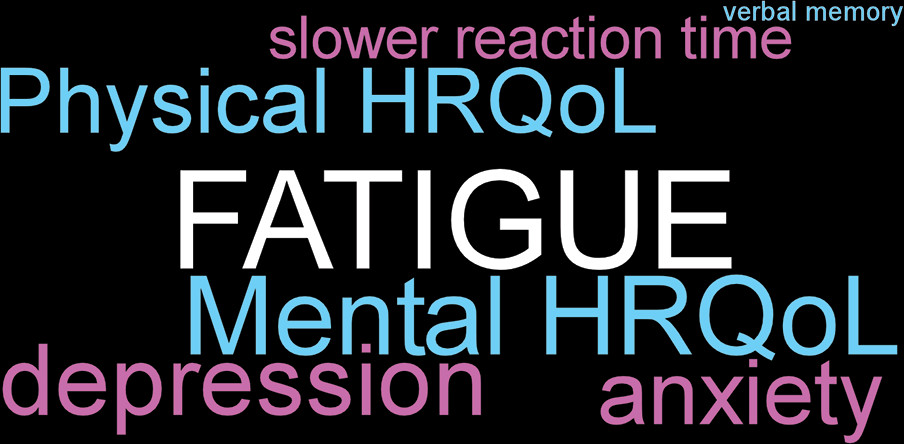
3,1925 Common functional problems include chronic fatigue, depression, and insomnia. As noted in a recent review, the substantial associations among these conditions (Figure 1) complicate identification and management.25 Although evidence varies, inattention, poor working memory, poor learning, and slowed psychomotor speed are the most commonly noted cognitive problems. When cognitive deficits are present, they tend to be mild.26,27 Notably, studies generally do not account for confounding factors present in this population (e.g., depressive symptoms, chronic fatigue) that can significantly influence cognitive performance, making it unclear whether HCV has any direct effects on cognition. A systematic review of studies found that HCV eradication led to improved functioning (e.g., HRQoL, cognition).28 However, patients who attained SVR (SVRs) were still impaired compared with healthy individuals. More recent studies have confirmed modestly improved functioning in SVRs posttreatment compared with individual baselines and to treatment nonresponders.2935 Patients with more severe liver disease may have greater functional gains.34 Overall, these results indicate that peripheral viral clearance decreases impairments but does not result in return to baseline levels of functioning in all HCV patients. The pathophysiological basis for these impairments is an area of debate.
FIGURE 1. Patients with mild chronic hepatitis C virus (HCV) infection report poorer health related quality of life (HRQoL) in multiple domains of functioning. Neuropsychiatric impairments (e.g., chronic fatigue, depression, insomnia) are reported much more consistently than cognitive impairments (e.g., inattention, reduced learning and/or memory, slowed psychomotor speed). Illustrated as a cloud diagram (phrases are scaled to magnitude of relationship) are the significant positive (purple) and negative (blue) associations between fatigue and aspects of functioning reported in a recent study.1
Having a diagnosis of HCV is proposed to be a psychosocial factor that negatively affects self-perceived HRQoL.24,27 A set of studies in voluntary blood donors obtained HRQoL from donors with positive initial tests at the time of confirmatory testing and after notification of the results.36,37 Prior to confirmatory testing, donors with initial positive tests reported lower scores on multiple HRQoL domains than matched (age, sex, race) blood donors with negative tests.36 When the group was divided based on the latter confirmatory test results, donors that were true positives had reported generally lower scores than donors who were false positives on multiple domains, but only physical role and emotional role components were statistically different.37 The groups differed significantly on the additional components of vitality and social functioning following notification of confirmatory test results, due primarily to improved scores in the false positive group, perhaps indicating relief from stress associated with initially testing positive. At clinical follow-up of the true positive group, improvements were observed in multiple domains. As noted by the authors, these results support the beneficial effects of receiving appropriate counseling and other clinical care.36,37
Studies assessing hepatic encephalopathy (HE) have reported presence of minimal HE in a substantial proportion of HCV patients.15,3841 Minimal HE is defined by impaired performance on cognitive tests in the absence of clinical manifestations of liver disease. A recent study utilizing hierarchical clustering analysis identified multiple patterns of deficits.41 The authors speculated that this symptomatic heterogeneity suggests involvement of different brain systems and potentially different underlying mechanisms across patients.41 This is consistent with an earlier proposal of neurocognitive disorder due to HCV that is distinct from minimal HE.15 Multiple pathophysiological mechanisms have been proposed for these impairments in brain functioning.15,3840,42 Some are related to liver dysfunction (e.g., hyperammonemia-induced brain edema, release of proinflammatory substances into the circulation). Others are secondary to the wide range of co-occurring conditions (e.g., renal dysfunction, hyponatremia, diabetes, sepsis, cardiovascular disease). Mechanisms secondary to HCV infection of the brain have also been proposed.
Functional neuroimaging provides multiple approaches to noninvasive assessment that may provide insight into pathophysiology. The methods that have been most commonly utilized in relevant studies are proton (1H) magnetic resonance spectroscopy (MRS), diffusion tensor imaging (DTI), and positron emission tomography (PET). Proton MRS provides noninvasive measures of several metabolites (after suppression of the signal from water) that are sensitive to changes in neuronal and glial state and/or density.2,3 The most commonly assessed metabolites include N-acetylaspartate (NAA; considered a marker for neurons/axons because it is located primarily in mature neurons), choline-containing compounds (Cho; considered a marker for cell turnover and membrane metabolism), creatine and phosphocreatine (Cr; considered a marker of cellular energy metabolism), and myoinositol (mI; considered a marker of glial cells because it serves as an intraglial osmolyte). In general, metabolites are normalized (reported as a ratio) to Cr. Cr is used as an internal standard because its level is thought to be stable; however, there is evidence that Cr level may be altered by some disease conditions, including neuroinflammation.2 Diffusion tensor imaging (DTI) is a type of magnetic resonance imaging that provides metrics for the speed and direction of water diffusion within the voxel. Mean diffusivity (MD) is an averaged measure of speed of diffusion in the three main directions. Fractional anisotropy (FA) is a measure of the degree to which diffusion is faster in one direction than others, and is highest in voxels containing highly parallel densely packed axons. Reductions in FA and elevations in MD are thought to indicate impaired white matter integrity. Microglial state is also assessed using the positron emission tomography (PET) ligand PK11195, which binds to the mitochondrial membrane translocator protein (TPSO) present in endothelial, astroglial, and microglial cells. It is generally considered a marker for microglial activation.
Several changes in neuroimaging metrics are considered to characterize individuals with minimal HE compared with healthy controls.
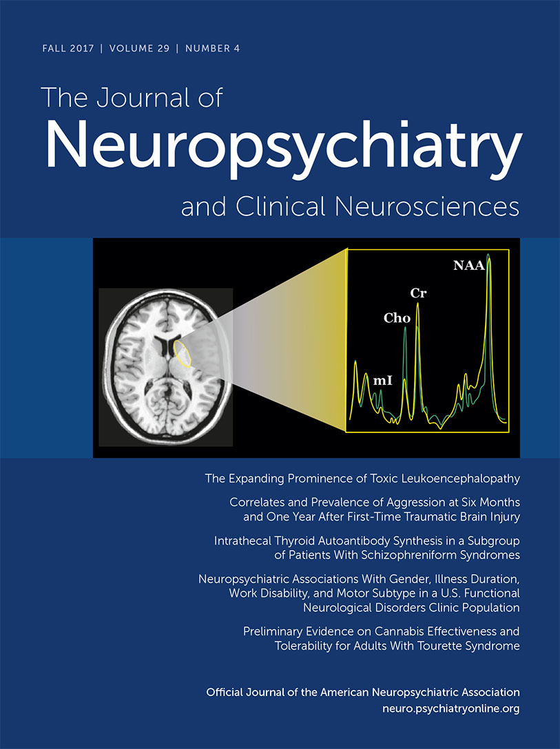
3,4,4246 Changes on proton MRS (expressed either as concentrations or referenced to Cr) include decreases in mI and Cho, and normal levels of NAA (Cover and Figure 2). For DTI, the most characteristic change is increased MD in most or all areas assessed, and normal FA. Two recent studies have assessed the value of specific metrics for differentiating patients with and without minimal HE.4,44 A study that compared MRS measures reported that the group with minimal HE had higher levels of glutamate (no difference in glutamine) than the group without; whereas mI did not differ significantly between groups.44 A study that included both MRS and DTI reported that greater changes in all metrics were present in the minimal HE group, but that higher MD discriminated presence of minimal HE better than any of the MRS metrics.
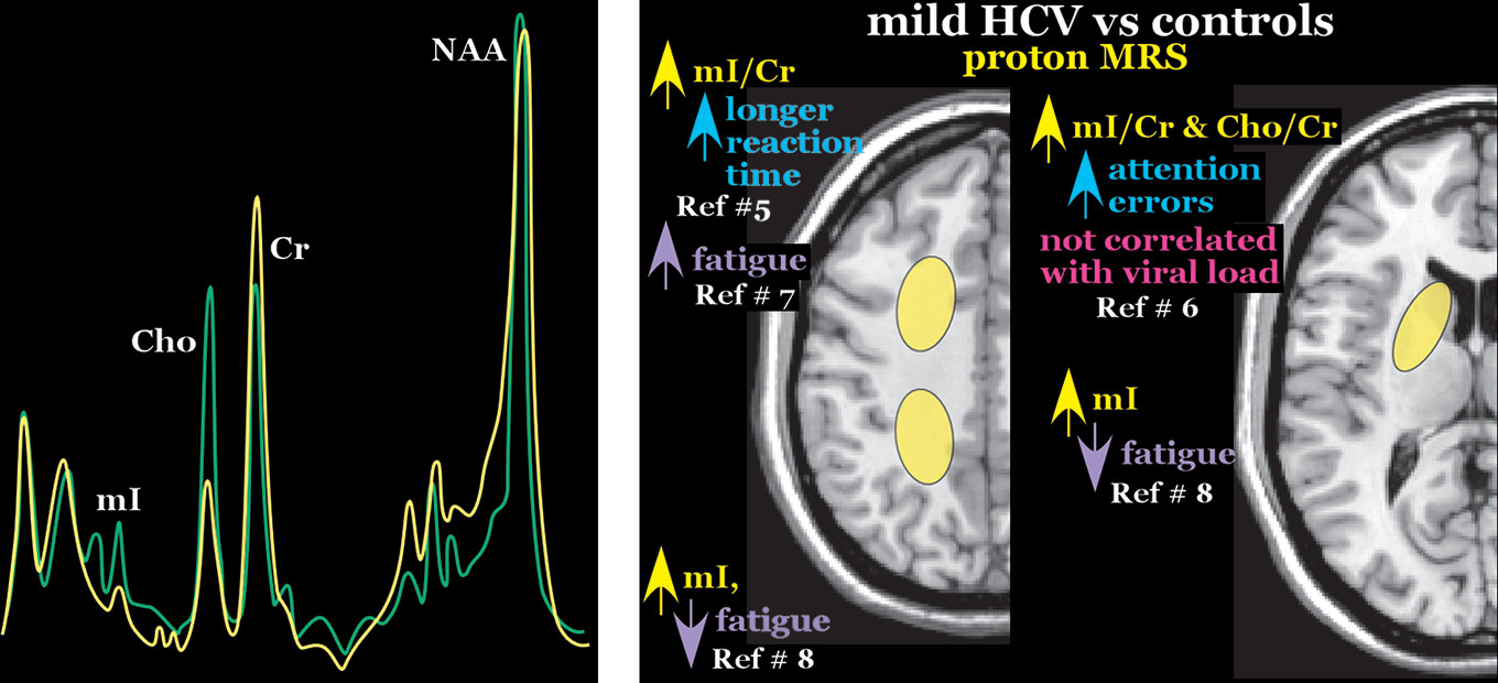
4
FIGURE 2 AND COVER. Proton magnetic resonance spectroscopy (MRS) provides noninvasive measures of several metabolites that are sensitive to changes in neuronal and glial state and/or density.2,3 Left and Cover. Simplified representative proton MRS contrast of commonly reported differences between healthy individuals (green) and patients with minimal hepatic encephalopathy (yellow).4 N-acetylaspartate (NAA) is considered a marker for neurons/axons. The choline-containing compound (Cho) peak is considered a marker for cell turnover and membrane metabolism. The creatine-phosphocreatine (Cr) peak is considered a marker of cellular energy metabolism. Myoinositol (mI) is considered a marker for glial cells and increases are thought to reflect microglial activation. In general, metabolites are normalized (reported as a ratio) to Cr. Although Cr is used as an internal standard because its level is thought to be stable, there is evidence that Cr level may be altered by some disease conditions, including neuroinflammation.2 Right. Two groups have reported that elevated mI/Cr (frontal white matter, basal ganglia) correlated with greater fatigue (purple) or more cognitive impairment (blue).57 Although viral load (pink) was associated with increasing fatigue in one study, it was not associated with any MRS metric.6 In contrast, another group reported significant elevations in parietal white matter mI only for the subset of HCV patients without pathological levels of fatigue and a negative correlation between fatigue and mI concentration.8 Overall, these studies suggest that microglial activation is associated with both beneficial and detrimental effects and is unrelated to viremia.
The changes on neuroimaging metrics reported in studies comparing patients with mild HCV (positive for the virus but no cirrhosis) to healthy individuals differ greatly from those considered to characterize minimal HE. Although studies vary considerably in the area(s) of brain most affected, mild HCV is characterized on proton MRS by higher mI (or mI/Cr), higher Cho (or Cho/Cr), and often lower NAA (or NAA/Cr).58,4749 Two groups have reported that elevated mI/Cr in the frontal white matter and/or basal ganglia correlated with greater fatigue and/or poorer cognitive performance in some domains (working memory reaction time, processing speed, language fluency), suggesting that microglial activation is detrimental (Figure 2).57 In contrast, another group reported significant elevations in parietal white matter mI only for the subset of HCV patients without pathological levels of fatigue. There were no differences when grouped by depression or attention performance.8 This study reported a negative correlation between fatigue and mI concentration, supporting a beneficial effect for microglial activation (Figure 2).8 Another study reported elevations in frontal white matter Cho/Cr and NAA/Cr in mild HCV that correlated with greater severity of depression and higher fatigue.26 Two studies compared MRS metrics prior-to and following treatment.29,30 One reported increased Cho/Cr in the basal ganglia in SVRs, but not improved cognitive performance.29 The other study reported decreased mI/Cr and Cho/Cr in the basal ganglia in SVRs accompanied by improvement on some domains of functioning.30 Both studies that utilized DTI reported that MD was increased in only a few of the areas assessed.7,47 One study reported decreased FA in all white matter areas measured.47 The other found no differences in FA of white matter areas, but increased FA in striatum, thalamus, and insula.7 Higher striatal FA correlated with poorer cognitive performance.7 Following treatment, only the SVRs with improved cognitive performance showed evidence of improved white matter integrity (increased FA and/or decreased MD).
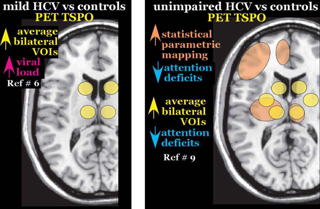
35 Studies from two research groups have compared PET TSPO binding in patients with mild HCV to healthy controls.6,9 One study reported significantly higher binding potential in all subcortical areas assessed (caudate, thalamus, pallidum) but in no cortical areas (frontal, parietal, temporal, occipital) (Figure 3).6 Higher binding potential in thalamus (trend for caudate) correlated with higher viral load. In turn, higher viral load correlated with greater fatigue. No PET metric correlated with cognitive performance or with MRS metrics.6 The other study reported no differences between patients with mild HCV and healthy controls.9 Higher binding potential was reported in frontal cortex only for the subgroup of patients no longer positive for the virus. The substantial differences across studies in changes of functional imaging metrics and direction of associations (i.e., positive, negative) with functional status in groups with mild HCV suggest that the underlying neurobiological states may vary considerably in this patient group.
FIGURE 3. Microglial state is also assessed using the positron emission tomography (PET) ligand PK11195, which binds to the mitochondrial membrane translocator protein (TPSO) present in endothelial, astroglial, and microglial cells. It is generally considered a marker for microglial activation. Left. Studies from two research groups have compared PET TSPO binding in patients with mild HCV to healthy controls.6,9 One study reported significantly higher binding potential in all subcortical (caudate, thalamus, pallidum) volumes of interest (VOIs) assessed but in no cortical VOIs (frontal, parietal, temporal, occipital).6 Higher binding potential in thalamus (trend for caudate) correlated with higher viral load (pink). No PET metric correlated with cognitive performance or with MRS. The other study reported no differences between patients with mild HCV and healthy controls in any VOIs assessed (amygdala, thalamus, caudate, putamen, pallidum, pons, cerebellum, and frontal, temporal and occipital cortices).9 Right. When the HCV group was subdivided by cognitive performance, TSPO binding potential was higher in the subgroup with intact attention (blue) than in healthy controls in some subcortical VOIs (yellow; putamen, caudate, thalamus) but not in cortical VOIs.9 The same comparison using statistical parametric mapping identified both subcortical and cortical regions with higher binding potential (orange). Though preliminary, these results provide further evidence that microglial activation may be beneficial in some circumstances.
Studies in cohorts of HCV patients without cirrhosis or relevant complicating conditions (e.g., other viral infections, history of substance use) have reported similarly high rates of impairment (e.g., HRQoL, sleep, depression, fatigue, cognition) in subgroupings based on viral testing (negative or positive)—even decades after initial infection.1,9,27,50 One study found 42% of the HCV group that had cleared the virus had impaired HRQoL, compared with 47% of the group that still tested positive.27 Logistic regression identified depression and fatigue as important factors influencing impaired HRQoL. Another research group has done a series of studies examining different aspects of functioning.1,9,50 In one study, they found that 95% of their HCV group reported disturbed sleep, despite 40% having cleared the virus.50 Measures of HRQoL, depression, and fatigue were significantly correlated with measures of sleep, suggesting depression as a possible mediator of impaired sleep quality. Their most recent study reported that a much greater proportion of HCV patients had pathological levels of neuropsychiatric symptoms (e.g., fatigue, anxiety, depression) than were impaired on cognitive testing regardless of viral status.1 Additionally, HRQoL was negatively associated with fatigue, depression, and attention (Figure 1). Notably, attention was also independently associated with fatigue. The cognitive measure with the highest rate of impaired performance was not correlated with any neuropsychiatric symptom domain or with attention, but was positively correlated with time since infection. These results support the importance of pathophysiological mechanisms other than viremia, and suggest independence between cognitive and functional impairments.1 Findings from their small study comparing PET TSPO between HCV patients and healthy individuals is consistent with this hypothesis.9 TSPO binding potential was higher in the HCV subgroup with intact attention than in healthy controls in subcortical (putamen, caudate, thalamus) volumes of interest (VOIs) (Figure 3). In contrast, TSPO binding was higher in the HCV subgroup without fatigue than in healthy individuals in the temporal cortex.9 As noted by the authors, these results support microglial activation as a beneficial process.
The pathophysiological mechanisms of cognitive and functional problems as well as sleep disturbance in HCV patients, in the absence of viremia, remain poorly understood. Proposed mechanisms include direct infection of the brain, chronic neuroinflammatory response, indirect stimulation of neurotoxic cytokine pathways, as well as the development of cerebral vasculitis.18,19,21,22,51 Most concerning, although sample sizes are relatively small, is the mounting evidence that HCV infection can spread into the brain.3,20,2224 The presence of HCV RNA in cerebrospinal fluid (CSF) of HCV-infected patients was the first indication. The possibility that HCV directly infects brain cells is supported by the presence of HCV negative strand RNA (a replicative intermediate) or other molecules linked to HCV replication in samples of postmortem brain tissue.52,53 Both studies reported glia as the major cell type infected, but differ on whether microglia or astroglia are more involved. HCV was relatively less abundant in brain than liver specimens.22 It has been suggested that the low levels of viral replication might not induce a sufficient immune response, therefore possibly supporting a chronic, persistent infection.24 In addition, these findings could also indicate passage of infected peripheral blood mononuclear cells (PBMCs) into the brain. The detection of different HCV strains (quasispecies) in brain and serum provide support for genetic compartmentalization and the hypothesis that there is independent viral evolution in the two sites of replication.3,20,22,23Most recently, the possibility of independent viral evolution was investigated using a deep sequencing approach in a small cohort of HCV patients without liver disease.3,54 In two-thirds of the group (4/6), HCV was detected in CSF. In half (2/4), different viral quasispecies were identified in CSF and plasma, further supporting the presence of independent viral evolution in some patients. Multiple authors have noted that the growing evidence of HCV neuroinfection in some patients brings the long-term efficacy of current therapies into question.
The precise method of HCV entry into the brain is speculative. Brain microvascular endothelial cells, a major component of the blood brain barrier (BBB), have been shown to express all receptors required for HCV internalization whereas other brain-derived cell lines (e.g., microglia, glioblastoma) did not.17,55,56 In vitro studies indicate that HCV infects brain microvascular endothelial cells that release the virus at low levels and also undergo apoptosis.55 If these processes occur in vivo, they would provide mechanisms (e.g., reservoir of infectious virus, altered BBB permeability) by which HCV could directly affect the brain.20,22,55 It has also been proposed that HCV may enter through a Trojan horse mechanism, similar to what is hypothesized for human immunodeficiency virus, as infected PBMCs can cross the BBB and serve as precursors of microglial cells.20,22,55 Increased viral load in the periphery might facilitate infiltration of the brain by this mechanism.54
Treatments for HCV are now very effective, with SVR increasingly likely with newer direct-acting antiviral agents.14 However, it is unclear if SVR equates to complete eradication of the virus. In addition, it is likely that antiviral therapies for HCV have limited ability to cross the BBB.14,54 Reactivation of HCV has been found to occur, especially in immunocompromised patients.57,58 It is possible that HCV-infected brain microvascular endothelial cells and/or glial cells may provide a reservoir for the virus to persist during antiviral treatment.22,55 Late relapses (up to 5 years after achieving SVR) following antiviral treatment might be explained by the slower replication rate of HCV in the brain.59 Studies assessing longer-term SVR with new therapies will be needed.
In conclusion, neuroimaging studies of subjects with HCV has contributed to the understanding of this complex viral infection. Early evidence indicates that in some patients HCV can pass through the BBB, that several cell types may serve as a viral reservoir, and that the virus can evolve independently necessitating further research to answer key questions. These include: What proportion of HCV-infected patients will have brain involvement; how extensive is this involvement; do the antivirals clear the brain of the virus; and are there lasting neuropsychiatric impairments following clearance of the virus?

References

1.
Dirks M, Pflugrad H, Haag K, et al: Persistent neuropsychiatric impairment in HCV patients despite clearance of the virus?! J Viral Hepat 2017; 24:541–550
2.
Chang L, Munsaka SM, Kraft-Terry S, et al: Magnetic resonance spectroscopy to assess neuroinflammation and neuropathic pain. J Neuroimmune Pharmacol 2013; 8:576–593
3.
Yarlott L, Heald E, Forton D: Hepatitis C virus infection, and neurological and psychiatric disorders: A review. J Adv Res 2017; 8:139–148
4.
Srivastava A, Chaturvedi S, Gupta RK, et al: Minimal hepatic encephalopathy in children with chronic liver disease: Prevalence, pathogenesis and magnetic resonance-based diagnosis. J Hepatol 2017; 66:528–536
5.
Forton DM, Hamilton G, Allsop JM, et al: Cerebral immune activation in chronic hepatitis C infection: a magnetic resonance spectroscopy study. J Hepatol 2008; 49:316–322
6.
Grover VPB, Pavese N, Koh S-B, et al: Cerebral microglial activation in patients with hepatitis C: in vivo evidence of neuroinflammation. J Viral Hepat 2012; 19:e89–e96
7.
Thames AD, Castellon SA, Singer EJ, et al: Neuroimaging abnormalities, neurocognitive function, and fatigue in patients with hepatitis C. Neurol Neuroimmunol Neuroinflamm 2015; 2:e59
8.
Bokemeyer M, Ding XQ, Goldbecker A, et al: Evidence for neuroinflammation and neuroprotection in HCV infection-associated encephalopathy. Gut 2011; 60:370–377
9.
Pflugrad H, Meyer GJ, Dirks M, et al: Cerebral microglia activation in hepatitis C virus infection correlates to cognitive dysfunction. J Viral Hepat 2016; 23:348–357
10.
Rosenberg ES, Hall EW, Sullivan PS, et al: Estimation of state-level prevalence of hepatitis C virus infection, US states and District of Columbia, 2010. Clin Infect Dis 2017; 64:1573–1581
11.
Institute of Medicine: Hepatitis and Liver Cancer: A National Strategy for Prevention and Control of Hepatitis B and C. Washington, DC, National Academies Press, 2010
12.
Maasoumy B, Wedemeyer H: Natural history of acute and chronic hepatitis C. Best Pract Res Clin Gastroenterol 2012; 26:401–412
13.
Tahir U, ul Ain Q, Waqas R, et al: Comparative prevalence studies of hepatitis c patients in developing vs developed countries. Int Curr Pharma J 2016; 5:106–110
14.
Kiser JJ, Burton JR Jr, Everson GT: Drug-drug interactions during antiviral therapy for chronic hepatitis C. Nat Rev Gastroenterol Hepatol 2013; 10:596–606
15.
Monaco S, Mariotto S, Ferrari S, et al: Hepatitis C virus-associated neurocognitive and neuropsychiatric disorders: Advances in 2015. World J Gastroenterol 2015; 21:11974–11983
16.
Murray J, Fishman SL, Ryan E, et al: Clinicopathologic correlates of hepatitis C virus in brain: A pilot study. J Neurovirol 2008; 14:17–27
17.
Bürgel B, Friesland M, Koch A, et al: Hepatitis C virus enters human peripheral neuroblastoma cells: Evidence for extra-hepatic cells sustaining hepatitis C virus penetration. J Viral Hepat 2011; 18:562–570
18.
Sherman AC, Sherman KE: Extrahepatic manifestations of hepatitis C infection: navigating CHASM. Curr HIV/AIDS Rep 2015; 12:353–361
19.
Cacoub P, Comarmond C, Domont F, et al: Extrahepatic manifestations of chronic hepatitis C virus infection. Ther Adv Infect Dis 2016; 3:3–14
20.
Adinolfi LE, Nevola R, Lus G, et al: Chronic hepatitis C virus infection and neurological and psychiatric disorders: An overview. World J Gastroenterol 2015; 21:2269–2280
21.
Laskus T, Radkowski M, Adair DM, et al: Emerging evidence of hepatitis C virus neuroinvasion. AIDS 2005; 19(Suppl 3):S140–S144
22.
Fletcher NF, McKeating JA: Hepatitis C virus and the brain. J Viral Hepat 2012; 19:301–306
23.
Gill K, Ghazinian H, Manch R, et al: Hepatitis C virus as a systemic disease: Reaching beyond the liver. Hepatol Int 2016; 10:415–423
24.
Weissenborn K, Tillmann HL: HCV encephalopathy: An artefact due to medical care? J Viral Hepat 2016; 23:580–583
25.
Golabi P, Sayiner M, Bush H, et al: Patient-reported outcomes and fatigue in patients with chronic hepatitis C infection. Clin Liver Dis 2017; 21:565–578
26.
Allah HMS, Ibrahim SA, Elyamany AS, et al: Neuropsychiatric disorders and magnetic resonance spectroscopy in chronic hepatitis C patients. Egypt J Neurol Psychiat Neurosurg 2014; 51:345–350
27.
Lowry D, Burke T, Galvin Z, et al: Is psychosocial and cognitive dysfunction misattributed to the virus in hepatitis C infection? Select psychosocial contributors identified. J Viral Hepat 2016; 23:584–595
28.
Spiegel BM, Younossi ZM, Hays RD, et al: Impact of hepatitis C on health-related quality of life: A systematic review and quantitative assessment. Hepatology 2005; 41:790–800
29.
Pattullo V, McAndrews MP, Damyanovich A, et al: Influence of hepatitis C virus on neurocognitive function in patients free from other risk factors: Validation from therapeutic outcomes. Liver Int 2011; 31:1028–1038
30.
Byrnes V, Miller A, Lowry D, et al: Effects of anti-viral therapy and HCV clearance on cerebral metabolism and cognition. J Hepatol 2012; 56:549–556
31.
Kraus MR, Schäfer A, Teuber G, et al: Improvement of neurocognitive function in responders to an antiviral therapy for chronic hepatitis C. Hepatology 2013; 58:497–504
32.
Huckans M, Fuller B, Wheaton V, et al: A longitudinal study evaluating the effects of interferon-alpha therapy on cognitive and psychiatric function in adults with chronic hepatitis C. J Psychosom Res 2015; 78:184–192
33.
Younossi ZM, Stepanova M, Marcellin P, et al: Treatment with ledipasvir and sofosbuvir improves patient-reported outcomes: Results from the ION-1, -2, and -3 clinical trials. Hepatology 2015; 61:1798–1808
34.
Younossi ZM, Stepanova M, Feld J, et al: Sofosbuvir and velpatasvir combination improves patient-reported outcomes for patients with HCV infection, without or with compensated or decompensated cirrhosis. Clin Gastroenterol Hepatol 2017; 15:421–430.e6
35.
Kuhn T, Sayegh P, Jones JD, et al: Improvements in brain and behavior following eradication of hepatitis C. J Neurovirol 2017
36.
Strauss E, Porto-Ferreira FA, de Almeida-Neto C, et al: Altered quality of life in the early stages of chronic hepatitis C is due to the virus itself. Clin Res Hepatol Gastroenterol 2014; 38:40–45
37.
Ferreira FA, de Almeida-Neto C, Teixeira MC, et al: Health-related quality of life among blood donors with hepatitis B and hepatitis C: Longitudinal study before and after diagnosis. Rev Bras Hematol Hemoter 2015; 37:381–387
38.
Solinas A, Piras MR, Deplano A: Cognitive dysfunction and hepatitis C virus infection. World J Hepatol 2015; 7:922–925
39.
Nardone R, Taylor AC, Höller Y, et al: Minimal hepatic encephalopathy: A review. Neurosci Res 2016; 111:1–12
40.
Ferenci P: Hepatic encephalopathy. Gastroenterol Rep (Oxf) 2017; 5:138–147
41.
Giménez-Garzó C, Garcés JJ, Urios A, et al: The PHES battery does not detect all cirrhotic patients with early neurological deficits, which are different in different patients. PLoS One 2017; 12:e0171211
42.
Grover VP, Tognarelli JM, Massie N, et al: The why and wherefore of hepatic encephalopathy. Int J Gen Med 2015; 8:381–390
43.
Alonso J, Córdoba J, Rovira A: Brain magnetic resonance in hepatic encephalopathy. Semin Ultrasound CT MR 2014; 35:136–152
44.
Meng LP, Chen YC, Li YH, et al: Viability assessment of magnetic resonance spectroscopy for the detection of minimal hepatic encephalopathy severity. Eur J Radiol 2015; 84:2019–2023
45.
Sureka B, Bansal K, Patidar Y, et al: Neurologic manifestations of chronic liver disease and liver cirrhosis. Curr Probl Diagn Radiol 2015; 44:449–461
46.
Yadav SK, Goel A, Saraswat VA, et al: Evaluation of cognitivity, proinflammatory cytokines, and brain magnetic resonance imaging in minimal hepatic encephalopathy induced by cirrhosis and extrahepatic portal vein obstruction. J Gastroenterol Hepatol 2016; 31:1986–1994
47.
Bladowska J, Zimny A, Knysz B, et al: Evaluation of early cerebral metabolic, perfusion and microstructural changes in HCV-positive patients: a pilot study. J Hepatol 2013; 59:651–657
48.
Forton DM, Allsop JM, Main J, et al: Evidence for a cerebral effect of the hepatitis C virus. Lancet 2001; 358:38–39
49.
Weissenborn K, Krause J, Bokemeyer M, et al: Hepatitis C virus infection affects the brain-evidence from psychometric studies and magnetic resonance spectroscopy. J Hepatol 2004; 41:845–851
50.
Heeren M, Sojref F, Schuppner R, et al: Active at night, sleepy all day--sleep disturbances in patients with hepatitis C virus infection. J Hepatol 2014; 60:732–740
51.
Kharabian Masouleh S, Herzig S, Klose L, et al: Functional connectivity alterations in patients with chronic hepatitis C virus infection: A multimodal MRI study. J Viral Hepat 2017; 24:216–225
52.
Letendre S, Paulino AD, Rockenstein E, et al: HIV Neurobehavioral Research Center Group: Pathogenesis of hepatitis C virus coinfection in the brains of patients infected with HIV. J Infect Dis 2007; 196:361–370
53.
Wilkinson J, Radkowski M, Laskus T: Hepatitis C virus neuroinvasion: identification of infected cells. J Virol 2009; 83:1312–1319
54.
Tully DC, Hjerrild S, Leutscher PD, et al: Deep sequencing of hepatitis C virus reveals genetic compartmentalization in cerebrospinal fluid from cognitively impaired patients. Liver Int 2016; 36:1418–1424
55.
Fletcher NF, Wilson GK, Murray J, et al: Hepatitis C virus infects the endothelial cells of the blood-brain barrier. Gastroenterology 2012; 142:634–643.e6
56.
Koyuncu OO, Hogue IB, Enquist LW: Virus infections in the nervous system. Cell Host Microbe 2013; 13:379–393
57.
Yazici O, Şendur MAN, Aksoy S: Hepatitis C virus reactivation in cancer patients in the era of targeted therapies. World J Gastroenterol 2014; 20:6716–6724
58.
Tagawa A, Ogawa T, Tetsuka S, et al: Hepatitis C virus (HCV) reactivation during fingolimod treatment for relapsing and remitting multiple sclerosis. Mult Scler Relat Disord 2016; 9:155–157
59.
Tillmann HL, Hepatitis C: Hepatitis C virus infection: when is a cure not a cure? J Infect Dis 2014; 209:6–8

Information & Authors

Information

Published In

Go to The Journal of Neuropsychiatry and Clinical Neurosciences
The Journal of Neuropsychiatry and Clinical Neurosciences
Pages: A6 - 307
PubMed: 28974163

History

Published in print: Fall 2017
Published online: 4 October 2017

Keywords

  1. Imaging Techniques
  2. Neurology

Authors

Affiliations

Sarah L. Martindale, Ph.D.
Dr. Martindale is affiliated with the Mental Health Service Line at the W.G. Hefner Veterans Affairs Medical Center in Salisbury, North Carolina and the Department of Psychiatry at Wake Forest School of Medicine in Winston-Salem, North Carolina. Drs. Martindale, Hurley, and Taber are affiliated with the Veterans Affairs Mid Atlantic Mental Illness Research, Education, and Clinical Center, and the Research and Academic Affairs Service Line at the W.G. Hefner Veterans Affairs Medical Center in Salisbury, North Carolina. Dr. Hurley is affiliated with the Departments of Psychiatry and Radiology at Wake Forest School of Medicine in Winston-Salem, North Carolina, and the Menninger Department of Psychiatry and Behavioral Sciences at Baylor College of Medicine in Houston, Texas. Dr. Taber is affiliated with the Division of Biomedical Sciences at the Via College of Osteopathic Medicine in Blacksburg, Virginia, and the Department of Physical Medicine and Rehabilitation at Baylor College of Medicine in Houston.
Robin A. Hurley, M.D. [email protected]
Dr. Martindale is affiliated with the Mental Health Service Line at the W.G. Hefner Veterans Affairs Medical Center in Salisbury, North Carolina and the Department of Psychiatry at Wake Forest School of Medicine in Winston-Salem, North Carolina. Drs. Martindale, Hurley, and Taber are affiliated with the Veterans Affairs Mid Atlantic Mental Illness Research, Education, and Clinical Center, and the Research and Academic Affairs Service Line at the W.G. Hefner Veterans Affairs Medical Center in Salisbury, North Carolina. Dr. Hurley is affiliated with the Departments of Psychiatry and Radiology at Wake Forest School of Medicine in Winston-Salem, North Carolina, and the Menninger Department of Psychiatry and Behavioral Sciences at Baylor College of Medicine in Houston, Texas. Dr. Taber is affiliated with the Division of Biomedical Sciences at the Via College of Osteopathic Medicine in Blacksburg, Virginia, and the Department of Physical Medicine and Rehabilitation at Baylor College of Medicine in Houston.
Katherine H. Taber, Ph.D.
Dr. Martindale is affiliated with the Mental Health Service Line at the W.G. Hefner Veterans Affairs Medical Center in Salisbury, North Carolina and the Department of Psychiatry at Wake Forest School of Medicine in Winston-Salem, North Carolina. Drs. Martindale, Hurley, and Taber are affiliated with the Veterans Affairs Mid Atlantic Mental Illness Research, Education, and Clinical Center, and the Research and Academic Affairs Service Line at the W.G. Hefner Veterans Affairs Medical Center in Salisbury, North Carolina. Dr. Hurley is affiliated with the Departments of Psychiatry and Radiology at Wake Forest School of Medicine in Winston-Salem, North Carolina, and the Menninger Department of Psychiatry and Behavioral Sciences at Baylor College of Medicine in Houston, Texas. Dr. Taber is affiliated with the Division of Biomedical Sciences at the Via College of Osteopathic Medicine in Blacksburg, Virginia, and the Department of Physical Medicine and Rehabilitation at Baylor College of Medicine in Houston.

Notes

Corresponding author: Send correspondence to Dr. Hurley; e-mail: [email protected].

Funding Information

This research was supported by the Department of Veterans Affairs Office of Academic Affiliations Advanced Fellowship Program in Mental Illness, Research, and Treatment

Metrics & Citations

Metrics

Citations

Export Citations

If you have the appropriate software installed, you can download article citation data to the citation manager of your choice. Simply select your manager software from the list below and click Download.

For more information or tips please see 'Downloading to a citation manager' in the Help menu.

Format
Citation style
Style
Copy to clipboard

There are no citations for this item

View Options

View options

PDF/ePub

View PDF/ePub

Get Access

Login options

Already a subscriber? Access your subscription through your login credentials or your institution for full access to this article.

Personal login Institutional Login Open Athens login
Purchase Options

Purchase this article to access the full text.

PPV Articles - Journal of Neuropsychiatry and Clinical Neurosciences

PPV Articles - Journal of Neuropsychiatry and Clinical Neurosciences

Not a subscriber?

Subscribe Now / Learn More

PsychiatryOnline subscription options offer access to the DSM-5-TR® library, books, journals, CME, and patient resources. This all-in-one virtual library provides psychiatrists and mental health professionals with key resources for diagnosis, treatment, research, and professional development.

Need more help? PsychiatryOnline Customer Service may be reached by emailing [email protected] or by calling 800-368-5777 (in the U.S.) or 703-907-7322 (outside the U.S.).

Media

Figures

Other

Tables

Share

Share

Share article link

Share