The parietal lobe has received little attention in schizophrenia despite its recognized importance in processes that are likely disturbed in schizophrenia, such as language
(1), spatial working memory, and attention
(2–
4). This lack of attention to parietal regions of the brain is further highlighted by the fact that only nine magnetic resonance imaging (MRI) studies of the parietal lobe, in contrast to more than 35 for both temporal and frontal lobes, have been conducted in subjects with schizophrenia
(5,
6).
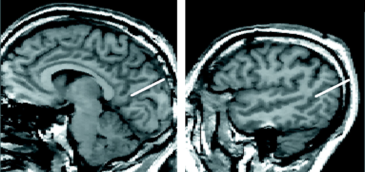
The parietal lobe itself consists of the postcentral gyrus, superior parietal gyrus, and inferior parietal lobule, which is further subdivided into the supramarginal gyrus (area 39) and angular gyrus (area 40). The inferior parietal lobule is part of the heteromodal association cortex, which has been proposed as the site of the key abnormality in schizophrenia
(7). Further, both the supramarginal gyrus and the angular gyrus have been described as part of a semantic-lexical network that supports “word meanings” represented by a “grid of connectivity” that constitutes a “final pathway for the chunking of words into thought”
(1). The role of the inferior parietal lobule, and especially the angular gyrus, in language comprehension has been further confirmed by functional MRI (fMRI) and positron emission tomography studies
(8–
10). Moreover, the semantic-lexical network proposed by Mesulam
(1) includes both the inferior parietal lobule and the planum temporale, the latter being located on the superioposterior surface of superior temporal gyrus, which, in turn, has been correlated with symptoms of thought disorder
(5,
6,
11). More anteriorly, the superior temporal gyrus has been correlated with auditory hallucinations
(5,
6,
12).
Of note, the inferior parietal lobule and neighboring cortical regions often exhibit marked lateral asymmetry
(13,
14) and belong to structures that support language. The presence of left-greater-than-right asymmetry also appears to be important for normal language development. For example, the absence of normal lateralization in these regions has been reported in subjects with autism
(15,
16) as well as in other language disorders
(17). In schizophrenia, a reversal of normal asymmetry in the superior temporal gyrus, including the planum temporale, has been associated with thought disorder
(5,
6). Thus, a reversal in normal asymmetry may be importantly related to schizophrenic pathology
(18–
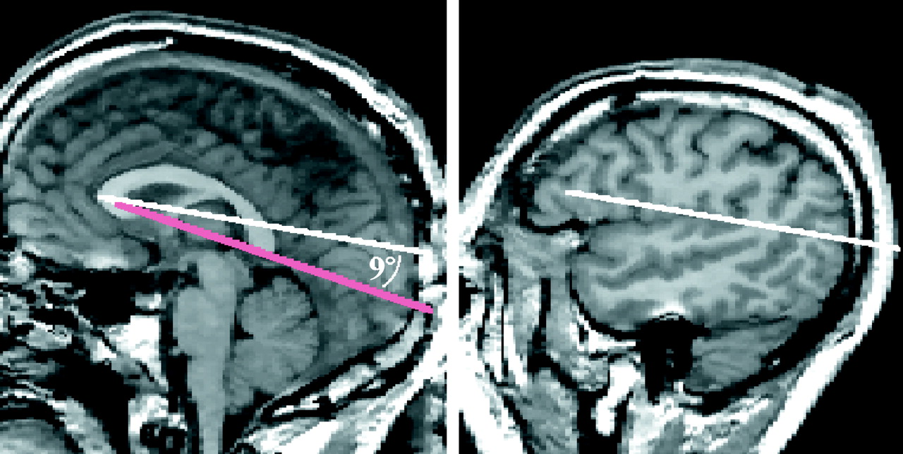
20). Consistent with these findings, we hypothesized that a reversal of normal asymmetry in the superior temporal gyrus would also be found in the inferior parietal lobule. Furthermore, since cortical asymmetries are present during fetal development
(21), finding an absence or a reversal of normal asymmetry might indicate a disruption in neuronal development.
The relevance of the angular gyrus and supramarginal gyrus to schizophrenia stems not only from their functions as part of the heteromodal association cortex but also from their reciprocal neuroanatomical connections to the prefrontal
(22) and temporal lobes
(23)—brain regions that have been shown to have disease-related abnormalities. We examined these neuroanatomical relationships by capitalizing on the opportunity of having volumetric data on prefrontal and temporal lobe regions in the same subjects
(11,
24,
25). If the hypothesis that schizophrenia is a disorder characterized primarily by heteromodal association cortex abnormalities is correct, then higher correlations between the inferior parietal lobule and other areas interconnected with the heteromodal association cortex would be predicted for schizophrenic patients but not for comparison subjects. Also, in an exploratory analysis, we examined the relationship between the heteromodal inferior parietal lobule and formal thought disorder, as well as neuropsychological measures of attention and memory, all of which are likely to contribute to thinking and judgment—functions commonly associated with the inferior parietal lobule.
RESULTS
MRI Volume Comparisons
For comparative purposes,
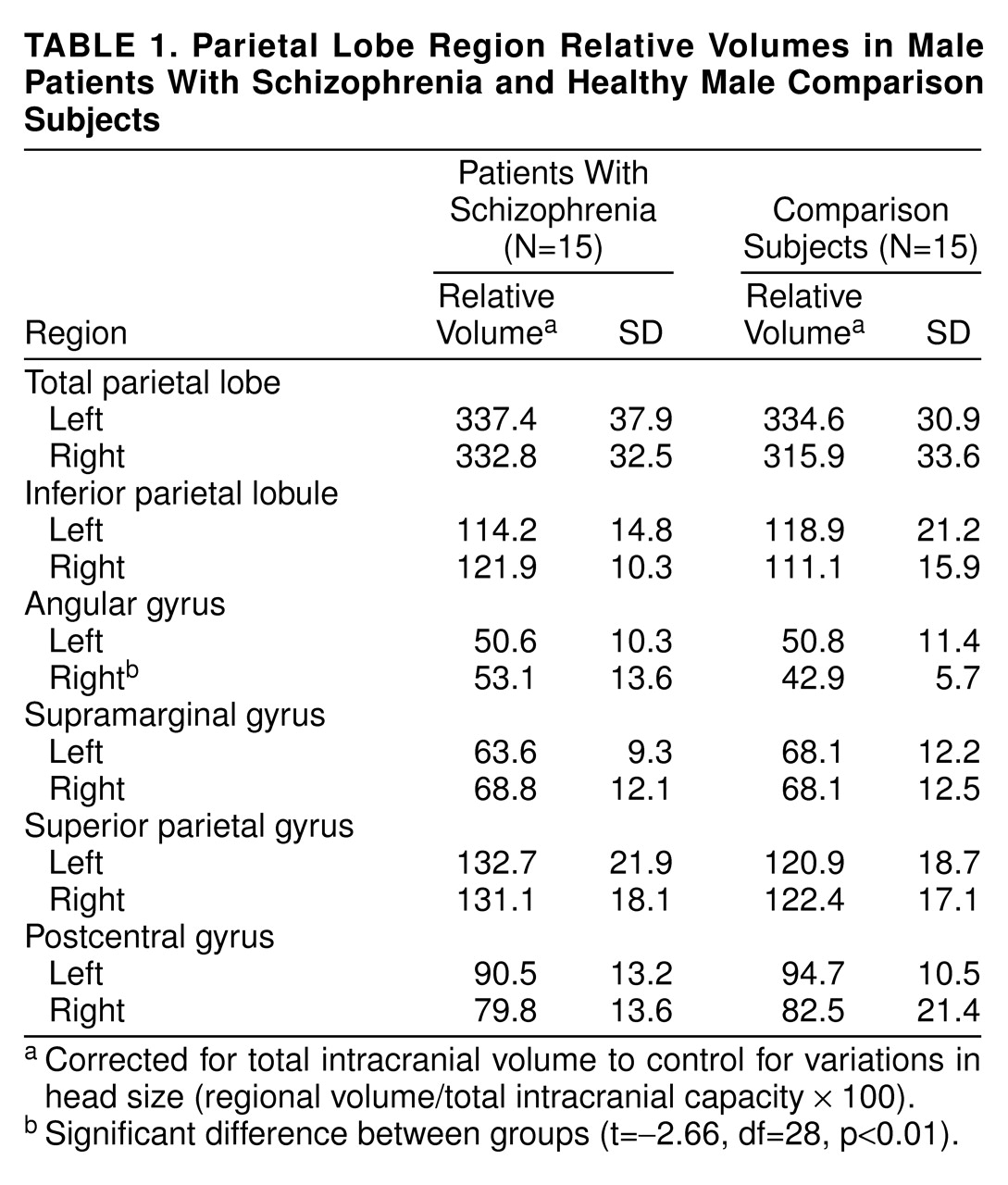
table 1 provides volumes for all parietal regions studied. As predicted, no group volume differences were found for the total parietal lobe, superior parietal gyrus, or the postcentral gyrus. However, volume differences were noted between the two groups for the inferior parietal lobule, specifically the angular gyrus.
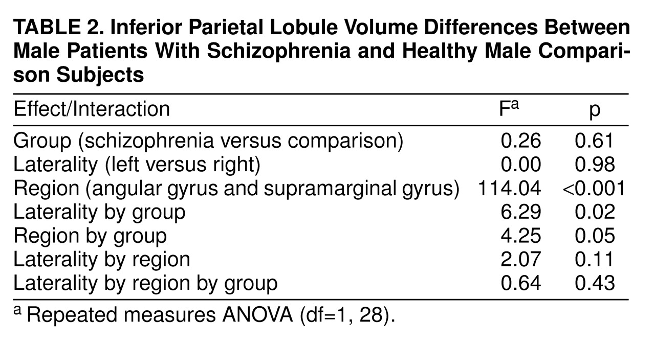
A repeated measures ANOVA showed no overall group volume differences or an overall laterality effect (
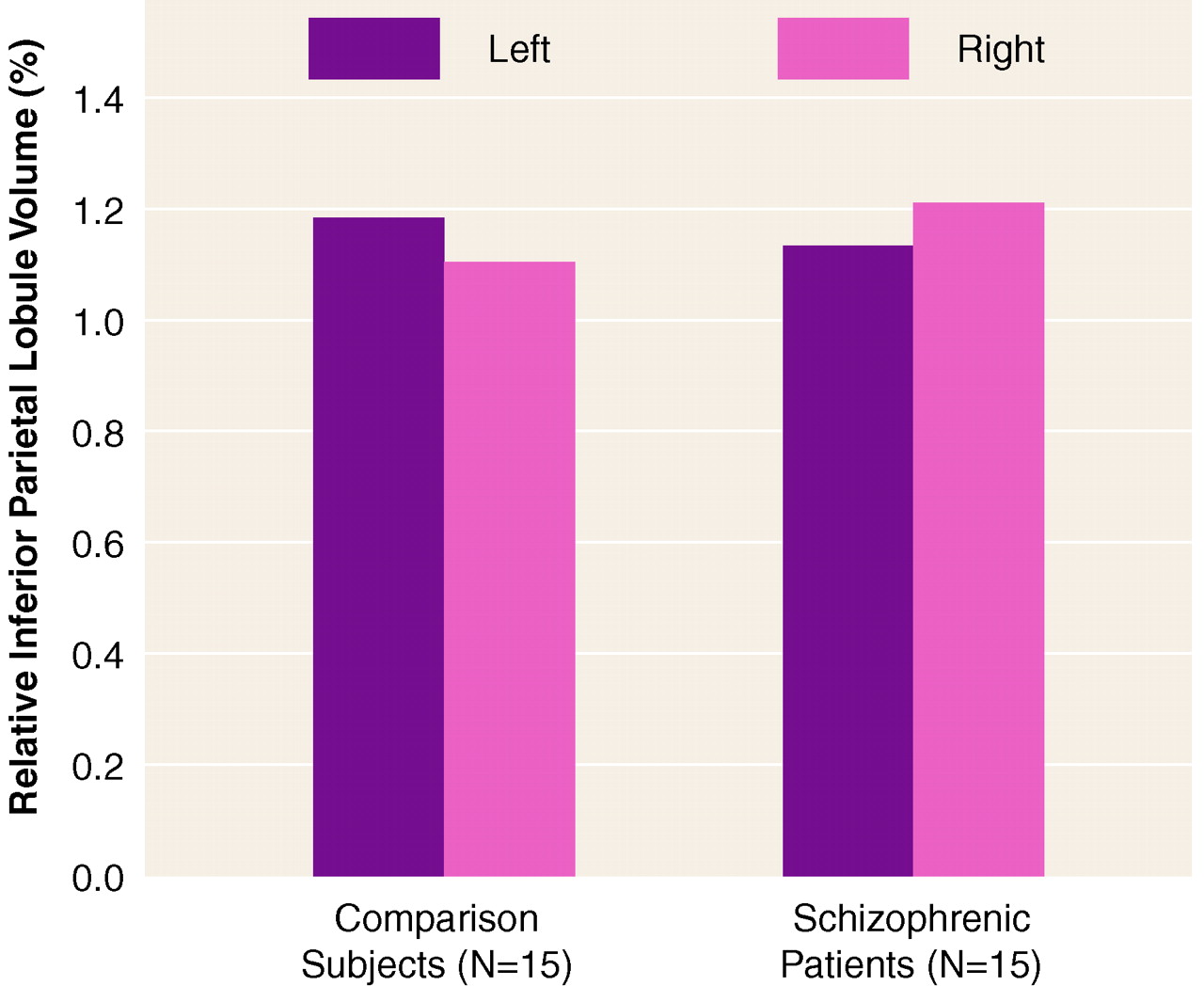
table 1). However, there was a significant laterality-by-group interaction that indicated a difference in asymmetry between the two groups. As shown in
figure 1, the comparison subjects had a leftward asymmetry (left inferior parietal lobule volume 7.0% larger than the right), and the schizophrenic patients showed a reversed asymmetry (left inferior parietal lobule volume 6.3% smaller than the right).
The volumes of the angular gyrus and the supramarginal gyrus for the two groups were significantly different (
table 2). There was also a significant region-by-group interaction. Post hoc tests indicated that schizophrenic patients had a larger right angular gyrus than the normal comparison group (
table 1). In the comparison subjects, the left angular gyrus volume was considerably larger (18.4%) than the right (paired t=2.71, df=14, p=0.02), whereas in the patients, the left angular gyrus was not significantly different (4.7% less volume) from the right (t=0.7, df=14, p>0.05).
Left-Right MRI Volume Asymmetries
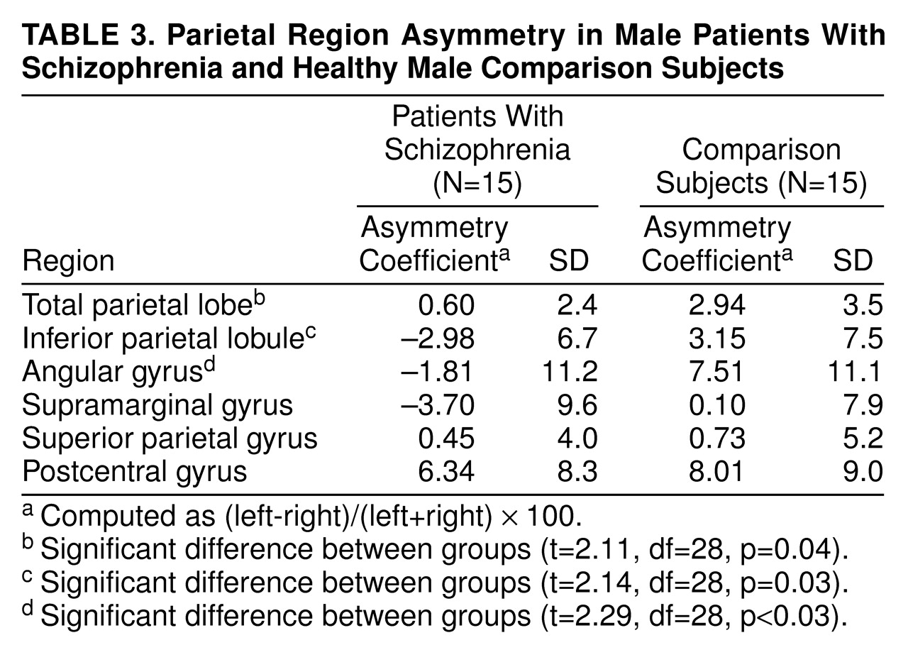
To evaluate further the aforementioned laterality-by-group interaction of the inferior parietal lobule as well as the laterality of the other parietal regions, we computed asymmetry coefficients by using the formula (left–right)/(left+right) × 100 (note that the asymmetry coefficient is dimensionless). A negative value indicates a larger right than left side volume. Student’s t tests were used to compare asymmetry coefficients between the two groups for all of the regions.
There was a significant difference between the two groups in the asymmetry coefficient for the total parietal lobe (
table 3). The comparison group exhibited a leftward asymmetry, with the left parietal lobe 6.0% larger than right (paired t=3.18, df=14, p=0.007), while the schizophrenic group exhibited virtually no total parietal asymmetry (t=–1.1, df=14, p>0.30).
Neither group exhibited significant asymmetry of the superior parietal gyrus, and both left and right postcentral gyrus volumes were similar in the schizophrenic and comparison groups. Both groups showed a significant leftward asymmetry, with the left postcentral gyrus being 14.8% larger than the right in the comparison subjects (paired t=2.76, df=14, p=0.02), and 13.4% larger in the schizophrenic patients (paired t=3.00, df=14, p=0.01).
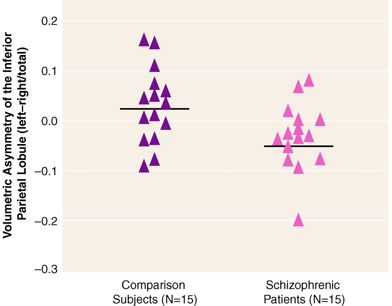
A significant group difference in asymmetry coefficient was evident for the inferior parietal lobule (
table 3), which was accounted for by a group difference in the angular gyrus. Note that all of the schizophrenic subjects except two had asymmetry coefficients below the mean (and median) of the subjects in the normal comparison group (
figure 2). The supramarginal gyrus did not show a significant asymmetry for either group. Thus, although the post hoc tests based on the ANOVA group-by-region interaction did not show statistically significant group differences between left and right angular gyrus in the schizophrenic patients, the differences between groups in the asymmetry coefficient suggests that there is a reversal of the normal asymmetry in the schizophrenic patients for the angular gyrus.
Correlations Between Parietal Gray Matter Volumes and Anatomically Connected Regions in Prefrontal and Temporal Cortex
For all correlations, significance levels were set at p≤0.05 (two-tailed), which corresponded to r>0.51 for the 15 schizophrenic subjects and r>0.53 for 14 comparison subjects (one comparison subject was dropped because artifact in the prefrontal cortex made this region too difficult to assess the volume accurately). In addition, the values reported here are for absolute volumes, but the correlations were considered significant only if they reached p≤0.05 for both relative and absolute volumes. For all of these correlations, parietal cortex regions included the right and left inferior parietal lobules, superior parietal gyrus, and postcentral gyrus; prefrontal cortex regions included right and left superior frontal, middle frontal, inferior frontal, and orbital frontal gyri; and temporal cortex regions included right and left anterior superior temporal gyrus, posterior superior temporal gyrus, parahippocampal gyrus, and amygdala-hippocampal complex.
High correlations were found between left and right inferior parietal lobule volumes in both schizophrenic (r=0.68, p<0.005) and comparison subjects (r=0.54, p<0.04), as well as between the left and right superior parietal gyrus (schizophrenic subjects: r=0.81, p<0.001; comparison subjects: r=0.75, p<0.001). Additionally, the comparison subjects showed a significant correlation between the left and right postcentral gyrus (r=0.63, p<0.01).
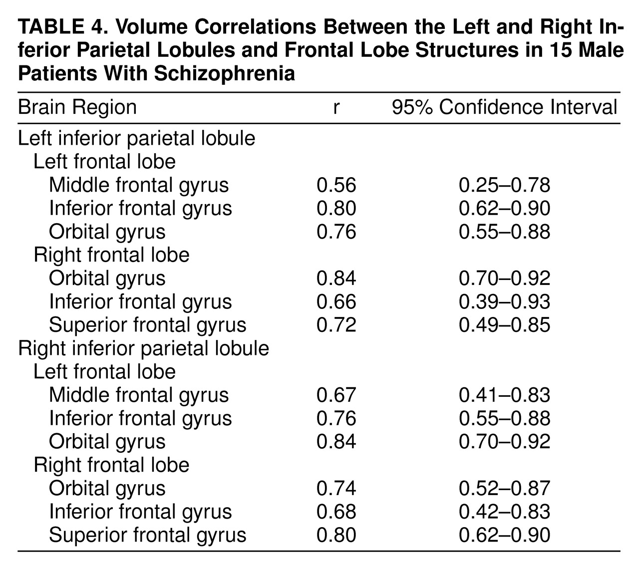
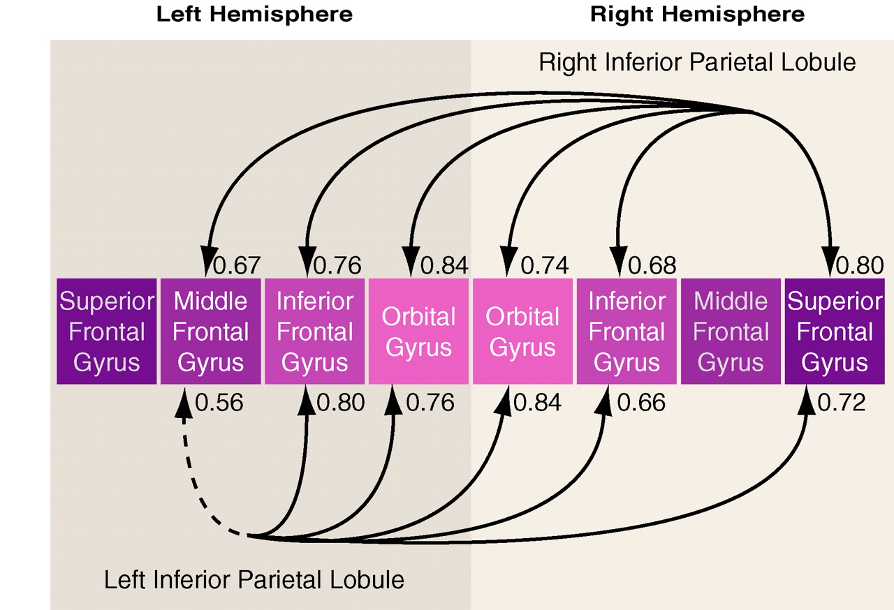
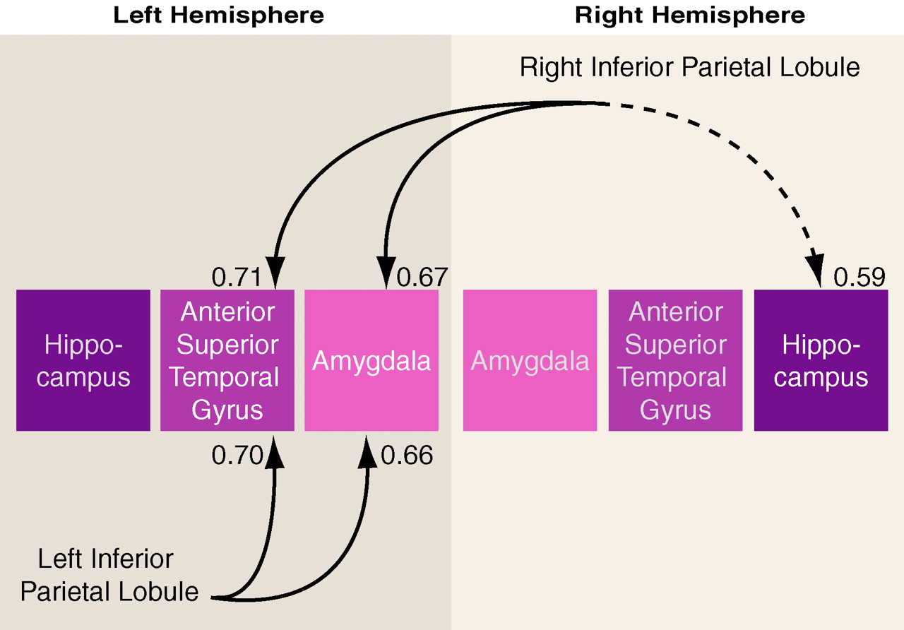
As hypothesized, the schizophrenic group showed several high correlations between gray matter volumes of the inferior parietal lobule and regions of the prefrontal cortex (
table 4).
Figure 3 provides an illustrative summary of these correlations, with the bottom arrows depicting prefrontal correlations with the left inferior parietal lobule, and the top arrows depicting prefrontal correlations with the right inferior parietal lobule (note: arrows do not imply direction). In addition to these correlations, the left postcentral gyrus correlated significantly with the left superior frontal gyrus (r=0.72, p<0.003) and with the right inferior frontal gyrus (r=0.70, p<0.005). In contrast, the comparison subjects showed no volumetric correlations between inferior parietal lobule volumes and prefrontal volumes at p<0.05.
The differences between groups in the correlations for respective brain areas were tested by using a Fisher’s z transformation. Of note, significant group differences in correlations emerged for the left inferior parietal lobule and prefrontal structures even though the correlations between left and right inferior parietal lobules and the prefrontal lobe structures were comparably high in the schizophrenic group. This result highlights the salience of the left inferior parietal lobule correlations with prefrontal measures in the schizophrenic group.
In the comparison subjects, but not in the schizophrenic subjects, the inferior parietal lobule asymmetry coefficient correlated inversely with all of the prefrontal structures (r=–0.50 to –0.76, p=0.03 to ≤0.001). In addition, the left postcentral gyrus correlated significantly with several prefrontal structures: the left superior frontal gyrus (r=0.83, p<0.001), left orbital gyrus (r=0.70, p<0.004), right superior frontal gyrus (r=0.64, p<0.01), and the right orbital gyrus (r=0.64, p<0.01).
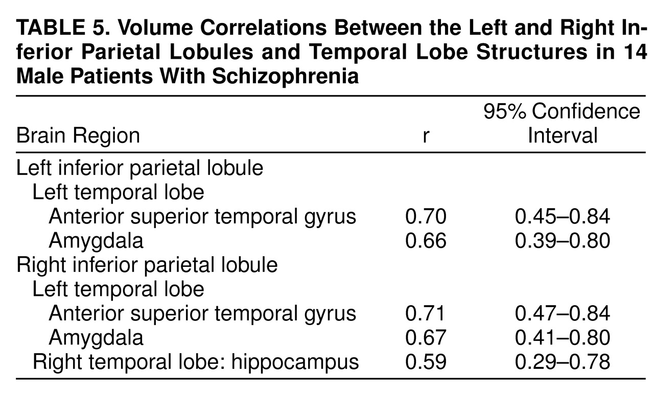
In the schizophrenic group, both the left and right inferior parietal lobules correlated significantly with the left amygdala (r=0.67, p<0.007) and the left anterior portion of the superior temporal gyrus (r=0.66, p<0.008 (
figure 4 and
table 5). In the normal comparison group, the one significant correlation was between left inferior parietal lobule and the left amygdala (r=0.53, p=0.04). The group differences in the respective, pairwise correlations, as tested with Fisher’s formula, did not reach statistical significance.
Correlations With Clinical Status and Clinical Measures and With Neuropsychological Data
Because of the exploratory nature of the correlations and the issue of multiple tests (i.e., some of the significant results might be due to chance), we report only correlations where p was below 0.01.
No correlations between regions of interest and clinical measures in schizophrenic patients reached the significance level of p<0.01. Exploratory analyses of correlations between neuropsychological measures and brain volume in schizophrenia patients indicated that reduced right inferior parietal lobule volume correlated with lower scores on tests of visual attention and visual memory (Trails B: rs=–0.76, p<0.002; visual reproduction I: rs=0.71, p<0.003; visual reproduction II: rs=0.84, p<0.001). Additionally, reduced inferior parietal lobule asymmetry was related to poor performance on Trails A (rs=–0.70, p<0.005) and Trails B (rs=–0.69, N=14, p<0.004).
DISCUSSION
The present study examined gray matter volume in individual gyri of the parietal lobe by using high resolution MRI (1.5-mm thick slices) and neuroanatomically based boundary definitions. The major findings from this study were that schizophrenic patients, in contrast to a normal comparison group, showed 1) a reversal of the normal left-greater-than-right asymmetry in the inferior parietal lobule that was localized to the angular gyrus and 2) significant volumetric correlations between parietal lobe regions and regions of the frontal and temporal cortex.
More specifically, gray matter abnormalities observed in the inferior parietal lobule consisted of a reversal of the normal left-greater-than-right asymmetry that was localized to the angular gyrus and further confirmed with a measure of asymmetry. The specificity of this asymmetry finding was underscored by the fact that no differences were observed between schizophrenic and comparison subjects for total parietal lobe, superior parietal gyrus, or postcentral gyrus. Further, there was also an absence of group differences in the lateralization patterns for superior parietal gyrus and postcentral gyrus. This finding supports previous reports of abnormality within the parietal lobe
(34–
37) and localizes the abnormality to the angular gyrus. Of note, schizophrenic patients did not show leftward lateralization of the total parietal lobe, which was present in the comparison subjects. This result was driven by the reversal of the normal left-greater-than-right asymmetry within the angular gyrus. This finding further underscores the utility of separate volumetric analyses for structures comprising the parietal lobe.
The present study is, to our knowledge, the first to report a reversal of normal asymmetry in the angular gyrus in schizophrenic patients, a brain region belonging to the neural circuitry that supports semantic aspects of language processing. As noted in our introduction, recent fMRI data suggest the involvement of the inferior parietal lobule, and especially the angular gyrus, in semantic processing. This region, in fact, is regarded as part of a semantic-lexical network that includes the planum temporale and is involved in both assigning meaning to strings of sounds and, at its output stage, in generating associative links responsible for constructing complex meanings and thought processes. Previous studies in schizophrenia have reported abnormal cortical asymmetry in superior temporal gyrus, especially in the region of the planum temporale
(38). Thus, the present finding, which extends the finding of abnormal asymmetry to the angular gyrus, enhances our understanding of the neural underpinnings of a core feature of schizophrenic syndrome: disordered thought and language processes.
The relationship between asymmetry of the planum temporale and that of the angular gyrus, the two structures belonging to the language network, has also been previously noted by Eidelberg and Galaburda
(13). These investigators reported a correlation between the degree of lateralization of the planum temporale and the angular gyrus. Thus, there appears to be a leftward lateralization for structures specialized for language
(7,
13,
18,
19). The presence of such asymmetries might have an evolutionary advantage in developing and supporting language in the human species, in which the specialized function of one hemisphere might confer additional advantage
(18–
20).
The reversal of normal asymmetry in the angular gyrus, in addition to previous reports of the reversed asymmetry in superior temporal gyrus, provides further support for the relationship between abnormal laterality patterns and the origins of schizophrenic pathology
(18–
20). Since cortical asymmetries are present during fetal development
(13,
21), it is thus quite possible that abnormal asymmetries in these two regions in schizophrenia may have a common neurodevelopmental origin.
The affected structures might be abnormal as a result of faulty developmental mechanisms such as gliosis or pruning, the disease process itself, or both. In fact, striking correlations between the inferior parietal lobule and neuroanatomically connected cortical regions of the prefrontal cortex and the temporal lobe, which were observed only in the schizophrenic subjects, provide support for the existence of a pathologic process (such as excitoxicity) that affects multiple, functionally interconnected brain regions in schizophrenia
(39–
41). The high correlations between regions that constitute primarily heteromodal association cortex regions, and observed only in the patient group, support the notion that schizophrenia might preferentially affect the association cortex.
Also of interest is the finding that in comparison subjects, the asymmetry coefficient was inversely correlated with frontal lobe volumes, which indicates that a normal brain’s development entails leftward lateralization of language structures
(13,
18,
19), and that this lateralization pattern is correlated in a healthy brain with the frontal lobes, which are intimately involved in mediating processes in parietal language areas.
A limitation of this study is the use of multiple tests, which elevates the risk of type I errors. However, we focused our attention on the inferior parietal lobule, where we had an a priori rationale for evaluating both the angular and supramarginal gyri. Furthermore, we used more conservative criteria for the significance level (i.e., p≤0.01) of the exploratory analyses as a compromise for the multiple correlations performed.
In summary, this study, which employed improved methods of parcellation and measurement of the gray matter of the parietal lobe, provides evidence for the absence of normal left-greater-than-right asymmetry in the angular gyrus, a brain region categorized functionally as part of the heteromodal association cortex and, importantly, linked to aspects of semantic processing. These findings, taken together, provide support for localized volumetric changes in schizophrenia associated with selective cognitive impairments and a possible neurodevelopmental component to schizophrenic pathogenesis. These findings also afford a more comprehensive understanding of schizophrenic pathology by demonstrating similar pathologic processes that affect functionally related brain areas: the inferior parietal lobule region and the temporal and frontal areas.