Abstract

OBJECTIVE: The authors examined the separate and combined effects of methamphetamine dependence and HIV infection on brain morphology. METHOD: Morphometric measures obtained from magnetic resonance imaging of methamphetamine-dependent and/or HIV-positive participants and their appropriate age- and education-matched comparison groups were analyzed. Main effects of age, HIV infection, methamphetamine dependence, and the interactions of these factors were examined in analyses of cerebral gray matter structure volumes. RESULTS: Independent of the effect of age, HIV infection was associated with reduced volumes of cortical, limbic, and striatal structures. There was also some evidence of an interaction between age and HIV infection such that older HIV-positive participants suffered disproportionate loss. Methamphetamine dependence was surprisingly associated with basal ganglia and parietal cortex volume increases, and in one of these structures—the nucleus accumbens—there appeared to be a larger effect in younger methamphetamine abusers. Neurocognitive impairment was associated with decreased cortical volumes in HIV-positive participants but with increased cortical volumes in methamphetamine-dependent participants. CONCLUSIONS: These results suggest significant brain structure alterations associated with both HIV infection and methamphetamine dependence. The regional patterns of the changes associated with these factors were distinct but overlapping, and the effects on brain volumes were opposing. Although the results of the present study provide little information about the specific mechanisms leading to the unexpected methamphetamine effects, they may be related to glial activation or neuritic growth, both of which have been associated with methamphetamine exposure in animal studies. These results have implications for the interpretation of brain morphological findings in methamphetamine-dependent, HIV-positive individuals, a group whose numbers are unfortunately increasing.
Substance abuse is associated with an increased frequency of high-risk behaviors for HIV exposure. This has led to an elevated prevalence of HIV infection among substance abusers. Methamphetamine is a common drug of abuse in the United States, and disproportionate numbers of methamphetamine abusers are HIV seropositive. Strong evidence exists for neurotoxic effects of both HIV infection and chronic methamphetamine use, and functional deficits have been observed in both populations (1, 2). Previous structural magnetic resonance imaging (MRI) studies have helped to define the pattern of neurodegeneration that occurs with progression of HIV-related disease (38). It is clear from these studies that even in the absence of CNS opportunistic infections, HIV infection is accompanied by damage to cerebral white matter (which results in progressive loss of white matter volume) and by more circumscribed progressive loss of cerebral gray matter, particularly in the striatum. These studies are complemented by magnetic resonance spectroscopy (MRS) studies that have shown widespread reductions in the neural marker N-acetylaspartate and increases in neuroinflammatory markers commensurate with the disease stage and degree of functional impairment (915). The MRS studies, like the structural MRI studies, suggest early damage to the striatum and subcortical white matter as well as eventual cortical gray matter degeneration.
It is unfortunate that methamphetamine abusers have only rarely been examined with structural MRI. In one study of stimulant abuse, amphetamine-dependent subjects were compared with cocaine abusers and nonabusing healthy subjects (16). In this study, total volume of the temporal lobe was reduced in the amphetamine abusers relative to the healthy comparison subjects. In a recent study that used computational morphometrics, Thompson et al. (17) examined active methamphetamine abusers and healthy subjects and reported methamphetamine-related decreases in the gray matter of the cingulate and other limbic cortices lateralized to the right hemisphere. They also observed bilateral hippocampal volume reductions that were correlated with memory performance on neuropsychological tests. In contrast, they observed significant bilateral hypertrophy of cerebral white matter, which they speculated might be due to compensatory glial responses such as astrogliosis.
No MR morphometry studies have been reported to date that clearly define brain morphological abnormalities in subjects recovering from methamphetamine abuse. Thus, if a regional pattern of neurodegenerative changes associated with methamphetamine abuse persists in recovery, it has not yet been established. This complicates the interpretation of brain structural abnormalities in HIV-infected methamphetamine abusers. It is possible that neurotoxic effects of methamphetamine are intensified in HIV-infected persons. To explore the individual and combined effects of these risks on brain structure, volume estimates of major gray matter structures of the cerebrum were compared in four age- and education-matched groups: 1) methamphetamine-dependent HIV-positive subjects; 2) methamphetamine-dependent HIV-negative subjects; 3) HIV-positive subjects with no methamphetamine dependence; and 4) HIV-negative subjects with no methamphetamine dependence.

Method

Subjects

This structural MRI study was conducted within the HIV Neurobehavioral Research Center of the University of California, San Diego, as part of a broader study of the central nervous system effects of methamphetamine dependence and HIV infection. In support of this study, the HIV Neurobehavioral Research Center recruits and maintains a participant cohort consisting of recovering methamphetamine-dependent individuals and HIV-infected (HIV-positive) individuals. The HIV Neurobehavioral Research Center establishes direct contact with prospective subjects and completes screening assessments. Methamphetamine dependence is defined on the basis of the Structured Clinical Interview for DSM-IV. Other key neuromedical variables obtained and procedures performed include the following: 1) a neuromedical history and review of systems, which generates an ICD-9-coded summary of HIV-related and unrelated medical diagnoses; 2) general medical, neuropsychological, psychiatric, and neurological examinations; 3) phlebotomy and lumbar puncture; 4) routine laboratory evaluations such as blood hematology and chemistry measurements and lymphocyte subsets; and 5) specialized laboratory evaluations such as plasma and CSF, HIV RNA levels, and urine toxicology for drugs of abuse. For the present study, participants with a history of schizophrenia, neurologic conditions unrelated to substance use or HIV (e.g., multiple sclerosis, epilepsy), central nervous system opportunistic infection, HIV-associated dementia, or head injury with loss of consciousness for greater than 30 minutes were excluded. This study was approved by the UCSD Human Research Protections Program, and informed consent was obtained from all participants after the study was explained by one of the investigators.
The 51 HIV-negative participants comprised 21 methamphetamine-dependent individuals and 30 individuals with no history of methamphetamine dependence. The 52 HIV-positive participants comprised 22 methamphetamine-dependent individuals and 30 individuals with no history of methamphetamine dependence. Demographic and other relevant characteristics of the participant groups are summarized in Table 1. Fifteen of the HIV-negative subjects with no methamphetamine dependence had participated as comparison subjects in non-HIV Neurobehavioral Research Center neuroimaging studies, and thus complete neuropsychological and neuromedical evaluations were not available for these subjects. Groups were well matched for age and education, but the gender distribution within the groups varied. Both HIV-positive groups exhibited the expected reduction in CD4 cells, those with no methamphetamine dependence to a greater extent than the methamphetamine-dependent group. Relative to the group of HIV-negative subjects with no methamphetamine dependence, all other groups were significantly impaired on clinical ratings of global neuropsychological impairment (18). On the basis of all historical information available, estimates were made for each HIV-positive participant of the years the participant was likely to have been infected at the time of study. For the HIV-positive subjects as a whole the mean was 6.9 years (SD=4.6). Estimates of group means were 6.6 (SD=5.6) and 7.0 (SD=3.7) for those with and without methamphetamine dependence, respectively.
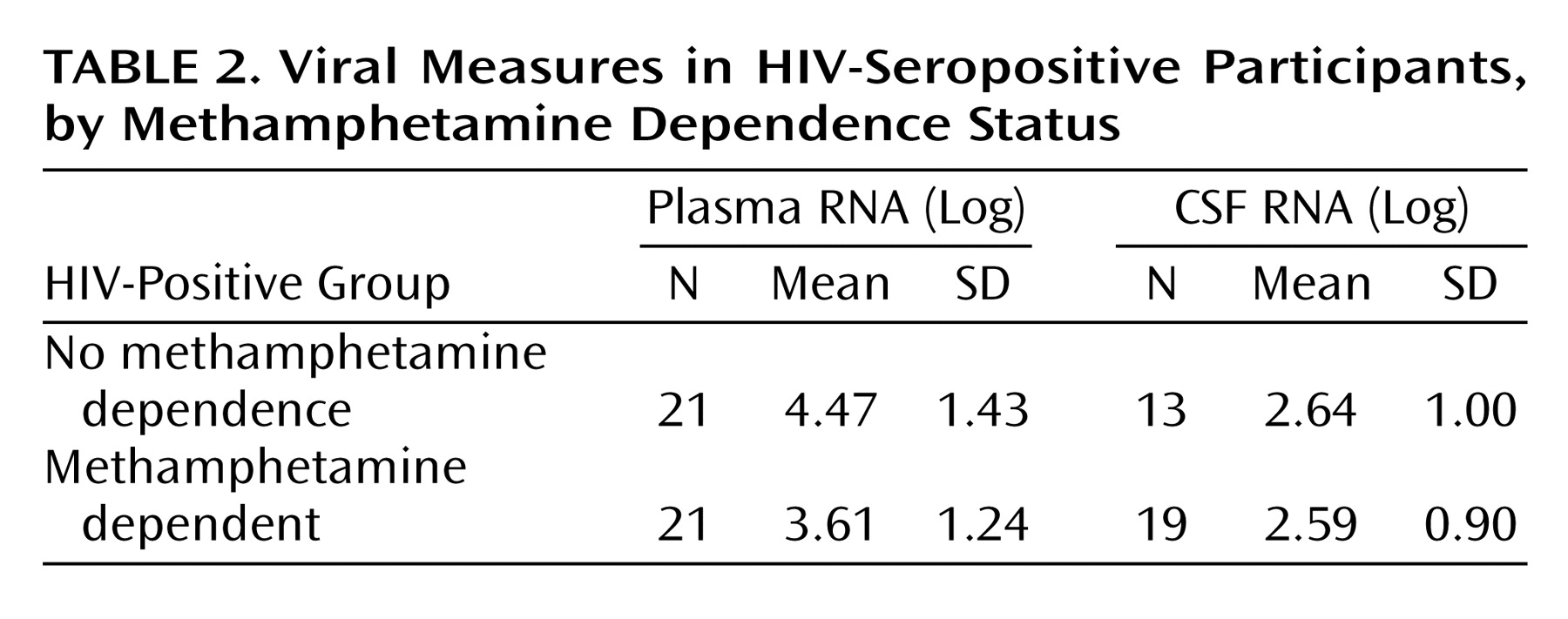
Measures of plasma and CSF viral RNA were available in a subset of the subjects. These data, summarized in Table 2, suggest that viral levels were comparable in the two groups at the time of study.
Among the methamphetamine-dependent participants of the present study, some met criteria for attention deficit hyperactivity disorder (ADHD) or antisocial personality disorder. Two (9.5%) of the HIV-negative/methamphetamine-dependent subjects and four (18.2%) of the HIV-positive/methamphetamine-dependent subjects had a lifetime diagnosis of ADHD. Four (19.0%) of the HIV-negative/methamphetamine-dependent subjects, but no HIV-positive/methamphetamine-dependent subject, met criteria for antisocial personality disorder.
A substance use questionnaire was administered to obtain a detailed history of quantity, frequency, and duration of methamphetamine use as well as most other classes of substances of abuse. This history was obtained for the last 30 days and 12 months, and in 5-year epochs covering the participant’s lifetime. Among the variables that this questionnaire yields are age at onset, total number of years of use, length of abstinence, preferred mode of use, lifetime grams of methamphetamine consumed, and average grams per year of use. Methamphetamine use characteristics of the methamphetamine-dependent subjects (estimated from interview data) are summarized in Table 3.
No subject was included who met criteria for dependence on any substance of abuse other than methamphetamine for the previous 5 years. However, subjects were included if they reported histories consistent with dependence in the years before that. Among the HIV-negative and HIV-positive subjects with methamphetamine dependence, 13 (61.9%) and four (18.2%), respectively, met criteria for previous alcohol dependence. Unfortunately, the data for establishing lifetime histories (i.e., prior to 5 years before study participation) of alcohol dependence were not available for the participants in the present study with no methamphetamine dependence.
Because some subjects were studied before complete assessments were collected, the data available for cigarette smoking were sparse in some groups. While assessments were available for all HIV-negative/methamphetamine-dependent participants and 18 (81.8%) of HIV-positive/methamphetamine-dependent participants, of those with no methamphetamine dependence only 11 HIV-positive participants (36.7%) and six HIV-negative participants (20.0%) had cigarette smoking data. However, percentage of smokers or previous smokers among the informative subjects of the different groups suggests heavier smoking in the substance abusing groups. Although 67% of the HIV-negative subjects with no methamphetamine dependence had smoked at some point, none was a present smoker. Among the HIV-positive participants with no methamphetamine dependence, 45% had ever smoked, and all of these remained smokers at the time of the study. In the HIV-negative/methamphetamine-dependent group, 62% had ever smoked, and all remained smokers at the time of study. In the HIV-positive/methamphetamine-dependent group, 100% had smoked and 83% remained smokers at the time of study.
Subjects underwent imaging only if they reported present abstinence from substances of abuse, and if their reports were confirmed by urine toxicology screen at the associated clinical follow-up visit. However, scanning was conducted on a different day, and urine toxicology screening was not repeated at the scanning session; thus abstinence on the day of scanning could not be definitely confirmed.
Among the HIV-positive participants, eight (26.7%) of those with no methamphetamine dependence and eight (36.4%) of the methamphetamine-dependent individuals were taking no antiretroviral medications at the time of study. The frequencies of different treatment combinations among the treated participants are summarized in Table 4 separately for the groups with and without methamphetamine dependence.
Finally, 14 of the 46 participants with no methamphetamine dependence for whom assessments were available were receiving psychoactive prescription medications at the time of study, while 18 of 43 assessed methamphetamine-dependent participants were. The distribution of anxiolytic, antidepressant, and antipsychotic medication use (by group) is given in Table 5. Although the use of such medications was common in both groups, the prevalence was somewhat higher among the methamphetamine-dependent participants (42% relative to 30% in participants with no methamphetamine dependence).

MRI Protocol

Three whole-brain image series were collected for each participant. The first two series were fast spin-echo acquisitions yielding two separate sets of coronal, 4 mm-thick images: TR=3000 msec, TE=17 msec, echo train=4 and TR=3800 msec, TE=102 msec, echo train=8. The third series was either 1) a sagittal gradient-echo T1-weighted series with TR=24 msec, TE=5 msec, number of excitations=2, flip angle=45°, section thickness=1.2 mm; or 2) a sagittal spiral fast spin-echo with TR=2000 msec, TE=60 msec, flip angle=90°, section thickness=1.3 mm. For all series, field of view was 24 cm.

Image Postprocessing

The image-analytic approach we used is similar to that used in our previous anatomical studies (5, 8) but represents a significant elaboration of these methods as described previously (19). Trained anatomists, who were blind to subject diagnosis, age, gender, or any other identifying information, subjected each image dataset to the following image analysis procedures: 1) interactive isolation of intracranial regions from surrounding extracranial tissue, 2) three-dimensional digital filtering of the matrix of pixel values to reduce spatial bias, 3) reslicing of the volume to a standard orientation, 4) tissue segmentation using semiautomated algorithms, and 5) neuroanatomical region of interest analysis.
First, the brain was isolated from extracranial areas in the image, i.e., from surrounding tissue that was in some instances contiguous with brain tissue and similar in signal value. This resulted in a new volume within which the positions of brain voxels were coded, i.e., a mask. Filtering was applied to reduce nonbiological signal drift across the field of view, which is presumably due to field inhomogeneity and susceptibility effects. A three-dimensional, high-pass filter was applied, with two iterations, separately to the “stripped” proton-density weighted and T2-weighted fast spin-echo image volumes.
The tissue classification procedure was an interactive supervised process. Trained operators manually designated the positions of three sets of tissue samples, one for each of the target tissues (gray, white, and CSF). Samples were selected in locations that appeared to be homogeneous and free of signal abnormalities. Sample voxel values were then analyzed using simple regression techniques to separate first all brain parenchymal voxels from CSF voxels then gray matter voxels from white matter voxels. The regression coefficients obtained in these simple analyses were then applied to classify each voxel within the volume as most similar to CSF, gray matter, or white matter.
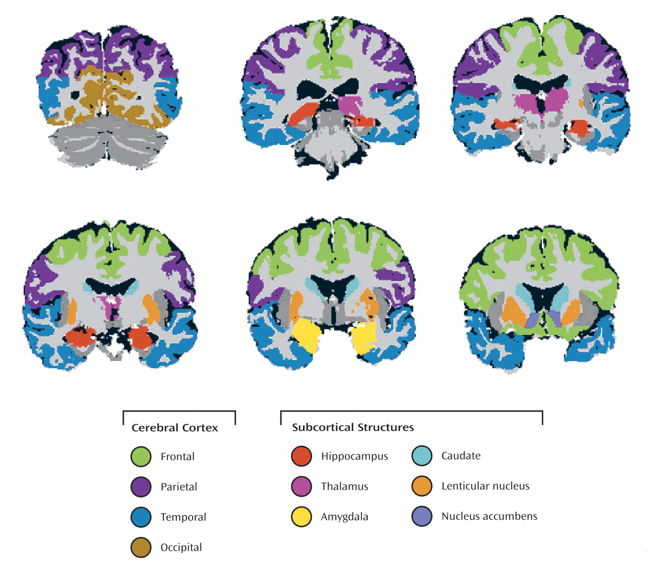
In order to facilitate the definition of regions of interest, datasets were aligned to a standardized stereotactic space defined relative to the decussations of the anterior and posterior commissures and the structural midline. Anatomists circumscribed regions on tissue-segmented images, guided by views from the high resolution T1-weighted images. Standardized rules were applied for delineating a set of subcortical and cortical regions within the cerebrum (Figure 1).
For the present study, the cerebral subcortical structures that were evaluated included the caudate nucleus, nucleus accumbens, lenticular nucleus, thalamus, amygdala (and adjacent entorhinal and perirhinal cortex), and hippocampus. Cerebral cortex was measured separately in four major lobes: occipital, temporal, parietal, and frontal. Structures in the mesial temporal lobe were not included in the temporal cortex volume. Estimates of the volumes of the structures were computed by summing the gray matter voxels within each of these regions. Significant artifacts distorted the images in the temporal lobe region in five of the 103 subjects of the study, and thus these subjects were assigned missing values for the measures of the hippocampus, amygdala, temporal cortex, and total cortex.

Statistical Methods

The effects of HIV seropositivity and methamphetamine dependence on volumes of cerebral structures were estimated with least squares regression after we controlled for the effects of several covariates. The dependent variables (brain volumes) were log transformed. Head size correction was accomplished by entering the log-transformed volume of the supratentorial cranial vault as a covariate. Other initial covariates of interest were age and gender. After controlling for cranial volume, no significant gender effects were observed in any region, and gender was dropped from the models. Secondary analyses were performed to examine interactions between age, HIV seropositivity, and methamphetamine dependence when significant main effects were present. To summarize, the significance of the main effects of the three major factors (age, HIV serostatus, and methamphetamine dependence) reported here is the significance (alpha=0.05, uncorrected) of the regression coefficients for these variables in a regression model predicting each log-transformed gray matter volume using the three factors (age, HIV seropositivity, and methamphetamine dependence) and the log-transformed cranial volume as predictors. For each gray matter volume, a secondary regression analysis was then conducted in which the interaction terms were added for each factor that yielded a significant main effect in the first analysis. Significant interactions reported here are those that were associated with significant (alpha=0.05, uncorrected) regression coefficients for the interaction terms in these secondary analyses. Furthermore, post hoc regression analyses were conducted 1) within the methamphetamine-dependent subjects to examine the relationship between substance use/psychiatric variables and brain volumes showing main effects of methamphetamine dependence; 2) to examine in greater detail the nature of observed interactions; and 3) to examine the degree of association between the brain volumes and neurocognitive impairment.

Results

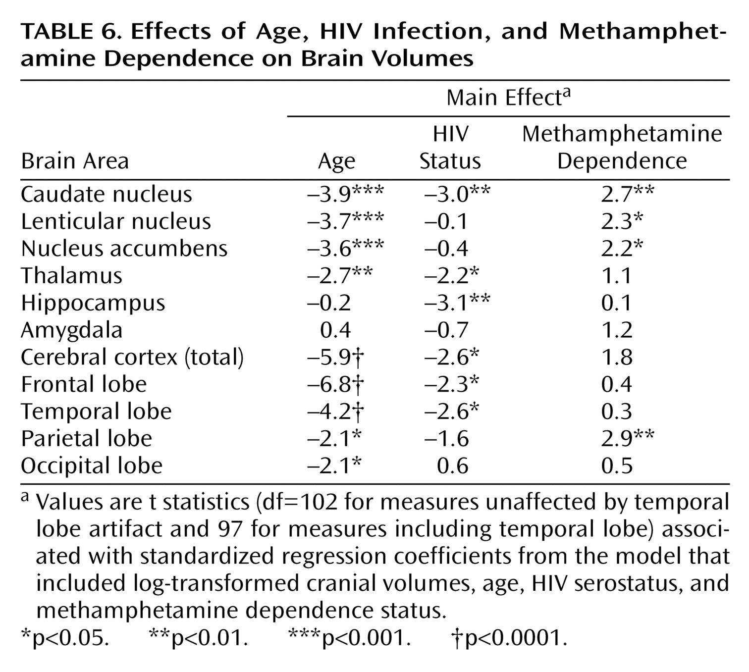
The results of the primary analyses (main effects) are summarized in Table 6. The statistics given are t statistics associated with standardized regression coefficients from the model including log-transformed cranial volumes, age, HIV serostatus, and methamphetamine dependence status. Positive t values indicate volume increases, negative t values indicate volume decreases.
Independent effects of age, HIV seropositivity, and methamphetamine dependence were observed for basal ganglia, diencephalon, and cerebral cortex volumes. Significant age-related volume reductions were present in all cerebral gray matter structures examined except those in the mesial temporal lobe.

Effects of HIV

Independent of age-related reductions, there were additional HIV-related volume losses in many structures. Within basal ganglia structures, the previously reported vulnerability of the caudate nucleus to HIV effects was again observed. The thalamus, hippocampus, and cerebral cortex were also reduced in volume in seropositive individuals. Within the cortical lobes, the HIV-related reductions within the frontal lobe and temporal lobe were significant. Secondary analyses revealed that there was also a significant age-by-HIV status interaction for the models predicting total (t=–2.6, df=97, p<0.05), frontal lobe (t=–2.8, df=97, p<0.01), and temporal lobe (t=–2.1, df=102, p<0.05) cortex volumes.
Post hoc analyses were conducted to determine whether the age-by-HIV status interactions might be mediated by the presence of more advanced disease in the older HIV-positive individuals. Two measures of disease progression were examined: the CD4 cell measure and the estimate of years HIV positive at the time of study. Of these, only the latter was significantly correlated with age within the HIV-positive participants (r=0.32, df=49, p<0.02). However, addition of neither of these variables as covariates eliminated the significant interaction effects.

Effects of Methamphetamine Dependence

Independent effects of methamphetamine dependence were observed for several structures. In all cases, methamphetamine dependence was associated with increased volumes. All three segments of the basal ganglia showed significant methamphetamine-related increases: caudate nucleus, lenticular nucleus, and nucleus accumbens. Secondary analyses revealed a significant age-by-methamphetamine interaction (t=–2.5, df=102, p<0.05) in the model predicting volume of the nucleus accumbens. This interaction was due to larger increases in younger than older methamphetamine-dependent individuals. There is also evidence for methamphetamine-related increase in the volume of the cerebral cortex. The estimated effect of methamphetamine on cortex volume fell just short of significance for the cortex as a whole. Examining the effects on separate lobes, only the methamphetamine effect on parietal lobe cortex reached significance.
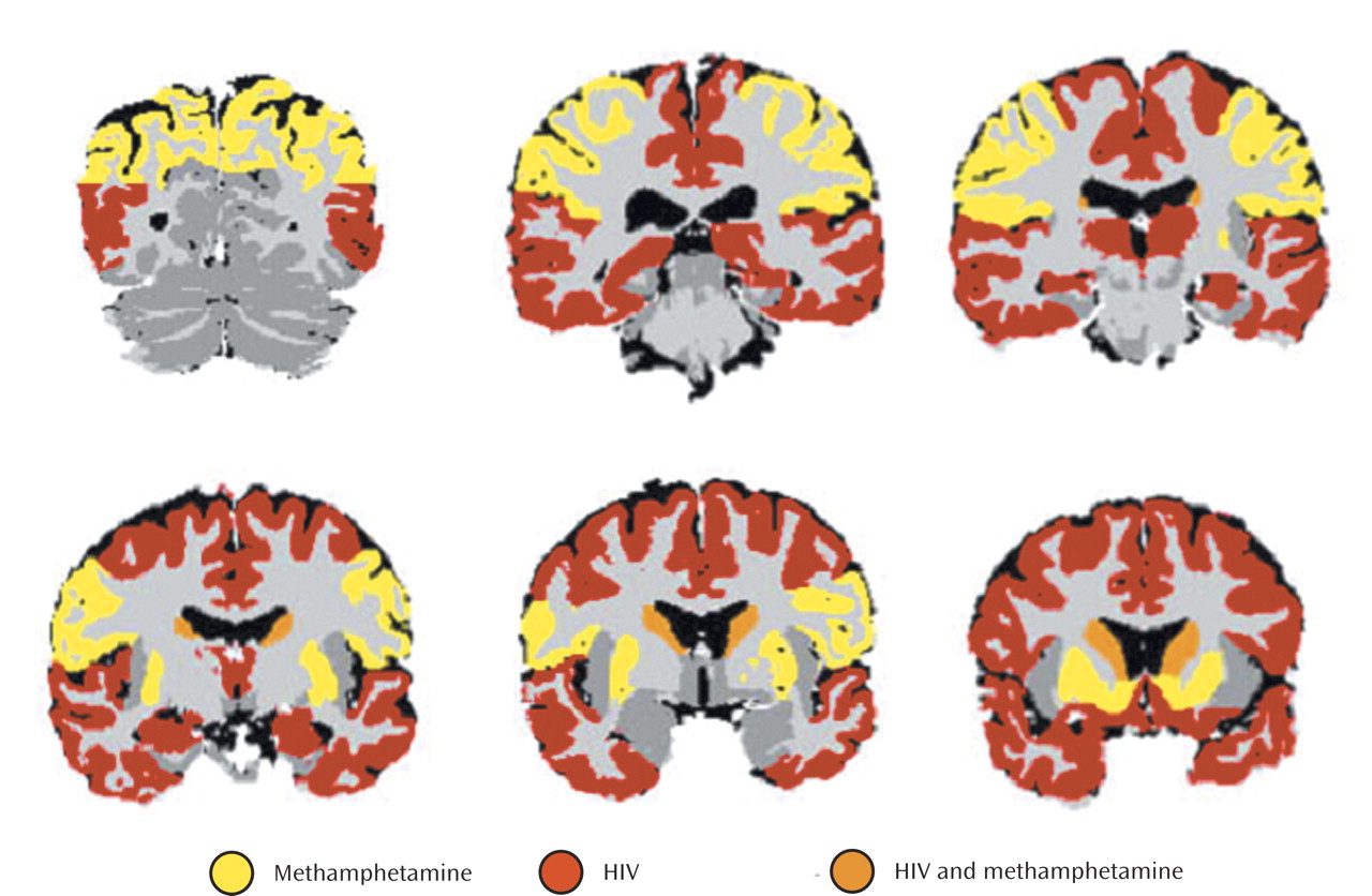
The differing patterns of HIV-related and methamphetamine-related volume alterations are shown graphically in Figure 2. Regions shown in red were reduced in volume in HIV-positive individuals. Those shown in yellow were increased in volume in methamphetamine-positive individuals. In the caudate nucleus, shown in orange, both effects were superimposed.

Substance Use and Psychiatric Variables

Post hoc analyses were performed to determine whether substance use variables listed in Table 3 were predictive of the magnitude of the methamphetamine-related volume alterations within the methamphetamine-dependent group. In no case did any use variable significantly improve the prediction of volumes of caudate nucleus, lenticular nucleus, nucleus accumbens, or parietal cortex after the effects of cranial volume and age were controlled. It should be noted, however, that the correlation between age at study and age at first abuse was high (r=0.68, df=41, p<0.0001) in the present group of methamphetamine-dependent subjects. Therefore, it was unclear whether the larger methamphetamine-related effects on the nucleus accumbens in younger subjects were more likely to be mediated by age at study or age at first abuse.
Since several of the methamphetamine-dependent participants met criteria for present or previous ADHD or antisocial personality disorder, we conducted post hoc analyses to determine whether such diagnoses could be related to the increased cortical and subcortical brain volumes. In no case did subjects with ADHD or antisocial personality disorder diagnoses have higher gray matter volumes than subjects without the diagnoses; in fact, normalized gray matter volumes tended to be lower in such subjects.

Neurocognitive Impairment

Additional post hoc analyses were conducted to examine the correlation between the ratings of neurocognitive impairment and volumes of the basal ganglia, hippocampus, and cerebral cortex. Since HIV and methamphetamine were associated with opposing effects on brain volumes, these associations were examined within the groups separately. Within neither of the single factor groups (HIV-positive only or methamphetamine-dependent only) were there any significant correlations between subcortical volumes and neurocognitive impairment. However, there was evidence that HIV-positive participants with no methamphetamine dependence who had more severe impairment had lower cortical volumes (r=–0.41, df=27, p=0.03), while HIV-negative/methamphetamine-dependent participants with more severe impairment had higher cortical volumes (r=0.46, df=18, p=0.04). Within the dually affected group, neither the cortical volumes nor the basal ganglia volumes showed any significant association with neurocognitive impairment, as might be expected given that the effects of the two factors on the volumes are opposing. However, there was a significant correlation between reductions in hippocampal volume and increasing neurocognitive impairment in the HIV-positive/methamphetamine-dependent group (r=–0.52, df=19, p=0.02).

Discussion

Effects of HIV Infection

The reductions in brain volumes noted in HIV-positive participants of the present study are similar to those reported previously (58). Again, the vulnerability of striatal and limbic structures to the effects of HIV were apparent. These results confirm that the frontotemporal cortex is also significantly affected in HIV-positive individuals.
The significant age-by-HIV status interaction observed in the analyses of the cortical measures has not been reported previously. Our first impression was that these effects were probably attributable to the association of age with disease progression. That is, since neurodegenerative changes are likely to accelerate during later stages of the illness, and older subjects are more likely to have reached that stage, the interactions could reflect accelerating changes in individuals with more advanced disease. This would be consistent with our earlier longitudinal study of HIV-related brain volume loss (8) in which greater volume loss over time was seen in CDC-C stage participants than in those in CDC-A and B stages. It is surprising, however, that although there was a modest association between estimated years of infection and age within HIV-positive participants, there was no evidence that more years of infection was associated with more cortical atrophy. The magnitude of the interaction effect was not reduced appreciably by the inclusion of this estimate of disease duration in the models predicting total cortex, frontal cortex, or temporal cortex. Greater reduction in CD4 cells was not related to increased age in the seropositive participants, and inclusion of this covariate also did not reduce the magnitude of the interaction effects. However, there was evidence of an independent effect of lower CD4 cells. That is, reduction of CD4 cells, independent of HIV seropositivity, methamphetamine dependence, or other covariates of interest, was associated with lower cortical volumes. In summary, the age-by-HIV status interaction remained significant in these models. There are at least two explanations for these findings. One is that these covariates are not sufficiently sensitive indices of disease progression to reveal the true effects, although chronicity of disease is in fact mediating the apparent age-by-HIV status interaction. Another explanation is that there are genuine interactions, such that alterations present in the older brain potentiate the effects of HIV. This issue deserves further scrutiny in future studies.

Methamphetamine Effects

In this study, comparisons of groups of individuals with and without methamphetamine dependence revealed unexpected basal ganglia and parietal cortex volume increases. Striatal structures, most notably the nucleus accumbens, have been implicated in previous studies of the CNS effects of stimulants. A number of studies have focused on mechanisms by which behavioral and neural sensitization occurs as a result of stimulant exposure (see Steketee [20] for a review), since such sensitization has putative links to the development of dependence and craving. These studies have demonstrated that low doses of stimulants such as cocaine and amphetamine result in increased dopamine release in the nucleus accumbens that is further enhanced as a result of repeated, intermittent exposure (2123). Animal studies (24, 25) have shown that acute doses of methamphetamine that mimic binge doses in human abusers result in neurotoxic effects on striatal dopamine terminals; i.e., reduced dopamine and metabolite levels and reduced dopamine transporter binding. However, when these doses are preceded by escalating lower doses, as plausibly occurs in human users, the effects on dopamine terminals, while still present, are substantially mitigated and show greater recovery.
In other animal studies, Robinson and Kolb (26, 27) have shown that exposure to low-dose amphetamine in rats is followed by long-lasting increases in the length of dendrites, density of dendritic spines, and number of branched spines on output cells of the nucleus accumbens. Similar effects were observed in the prefrontal cortex. In previous studies (28, 29) the effects of methamphetamine exposure on plasticity-related genes in the rat brain were examined. Such exposure resulted in increases by 20%–40% in the expression of synaptophysin mRNA in the nucleus accumbens and in prefrontal and temporal cortices within 1 day of administration. Increases in stathmin mRNA were also observed in the prefrontal cortex. Thus, increases in expression of plasticity-related genes may lead to previously reported neurite extension in the striatum, and these changes may contribute to the volume increases observed in the present study.
Another relevant study (30) examined the effects of MPTP-induced dopaminergic lesions on tyrosine hydroxylase fibers in monkeys. It is interesting that some fibers became thicker and increased their branching, and mRNA for growth-associated protein-43 was upregulated in midbrain dopamine cells. The authors concluded that sprouting in the striatum was a compensatory response to partial dopaminergic depletion. Thus, multiple lines of evidence suggest that some progressive compensatory changes, perhaps associated with the depletion of dopamine transporters or damage to dopamine terminals, could contribute to the volume increases observed in methamphetamine-dependent individuals.
In addition to these animal studies of sprouting in the striatum, several studies have demonstrated pronounced effects on microglial activation in methamphetamine-exposed rodents (31, 32), both in the striatum and, specifically, in the somatosensory cortex; these studies have suggested a role for microglial activation in methamphetamine neurotoxicity. Microglial activation has also been linked to astrocytosis (33), and both astrocytosis and depletion of glutamate-staining neurons have been observed in the somatosensory cortex of methamphetamine-exposed animals (34). Finally, Eisch et al. (35) examined [3H]mazindol ([3H]MAZ) binding to striatal dopamine transporters and [3H]GLU binding to N-methyl-d-aspartate (NMDA) receptors in the striatum and cortex 1 week and 1 month after a neurotoxic methamphetamine regimen. Dopamine transporters and NMDA receptors were reduced in the striatum after 1 week, and decreases in dopamine transporters but not NMDA receptors persisted at 1 month. Of interest is that increases in NMDA receptors in the somatosensory cortex (but not the frontal cortex) were present at both time points, possibly increasing the vulnerability of this area to excitotoxicity.
In summary, methamphetamine exposure in rodents has been shown to 1) reduce striatal and frontocortical dopamine transporters, 2) lead to neuroplastic changes in the striatum as well as microglial and astrocytic activation in the striatum and parietal cortex, and 3) affect parietal lobe NMDA receptors and glutamatergic neurons. Thus, it appears that microgliosis, astrocytosis, and neuroplastic changes associated with methamphetamine exposure may underlie the striatal volume increases we observed. Methamphetamine-related microgliosis, astrocytosis, and glutamatergic excitotoxicity, observed specifically in parietal regions in animal studies, may occur in the methamphetamine abusers studied here and may contribute to their parietal lobe volume increases.
Previous PET studies in stimulant-dependent individuals have also specifically implicated striatal and parietal lobe structures, where depletion of striatal dopamine transporters and reduced striatal metabolism have been observed, as well as some recovery of striatal dopamine transporters with abstinence (3640). Although in the PET studies striatal regions showed significantly reduced relative cerebral metabolism on PET, global cerebral metabolism was shown to be increased in recovering methamphetamine abusers, and of interest is that this increase appeared to be disproportionately due to increased metabolism in the parietal cortex (37).
MR spectroscopy studies in human abusers also suggest that methamphetamine-related neurotoxicity may result in neuroinflammatory responses and astrogliosis (41). Such factors could contribute to apparent volume increases in affected structures on MRI, either through true volume increases or by affecting the MR signal of the local white or gray matter such that the volumes appear to be larger.
Last, it is also possible that the structural anomalies observed here in methamphetamine-dependent participants do not result from stimulant exposure but instead are markers of the vulnerability of these individuals for the development of substance use disorders. Of particular concern in this regard is the high comorbidity of substance dependence and developmental disorders such as attention deficit hyperactivity disorder and antisocial personality disorder. Although such comorbidity was present in the current sample, no evidence emerged for a link between either and the volume increases noted. However, developmental factors that preceded the stimulant exposure in these participants cannot be ruled out as an explanation. Longitudinal studies of these subjects may yield additional information, if the changes can be linked to use patterns over time.
While increased structural volumes have not been reported previously in methamphetamine abusers, it should be noted that similar findings have been reported in cocaine abusers. Jacobsen et al. (42) measured caudate and putamen volumes in cocaine abusers and reported increases of 3.4% and 9.2%, respectively, in the abusers relative to comparison subjects. These authors also speculated that the volume increases they observed might be related to reductions of striatal dopamine transporters.
It is interesting that other studies focusing on the cerebral cortex have provided evidence of volume loss in cocaine abusers, specifically in prefrontal and temporal lobe regions (16, 43). Thompson et al. (17) found limbic volume decreases in active methamphetamine users. Reductions in cortical and limbic structures were not observed in this sample of methamphetamine-dependent individuals. It is possible that the effects of methamphetamine dependence on the cerebral cortex may differ from those of other stimulants, and effects in active users may differ from those in recovering abusers. Alternatively, neurodegenerative changes in the present methamphetamine-positive group may have been obscured by mechanisms related to the volume increases we observed in the parietal cortex.
There was evidence in the present study that the volume increases in the nucleus accumbens were more pronounced in younger methamphetamine-dependent participants. Such age-related variability in the effect of methamphetamine dependence warrants further examination. It is possible that methamphetamine effects on brain volumes are biphasic, with more prominent “trophic” effects during the early course of dependence, and more prominent neurodegenerative effects with longer periods of dependence. However, estimates of duration of dependence did not correlate with brain volumes in our study.
It is also possible that the prominence of volume increases in younger subjects occurs because the younger brain responds differently to methamphetamine exposure. In the present study, estimated age at first exposure was strongly correlated with the current age of the subjects. Thus, it was difficult to assess whether the apparent age dependence of the changes was mediated by the subjects’ present age or by the age at which regular exposure began.
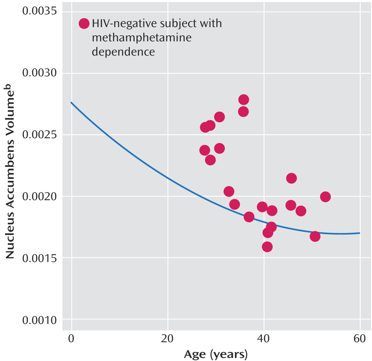
Previous studies of late brain maturation during adolescence and early adulthood (4446) have provided evidence for continuing myelination in white matter, and for continuing volume loss in cerebral gray matter structures—notably in the striatum. Methamphetamine exposure during adolescence may alter the course of ongoing brain maturation in these structures. In Figure 3, the (proportionalized) nucleus accumbens volumes of the HIV-negative/methamphetamine-dependent subjects in the present study are plotted against age. The curve shows the function relating the normalized nucleus accumbens volumes (measured using the same methods applied in the present study) to age in normally developing youngsters and young adults found in our previous studies (19, 45). As can be seen, significant volume reduction in the nucleus accumbens occurs in normally developing children. This figure illustrates that while the volumes observed in the younger methamphetamine-dependent subjects are increased relative to those of their nonabusing age-peers, they are more similar to volumes measured in 10–15-year-old normal youngsters.

Neurocognitive Impairment

Results of post hoc analyses suggest that among HIV-positive participants with no history of methamphetamine dependence, in whom we found significant cortical volume loss, more severe neurocognitive impairment is associated with lower cortical volumes. However, among HIV-negative/methamphetamine-dependent participants, in whom we found evidence for cortical volume increases, those with more severe impairment had higher cortical volumes. This pattern is consistent with the notion that both the cortical volume loss in HIV-positive individuals and the cortical volume increases in methamphetamine-dependent individuals reflect pathological tissue alterations that are accompanied by cortical dysfunction. It is not surprising that few significant correlations between brain volumes and impairment were noted in the HIV-positive/methamphetamine-dependent group, presumably because of combined (and opposing) effects of the two factors. However, it is interesting that hippocampal volume decreases, which were present in the HIV-positive individuals but not in the methamphetamine-dependent individuals of this study, were associated with neuropsychological impairment in the dually affected group.

Combined Effects of HIV and Methamphetamine

These results suggest overlapping, and opposing, effects of methamphetamine dependence and HIV infection on brain volumes. Apparently, in some brain structures, the effects of one or the other factor is dominant and this results in our report of different regional patterns associated with HIV effects and methamphetamine effects. In at least one site within the brain, the caudate nucleus, the effects of both factors are sufficiently strong to be detectable. In this structure, the effects, when combined, are likely to yield apparently normal volumes. This complicates the interpretation of striatal volumes in dually affected individuals. Furthermore, whatever the mechanisms underlying the volume increases and decreases we observe, they are likely to be associated with highly complex functional interactions within both affected and unaffected structures.
We did not observe statistically significant HIV status-by-methamphetamine interaction effects. That is, we did not obtain evidence for a significantly different effect of methamphetamine in HIV-positive than in HIV-negative individuals, nor of a significantly different effect of HIV in individuals with and without methamphetamine dependence. Rather, we obtained evidence for an additive effect of the two factors in the dually affected group. It is not possible to determine from these data whether the effects of methamphetamine and HIV are mediated by similar or completely different mechanisms. It is even possible, given these data, that the effects of HIV and methamphetamine are competitive (i.e., the effects of methamphetamine reverse those of HIV), although this seems very unlikely, since evidence from this study and a previous study (1) suggests that dually affected individuals have more severe neurocognitive impairment. The lack of significant HIV status-by-methamphetamine interaction effects on brain structure does not imply that functional interaction effects do not occur in association with those mechanisms that alter morphology. It is even possible that significant interaction effects on brain volume could emerge from analyses with larger samples.

Conclusions

The present study reveals distinct patterns of brain structural alterations associated with methamphetamine dependence and AIDS, two conditions observed with increasing frequency in the same individuals. The results help to define the most heavily affected structures and the regional extent of each of these separate patterns. This information may be useful in guiding future studies aimed at explaining the anomalies. Unfortunately, the present study yields only limited information about the cause of the changes. No relationship between measures of duration or pattern of use and the methamphetamine-related anomalies was observed. Furthermore, there was no apparent association between other psychiatric diagnoses and the anomalies present in methamphetamine-dependent individuals. Thus, there was no evidence that the alterations are markers for premorbid factors associated with the development of dependence. An important clue to the cause of the methamphetamine-related alterations, however, may be the association of less volume increase with increased age of the individuals. Further investigation of possible interactions between brain maturation and methamphetamine exposure is warranted.
Future studies should focus on defining the mechanisms that mediate the anomalies reported here. Previous studies with other imaging modalities have revealed a number of abnormalities in abstinent methamphetamine abusers. For example, a study employing MR spectroscopy reported decreased concentrations of a neuronal marker in frontal white matter and basal ganglia, and increased markers of gliosis in frontal cortex (41), suggesting neurotoxic effects. PET studies of dopamine transporter binding have suggested decreases in the striatum (37, 38, 40), and a PET study of cerebral glucose metabolism revealed decreased metabolism in the striatum and thalamus (37). Results of an MR perfusion study (47) also indicated an abnormal pattern of perfusion in abstinent methamphetamine users. The aforementioned MRS studies indicated early neuroinflammatory changes followed by neurodegeneration in HIV-infected individuals, and a recent study combining MR spectroscopy with fMRI suggests a direct role of glial activation in the cognitive dysfunction associated with HIV, as reflected in abnormal patterns of brain activation. Further studies of dually affected subjects using MR morphometric techniques in combination with different imaging modalities, such as MR spectroscopy, diffusion tensor imaging, and PET neuroreceptor mapping may implicate particular neural processes. For example, results from these studies may link the morphological alterations to MRS markers for gliosis or neurodegeneration, or to changes in receptor binding. Finally, diffusion tensor imaging studies may help to determine whether the volume changes are associated with changes in specific afferent or efferent projections of the affected structures.
TABLE 1
TABLE 2
TABLE 3
TABLE 4
TABLE 5
TABLE 6
Figure 1. Structural Boundaries for Gray Matter Regions
Figure 2. Volume Changes Related to Methamphetamine, HIV, and Methamphetamine and HIV
Figure 3. Relationship Between Nucleus Accumbens Volume and Age in HIV-Negative/Methamphetamine-Dependent Subjectsa
aThe healthy comparison curve is shown in blue.
bAs a proportion of supratentorial cranial volume.

Footnote

Received Sept. 8, 2003; revision received Oct. 15, 2004; accepted Oct. 22, 2004. From the Department of Psychiatry and Department of Neurosciences, UC San Diego, La Jolla; the Veterans Affairs San Diego Healthcare System, San Diego; and Fordham University, New York. Address correspondence and reprint requests to Dr. Jernigan, Laboratory of Cognitive Imaging, 9151B, University of California, San Diego, 9500 Gilman Dr., La Jolla, CA 92093–9151B; [email protected] (e-mail). This research was supported by the Medical Research Service of the Department of Veterans Affairs and by NIH grants from the National Institute on Drug Abuse (DA-12065) and the HIV Neurobehavioral Research Center (MH-45294, MH-62512).

References

1.
Rippeth JD, Heaton RK, Carey CL, Marcotte TD, Moore DJ, Gonzalez R, Wolfson T, Grant I (HNRC Group): Methamphetamine dependence increases risk of neuropsychological impairment in HIV infected persons. J Int Neuropsychol Soc 2004; 10:1–14
2.
Kalechstein AD, Newton TF, Green M: Methamphetamine dependence is associated with neurocognitive impairment in the initial phases of abstinence. J Neuropsychiatry Clin Neurosci 2003; 15:215–220
3.
Levin HS, Williams DH, Borucki MJ, Hillman GR, Williams JB, Guinto FC Jr, Amparo EG, Crow WN, Pollard RB: Magnetic resonance imaging and neuropsychological findings in human immunodeficiency virus infection. J Acquir Immune Defic Syndr 1990; 3:757–762
4.
Post MJ, Berger JR, Quencer RM: Asymptomatic and neurologically symptomatic HIV-seropositive individuals: prospective evaluation with cranial MR imaging. Radiology 1991; 178:131–139
5.
Jernigan TL, Archibald S, Hesselink JR, Atkinson JH, Velin RA, McCutchan JA, Chandler J, Grant I (HNRC Group): Magnetic resonance imaging morphometric analysis of cerebral volume loss in human immunodeficiency virus infection. Arch Neurol 1993; 50:250–255
6.
Aylward EH, Henderer JD, McArthur JC, Brettschneider PD, Harris GJ, Barta PE, Pearlson GD: Reduced basal ganglia volume in HIV-1-associated dementia: results from quantitative neuroimaging. Neurology 1993; 43:2099–2104
7.
Henderer JD, Barta PE, Tien AY, Pearlson GD: Magnetic resonance imaging measurement of gray matter volume reductions in HIV dementia. Am J Psychiatry 1995; 152:987–994
8.
Stout JC, Ellis RJ, Jernigan TL, Archibald SL, Abramson I, Wolfson T, McCutchan JA, Wallace MR, Atkinson JH, Grant I (HIV Neurobehavioral Research Center Group): Progressive cerebral volume loss in human immunodeficiency virus infection: a longitudinal volumetric magnetic resonance imaging study. Arch Neurol 1998; 55:161–168
9.
Chong WK, Sweeney B, Wilkinson ID, Paley M, Hall-Craggs MA, Kendall BE, Shepard JK, Beecham M, Miller RF, Weller IV: Proton spectroscopy of the brain in HIV infection: correlation with clinical, immunologic, and MR imaging findings. Radiology 1993; 188:119–124
10.
Meyerhoff DJ, MacKay S, Bachman L, Poole N, Dillon WP, Weiner MW, Fein G: Reduced brain N-acetylaspartate suggests neuronal loss in cognitively impaired human immunodeficiency virus-seropositive individuals: in vivo 1H magnetic resonance spectroscopic imaging. Neurology 1993; 43:509–515
11.
Jarvik JG, Lenkinski RE, Grossman RI, Gomori JM, Schnall MD, Frank I: Proton MR spectroscopy of HIV-infected patients: characterization of abnormalities with imaging and clinical correlation. Radiology 1993; 186:739–744
12.
Paley M, Cozzone PJ, Alonso J, Vion-Dury J, Confort-Gouny S, Wilkinson ID, Chong WK, Hall-Craggs MA, Harrison MJ, Gili J, Rovira A, Capellades J, Rio J, Ocana I, Nicoli F, Dhiver C, Gastaut JL, Gastaut JA, Wicklow K, Sauter R: A multicenter proton magnetic resonance spectroscopy study of neurological complications of AIDS. AIDS Res Hum Retroviruses 1996; 12:213–222
13.
Tracey I, Carr CA, Guimaraes AR, Worth JL, Navia BA, González RG: Brain choline-containing compounds are elevated in HIV-positive patients before the onset of AIDS dementia complex: a proton magnetic resonance spectroscopic study. Neurology 1996; 46:783–788; correction, 46:1787
14.
Laubenberger J, Häussinger D, Bayer S, Thielemann S, Schneider B, Mundinger A, Hennig J, Langer M: HIV-related metabolic abnormalities in the brain: depiction with proton MR spectroscopy with short echo times. Radiology 1996; 199:805–810
15.
Wilkinson ID, Miller RF, Miszkiel KA, Paley MN, Hall-Craggs MA, Baldeweg T, Williams IG, Carter S, Newman SP, Kendall BE, Catalan J, Chinn RJ, Harrison MJ: Cerebral proton magnetic resonance spectroscopy in asymptomatic HIV infection. AIDS 1997; 11:289–295
16.
Bartzokis G, Beckson M, Lu PH, Edwards N, Rapoport R, Wiseman E, Bridge P: Age-related brain volume reductions in amphetamine and cocaine addicts and normal controls: implications for addiction research. Psychiatry Res 2000; 98:93–102
17.
Thompson PM, Hayashi KM, Simon SL, Geaga JA, Hong MS, Sui Y, Lee JY, Toga AW, Ling W, London ED: Structural abnormalities in the brains of human subjects who use methamphetamine. J Neurosci 2004; 24:6028–6036
18.
Heaton RK, Grant I, Butters N, White DA, Kirson D, Atkinson JH, McCutchan JA, Taylor MJ, Kelly MD, Ellis RJ, Wofson T, Velin R, Marcotte TD, Hesselink JR, Jernigan TL, Chandler J, Wallace M, Abramson I: The HNRC500—neuropsychology of HIV infection at different disease stages. J Int Neuropsychol Soc 1995; 1:231–251
19.
Jernigan TL, Archibald SL, Fennema-Notestine C, Gamst AC, Stout JC, Bonner J, Hesselink JR: Effects of age on tissues and regions of the cerebrum and cerebellum. Neurobiol Aging 2001; 22:581–594
20.
Steketee JD: Neurotransmitter systems of the medial prefrontal cortex: potential role in sensitization to psychostimulants. Brain Res Brain Res Rev 2003; 41:203–228
21.
Robinson TE, Becker JB: Behavioral sensitization is accompanied by an enhancement in amphetamine-stimulated dopamine release from striatal tissue in vitro. Eur J Pharmacol 1982; 85:253–254
22.
Wilcox RA, Robinson TE, Becker JB: Enduring enhancement in amphetamine-stimulated striatal dopamine release in vitro produced by prior exposure to amphetamine or stress in vivo. Eur J Pharmacol 1986; 124:375–376
23.
Iwata SI, Hewlett GH, Ferrell ST, Kantor L, Gnegy ME: Enhanced dopamine release and phosphorylation of synapsin I and neuromodulin in striatal synaptosomes after repeated amphetamine. J Pharmacol Exp Ther 1997; 283:1445–1452
24.
Kuczenski R, Melega WP, Cho AK, Segal DS: Extracellular dopamine and amphetamine after systemic amphetamine administration: comparison to the behavioral response. J Pharmacol Exp Ther 1997; 282:591–596
25.
Segal DS, Kuczenski R, O’Neil ML, Melega WP, Cho AK: Escalating dose methamphetamine pretreatment alters the behavioral and neurochemical profiles associated with exposure to a high-dose methamphetamine binge. Neuropsychopharmacology 2003; 28:1730–1740
26.
Robinson TE, Kolb B: Persistent structural modifications in nucleus accumbens and prefrontal cortex neurons produced by previous experience with amphetamine. J Neurosci 1997; 17:8491–8497
27.
Robinson TE, Kolb B: Alterations in the morphology of dendrites and dendritic spines in the nucleus accumbens and prefrontal cortex following repeated treatment with amphetamine or cocaine. Eur J Neurosci 1999; 11:1598–1604
28.
Takaki M, Ujike H, Kodama M, Takehisa Y, Yamamoto A, Kuroda S: Increased expression of synaptophysin and stathmin mRNAs after methamphetamine administration in rat brain. Neuroreport 2001; 12:1055–1060
29.
Ujike H, Takaki M, Kodama M, Kuroda S: Gene expression related to synaptogenesis, neuritogenesis, and MAP kinase in behavioral sensitization to psychostimulants. Ann NY Acad Sci 2002; 965:55–67
30.
Song DD, Haber SN: Striatal responses to partial dopaminergic lesion: evidence for compensatory sprouting. J Neurosci 2000; 20:5102–5114
31.
Thomas DM, Francescutti-Verbeem DM, Liu X, Kuhn DM: Identification of differentially regulated transcripts in mouse striatum following methamphetamine treatment—an oligonucleotide microarray approach. J Neurochem 2004; 88:380–393
32.
LaVoie MJ, Card JP, Hastings TG: Microglial activation precedes dopamine terminal pathology in methamphetamine-induced neurotoxicity. Exp Neurol 2004; 187:47–57
33.
Giulian D, Li J, Leara B, Keenen C: Phagocytic microglia release cytokines and cytotoxins that regulate the survival of astrocytes and neurons in culture. Neurochem Int 1994; 25:227–233
34.
Pu C, Broening HW, Vorhees CV: Effect of methamphetamine on glutamate-positive neurons in the adult and developing rat somatosensory cortex. Synapse 1996; 23:328–334
35.
Eisch AJ, O’Dell SJ, Marshall JF: Striatal and cortical NMDA receptors are altered by a neurotoxic regimen of methamphetamine. Synapse 1996; 22:217–225
36.
Sekine Y, Iyo M, Ouchi Y, Matsunaga T, Tsukada H, Okada H, Yoshikawa E, Futatsubashi M, Takei N, Mori N: Methamphetamine-related psychiatric symptoms and reduced brain dopamine transporters studied with PET. Am J Psychiatry 2001; 158:1206–1214
37.
Volkow ND, Chang L, Wang G-J, Fowler JS, Franceschi D, Sedler MJ, Gatley SJ, Hitzemann R, Ding Y-S, Wong C, Logan J: Higher cortical and lower subcortical metabolism in detoxified methamphetamine abusers. Am J Psychiatry 2001; 158:383–389
38.
Volkow ND, Chang L, Wang G-J, Fowler JS, Leonido-Yee M, Franceschi D, Sedler MJ, Gatley SJ, Hitzemann R, Ding Y-S, Logan J, Wong C, Miller EN: Association of dopamine transporter reduction with psychomotor impairment in methamphetamine abusers. Am J Psychiatry 2001; 158:377–382
39.
Volkow ND, Chang L, Wang GJ, Fowler JS, Franceschi D, Sedler M, Gatley SJ, Miller E, Hitzemann R, Ding YS, Logan J: Loss of dopamine transporters in methamphetamine abusers recovers with protracted abstinence. J Neurosci 2001; 21:9414–9418
40.
McCann UD, Wong DF, Yokoi F, Villemagne V, Dannals RF, Ricaurte GA: Reduced striatal dopamine transporter density in abstinent methamphetamine and methcathinone users: evidence from positron emission tomography studies with [11C]WIN-35,428. J Neurosci 1998; 18:8417–8422
41.
Ernst T, Chang L, Leonido-Yee M, Speck O: Evidence for long-term neurotoxicity associated with methamphetamine abuse: a 1H MRS study. Neurology 2000; 54:1344–1349
42.
Jacobsen LK, Giedd JN, Gottschalk C, Kosten TR, Krystal JH: Quantitative morphology of the caudate and putamen in patients with cocaine dependence. Am J Psychiatry 2001; 158:486–489
43.
Franklin TR, Acton PD, Maldjian JA, Gray JD, Croft JR, Dackis CA, O’Brien CP, Childress AR: Decreased gray matter concentration in the insular, orbitofrontal, cingulate, and temporal cortices of cocaine patients. Biol Psychiatry 2002; 51:134–142
44.
Jernigan TL, Trauner DA, Hesselink JR, Tallal PA: Maturation of human cerebrum observed in vivo during adolescence. Brain 1991; 114:2037–2049
45.
Sowell ER, Trauner DA, Gamst A, Jernigan TL: Development of cortical and subcortical brain structures in childhood and adolescence: a structural MRI study. Dev Med Child Neurol 2002; 44:4–16
46.
Sowell ER, Thompson PM, Holmes CJ, Jernigan TL, Toga AW: In vivo evidence for post-adolescent brain maturation in frontal and striatal regions (letter). Nat Neurosci 1999; 2:859–861
47.
Chang L, Ernst T, Speck O, Patel H, DeSilva M, Leonido-Yee MV: Perfusion MRI and computerized cognitive test abnormalities in abstinent methamphetamine users. Psychiatry Res 2002; 114:65–79

Information & Authors

Information

Published In

Go to American Journal of Psychiatry
American Journal of Psychiatry
Pages: 1461 - 1472
PubMed: 16055767

History

Published online: 1 August 2005
Published in print: August 2005

Authors

Details

Terry L. Jernigan, Ph.D.
Anthony C. Gamst, Ph.D.
Sarah L. Archibald, M.A.
Christine Fennema-Notestine, Ph.D.
Monica Rivera Mindt, Ph.D.
Thomas L. Marcotte, Ph.D.
Robert K. Heaton, Ph.D.

Metrics & Citations

Metrics

Citations

Export Citations

If you have the appropriate software installed, you can download article citation data to the citation manager of your choice. Simply select your manager software from the list below and click Download.

For more information or tips please see 'Downloading to a citation manager' in the Help menu.

Format
Citation style
Style
Copy to clipboard

View Options

View options

PDF/EPUB

View PDF/EPUB

Login options

Already a subscriber? Access your subscription through your login credentials or your institution for full access to this article.

Personal login Institutional Login Open Athens login
Purchase Options

Purchase this article to access the full text.

PPV Articles - American Journal of Psychiatry

PPV Articles - American Journal of Psychiatry

Not a subscriber?

Subscribe Now / Learn More

PsychiatryOnline subscription options offer access to the DSM-5-TR® library, books, journals, CME, and patient resources. This all-in-one virtual library provides psychiatrists and mental health professionals with key resources for diagnosis, treatment, research, and professional development.

Need more help? PsychiatryOnline Customer Service may be reached by emailing [email protected] or by calling 800-368-5777 (in the U.S.) or 703-907-7322 (outside the U.S.).

Media

Figures

Other

Tables

Share

Share

Share article link

Share