Abstract

Objective:

Behavioral inhibition is an early childhood temperament recently associated with altered striatal response in adolescence to incentives of increasing magnitudes. Since early childhood behavioral inhibition is also associated with risk for adolescent social phobia, a similar pattern of striatal activation may manifest in social phobia. The present study compares striatal function in healthy adolescents, adolescents with social phobia, and adolescents with generalized anxiety disorder.

Method:

Blood-oxygen-level-dependent signal in striatal regions was examined in 58 medication-free adolescents—14 with social phobia, 18 with generalized anxiety disorder but not social phobia, and 26 with no psychiatric disorder—matched on sex, age, puberty, IQ, and socioeconomic status. During functional magnetic resonance imaging, participants responded to incentive cues depicting potential monetary gains or losses of varying magnitudes.

Results:

While anticipating incentives of increasing magnitude, adolescents with social phobia showed increasingly heightened caudate and putamen activation at a level greater than that seen in the healthy comparison and generalized anxiety disorder groups. The generalized anxiety disorder group showed a unique valence-specific putamen response relative to the healthy comparison or social phobia group. Both patient groups displayed more complex patterns in the nucleus accumbens than in the caudate or putamen.

Conclusions:

Caudate and putamen hypersensitivity to incentives of increasing magnitudes characterizes adolescent social phobia, relative to activation in this region in adolescents with generalized anxiety disorder as well as healthy adolescents. Thus, these findings resemble the pattern previously found in adolescents with early childhood behavioral inhibition, thereby implicating similar neural responses to anticipation of incentives in both early childhood behavioral inhibition and adolescent social phobia.
Adolescent anxiety is highly prevalent and predicts adverse outcomes such as adult anxiety and depression (13). Four sets of key findings guide questions on the precursors, mechanisms, and consequences of adolescent anxiety. First, adolescent anxiety disorders are often co-occurring, with particularly high comorbidity between social phobia and generalized anxiety disorder (4). Second, some anxiety disorders share risk factors, which could explain their high comorbidity levels (46). Third, despite comorbidity and shared risks, evidence of specificity still emerges (e.g., adolescent social phobia predicts risk for adult social phobia but not adult generalized anxiety disorder) (2). Similarly, behavioral inhibition is an early childhood temperament associated with heightened risk for social phobia (69) but not generalized anxiety disorder (10). Finally, based on prior neuroimaging data on behavioral inhibition, a heightened neural response to anticipated incentives may be a marker that links early childhood behavioral inhibition specifically to later social phobia but not generalized anxiety disorder (11). We used methods previously employed in a study of adolescents with early childhood behavioral inhibition (11) to test this possibility and compared neural responses to anticipated incentives in adolescents with social phobia and adolescents with generalized anxiety disorder.
Amygdala and ventral prefrontal cortex responses to threat are altered in adolescents with early childhood behavioral inhibition (12, 13) and in those with anxiety disorders (1419). These findings are nonspecific, however, since they are seen in behavioral inhibition, social phobia, generalized anxiety disorder, and to a degree, major depression (1220). Altered neural response to potential incentives may occur in a more restricted, specific fashion given unique responses to anticipated incentives in behavioral inhibition (11, 21, 22) that are distinct from responses in adolescent depression (23). However, few studies have extended such research to clinically anxious and healthy adolescents, and none have compared social phobia with generalized anxiety disorder. Direct comparison of social phobia with generalized anxiety disorder in adolescents may reveal unique risk mechanisms shared between social phobia and early childhood behavioral inhibition.
Several findings link anxiety and incentive processing. An initial study found that high state anxiety correlates positively with a hypersensitive behavioral response to rewards (24). Subsequent studies examined neural manifestations of incentive hypersensitivity in behavioral inhibition (11, 21). Adolescents characterized by early childhood behavioral inhibition, relative to those characterized as noninhibited, showed greater striatal response modulation with increasing incentive magnitudes (11). A later study showed striatal hyperactivation in behaviorally inhibited adolescents, specifically when anticipated incentives were contingent on participants' choices (21). Consistent with these data, research in adult social phobia has found altered striatal dopamine function (25, 26) and task-elicited striatal perturbations (27); no such work has examined striatal function in adolescent social phobia. These findings suggest that striatal hyperactivation may manifest in adolescent social phobia and raise questions about the specificity of this functional alteration in social phobia relative to generalized anxiety disorder.
In the present study, we compared striatal function in healthy adolescents, adolescents with social phobia, and adolescents with generalized anxiety disorder on the same monetary incentive delay task used previously to show striatal hyperactivation in adolescents with early childhood behavioral inhibition (11). Given data linking behavioral inhibition to adolescent social phobia (10), we hypothesized that incentive magnitude would modulate striatal response more strongly in adolescents with social phobia than in healthy adolescents or adolescents with generalized anxiety disorder.

Method

Participants

Participants were 26 healthy adolescent volunteers, 18 adolescent patients diagnosed with generalized anxiety disorder but not social phobia, and 14 adolescent patients diagnosed with social phobia (Table 1). Three social phobia patients met criteria for generalized anxiety disorder as a secondary diagnosis. Three patients with generalized anxiety disorder and two with social phobia met criteria for depression; all five of these patients were included because anxiety was the primary reason for referral.
TABLE 1. Demographic and Clinical Characteristics of Adolescents With Generalized Anxiety Disorder or Social Phobia and Age-Matched Healthy Comparison Subjects
 Participant Group
CharacteristicGeneralized Anxiety Disorder (N=18)Social Phobia (N=14)Healthy Comparison (N=26)
 MeanSDMeanSDMeanSD
Age (years)12.912.6713.133.0213.992.44
Wechsler Abbreviated Scale of Intelligence full-scale IQ11211.1811116.0511110.80
Socioeconomic status4.751.074.621.334.051.33
Tanner puberty stage rating3.001.363.381.193.101.26
Screen for Child Anxiety Related Emotional Disorders scorea28.0612.7431.2110.0810.846.94
 N%N%N%
Female10569641142
Major depressive disorder317214  
Generalized anxiety disorder18100321  
Social phobia0014100  
Separation anxiety528321  
Specific phobia528321  
a
Scores ranged from 0 to 52; scores ≥24 indicate presence of an anxiety disorder. Significant difference among groups (F=23.92, df=2, 53, p<0.001); patient groups had similar scores and healthy comparison subjects had lower scores than patients.
Patients sought treatment for anxiety symptoms (Pediatric Anxiety Rating Scale [28] score: ≥10; Child Global Assessment Scale score: <60). Diagnoses were determined using clinician-based interviews (29). Anxiety severity was indexed by scores on the Screen for Child Anxiety Related Emotional Disorders scale (30) averaged from adolescent and parent reports. Exclusion criteria were current Tourette's syndrome, obsessive-compulsive disorder, conduct disorder, or suicidal ideation; history of mania, psychosis, or pervasive developmental disorder; traumatic exposure; an IQ <70; or psychoactive substance use in the past month (2 months for fluoxetine). The National Institute of Mental Health Institutional Review Board approved the study. After receiving complete description of the study, parents/legal guardians provided written informed consent, and participants provided written informed assent.
Groups were well matched on demographic characteristics (Table 1). Both patient groups had similarly extreme elevations in their scores on the Screen for Child Anxiety Related Emotional Disorders scale relative to healthy comparison subjects (p<0.001).

Task Paradigm

The monetary incentive delay task engages the striatum during anticipation of potential monetary gain or loss (31, 32), with a parametric version varying the monetary amount at stake (31). A cue indicates trial incentive magnitude. Participants respond as quickly as possible during target presentation. Successful performance leads to winning or avoiding loss. Based on our prior work (11), we focused on the anticipatory phase, which is during the cue presentation before motor response. Participants practiced the task prescan to standardize performance and task difficulty by individually tailoring success on approximately 66% of trials (31).
Participants completed two runs of 72 contiguous 6-second trials (see Figure 1 in the data supplement accompanying the online version of this article). Trials began with a cue presentation (250 msec) followed by cross-hair fixation points (2,000–2,500 msec) and target response cues (160–250 msec). Circle cues (N=64) indicated monetary gain (U.S. dollars) if the button press occurred quickly enough at target onset. Square cues (N=64) signified monetary loss if the button press did not occur quickly enough at target onset. Triangle “neutral” cues (N=16) indicated $0 at stake. Incentive magnitude was represented by a single line ($0.20; N=32), two lines ($1.00; N=32), or three lines ($5.00; N=32) within the cue. After the target's disappearance, feedback (1,650 msec) notified participants of a gain, a loss, or no change and of cumulative winnings. Trial-type order was fully randomized. Participants could win up to $50. Postscan, they rated their cue preferences from –5 (dislike very much) to +5 (like very much).

Behavioral Data Analysis

Dependent variables were accuracy (i.e., proportion of successful button presses during target presentation), reaction time for correct hits (i.e., time between target onset and successful button presses), and postscan affective ratings. Accuracy, reaction time, and affective ratings were examined with a group-by-magnitude-by-valence repeated-measures analysis of variance (ANOVA). To parallel imaging analyses, which used $0 trials as a baseline, response to $0 cues was a covariate (except for affective ratings).

Functional Magnetic Resonance Imaging (fMRI) Data Acquisition

Scanning occurred on a General Electric Signa 3T magnet (General Electric Co., Waukesha, Wisc.), with a standard birdcage head-coil and Cedrus Lumina response box (Cedrus, San Pedro, Calif.). Stimuli were projected onto a screen at the foot of the scanner bed and viewed with mirrors. Functional imaging parameters were as follows: 30 interleaved 4-mm thick slices acquired in the sagittal plane using a T2-weighted gradient echo sequence; TR=2,500 msec, TE=23 msec, flip angle=90°, voxel dimension=3.75×3.75×4.0 mm, matrix size=64×64, and field of view=24 cm. Four acquisitions were obtained before task onset to stabilize the signal. A high-resolution structural image was acquired for spatial normalization (T1-weighted standardized magnetization-prepared spoiled gradient-recalled echo sequence with 124 1-mm slices; TR=8,100 msec, TE=32 msec, flip angle=15°, matrix size=256×256, field of view=24 cm).

fMRI Data Preprocessing

Analysis of Functional and Neural Images software (33) was used. Preprocessing included slice time correction, motion correction, and spatial smoothing (6-mm full-width half-maximum kernel). A despiking algorithm applied on a voxelwise basis smoothed signal deviations >2.5 standard deviations from the mean. A band-pass filtering algorithm smoothed cyclical fluctuations in signals (either >0.011 second or <0.15 second) not temporally indicative of a hemodynamic response. Data for each participant were converted to percent signal change using each participant's voxelwise time-series mean as a baseline.
Time-series data for each participant were analyzed with multiple regression using a region-of-interest approach that followed past procedures (11). The model included event-type regressors of interest (incentive cues, target cue, feedback), six regressors modeling effects as a result of residual motion (in the x, y, and z planes and yaw, pitch, and roll dimensions), and two regressors modeling baseline and linear trends per run. Regressors of interest were convolved with a gamma variate function that modeled a prototypical hemodynamic response (34). Idealized signal time courses were estimated from the onset time of event type.

fMRI Data Analysis

Following past methods (11), six contrasts of blood-oxygen-level-dependent (BOLD) activation were created individually for the three monetary gain cues and three monetary loss cues, each compared with no-monetary neutral cues. Activation was calculated as the net signal difference between each incentive magnitude and no incentive at the acquisition of the event-related hemodynamic response function during cue presentation. Mean contrast values were generated for all voxels located within each of the following three striatal structures: nucleus accumbens, caudate nucleus (encompassing the head and body), and putamen. Talairach anatomical boundaries, provided by Analysis of Functional and Neural Images software, defined voxels within each region after spatial normalization (35). One contrast value was generated for each participant per region to minimize type I errors.
Contrast values were analyzed at the group level using SPSS software (SPSS, Inc., Chicago). Based on previous findings (11), we hypothesized that the social phobia group would show increased striatal activation as a function of increased incentive magnitude. We tested this hypothesis using an omnibus repeated-measures ANOVA that included group (social phobia, generalized anxiety disorder, healthy comparison), magnitude (small, medium, large), valence (gain, loss), and region (bilateral nucleus accumbens, caudate, putamen).
Although previous research on behavioral inhibition showed reactivity to incentive magnitudes across striatal regions, we retained specific striatal region as a factor for the following reasons. Because few studies examine reward function in clinical anxiety, it is important to generate initial data for specific regions. Moreover, because our prior study of adolescents with early-life behavioral inhibition was moderately sized (N=32), power on higher-order interaction tests was restricted. Similarly, a second, larger study of adolescents with early-life behavioral inhibition using a different task found region-specific striatal responses (21). Finally, including region as a factor is consistent with documented functional specialization within the striatal structures examined in the present study (31, 36, 37).
Dependent measures encompassed BOLD signal change values of each event-related contrast extracted from anatomically defined masks of a priori striatal regions. Based on past findings (11), we expected to observe group-by-magnitude interactions, either within or across striatal regions. Least significant difference comparisons identified specific differences driving significant group main and interaction effects.

Results

In-Scanner Task Performance

None of the two- or three-way interactions with group were significant for reaction time or accuracy. No significant group main effects emerged for reaction time or accuracy. Thus, the three groups showed similar task performance.

Affective Response to Task Cues

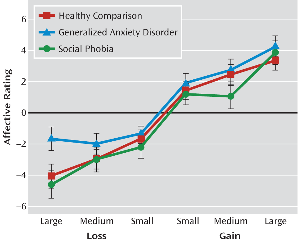
A significant valence-by-magnitude interaction was found on postscan cue preferences. Collapsed across groups, participants' cue preference increased as the gain cue magnitude increased and decreased as the loss cue magnitude increased (F=35.06, df=2, 92, p<0.001 [Figure 1]). A significant group-by-magnitude interaction revealed group differences in cue preference as a function of magnitude (F=2.51, df=2, 92, p=0.04). Post hoc tests indicated a unique profile in the generalized anxiety disorder group, where large incentives were more preferred than in the social phobia (p=0.01) or healthy comparison (p=0.005) group. Valence effects on cue preference did not differ between groups. Nevertheless, these tests have low statistical power to detect significant interactions. Despite the nonsignificant interactions, however, the pattern of mean rating levels suggests that large incentives in the loss, but not gain, condition may be more aversive to the social phobia group than to the generalized anxiety disorder group.
FIGURE 1. Postscan Affective Ratings of Cues Depicting Small, Medium, and Large Incentives for Loss and Gain Trials Among Adolescents With Social Phobia or Generalized Anxiety Disorder and Age-Matched Healthy Comparison Subjectsa
a Ratings ranged from –5 (dislike very much) to +5 (like very much). Participants' preference of cues increased as the gain cue magnitude increased and decreased as the loss cue magnitude increased (F=35.06, df=2, 92, p<0.001). Error bars represent the standard error of the mean.

Striatal Response

The omnibus ANOVA showed a significant group-by-magnitude-by-valence-by-region interaction (F=2.70, df=4.73, 130.11, p=0.03, Greenhouse-Geisser corrected). Region-specific responding occurred in the three groups as a function of magnitude and valence. The four-way interaction was decomposed with repeated-measures ANOVAs examining lower-order interactions per region. Group and task parameter interaction effects were examined. Post hoc comparisons focused on group differences. Valence, magnitude, and valence-by-magnitude effects are presented in Table 1 of the data supplement.

Caudate nucleus.

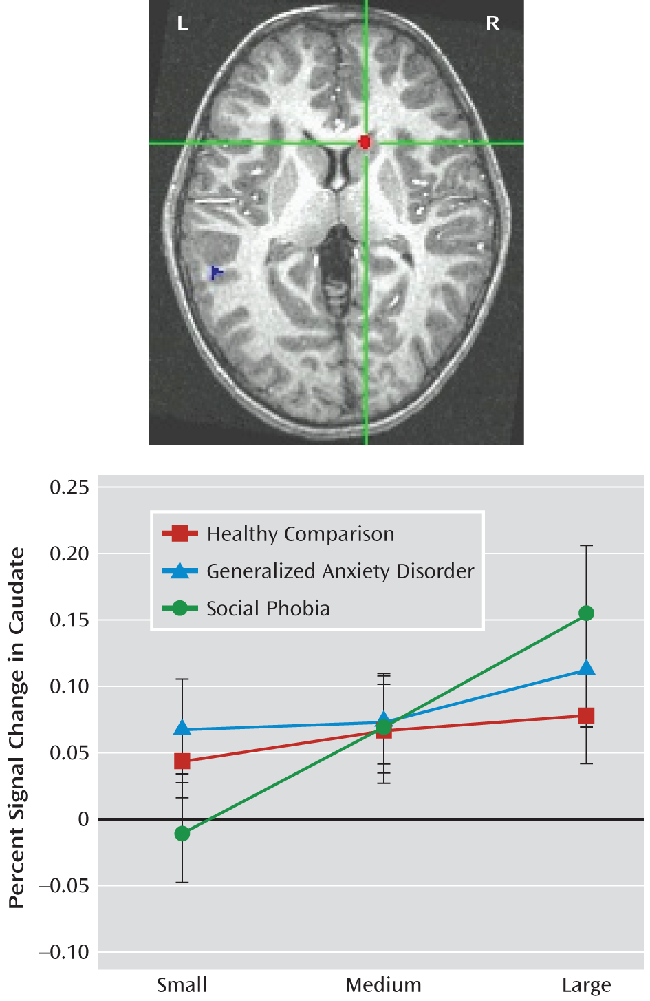
There was a significant group-by-magnitude interaction on caudate activation (within-subjects linear effect: F=3.52, df=2, 55, p=0.04). Post hoc tests conducted for each group showed that within the social phobia group, caudate activation increased as potential wins or losses increased from small to medium to large (small versus medium, p=0.02; small versus large, p<0.001; medium versus large, p=0.02 [Figure 2]). In contrast, incentive magnitude did not significantly modulate caudate activation within the generalized anxiety disorder or healthy comparison group, indicating greater caudate sensitivity to incentive magnitude in the social phobia group. The generalized anxiety disorder and healthy comparison groups showed striatal activation in response to all three magnitudes relative to the neutral-cue baseline. Thus, between-group differences were evident in the caudate, with greater magnitude sensitivity in the social phobia group compared with the generalized anxiety disorder or healthy comparison group.
FIGURE 2. Activation in the Caudate Nucleus in Response to Incentive Cues Among Adolescents With Social Phobia Relative to Adolescents With Generalized Anxiety Disorder and Age-Matched Healthy Comparison Subjectsa
a For illustrative purposes, the top image is based on results from a voxelwise one-sample t test within the social phobia group showing right caudate activation in response to high gain cues (planes: x=13, y=19, z=9; t=5.01, df=14, p<0.001). Caudate response to high gain cues was not significantly different from zero within the generalized anxiety disorder and healthy comparison groups. The bottom graph depicts event-related percent BOLD signal change extracted from the caudate region of interest. A significant group-by-magnitude interaction effect was found (F=3.52, df=2, 55, p=0.04). Only within the social phobia group did caudate activation increase as the incentive increased (small versus medium, p=0.02; small versus large, p<0.001; medium versus large, p=0.02). Error bars represent the standard error of the mean. Abbreviations: L=left; R=right.

Putamen.

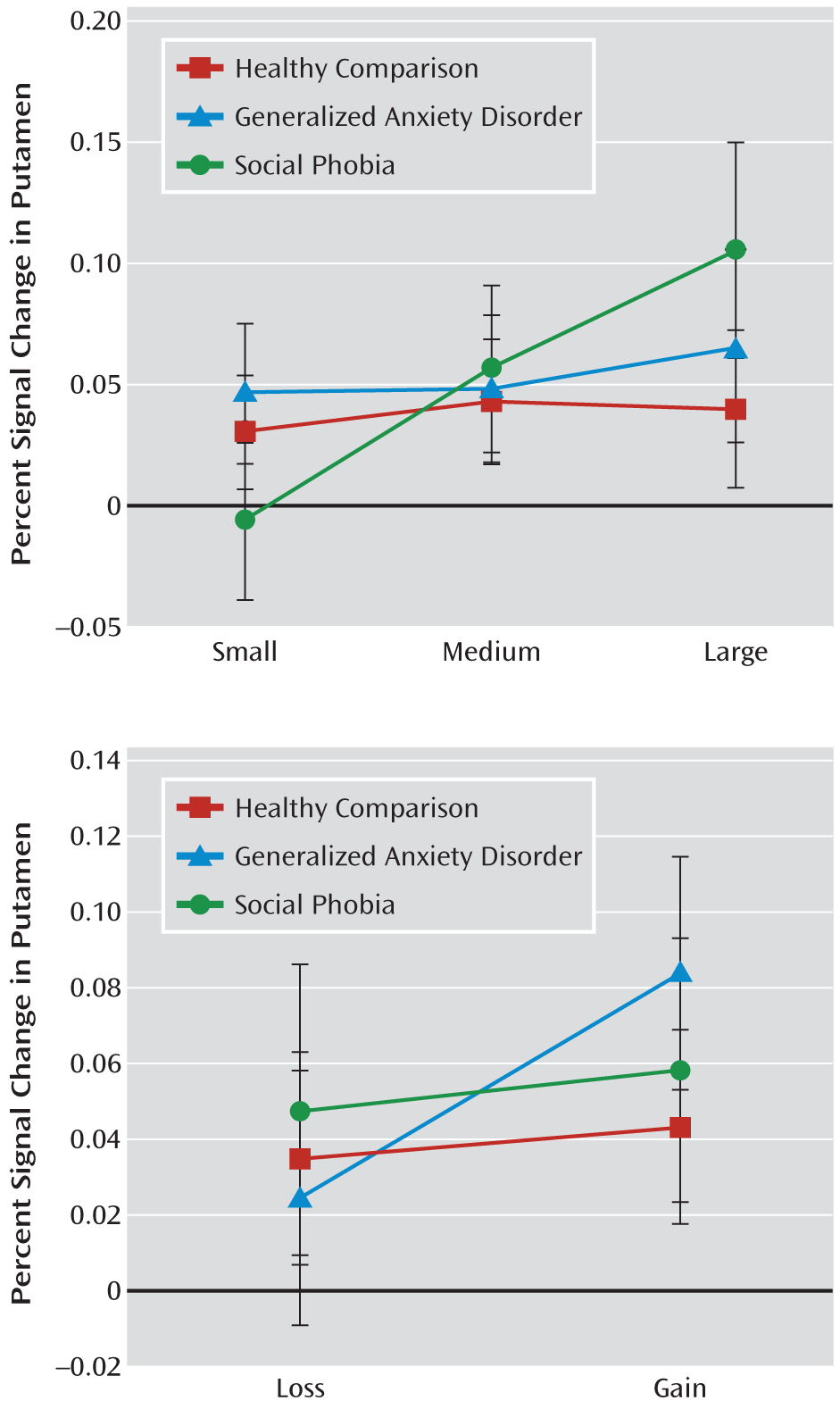
There was also a significant group-by-magnitude interaction on putamen activation (within-subjects linear effect: F=3.94, df=2, 55, p=0.03 [Figure 3]). Post hoc tests within each group showed that the putamen response was similar to the caudate pattern, with increasing activation as a function of increasing incentive magnitude only in the social phobia group (small versus medium [p=0.02]; small versus large [p=0.001]). As with the caudate, these contrasts were not significant for the generalized anxiety disorder or healthy comparison group.
FIGURE 3. Event-Related Percent BOLD Signal Change Extracted From the Putamen Region of Interest Among Adolescents With Social Phobia or Generalized Anxiety Disorder and Age-Matched Healthy Comparison Subjectsa
a The top graph depicts a significant group-by-magnitude interaction effect (F=3.94, df=2, 55, p=0.03). Within the social phobia group, but not the generalized anxiety disorder or healthy comparison group, putamen activation increased as incentive magnitude increased from small to medium (p=0.02) and small to large (p=0.001). The bottom graph depicts a significant group-by-valence interaction effect (F=3.21, df=4, 55, p<0.05). Within the generalized anxiety disorder group, but not the social phobia or healthy comparison group, putamen activation was significantly greater on gain versus loss trials (p=0.001). Error bars represent the standard error of the mean.
Unlike the caudate pattern, the group effect on the putamen was modulated by valence (F=3.21, df=4, 55, p<0.05 [Figure 3]). Valence was only a significant factor for the generalized anxiety disorder group. Post hoc comparisons showed significantly greater putamen activation during potential gain versus loss trials (p=0.001). Post hoc tests for the social phobia and healthy comparison groups revealed no significant modulation by valence on the putamen.

Nucleus accumbens.

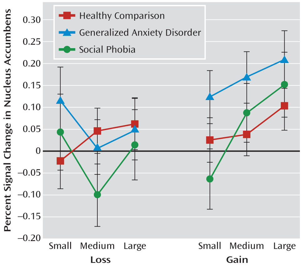
A significant group-by-magnitude-by-valence interaction on nucleus accumbens activation was found (F=2.69, df=4, 110, p=0.04 [Figure 4]). Post hoc between-group comparisons indicated that the generalized anxiety disorder group relative to the social phobia group had greater nucleus accumbens activation in response to anticipated small gains (p<0.05), with no other significant group differences. Subsequent ANOVAs tested the magnitude-by-valence interaction within each group. Within the social phobia group, this interaction was significant (F=4.02, df=2, 26, p=0.03). Post hoc tests revealed that potential small losses versus medium losses (p=0.02) and potential large gains versus small gains (p=0.001) elicited greater nucleus accumbens activation. Only for medium incentives did gains versus losses (p=0.02) elicit greater nucleus accumbens activation. Within the generalized anxiety disorder group, a significant magnitude-by-valence interaction emerged (F=4.53, df=2, 34, p=0.02); for medium (p=0.001) and large (p=0.01) incentives, gains versus losses elicited greater nucleus accumbens activation. Significant differences were not found comparing magnitude levels within each valence. Within the healthy comparison group, nucleus accumbens activation did not vary by valence or magnitude.
FIGURE 4. Event-Related Percent BOLD Signal Change Extracted From the Nucleus Accumbens Region of Interest Among Adolescents With Social Phobia or Generalized Anxiety Disorder and Age-Matched Healthy Comparison Subjectsa
a The graph depicts a significant group-by-magnitude-by-valence interaction effect (F=2.69, df=4, 110, p=0.04). Within the social phobia group, small losses versus medium losses (p=0.02), large gains versus small gains (p=0.001), and medium gains versus medium losses (p=0.02) elicited greater activation. Within the generalized anxiety disorder group, greater activation was seen for medium (p=0.001) and large (p=0.01) gains versus losses. Error bars represent the standard error of the mean.

Discussion

The present study tested hypotheses about striatal circuitry alterations among healthy and clinically anxious adolescents. This work extends previous findings documenting striatal hypersensitivity to anticipated incentives in adolescents with early childhood behavioral inhibition (11, 21). Because early childhood behavioral inhibition predicts risk for later anxiety disorders, particularly social phobia (610), we expected striatal circuitry hypersensitivity to be evident in adolescents with social phobia. Specifically, we expected incentive magnitude to modulate striatal activation more strongly in adolescents with social phobia than in healthy adolescents or adolescents with generalized anxiety disorder.
The caudate and putamen showed the expected pattern of increased activation as incentive magnitude increased in the social phobia group but not in the generalized anxiety disorder or healthy comparison group. This finding suggests a striatal circuitry functional profile shared by both a behaviorally inhibited temperament and social phobia in adolescence. Previous research has suggested that adolescents characterized by early childhood behavioral inhibition find cues indicating potential for reward or punishment to be highly salient because of performance-related concerns (11). This interpretation was supported in work documenting striatal hyperactivity in adolescents with early childhood behavioral inhibition when anticipated reward outcomes resulted directly from the participants' actions (21) and when anticipated rewards were not received (22). In the present study, adolescents with social phobia also showed striatal sensitivity to stakes associated with performance, suggesting that rewards might engage similar psychological processes in adolescent social phobia and early-life behavioral inhibition.
Altered caudate and putamen function distinguished the social phobia group from the other two groups, but findings in the generalized anxiety disorder group did not quite mirror those in the healthy comparison group. Adolescents with generalized anxiety disorder showed putamen hyperactivation in response to valence (gain versus loss), findings not observed in those with social phobia or in healthy adolescents. Thus, while behavioral inhibition and social phobia are associated with one pattern of perturbed striatal response to incentives, adolescents with generalized anxiety disorder displayed a distinctly different perturbed neural response relative to healthy adolescents. In addition, adolescents with generalized anxiety disorder exhibited a different profile of cue preference that was sensitive to loss. It would be worthwhile for future research to test new hypotheses based on these results, which suggest that adolescents with generalized anxiety disorder may be more influenced by valence, particularly for anticipated losses, than their peers without generalized anxiety disorder. Collectively, these findings reflect reward-related perturbations in both generalized anxiety disorder and social phobia but with distinct patterns. Furthermore, in adolescents with early-life behavioral inhibition, similar to adolescents with social phobia, striatal sensitivity to valence or self-reported affective sensitivity to incentive magnitude was not seen (11), supporting specificity in features of generalized anxiety disorder relative to social phobia or behavioral inhibition.
Distinct striatal subregion responses might provide clues about diagnostic specificity and the differential role of incentive processing in anxiety states. Relative to adolescents with generalized anxiety disorder, a more generalized pattern of striatal response was seen in adolescents with social phobia when compared with their healthy peers. This pattern emerged across the caudate, putamen, and nucleus accumbens. Similar to the pattern of magnitude-related hypersensitivity, this cross-region involvement echoes findings in behavioral inhibition (11, 21). Widespread magnitude-related incentive activations may underlie psychological states common to behavioral inhibition and social phobia, such as performance monitoring or sensitivity to feedback (22, 38). Finally, caudate hyperactivation in social phobia and behavioral inhibition suggests anomalies in goal-based processes (39), which are more strongly modulated in the caudate than in the putamen or accumbens. By comparison, restriction of striatal abnormality to the putamen and nucleus accumbens in generalized anxiety disorder suggests more delimited incentive dysfunction.
Although early childhood behavioral inhibition is associated with risk for social phobia, longitudinal data suggest heterogeneous outcomes for behaviorally inhibited children. Some inhibited children develop social phobia, whereas others do not (40). Similarly, only a subset of adolescents with social phobia likely exhibited early childhood behavioral inhibition. Such heterogeneity could explain why temperament-based group differences did not vary by striatal region in our previous work, whereas they did in the present study. Our inclusion of a generalized anxiety disorder group in the present study could further contribute to these differences. For example, we found previously that among adolescents with early childhood behavioral inhibition, striatal response did not differ by incentive valence. In contrast, in the present study we report heightened sensitivity to gains versus losses in the generalized anxiety disorder group but not in the social phobia or healthy comparison group. While these differences warrant further study, they suggest that risk for and expressions of adolescent anxiety disorders have different neural signatures. Collectively, adolescents with early childhood behavioral inhibition and those with current social phobia or generalized anxiety disorder show both commonalities and differences in striatal activation modulation by incentive magnitude and valence. Longitudinally tracking these patterns may elucidate processes that differentiate increased risk, as manifested in temperament (i.e., behavioral inhibition status), from overt expressions of psychopathology (i.e., anxiety disorder) and clarify neural mechanisms underlying shifts from nonpathological to pathological anxiety.
Our study has some limitations. First, because each group had a relatively small sample size, the results are vulnerable to type I errors. Thus, replication is important in larger samples. Additionally, sample size limited our ability to examine moderating factors such as sex or age/puberty. Future studies with larger samples should identify potential moderators of clinical anxiety and incentive-related striatal function. Second, the generalized anxiety disorder and social phobia groups included adolescents with comorbid depression. Analyses excluding these patients (data not reported) showed similar results. Thus, findings reported in the present study were related to the primary generalized anxiety disorder or social phobia diagnosis rather than the few comorbid depression cases. Because findings in depression typically reveal striatal hyposensitivity rather than hypersensitivity, comorbid depression would likely have muted our between-group differences. Again, studies of larger groups of adolescents with pure social phobia or generalized anxiety disorder need to replicate and extend the present findings. Finally, we focused specifically on striatal function using a model from prior work. Other brain regions may show differential incentive-modulated responses, particularly regions underlying executive function, and other neuroimaging paradigms should be used to probe interactions between incentive processing and executive function.
We found unique neural correlates of adolescent social phobia and generalized anxiety disorder and suggest that incentive-related brain hyperactivation may be an important target for the treatment of adolescent anxiety. Prospective longitudinal studies of incentive processing in adolescents at risk for anxiety disorders (e.g., by virtue of temperament or family history of anxiety) are needed. Such studies can help identify which subgroups of adolescents develop social phobia, generalized anxiety disorder, or related disorders (e.g., depression) and, through repeated neuroimaging assays, what neural mechanisms predict the shift to psychopathology.
Footnote

Received Jan. 3, 2011; revisions received March 25 and April 22, 2011; accepted May 12, 2011.

Supplementary Material

File (appi.ajp.2011.11010006.ds001.pdf)

References

1.
Kessler RC, Berglund P, Demler O, Jin R, Merikangas KR, Walters EE: Lifetime prevalence and age-of-onset distributions of DSM-IV disorders in the National Comorbidity Survey Replication. Arch Gen Psychiatry 2005; 62:593–602
2.
Pine DS, Cohen P, Gurley D, Brook J, Ma Y: The risk for early-adulthood anxiety and depressive disorders in adolescents with anxiety and depressive disorders. Arch Gen Psychiatry 1998; 55:56–64
3.
Stein MB, Fuetsch M, Muller N, Hofler M, Lieb R, Wittchen HU: Social anxiety disorder and the risk of depression: a prospective community study of adolescents and young adults. Arch Gen Psychiatry 2001; 58:251–256
4.
Kendall PC, Compton SN, Walkup JT, Birmaher B, Albano AM, Sherrill J, Ginsburg G, Rynn M, McCracken J, Gosch E, Keeton C, Bergman L, Sakolsky D, Suveg C, Iyengar S, March J, Piacentini J: Clinical characteristics of anxiety disordered youth. J Anxiety Disord 2010; 24:360–365
5.
Beesdo K, Pine DS, Lieb R, Wittchen HU: Incidence and risk patterns of anxiety and depressive disorders and categorization of generalized anxiety disorder. Arch Gen Psychiatry 2010; 67:47–57
6.
Essex MJ, Klein MH, Slattery MJ, Goldsmith HH, Kalin NH: Early risk factors and developmental pathways to chronic high inhibition and social anxiety disorder in adolescence. Am J Psychiatry 2010; 167:40–46
7.
Hirshfeld-Becker DR, Biederman J, Henin A, Faraone SV, Davis S, Harrington K, Rosenbaum JF: Behavioral inhibition in preschool children at risk is a specific predictor of middle childhood social anxiety: a five-year follow-up. J Dev Behav Pediatr 2007; 28:225–233
8.
Schwartz CE, Snidman N, Kagan J: Adolescent social anxiety as an outcome of inhibited temperament in childhood. J Am Acad Child Adolesc Psychiatry 1999; 38:1008–1015
9.
Biederman J, Hirshfeld-Becker DR, Rosenbaum JF, Herot C, Friedman D, Snidman N, Kagan J, Faraone SV: Further evidence of association between behavioral inhibition and social anxiety in children. Am J Psychiatry 2001; 158:1673–1679
10.
Chronis-Tuscano A, Degnan KA, Pine DS, Perez-Edgar K, Henderson HA, Diaz Y, Raggi VL, Fox NA: Stable early maternal report of behavioral inhibition predicts lifetime social anxiety disorder in adolescence. J Am Acad Child Adolesc Psychiatry 2009; 48:928–935
11.
Guyer AE, Nelson EE, Perez-Edgar K, Hardin MG, Roberson-Nay R, Monk CS, Bjork JM, Henderson HA, Pine DS, Fox NA, Ernst M: Striatal functional alteration in adolescents characterized by early childhood behavioral inhibition. J Neurosci 2006; 26:6399–6405
12.
Schwartz CE, Wright CI, Shin LM, Kagan J, Rauch SL: Inhibited and uninhibited infants grown up: adult amygdalar response to novelty. Science 2003; 300:1952–1953
13.
Perez-Edgar K, Roberson-Nay R, Hardin MG, Poeth K, Guyer AE, Nelson EE, McClure EB, Henderson HA, Fox NA, Pine DS, Ernst M: Attention alters neural responses to evocative faces in behaviorally inhibited adolescents. Neuroimage 2007; 35:1538–1546
14.
Monk CS, Telzer EH, Mogg K, Bradley BP, Mai X, Louro HM, Chen G, McClure-Tone EB, Ernst M, Pine DS: Amygdala and ventrolateral prefrontal cortex activation to masked angry faces in children and adolescents with generalized anxiety disorder. Arch Gen Psychiatry 2008; 65:568–576
15.
McClure EB, Monk CS, Nelson EE, Parrish JM, Adler A, Blair RJ, Fromm SJ, Charney DS, Leibenluft E, Ernst M, Pine DS: Abnormal attention modulation of fear circuit function in pediatric generalized anxiety disorder. Arch Gen Psychiatry 2007; 64:97–106
16.
Thomas KM, Drevets WC, Dahl RE, Ryan ND, Birmaher B, Eccard CH, Axelson D, Whalen PJ, Casey BJ: Amygdala response to fearful faces in anxious and depressed children. Arch Gen Psychiatry 2001; 58:1057–1063
17.
Guyer AE, Lau JY, McClure-Tone EB, Parrish J, Shiffrin ND, Reynolds RC, Chen G, Blair RJ, Leibenluft E, Fox NA, Ernst M, Pine DS, Nelson EE: Amygdala and ventrolateral prefrontal cortex function during anticipated peer evaluation in pediatric social anxiety. Arch Gen Psychiatry 2008; 65:1303–1312
18.
Beesdo K, Lau JY, Guyer AE, McClure-Tone EB, Monk CS, Nelson EE, Fromm SJ, Goldwin MA, Wittchen HU, Leibenluft E, Ernst M, Pine DS: Common and distinct amygdala-function perturbations in depressed vs anxious adolescents. Arch Gen Psychiatry 2009; 66:275–285
19.
Killgore WD, Yurgelun-Todd DA: Social anxiety predicts amygdala activation in adolescents viewing fearful faces. Neuroreport 2005; 16:1671–1675
20.
Roberson-Nay R, McClure EB, Monk CS, Nelson EE, Guyer AE, Fromm SJ, Charney DS, Leibenluft E, Blair J, Ernst M, Pine DS: Increased amygdala activity during successful memory encoding in adolescent major depressive disorder: an fMRI study. Biol Psychiatry 2006; 60:966–973
21.
Bar-Haim Y, Fox NA, Benson B, Guyer AE, Williams A, Nelson EE, Perez-Edgar K, Pine DS, Ernst M: Neural correlates of reward processing in adolescents with a history of inhibited temperament. Psychol Sci 2009; 20:1009–1018
22.
Helfinstein SM, Benson B, Perez-Edgar K, Bar-Haim Y, Detloff A, Pine DS, Fox NA, Ernst M: Striatal responses to negative monetary outcomes differ between temperamentally inhibited and non-inhibited adolescents. Neuropsychologia 2011; 49:479–485
23.
Forbes EE, May JC, Siegle GJ, Ladouceur CD, Ryan ND, Carter CS, Birmaher B, Axelson DA, Dahl RE: Reward-related decision-making in pediatric major depressive disorder: an fMRI study. J Child Psychol Psychiatry 2006; 47:1031–1040
24.
Hardin MG, Perez-Edgar K, Guyer AE, Pine DS, Fox NA, Ernst M: Reward and punishment sensitivity in shy and non-shy adults: relations between social and motivated behavior. Pers Individ Dif 2006; 40:699–711
25.
Schneier FR, Liebowitz MR, Abi-Dargham A, Zea-Ponce Y, Lin SH, Laruelle M: Low dopamine D2 receptor binding potential in social phobia. Am J Psychiatry 2000; 157:457–459
26.
Tiihonen J, Kuikka J, Bergstrom K, Lepola U, Koponen H, Leinonen E: Dopamine reuptake site densities in patients with social phobia. Am J Psychiatry 1997; 154:239–242
27.
Lorberbaum JP, Kose S, Johnson MR, Arana GW, Sullivan LK, Hamner MB, Ballenger JC, Lydiard RB, Brodrick PS, Bohning DE, George MS: Neural correlates of speech anticipatory anxiety in generalized social phobia. Neuroreport 2004; 15:2701–2705
28.
Research Units on Pediatric Psychoparmacology Anxiety Study Group: The Pediatric Anxiety Rating Scale (PARS): development and psychometric properties. J Am Acad Child Adolesc Psychiatry 2002; 41:1061–1069
29.
Kaufman J, Birmaher B, Brent D, Rao U, Flynn C, Moreci P, Williamson D, Ryan N: Schedule for Affective Disorders and Schizophrenia for School-Age Children–Present and Lifetime Version (K-SADS-PL): initial reliability and validity data. J Am Acad Child Adolesc Psychiatry 1997; 36:980–988
30.
Birmaher B, Khetarpal S, Brent D, Cully M, Balach L, Kaufman J, Neer SM: The Screen for Child Anxiety Related Emotional Disorders (SCARED): scale construction and psychometric characteristics. J Am Acad Child Adolesc Psychiatry 1997; 36:545–553
31.
Knutson B, Adams CM, Fong GW, Hommer D: Anticipation of increasing monetary reward selectively recruits nucleus accumbens. J Neurosci 2001; 21:RC159
32.
Bjork JM, Knutson B, Fong GW, Caggiano DM, Bennett SM, Hommer DW: Incentive-elicited brain activation in adolescents: similarities and differences from young adults. J Neurosci 2004; 24:1793–1802
33.
Cox RW: AFNI: software for analysis and visualization of functional magnetic resonance neuroimages. Comput Biomed Res 1996; 29:162–173
34.
Cohen MS: Parametric analysis of fMRI data using linear systems methods. Neuroimage 1997; 6:93–103
35.
Talairach J, Tournoux P: Co-planar Stereotaxic Altas of the Human Brain. Stuttgart, Germany, Georg Thieme Verlag, 1988
36.
O'Doherty J, Dayan P, Schultz J, Deichmann R, Friston K, Dolan RJ: Dissociable roles of ventral and dorsal striatum in instrumental conditioning. Science 2004; 304:452–454
37.
Delgado MR, Locke HM, Stenger VA, Fiez JA: Dorsal striatum responses to reward and punishment: effects of valence and magnitude manipulations. Cogn Affect Behav Neurosci 2003; 3:27–38
38.
McDermott JM, Perez-Edgar K, Henderson HA, Chronis-Tuscano A, Pine DS, Fox NA: A history of childhood behavioral inhibition and enhanced response monitoring in adolescence are linked to clinical anxiety. Biol Psychiatry 2009; 65:445–448
39.
Yin HH, Knowlton BJ: The role of the basal ganglia in habit formation. Nat Rev Neurosci 2006; 7:464–476
40.
Degnan KA, Fox NA: Behavioral inhibition and anxiety disorders: multiple levels of a resilience process. Dev Psychopathol 2007; 19:729–746

Information & Authors

Information

Published In

Go to American Journal of Psychiatry
American Journal of Psychiatry
Pages: 205 - 212
PubMed: 22423352

History

Received: 3 January 2011
Revision received: 25 March 2011
Revision received: 22 April 2011
Accepted: 12 May 2011
Published online: 1 February 2012
Published in print: February 2012

Authors

Details

Amanda E. Guyer, Ph.D.
From the Department of Human and Community Development, Center for Mind and Brain, University of California Davis, Davis, Calif.; the Mood and Anxiety Program, National Institute of Mental Health, Bethesda, Md.; the Department of Psychology, George Mason University, Fairfax, Va.; and the Department of Human Development, University of Maryland, College Park, Md.
Victoria R. Choate, B.A.
From the Department of Human and Community Development, Center for Mind and Brain, University of California Davis, Davis, Calif.; the Mood and Anxiety Program, National Institute of Mental Health, Bethesda, Md.; the Department of Psychology, George Mason University, Fairfax, Va.; and the Department of Human Development, University of Maryland, College Park, Md.
Allison Detloff, B.S.
From the Department of Human and Community Development, Center for Mind and Brain, University of California Davis, Davis, Calif.; the Mood and Anxiety Program, National Institute of Mental Health, Bethesda, Md.; the Department of Psychology, George Mason University, Fairfax, Va.; and the Department of Human Development, University of Maryland, College Park, Md.
Brenda Benson, Ph.D.
From the Department of Human and Community Development, Center for Mind and Brain, University of California Davis, Davis, Calif.; the Mood and Anxiety Program, National Institute of Mental Health, Bethesda, Md.; the Department of Psychology, George Mason University, Fairfax, Va.; and the Department of Human Development, University of Maryland, College Park, Md.
Eric E. Nelson, Ph.D.
From the Department of Human and Community Development, Center for Mind and Brain, University of California Davis, Davis, Calif.; the Mood and Anxiety Program, National Institute of Mental Health, Bethesda, Md.; the Department of Psychology, George Mason University, Fairfax, Va.; and the Department of Human Development, University of Maryland, College Park, Md.
Koraly Perez-Edgar, Ph.D.
From the Department of Human and Community Development, Center for Mind and Brain, University of California Davis, Davis, Calif.; the Mood and Anxiety Program, National Institute of Mental Health, Bethesda, Md.; the Department of Psychology, George Mason University, Fairfax, Va.; and the Department of Human Development, University of Maryland, College Park, Md.
Nathan A. Fox, Ph.D.
From the Department of Human and Community Development, Center for Mind and Brain, University of California Davis, Davis, Calif.; the Mood and Anxiety Program, National Institute of Mental Health, Bethesda, Md.; the Department of Psychology, George Mason University, Fairfax, Va.; and the Department of Human Development, University of Maryland, College Park, Md.
Daniel S. Pine, M.D.
From the Department of Human and Community Development, Center for Mind and Brain, University of California Davis, Davis, Calif.; the Mood and Anxiety Program, National Institute of Mental Health, Bethesda, Md.; the Department of Psychology, George Mason University, Fairfax, Va.; and the Department of Human Development, University of Maryland, College Park, Md.
Monique Ernst, M.D., Ph.D.
From the Department of Human and Community Development, Center for Mind and Brain, University of California Davis, Davis, Calif.; the Mood and Anxiety Program, National Institute of Mental Health, Bethesda, Md.; the Department of Psychology, George Mason University, Fairfax, Va.; and the Department of Human Development, University of Maryland, College Park, Md.

Notes

Address correspondence to Dr. Guyer ([email protected]).

Funding Information

The authors report no financial relationships with commercial interests.Supported by the Intramural Research Program of the National Institute of Mental Health and a National Institutes of Health Career Development Award (K99/R00 MH-080076) (Dr. Guyer).

Metrics & Citations

Metrics

Citations

Export Citations

If you have the appropriate software installed, you can download article citation data to the citation manager of your choice. Simply select your manager software from the list below and click Download.

For more information or tips please see 'Downloading to a citation manager' in the Help menu.

Format
Citation style
Style
Copy to clipboard

View Options

View options

PDF/EPUB

View PDF/EPUB

Get Access

Login options

Already a subscriber? Access your subscription through your login credentials or your institution for full access to this article.

Personal login Institutional Login Open Athens login
Purchase Options

Purchase this article to access the full text.

PPV Articles - American Journal of Psychiatry

PPV Articles - American Journal of Psychiatry

Not a subscriber?

Subscribe Now / Learn More

PsychiatryOnline subscription options offer access to the DSM-5-TR® library, books, journals, CME, and patient resources. This all-in-one virtual library provides psychiatrists and mental health professionals with key resources for diagnosis, treatment, research, and professional development.

Need more help? PsychiatryOnline Customer Service may be reached by emailing [email protected] or by calling 800-368-5777 (in the U.S.) or 703-907-7322 (outside the U.S.).

Media

Figures

Other

Tables

Share

Share

Share article link

Share