Full access
New Research
Published Online: 1 November 2014

Association of Gene Variants of the Renin-Angiotensin System With Accelerated Hippocampal Volume Loss and Cognitive Decline in Old Age

Abstract

Objective:

Genetic factors confer risk for neuropsychiatric phenotypes, but the polygenic etiology of these phenotypes makes identification of genetic culprits challenging. An approach to this challenge is to examine the effects of genetic variation on relevant endophenotypes, such as hippocampal volume loss. A smaller hippocampus is associated with gene variants of the renin-angiotensin system (RAS), a system implicated in vascular disease. However, no studies to date have investigated longitudinally the effects of genetic variation of RAS on the hippocampus.

Method:

The authors examined the effects of polymorphisms of AGTR1, the gene encoding angiotensin-II type 1 receptor of RAS, on longitudinal hippocampal volumes of older adults. In all, 138 older adults (age ≥60 years) were followed for an average of about 4 years. The participants underwent repeated structural MRI and comprehensive neurocognitive testing, and they were genotyped for four AGTR1 single-nucleotide polymorphisms (SNPs) with low pairwise linkage disequilibrium values and apolipoprotein E (APOE) genotype.

Results:

Genetic variants at three AGTR1 SNPs (rs2638363, rs1492103, and rs2675511) were independently associated with accelerated hippocampal volume loss over the 4-year follow-up period in the right but not left hemisphere. Intriguingly, these AGTR1 risk alleles also predicted worse episodic memory performance but were not related to other cognitive measures. Two risk variants (rs2638363 and rs12721331) interacted with the APOE4 allele to accelerate right hippocampal volume loss.

Conclusions:

Risk genetic variants of the RAS may accelerate memory decline in older adults, an effect that may be conferred by accelerated hippocampal volume loss. Molecules involved in this system may hold promise as early therapeutic targets for late-life neuropsychiatric disorders.
Genetic factors have long been hypothesized to confer risk for neuropsychiatric phenotypes, but it has been challenging to identify genetic culprits. The failure to identify risk genes may be in large part a result of the polygenic etiology and phenotypic heterogeneity of neuropsychiatric disorders (1). These disorders result from complex interactions among multiple genes, tissue-specific epigenetic regulation, and environmental influences, which make identifying the relationships between single genes and distal phenotypes challenging.
An alternative approach is to examine endophenotypes, measurable constructs that confer risk for complex disorders and are thought to lie in greater etiological proximity to genetic factors (2). One promising endophenotype for late-life neuropsychiatric disorders is small hippocampal volume (3). Smaller hippocampal volume has been associated with treatment resistance in late-life depression (4) and predicts progressive cognitive decline in elderly individuals (5). Therefore, efforts to link hippocampal volume reduction with risk genes may be particularly relevant for late-life neuropsychiatric syndromes.
A novel and biologically plausible gene to investigate in these relationships is AGTR1, the gene encoding angiotensin-II type 1 (AT1) receptor in humans. AT1 receptors are the primary effector of the renin-angiotensin system (RAS) in several organs, including the brain. The RAS is an important regulator of the stress response, and AT1 receptors are expressed in brain regions that modulate stress and emotion, including the hypothalamus, amygdala, and hippocampus (6, 7). In animal studies, RAS activation leads to hyperactivity of the stress system and heightened anxious behavior, whereas blockade of AT1 receptors dampens stress responses and ameliorates anxious and depressive behavior (6, 8). As the RAS also plays a central role in blood pressure regulation and has been implicated in vascular disease (9), examining this system may be particularly relevant for older adults with depression, since late-life depression is often characterized by vascular comorbidity (10).
Despite these theoretical implications, few studies have examined the relationship between genetic variation in AGTR1 and either neuropsychiatric phenotypes or hippocampal morphology. Studies on the common rs5186 AGTR1 (A-to-C) polymorphism have reported antidepressant response differences between genotypes in elderly depressed individuals (1113). In a broader analysis of single-nucleotide polymorphisms (SNPs) in AGTR1 (14), our group reported that allele frequency differences in two AGTR1 SNPs increased the odds of late-life depression. We also found cross-sectional associations between right hippocampal volume and four AGTR1 intronic SNPs, namely rs2638363, rs1492103, rs2675511, and rs12721331 (14). To our knowledge, no studies have examined the effects of AGTR1 polymorphisms on longitudinal changes in hippocampal morphology in older adults. This is particularly important as reduction in hippocampal volume is associated with subsequent cognitive decline (5).
To extend our previous findings demonstrating cross-sectional relationships between AGTR1 and hippocampal morphology (14), we examined the effects of AGTR1 genotype on longitudinal change in hippocampal volume. Our a priori hypothesis was that the gene variants we previously associated with smaller cross-sectional hippocampus volume would also be associated with greater hippocampal volume loss over time. We also sought to determine if these gene variants were associated with differences in cognitive function over time, particularly in domains involving the hippocampus, such as episodic memory. To control for the effects of depression that have been shown to lead to smaller hippocampal volumes via chronic hyperactivity of the stress system (15), we examined two cohorts of elderly depressed and nondepressed individuals. In secondary analyses, we examined whether depression diagnosis or apolipoprotein E (APOE) genotype had synergistic effects with AGTR1 genotypes on hippocampal volume change.

Method

Study Participants and Clinical Care

All participants were age 60 years or older and participated in the Conte Center for the Neuroscience of Depression in Late Life and the Neurocognitive Outcomes of Depression in the Elderly studies conducted at Duke University Medical Center. The Duke University Medical Center institutional review board approved these studies, and eligible individuals provided written informed consent.
The two cohorts were made up of depressed patients and nondepressed comparison subjects. Depressed patients were recruited primarily by clinical referral and secondarily by advertisements, and they met DSM-IV criteria for major depressive disorder. Diagnosis was based on the NIMH Diagnostic Interview Schedule (DIS) (16) and was confirmed by a geriatric psychiatrist during baseline clinical evaluation. Nondepressed individuals were community dwelling older adults recruited either from the Duke University Aging Center Subject Registry or through advertisements. All particpants underwent cognitive screening with the Mini-Mental State Examination (MMSE) (17). Medical comorbidity was assessed with a previously used self-report questionnaire (18).
Exclusion criteria for all individuals were as follows: 1) presence of other major psychiatric disorder such as schizophrenia or bipolar disorder; 2) history of substance abuse or dependence; 3) presence of neurological disease; 4) metal in the body or other contraindication for MRI; and 5) a screening MMSE score lower than 25. Furthermore, nondepressed individuals were excluded if they had evidence of past psychiatric disorder based on the DIS.
Antidepressant treatment followed the Duke Somatic Treatment Algorithm for Geriatric Depression (19), which allows step-wise use of commercially available antidepressant modalities. The majority of depressed patients were prescribed sertraline on study entry, but treatment differed among participants and was guided by previous medication trials and depression severity. Following failed trials, switches to other antidepressant agents and augmentation strategies were allowed as clinically indicated. Treatment alternatives included psychotherapy and electroconvulsive therapy.
The current longitudinal study extends our past research (14), where in a separate cohort we found a relationship between four AGTR1 SNPs and right hippocampal volumes. Although there was some overlap in participants, the present sample is much larger (138 compared with 70 individuals) and imaging data differ from our previous study. The previous study used 3-T MRI data; the present study used 1.5-T MRI data, as longitudinal 3-T data were not available for the majority of participants. Similar to our previous report and based on research showing racial differences in AGTR1 allele frequencies (14, 20), we limited the current analyses to Caucasian individuals who had genotype data for all four AGTR1 SNPs and at least two hippocampal volume measurements (a baseline and at least one follow-up measurement).

Neuropsychological Assessments

Neuropsychological testing was administered to study participants at baseline and then annually, regardless of the presence or absence of depressive symptoms. The battery is described fully elsewhere (21) and has been successfully employed in a number of clinical and epidemiological settings (22). Testing was administered by a trained psychometric technician supervised by a licensed clinical psychologist.
We created composite variables from the broader neuropsychological test battery that represented cognitive domains that may be adversely affected by aging. This was achieved by grouping neuropsychological tests into rational constructs, similar to previously published studies (23). We created Z scores for each measure based on the performance of all participants and summed the Z scores for all tests within each domain. Internal consistency for each domain was assured using Cronbach’s coefficient alpha. Following this approach, we created four composite neurocognitive measures: 1) episodic memory (logical memory, Benton visual retention test, immediate word learning, and word list recall; Cronbach’s coefficient alpha=0.88); 2) executive speed (speed in Trails A and Trails B and symbol-digit modality test; Cronbach’s coefficient alpha=0.86); 3) verbal fluency (verbal fluency test and controlled oral word association test; Cronbach’s coefficient alpha=0.74); and d) working memory (digits forward, digits backward, and digits ascending; Cronbach’s coefficient alpha=0.74).

Genotyping and Genetic Analyses

All genotyping was performed on DNA from whole peripheral blood using the Gentra PureGene system (Qiagen, Valencia, Calif.). Assays employed quality control procedures, which included serial genotyping of blinded duplicate samples. Quality requirements for each assay were met only if duplicate samples matched 100%, and efficiency of 95% was further required for each assay before statistical analyses. Deviations from Hardy-Weinberg equilibrium were previously tested for all SNPs separately in the depressed and nondepressed cohorts (14) using exact tests per the Genetic Data Analysis program (24). Genotyped SNPs were identified using Linkage Disequilibrium Select (25). Selected SNPs had linkage disequilibrium of r2<0.64 and minor allele frequency of at least 0.10 in the HapMap project (www.hapmap.org), which was based on Utah residents of European ancestry and provided genetic coverage of the entire AGTR1. In this study, we focused on the four SNPs that displayed significant cross-sectional relationships with hippocampal volumes among the 10 SNPs tested in our previous report (14). To examine whether these four SNPs could be treated as independent signals in our analyses, we estimated the pairwise linkage disequilibrium between all SNPs in our study population. All pairwise r2 values were found to be below 0.5. APOE genotype was determined using previously published methods (26).

MRI Acquisition and Analysis

Each participant was screened for contraindications and scanned with a 1.5-T whole-body MRI system (Signa, GE Medical Systems, Milwaukee) approximately every 2 years. All acquisitions were performed with the standard head (volumetric) radiofrequency coil. Using a previously described MRI acquisition protocol (27, 28), we first confirmed alignment by a rapid sagittal localizer scan and then obtained two dual-echo fast spin-echo acquisitions: one in the axial plane for cerebral morphometry and one in a coronal oblique plane for hippocampal morphometry.
The images were then analyzed at the Duke Neuropsychiatric Imaging Research Laboratory. Segmentation of tissue and measurement of total cerebral volume, which included total white and gray matter and CSF volumes in both hemispheres, were performed as previously described (27). Image analysts received extensive training, and reliability was established before any data processing by repeated measurements on multiple MRIs separated by at least a week. Intraclass correlation coefficients were as follows: left hippocampus=0.8; right hippocampus=0.7; and total cerebral volume=0.997.
Delineation of the hippocampus was based on previously described methods (28). Analysts began with the most posterior coronal slice and moved anteriorly, measuring the hippocampus where the pulvinar nucleus of the thalamus obscured the crura fornicis on each side. The fimbria and the thin strip of gray matter along the medial border of the hippocampus were transected at their narrowest points. Tracing continued around the hippocampal body to the starting point. The anterior border of the hippocampus was defined as the slice on which the inferolateral ventricle appeared horizontally without any body of gray matter visible below it. The amygdala-hippocampal transition zone, which was transected at its narrowest point, appeared as a diffuse area of gray matter between the anterior portion of the hippocampus and the posterior portion of the amygdala.

Statistical Analyses

All statistical tests were performed with SAS, version 9.2 (Cary, N.C.). The level of statistical significance was set a priori at α=0.05 and all tests were two tailed. To maximize power in our analyses, we dichotomized each SNP into two genotype groups, the major allele homozygotes and the minor allele carriers. This further increased comparability with our previous report (14), where the same genotype groups were used. The two diagnostic cohorts (depressed and nondepressed) were compared at baseline for differences in demographic variables and baseline measures. Categorical variables were compared with chi-square tests, equal-variance continuous variables with pooled two-sample t tests, and unequal-variance continuous variables with Satterthwaite t tests.
We next examined the longitudinal effects of AGTR1 polymorphisms on hippocampal volumes and composite cognitive measures. We used linear mixed-effects models (29) using the PROC MIXED command in SAS 9.3 to analyze these longitudinal data. Analyses included the maximum number of participants with data for longitudinal time points and for all variables included in the models. In these models, each individual was the independent sampling unit, and each measurement at a particular time point was the observation unit. Separate models were created for the right and left hippocampus and for each composite cognitive measure, with each AGTR1 SNP genotype group, time (as a continuous variable), and genotype-by-time interaction as the main independent variables. In order to account for between-subject variability in brain volume, we included cerebral volume as a covariate in models that used hippocampal volume as the dependent variable. Other covariates included in all models were sex, age, diagnostic group (depressed or nondepressed), and the respective baseline hippocampal volume or cognitive measurement. The primary effects of interest were the genotype main effect, which examines the genotype effect irrespective of time, and the genotype-by-time interaction, which examines the rate of change in the mean hippocampal volume or cognitive measure over time between the two genotype groups for each AGTR1 SNP. Secondary analyses tested the three-way depression-by-genotype-by-time interaction and the AGTR1 genotype-by-APOE genotype-by-time interaction. All models were initially run with the interaction terms, and these terms remained in the model if significant, but were excluded if nonsignificant.

Results

Sample Demographics and Baseline Measures

The primary sample included 138 elderly individuals (79 depressed and 59 nondepressed) with genotype data for AGTR1 SNPs and at least two MRI scans (a baseline and one follow-up assessment). The demographic characteristics and clinical data are summarized in Table 1. Their ages ranged from 60 to 84 years for the depressed cohort and from 60 to 82 years for the nondepressed cohort. The two groups were similar in genotype frequencies, age, MMSE scores, and baseline left and right hippocampal volumes. However, the percentage of female participants and educational levels were higher in the nondepressed group, and the percentage of patients who reported hypertension was higher in the depressed group. Finally, we found that the number of patients who reported a history of either cardiac complaints or hypertension did not significantly differ between the four AGTR1 or APOE genotype groups (data not shown). The maximum number of MRI measures per participant was five, and no significant difference was found for length of study follow-up between diagnostic cohorts (a mean of 1,435.3 days [SD=502.5] for depressed patients and a mean of 1,495.9 days [SD=785.8] for nondepressed patients; Satterthwaite t test=130, df=2, t=0.55, p=0.5855).
TABLE 1. Demographic Variables and Genotype Frequencies in a Study of the Renin-Angiotensin System, Accelerated Hippocampal Volume Loss, and Cognitive Decline in Old Agea
VariableDepressed Patients (N=79)Nondepressed Comparison Subjects (N=59)Analysis
 MeanSDMeanSDtdfp
Age (years)69.66.769.75.80.051360.9611
Education (years)14.52.115.71.64.081340.0004
Mini-Mental State Examination score28.81.329.11.11.351320.1808
Baseline right hippocampal volume, ml3.080.393.130.420.851360.3968
Baseline left hippocampal volume, ml2.970.43.000.450.471360.6395
Cerebrum, ml1,164.8128.31,140.6117.21.121360.2645
 N N χ2  
Sex    4.0810.0433
 Female52 48    
 Male27 11    
History of cardiac complaint    0.210.6517
 Yes14 9    
 No63 50    
History of hypertension    5.8210.0158
 Yes29 11    
 No48 48    
rs2638363 (A/G)    2.3910.1220
 GG58 36    
 AA/AG21 23    
rs1492103 (C/T)    0.8710.3522
 TT58 39    
 CC/CT21 20    
rs12721331 (C/T)    1.4310.2312
 TT70 48    
 CC/CT9 11    
rs2675511 (A/G)    1.0310.3107
 AA40 35    
 GG/GA39 24    
APOE    0.4610.4994
 APOE4 carriers20 18    
 No APOE459 41    
a
All continuous variables had equal variances and were tested with pooled t test, except for education, which exhibited unequal variances between the two diagnostic groups and was thus tested using Satterthwaite t test. Reported p values are two-tailed.

Longitudinal Effects of AGTR1 SNPs on Hippocampal Volumes

We created mixed models examining hippocampus volume in the left or right hemisphere as repeatedly measured dependent variables. Independent variables included diagnosis (depressed or nondepressed), age, sex, cerebral volume, baseline hippocampal volume, time, and AGTR1 SNP genotype. To test the hypothesis that SNPs would differentially affect hippocampal volume over time, we included a SNP-by-time interaction term. Table 2 summarizes the effects of the four AGTR1 SNPs examined and their interactions with time on left and right hippocampal volumes. When examining models of right hippocampal volume, three of the four SNPs (rs2638363, rs1492103, rs2675511) showed both a statistically significant primary effect and an interactive effect with time (Table 2). The primary effect of AGTR1 SNPs replicated our previous findings (14), despite nearly double the sample size and different MRI field strength. Again, individuals homozygous for the vascular risk alleles (rs2638363 GG, rs1492103 TT, and rs2675511 AA) exhibited smaller hippocampal volumes when compared with individuals who were heterozygous or homozygous for the alternate allele. The SNP-by-time interaction tested for differences between SNP alleles on change in hippocampal volume over time. When examining these interactions, individuals homozygous for the risk alleles exhibited an accelerated decrease in right hippocampal volume over time relative to individuals with alternate genotypes. However, the four SNPs exhibited neither a significant primary effect nor a significant gene-by-time interaction on left hippocampal volume. In other words, the effect of all three SNPs was selective and lateralized to the right hemisphere.
TABLE 2. Longitudinal Effects of AGTR1 Single-Nucleotide Polymorphisms (SNPs) on Hippocampal Volumesa
GenotypeRight Hippocampal VolumeLeft Hippocampal Volume
FpFp
rs26383635.960.01600.810.3693
rs2638363 by time6.430.01240.110.7400
rs2638363 by APOE by time4.800.03030.140.7115
rs14921035.010.02690.720.3967
rs1492103 by time5.900.01660.100.7558
rs1492103 by APOE by time3.780.05410.010.9100
rs127213310.200.65640.960.3297
rs12721331 by time1.290.25740.090.7709
rs12721331 by APOE by time6.450.01230.070.7982
rs26755114.060.04590.300.5842
rs2675511 by time4.620.03350.230.6352
rs2675511 by APOE by time0.750.38900.30.5876
a
Analyses conducted using linear mixed-effects models. These models tested for the effect of AGTR1 SNP genotype on hippocampal volume and also examined AGTR1-by-time and AGTR1-by-APOE-by-time interactions. All models included 138 participants and controlled for sex, age, diagnostic group (depressed or nondepressed), baseline hippocampal volume, and total cerebral volume. Values in bold denote statistically significant parameters, defined as p<0.05. Reported p values are two-tailed.
In secondary analyses we tested whether AGTR1 genetic variation has synergistic effects with depression or APOE genotype on the rate of hippocampal volume change. The three-way depression-by-AGTR1-by-time interactions did not reach significance for any of the SNPs for either the left or right hippocampus. However, we did observe significant gene-gene interactions. The APOE genotype had statistically significant epistatic effects with rs2638363 and rs12721331 for the right hippocampus volume over time (Table 2). In both cases, the presence of both the risk AGTR1 allele and the APOE4 allele was associated with accelerated hippocampal volume loss. We observed a similar trend that did not achieve statistical significance for the interaction between rs1492103, APOE, and time (Table 2).

Longitudinal Effects of AGTR1 SNPs on Composite Cognitive Measures

We next tested whether AGTR1 variants confer risk for cognitive decline. A total of 138 elderly participants (63 depressed and 75 nondepressed) had both genotype and neurocognitive assessment data and were included in these analyses. This sample partially overlaps with the sample examining hippocampal volumes, with 81 participants (36 depressed and 45 nondepressed) included in both analyses. As expected, depressed individuals performed significantly worse than nondepressed individuals on univariate comparisons of the four composite cognitive measures of episodic memory (t=5.14, df=106, p<0.0001), executive speed (t=4.36, df=67, p<0.0001), verbal fluency (t=5.18, df=136, p<0.0001), and working memory (t=2.97, df=114, p=0.0037).
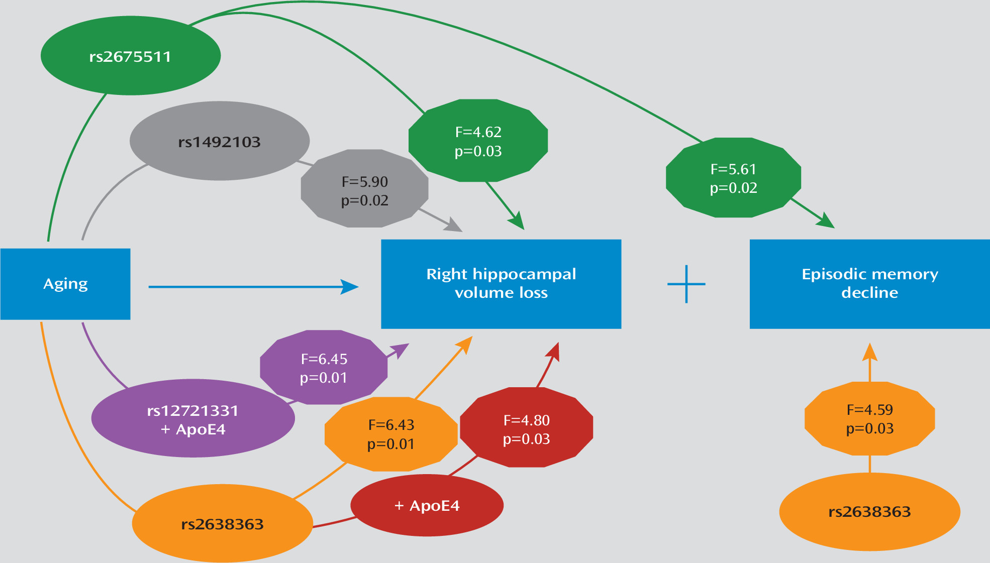
We used mixed models to examine the effect of AGTR1 genotypes and their interaction with time on each of the composite cognitive measures. All models controlled for diagnosis (depressed or nondepressed), age, sex, education, time, and baseline cognitive measure score. In these models, only rs2638363 displayed a significant main effect of genotype on composite episodic memory performance (F=4.59, df=1, 126, p=0.0340), but the genotype-by-time interaction did not reach significance. Similarly, rs1492103 exhibited a trend for a direct effect on episodic memory (F=3.85, df=1, 126, p=0.0519), but this did not reach statistical significance. Finally, rs2675511 exhibited a gene-by-time interaction (F=5.61, df=1, 125, p=0.0194) predicting worsening episodic memory performance. In accordance with SNP effects on hippocampal volumes, individuals homozygous for the vascular risk alleles (rs2638363 GG, rs1492103 TT, and rs2675511 AA) exhibited lower episodic memory scores. None of the four genetic variants exhibited either a statistically significant direct effect or interaction with time effect on executive speed, verbal fluency, or working memory.

Discussion

Replicating and extending our previous findings of AGTR1 effects on right hippocampal morphology (14), three of the same SNPs predicted longitudinal hippocampal volume change in the right but not the left hemisphere in elderly individuals. The direction of the observed relationships was similar to results from the previous cross-sectional study, that is, each of the previously identified “risk” genotypes (rs2638363 GG, rs1492103 TT, and rs2675511 AA) predicted longitudinally greater shrinkage of the right hippocampus when compared with the alternate genotypes. Intriguingly, these AGTR1 risk alleles also predicted poorer performance in episodic memory, a neurocognitive measure mediated by the hippocampus, but had no effects on other cognitive measures. Additionally, two risk variants (rs2638363 and rs12721331) showed epistatic effects with the APOE4 allele, a known risk factor for dementia (26), on right hippocampal volume loss over time. Importantly, the SNPs examined are in low pairwise linkage disequilibrium (r2<0.5) and thus represent independent signals. The presence of several signals within the AGTR1 locus that are independently associated with hippocampal volume changes and memory decline lends further support to the reliability of these associations. Taken together, our findings strongly support that AGTR1 gene variants may confer vulnerability via both direct and epistatic effects for progressive memory decline by accelerating hippocampal volume loss. The main findings and the supported model are schematically summarized in Figure 1.
FIGURE 1. Graphic Summary of AGTR1 Single-Nucleotide Polymorphism (SNP) Associations With Accelerated Right Hippocampal Volume Loss and Episodic Memory Decline in Old Agea
a Genotype-by-time interactions are shown as arrows connecting aging and outcomes (right hippocampal volume loss and episodic memory decline) after moderation by encircled SNPs. Direct genotype effects are shown on the right side of the figure as arrows directly connecting SNPs with episodic memory decline. The encircled numbers on each arrow represent the F values and p values as estimated from the respective mixed regression models. Further details are provided in text and Table 2.
Our study offers novel insights into the role of the renin-angiotensin system (RAS) in hippocampal atrophy and cognitive decline. Hippocampal atrophy has been shown to predict cognitive decline in late life and time-to-conversion to Alzheimer’s disease (5, 30). Previous animal and human studies support involvement of the RAS in cognitive syndromes (6, 14, 31). RAS activation modulates cerebral blood flow, increases brain vulnerability to ischemia, and promotes brain inflammation (6, 32). These effects render this system particularly relevant in late life, where vascular and inflammatory processes are hypothesized to contribute to depressive and cognitive syndromes (10, 33). On the other hand, AT1 receptor blockade dampens stress responses, ameliorates anxious and depressive behaviors, and reduces brain inflammation and vulnerability to ischemia (6, 8, 32). Thus, it is plausible that perturbations in the RAS could heighten the risk for developing dementia by accelerating age-related changes in brain morphology, particularly in brain regions most vulnerable to the effects of aging, such as the hippocampus. Although RAS activity may be modulated by functional variants at the AGTR1 locus, the molecular effects of the risk AGTR1 SNPs are currently not clear. Despite their intronic location, these SNPs could have functional effects. For instance, intronic SNPs may be located in enhancer regions that influence the three-dimensional changes in chromatin conformation necessary for transcription regulation (34). Alternately, these SNPs may be tagging other functional variants in nearby regions of the AGTR1 locus. Finally, as supported by the epistatic effects with APOE observed in our study, these SNPs may have functional effects via interaction with other genes that lie in common biological pathways central for the development of brain pathology and dementia. Future studies on the functional effects of these genetic variants are warranted and may shed light on the mechanisms linking AGTR1 and cognitive syndromes.
Intriguingly, the effects of AGTR1 variation were lateralized to the right hippocampus, which is in accordance with our previous cross-sectional study (14). Although the significance of this lateralization is unclear, hippocampal asymmetry may be an important endophenotype that has been underemphasized by previous studies. Hippocampal asymmetry has been observed in patients with severe depression and in nondepressed relatives of depressed individuals (35, 36). Furthermore, unilateral hippocampal volume changes may be associated with decline in specific cognitive outcomes (5, 37) and with decreased likelihood of achieving antidepressant remission (4). Thus, hippocampal asymmetry may be an early step in the pathogenesis of some late-life mood and cognitive syndromes. This asymmetry could be induced by differential hemispheric effects of the RAS on the hippocampus. Notably, activity of angiotensinase, an enzyme that metabolizes angiotensin, has been found to be distributed asymmetrically between the left and right hippocampi of the rat brain (38, 39). This may also reflect differential activity of angiotensin and potentially asymmetric effects of AGTR1 genetic variants between the two hemispheres. Whether this mechanism holds true, and the clinical significance of these asymmetries, remains to be determined.
Several limitations should be considered when interpreting our findings. Although we controlled for the effects of depression diagnosis and basic demographic factors on hippocampal morphology, we did not control for antidepressant treatments that may have neurotrophic effects on the hippocampus (40) and may even reverse hippocampal volume loss in some cases of depression (37). Depressed participants were treated based on an algorithm rather than a rigid clinical trial. Although this makes our approach comparable to clinical practice, it makes it challenging to elucidate the effects of antidepressants. Data on antidepressant use prior to enrollment, as well as specific treatment modalities, duration, and dosages used over the study period, were not known for each participant. Thus, it is possible that variable treatments between genotype groups may have influenced our results. However, the assignment of treatment modalities occurred randomly and treating clinicians were blind to genotype groups, and thus systematic errors cannot account for our findings. Another limitation was our focus on variation at a single genetic locus and a single brain region. This was based on our sample size and the a priori plausible involvement of AGTR1 and hippocampal pathology in late-life neuropsychiatric syndromes. Larger longitudinal studies are warranted for a more systematic examination of the multiple brain regions, genes, and biological pathways involved in the pathogenesis of these syndromes. Our study shows that the RAS is one such pathway that should be included in future pathogenetic models.
In summary, this is the first study to our knowledge to explore the longitudinal effects of AGTR1 genetic variation on hippocampal morphology and cognitive decline. In contrast with cross-sectional approaches that cannot establish temporal relationships, the present study reveals that older adults homozygous for AGTR1 risk variants exhibit accelerated hippocampal volume loss and memory decline. Our study exemplifies how examining the longitudinal effects of biologically plausible genotypes on relevant endophenotypes may provide novel insights into the pathogenesis of complex phenotypes. It further suggests that molecules involved in the RAS may serve as early therapeutic targets for late-life neuropsychiatric syndromes.

Acknowledgments

The authors acknowledge Dr. Torsten Klengel for helpful discussions on interpreting the single-nucleotide polymorphism data.

References

1.
Sullivan PF, Daly MJ, O’Donovan M: Genetic architectures of psychiatric disorders: the emerging picture and its implications. Nat Rev Genet 2012; 13:537–551
2.
Gottesman II, Gould TD: The endophenotype concept in psychiatry: etymology and strategic intentions. Am J Psychiatry 2003; 160:636–645
3.
Hasler G, Drevets WC, Manji HK, Charney DS: Discovering endophenotypes for major depression. Neuropsychopharmacology 2004; 29:1765–1781
4.
Hsieh MH, McQuoid DR, Levy RM, Payne ME, MacFall JR, Steffens DC: Hippocampal volume and antidepressant response in geriatric depression. Int J Geriatr Psychiatry 2002; 17:519–525
5.
Steffens DC, McQuoid DR, Payne ME, Potter GG: Change in hippocampal volume on magnetic resonance imaging and cognitive decline among older depressed and nondepressed subjects in the neurocognitive outcomes of depression in the elderly study. Am J Geriatr Psychiatry 2011; 19:4–12
6.
Saavedra JM, Sánchez-Lemus E, Benicky J: Blockade of brain angiotensin II AT1 receptors ameliorates stress, anxiety, brain inflammation and ischemia: therapeutic implications. Psychoneuroendocrinology 2011; 36:1–18
7.
Tsutsumi K, Saavedra JM: Characterization and development of angiotensin II receptor subtypes (AT1 and AT2) in rat brain. Am J Physiol 1991; 261:R209–R216
8.
Nayak V, Patil PA: Antidepressant activity of fosinopril, ramipril and losartan, but not of lisinopril in depressive paradigms of albino rats and mice. Indian J Exp Biol 2008; 46:180–184
9.
Duprez DA: Role of the renin-angiotensin-aldosterone system in vascular remodeling and inflammation: a clinical review. J Hypertens 2006; 24:983–991
10.
Taylor WD, Aizenstein HJ, Alexopoulos GS: The vascular depression hypothesis: mechanisms linking vascular disease with depression. Mol Psychiatry 2013; 18:963–974
11.
Bondy B, Baghai TC, Zill P, Schule C, Eser D, Deiml T, Zwanzger P, Ella R, Rupprecht R: Genetic variants in the angiotensin I-converting-enzyme (ACE) and angiotensin II receptor (AT1) gene and clinical outcome in depression. Prog Neuropsychopharmacol Biol Psychiatry 2005; 29:1094–1099
12.
Kondo DG, Speer MC, Krishnan KR, McQuoid DR, Slifer SH, Pieper CF, Billups AV, Steffens DC: Association of AGTR1 with 18-month treatment outcome in late-life depression. Am J Geriatr Psychiatry 2007; 15:564–572
13.
Saab YB, Gard PR, Yeoman MS, Mfarrej B, El-Moalem H, Ingram MJ: Renin-angiotensin-system gene polymorphisms and depression. Prog Neuropsychopharmacol Biol Psychiatry 2007; 31:1113–1118
14.
Taylor WD, Benjamin S, McQuoid DR, Payne ME, Krishnan RR, MacFall JR, Ashley-Koch A: AGTR1 gene variation: association with depression and frontotemporal morphology. Psychiatry Res 2012; 202:104–109
15.
Sheline YI, Gado MH, Kraemer HC: Untreated depression and hippocampal volume loss. Am J Psychiatry 2003; 160:1516–1518
16.
Robins LN, Helzer JE, Croughan J, Ratcliff KS: National Institute of Mental Health Diagnostic Interview Schedule: its history, characteristics, and validity. Arch Gen Psychiatry 1981; 38:381–389
17.
Folstein MF, Folstein SE, McHugh PR: “Mini-mental state”: a practical method for grading the cognitive state of patients for the clinician. J Psychiatr Res 1975; 12:189–198
18.
Taylor WD, McQuoid DR, Krishnan KR: Medical comorbidity in late-life depression. Int J Geriatr Psychiatry 2004; 19:935–943
19.
Steffens DC, McQuoid DR, Krishnan KRR: The Duke Somatic Treatment Algorithm for Geriatric Depression (STAGED) approach. Psychopharmacol Bull 2002; 36:58–68
20.
Hindorff LA, Heckbert SR, Tracy R, Tang Z, Psaty BM, Edwards KL, Siscovick DS, Kronmal RA, Nazar-Stewart V: Angiotensin II type 1 receptor polymorphisms in the cardiovascular health study: relation to blood pressure, ethnicity, and cardiovascular events. Am J Hypertens 2002; 15:1050–1056
21.
Steffens DC, Welsh-Bohmer KA, Burke JR, Plassman BL, Beyer JL, Gersing KR, Potter GG: Methodology and preliminary results from the neurocognitive outcomes of depression in the elderly study. J Geriatr Psychiatry Neurol 2004; 17:202–211
22.
Tschanz JT, Welsh-Bohmer KA, Skoog I, West N, Norton MC, Wyse BW, Nickles R, Breitner JC: Dementia diagnoses from clinical and neuropsychological data compared: the Cache County study. Neurology 2000; 54:1290–1296
23.
Sheline YI, Pieper CF, Barch DM, Welsh-Bohmer K, McKinstry RC, MacFall JR, D’Angelo G, Garcia KS, Gersing K, Wilkins C, Taylor W, Steffens DC, Krishnan RR, Doraiswamy PM: Support for the vascular depression hypothesis in late-life depression: results of a 2-site, prospective, antidepressant treatment trial. Arch Gen Psychiatry 2010; 67:277–285
24.
Zaykin D, Zhivotovsky L, Weir BS: Exact tests for association between alleles at arbitrary numbers of loci. Genetica 1995; 96:169–178
25.
Carlson CS, Eberle MA, Rieder MJ, Yi Q, Kruglyak L, Nickerson DA: Selecting a maximally informative set of single-nucleotide polymorphisms for association analyses using linkage disequilibrium. Am J Hum Genet 2004; 74:106–120
26.
Saunders AM, Strittmatter WJ, Schmechel D, George-Hyslop PH, Pericak-Vance MA, Joo SH, Rosi BL, Gusella JF, Crapper-MacLachlan DR, Alberts MJ et al: Association of apolipoprotein E allele epsilon 4 with late-onset familial and sporadic Alzheimer’s disease. Neurology 1993; 43:1467–1472
27.
Payne ME, Fetzer DL, MacFall JR, Provenzale JM, Byrum CE, Krishnan KRR: Development of a semi-automated method for quantification of MRI gray and white matter lesions in geriatric subjects. Psychiatry Res 2002; 115:63–77
28.
Steffens DC, Byrum CE, McQuoid DR, Greenberg DL, Payne ME, Blitchington TF, MacFall JR, Krishnan KR: Hippocampal volume in geriatric depression. Biol Psychiatry 2000; 48:301–309
29.
Cheng J, Edwards LJ, Maldonado-Molina MM, Komro KA, Muller KE: Real longitudinal data analysis for real people: building a good enough mixed model. Stat Med 2010; 29:504–520
30.
Devanand DP, Pradhaban G, Liu X, Khandji A, De Santi S, Segal S, Rusinek H, Pelton GH, Honig LS, Mayeux R, Stern Y, Tabert MH, de Leon MJ: Hippocampal and entorhinal atrophy in mild cognitive impairment: prediction of Alzheimer disease. Neurology 2007; 68:828–836
31.
Wright JW, Harding JW: The brain RAS and Alzheimer’s disease. Exp Neurol 2010; 223:326–333
32.
Saavedra JM, Nishimura Y: Angiotensin and cerebral blood flow. Cell Mol Neurobiol 1999; 19:553–573
33.
Alexopoulos GS, Morimoto SS: The inflammation hypothesis in geriatric depression. Int J Geriatr Psychiatry 2011; 26:1109–1118
34.
Zannas AS, Binder EB: Gene-environment interactions at the FKBP5 locus: sensitive periods, mechanisms and pleiotropism. Genes Brain Behav 2014; 13:25–37
35.
Boccardi M, Almici M, Bresciani L, Caroli A, Bonetti M, Monchieri S, Gennarelli M, Frisoni GB: Clinical and medial temporal features in a family with mood disorders. Neurosci Lett 2010; 468:93–97
36.
Mervaala E, Föhr J, Könönen M, Valkonen-Korhonen M, Vainio P, Partanen K, Partanen J, Tiihonen J, Viinamäki H, Karjalainen AK, Lehtonen J: Quantitative MRI of the hippocampus and amygdala in severe depression. Psychol Med 2000; 30:117–125
37.
Hou Z, Yuan Y, Zhang Z, Bai F, Hou G, You J: Longitudinal changes in hippocampal volumes and cognition in remitted geriatric depressive disorder. Behav Brain Res 2012; 227:30–35
38.
Banegas I, Prieto I, Alba F, Vives F, Araque A, Segarra AB, Durán R, de Gasparo M, Ramírez M: Angiotensinase activity is asymmetrically distributed in the amygdala, hippocampus and prefrontal cortex of the rat. Behav Brain Res 2005; 156:321–326
39.
Wu HM, Wang C, Wang XL, Wang L, Chang CW, Wang P, Gao GD: Correlations between angiotensinase activity asymmetries in the brain and paw preference in rats. Neuropeptides 2010; 44:253–259
40.
Castrén E: Neurotrophic effects of antidepressant drugs. Curr Opin Pharmacol 2004; 4:58–64

Information & Authors

Information

Published In

Go to American Journal of Psychiatry
American Journal of Psychiatry
Pages: 1214 - 1221
PubMed: 25124854

History

Received: 23 November 2013
Revision received: 21 April 2014
Accepted: 6 May 2014
Published online: 1 November 2014
Published in print: November 01, 2014

Authors

Affiliations

Anthony S. Zannas, M.D., M.Sc.
From the Department of Psychiatry and Behavioral Sciences, the Neuropsychiatric Imaging Research Laboratory, the Department of Radiology, the Center for Human Genetics, and the Department of Medicine, Duke University Medical Center, Durham, N.C.; the Department of Translational Research in Psychiatry, Max Planck Institute of Psychiatry, Munich, Germany; the Department of Psychiatry, University of Connecticut Health Sciences Center, Farmington, Conn.; and the Department of Psychiatry, Vanderbilt University Medical Center and The Geriatric Research, Education, and Clinical Center (GRECC), Department of Veterans Affairs Medical Center, Tennessee Valley Healthcare System, Nashville, Tenn.
Douglas R. McQuoid, M.P.H.
From the Department of Psychiatry and Behavioral Sciences, the Neuropsychiatric Imaging Research Laboratory, the Department of Radiology, the Center for Human Genetics, and the Department of Medicine, Duke University Medical Center, Durham, N.C.; the Department of Translational Research in Psychiatry, Max Planck Institute of Psychiatry, Munich, Germany; the Department of Psychiatry, University of Connecticut Health Sciences Center, Farmington, Conn.; and the Department of Psychiatry, Vanderbilt University Medical Center and The Geriatric Research, Education, and Clinical Center (GRECC), Department of Veterans Affairs Medical Center, Tennessee Valley Healthcare System, Nashville, Tenn.
Martha E. Payne, Ph.D., M.P.H.
From the Department of Psychiatry and Behavioral Sciences, the Neuropsychiatric Imaging Research Laboratory, the Department of Radiology, the Center for Human Genetics, and the Department of Medicine, Duke University Medical Center, Durham, N.C.; the Department of Translational Research in Psychiatry, Max Planck Institute of Psychiatry, Munich, Germany; the Department of Psychiatry, University of Connecticut Health Sciences Center, Farmington, Conn.; and the Department of Psychiatry, Vanderbilt University Medical Center and The Geriatric Research, Education, and Clinical Center (GRECC), Department of Veterans Affairs Medical Center, Tennessee Valley Healthcare System, Nashville, Tenn.
James R. MacFall, Ph.D., M.S.
From the Department of Psychiatry and Behavioral Sciences, the Neuropsychiatric Imaging Research Laboratory, the Department of Radiology, the Center for Human Genetics, and the Department of Medicine, Duke University Medical Center, Durham, N.C.; the Department of Translational Research in Psychiatry, Max Planck Institute of Psychiatry, Munich, Germany; the Department of Psychiatry, University of Connecticut Health Sciences Center, Farmington, Conn.; and the Department of Psychiatry, Vanderbilt University Medical Center and The Geriatric Research, Education, and Clinical Center (GRECC), Department of Veterans Affairs Medical Center, Tennessee Valley Healthcare System, Nashville, Tenn.
Allison Ashley-Koch, Ph.D.
From the Department of Psychiatry and Behavioral Sciences, the Neuropsychiatric Imaging Research Laboratory, the Department of Radiology, the Center for Human Genetics, and the Department of Medicine, Duke University Medical Center, Durham, N.C.; the Department of Translational Research in Psychiatry, Max Planck Institute of Psychiatry, Munich, Germany; the Department of Psychiatry, University of Connecticut Health Sciences Center, Farmington, Conn.; and the Department of Psychiatry, Vanderbilt University Medical Center and The Geriatric Research, Education, and Clinical Center (GRECC), Department of Veterans Affairs Medical Center, Tennessee Valley Healthcare System, Nashville, Tenn.
David C. Steffens, M.D., M.H.Sc.
From the Department of Psychiatry and Behavioral Sciences, the Neuropsychiatric Imaging Research Laboratory, the Department of Radiology, the Center for Human Genetics, and the Department of Medicine, Duke University Medical Center, Durham, N.C.; the Department of Translational Research in Psychiatry, Max Planck Institute of Psychiatry, Munich, Germany; the Department of Psychiatry, University of Connecticut Health Sciences Center, Farmington, Conn.; and the Department of Psychiatry, Vanderbilt University Medical Center and The Geriatric Research, Education, and Clinical Center (GRECC), Department of Veterans Affairs Medical Center, Tennessee Valley Healthcare System, Nashville, Tenn.
Guy G. Potter, Ph.D.
From the Department of Psychiatry and Behavioral Sciences, the Neuropsychiatric Imaging Research Laboratory, the Department of Radiology, the Center for Human Genetics, and the Department of Medicine, Duke University Medical Center, Durham, N.C.; the Department of Translational Research in Psychiatry, Max Planck Institute of Psychiatry, Munich, Germany; the Department of Psychiatry, University of Connecticut Health Sciences Center, Farmington, Conn.; and the Department of Psychiatry, Vanderbilt University Medical Center and The Geriatric Research, Education, and Clinical Center (GRECC), Department of Veterans Affairs Medical Center, Tennessee Valley Healthcare System, Nashville, Tenn.
Warren D. Taylor, M.D., M.H.Sc.
From the Department of Psychiatry and Behavioral Sciences, the Neuropsychiatric Imaging Research Laboratory, the Department of Radiology, the Center for Human Genetics, and the Department of Medicine, Duke University Medical Center, Durham, N.C.; the Department of Translational Research in Psychiatry, Max Planck Institute of Psychiatry, Munich, Germany; the Department of Psychiatry, University of Connecticut Health Sciences Center, Farmington, Conn.; and the Department of Psychiatry, Vanderbilt University Medical Center and The Geriatric Research, Education, and Clinical Center (GRECC), Department of Veterans Affairs Medical Center, Tennessee Valley Healthcare System, Nashville, Tenn.

Notes

Address correspondence to Dr. Zannas ([email protected]).

Funding Information

The authors report no financial relationships with commercial interests.Supported by research grants R01 MH077745, R01 MH054846, and K24 MH070027.

Metrics & Citations

Metrics

Citations

Export Citations

If you have the appropriate software installed, you can download article citation data to the citation manager of your choice. Simply select your manager software from the list below and click Download.

For more information or tips please see 'Downloading to a citation manager' in the Help menu.

Format
Citation style
Style
Copy to clipboard

There are no citations for this item

View Options

View options

PDF/ePub

View PDF/ePub

Get Access

Login options

Already a subscriber? Access your subscription through your login credentials or your institution for full access to this article.

Personal login Institutional Login Open Athens login
Purchase Options

Purchase this article to access the full text.

PPV Articles - American Journal of Psychiatry

PPV Articles - American Journal of Psychiatry

Not a subscriber?

Subscribe Now / Learn More

PsychiatryOnline subscription options offer access to the DSM-5-TR® library, books, journals, CME, and patient resources. This all-in-one virtual library provides psychiatrists and mental health professionals with key resources for diagnosis, treatment, research, and professional development.

Need more help? PsychiatryOnline Customer Service may be reached by emailing [email protected] or by calling 800-368-5777 (in the U.S.) or 703-907-7322 (outside the U.S.).

Media

Figures

Other

Tables

Share

Share

Share article link

Share