Abstract

Objective:

The norepinephrine transporter has been suggested to play a crucial role in major depressive disorder. However, norepinephrine transporter availability in major depressive disorder and its role with clinical symptoms are not known. The authors tested norepinephrine transporter availability in patients with major depressive disorder with the aim to identify any associations between test results and clinical symptoms.

Method:

The present research was a cross-sectional study in which 19 patients with major depressive disorder and 19 age- and sex-matched healthy comparison subjects underwent positron emission tomography scanning to evaluate the norepinephrine transporter availability measured by the radioligand (S,S)-[18F]FMeNER-D2. Norepinephrine transporter availability in the thalamus and its subregions was quantified in terms of nondisplaceable binding potential (BPND). The authors also analyzed the association between norepinephrine transporter availability and clinical symptoms.

Results:

Compared with healthy subjects, patients with major depressive disorder showed 29.0% higher BPND values in the thalamus and, in particular, 28.2% higher values in the thalamic subregion anatomically connected to the prefrontal cortex. Elevated norepinephrine transporter availability in the thalamus in patients was positively correlated with attention, as measured by the Trail Making Test, part A.

Conclusions:

These findings revealed altered norepinephrine transmission in patients with major depressive disorder, suggesting that this alteration could be related to attention in this patient population.
Major depressive disorder is a globally prevalent psychiatric disorder characterized by affective, cognitive, and somatic symptoms. Multiple pathologies are implicated in major depressive disorder, including dysregulation of monoamine neurotransmitters such as serotonin, norepinephrine, and dopamine (1).
The critical role of norepinephrine in neural systems underlying the generation of major depressive disorder symptoms has been recognized for many years. There have been several studies on the relationship between norepinephrine systems and major depressive disorder. Norepinephrine transporter binding in the locus ceruleus, which reflects the dynamics of norepinephrine (2), has been found to be decreased in postmortem tissues from patients with major depressive disorder (3), suggesting the alteration of norepinephrine systems. Postmortem studies of depressed patients have shown altered density and affinity state of α2A-adrenoceptors, which modulate norepinephrine release (4). Another study found increased β-adrenergic receptor binding in the cortex density of suicide subjects (5). Other research found that α2- and β1-adrenoceptor densities in the frontal cortex were higher in antidepressant-free patients with major depressive disorder compared with matched comparison subjects (6). These studies suggest the critical role of norepinephrine in neural systems underlying the generation of major depressive disorder. The norepinephrine transporter has been targeted as a treatment pathway for major depressive disorder (7). For instance, antidepressants with dual-target sites that include the norepinephrine transporter, such as venlafaxine, milnacipran, and duloxetine, are equivalent or are even more effective than pure selective serotonin reuptake inhibitors in achieving remission (8).
Animal studies have revealed that the proportion of messenger RNA and protein levels of the norepinephrine transporter are increased in rat models of depression (9, 10). While two in vivo positron emission tomography (PET) studies have reported on norepinephrine transporter availability in patients with posttraumatic stress disorder (PTSD) and attention deficit hyperactivity disorder (11, 12), to our knowledge there has been no in vivo study examining norepinephrine transporter availability in patients with major depressive disorder.
In the present study, we evaluated in vivo norepinephrine transporter availability in patients with major depressive disorder in the thalamus and in the locus ceruleus using (S,S)-[18F]FMeNER-D2 PET (1316). We then tried to find the possible association between neuropsychological test results and clinical symptoms and norepinephrine transporter availability (17).

Method

Participants

Nineteen patients (11 men and eight women; mean age=40.1 years [SD=10.7], range=20–56 years) fulfilling DSM-IV criteria for major depressive disorder were recruited from two affiliated psychiatric hospitals and two clinics in the Chiba, Japan, area. The participants had no other history of major medical illness. S.M., H.T., K.T., and T.N. evaluated lifetime psychiatric disorders based on the Mini-International Neuropsychiatric Interview (18), and patients were determined to be free of comorbid psychiatric disorders. Clinical characteristics of the patients are summarized in Table 1. Seventeen patients were categorized with melancholic depression based on the Mini-International Neuropsychiatric Interview, and none of the patients had received psychopharmacologic treatment for at least 1 year before the study. Sixteen of the 19 patients were antidepressant-naive, and the remaining three patients were antidepressant-free at the time of the study; one patient had taken duloxetine 3 years earlier, one had taken sulpiride 2 years earlier, and the other had taken antidepressants (the specific agents were not identified) 5 years earlier. The lifetime number of major depressive episodes ranged from one to two.
TABLE 1. Characteristics of Participants in a Study of Norepinephrine Transporter in Major Depressive Disorder
MeasurePatients (N=19)Healthy Subjects (N=19) 
 MeanSDMeanSDp
Age (years)40.110.740.712.20.88
Hamilton Depression Rating Scale score20.94.7   
Beck Depression Inventory–II score30.09.2   
Trail Making Test, part A (reaction time, seconds)30.2a9.724.66.60.054
Trail Making Test, part B (reaction time, seconds)66.2a20.650.713.40.013b
Symbol Digit Modalities Test (correct number)56.2a13.973.58.5<0.001b
Past episodes (number)1.2c0.41   
Duration of current episode (months)5.4d4.9   
 N%N% 
Smoking status15211 
Melancholic depression1789   
Antidepressant free19100   
Antidepressant naive1684   
a
Data available for 17 patients.
b
p<0.017=0.05/3; Bonferroni correction for three neuropsychological tests.
c
Range=1–2.
d
Range=1–18.
Age- and sex-matched healthy subjects (11 men and eight women; mean age=40.7 years [SD=12.2], range=22–61 years) participated in the study as a comparison group. All healthy subjects were free of any somatic, neurological, or psychiatric disorders and had no history of current or previous drug abuse.
Studies were performed and analyzed at the National Institute of Radiological Sciences (Chiba, Japan). All participants provided written informed consent before participating in the study, which was approved by the Radiation Drug Safety Committee and by the institutional review board of the National Institute of Radiological Sciences, and which was performed in accordance with the ethical standards laid down in the 1964 Declaration of Helsinki and its later amendments.

Clinical and Neuropsychological Assessment

The severity of symptoms for all patients was assessed using the Japanese version of the 17-item Hamilton Depression Rating Scale (HAM-D) and the Beck Depression Inventory–II (BDI-II) on the day of the PET scan (19, 20). All participants performed three neuropsychological tests: the Trail Making Test, parts A and B, and the Symbol Digit Modalities Test (21, 22). Trails A requires visual attention, and Trails B requires not only visual attention but also working memory. The Symbol Digit Modalities Test assesses executive functions such as complex scanning and visual tracking (22, 23). Two patients failed to take the above-mentioned tests because of fatigue.

PET and MRI Acquisition

An ECAT EXACT HR+ (CTI-Siemens, United States) PET scanner system, which provides a 15.5-cm axial field of view, was used for all patients and healthy subjects with a head fixation device to minimize head movement. A 10-minute transmission scan was acquired after the emissions scan using a 68Ge-68Ga source for subsequent attenuation correction. (S,S)-[18F]FMeNER-D2 was synthesized by fluoromethylation of norethylreboxetine with 18F-bromofluoro-methane-d2 (24), yielding a radiochemical purity of higher than 95%. Emission data were acquired from 120 minutes to 180 minutes (10 minutes × 6 frames) in three-dimensional mode, following an intravenous bolus injection of (S,S)-[18F]FMeNER-D2. Injected radioactivity averaged 186.2 MBq (SD=7.9) for patients and 188.0 MBq (SD=13.3) for healthy subjects, while specific radioactivity averaged 675.6 GBq/μmol (SD=388.7) for patients and 584.0 GBq/μmol (SD=394.5) for healthy subjects at the time of injection. Data were reconstructed by a filtered back-projection using the Hanning filter (6.3 mm, full width at half maximum).
MR images were acquired with a 3.0-T scanner, MAGNETOM Verio (SIEMENS, Germany). Three-dimensional volumetric acquisition of a T1-weighted gradient echo sequence produced a gapless series of thin sagittal sections (TE=1.95 ms, TR=2,300 ms, TI=900 ms, flip angle=9°, field of view=250 mm, acquisition matrix=256×256, slice thickness=1 mm, voxel size=1 mm×1 mm×1 mm). The MRI results revealed no apparent structural abnormalities.

Image Analysis

T1-weighted MRIs were coregistered to the corresponding PET images using SPM8 (Wellcome Trust Centre for Neuroimaging, London). MR and PET images were then spatially normalized to Montreal Neurological Institute space (MNI 152 2 mm template) based on the transformation parameters estimated from the Diffeomorphic Anatomical Registration Through Exponentiated Lie Algebra (DARTEL) algorithm in SPM8 (25). A template of regions of interest (Oxford thalamic connectivity atlas [17]) was applied to the spatially normalized PET images to extract time activity curves for the subregions of the thalamus. The Oxford thalamic connectivity atlas is based on diffusion weighted imaging conducted by Behrens (17). This atlas thus separates the thalamus into seven subregions that are anatomically connected with seven cerebral cortical regions. Manually drawn regions of interest were applied for the locus ceruleus according to Keren et al. (26), as well as for the caudate, which was used as a reference region because it is almost devoid of norepinephrine transporter availability (15, 27). Nondisplaceable binding potential (BPND) values were calculated using the area under the curve ratio (AUC) method (13) from 120 minutes to 180 minutes using the following equation:

Statistical Analysis

Statistical analysis was performed using SPSS, version 22 (IBM, Armonk, N.Y.). Group differences in BPND values and neuropsychological performance between patients with major depressive disorder and healthy subjects were analyzed using independent sample t statistics (two-tailed) with the Bonferroni correction for multiple comparisons. The correlations between regional BPND values and neuropsychological performance, as well as between clinical variables (HAM-D total items, HAM-D insomnia items, HAM-D anxiety items, and BDI-II), were analyzed using age as a covariate followed by the Benjamini-Hochberg method for multiple comparisons using the false discovery rate controlling procedure with q*=0.05.

Results

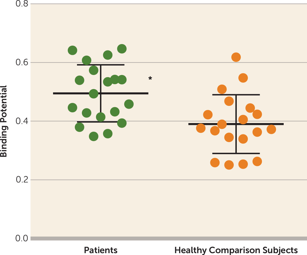
Table 1 presents clinical variables and neuropsychological performances. Relative to healthy comparison subjects, patients with major depressive disorder showed 29.0% higher BPND values in the thalamus (t=2.85, df=36, p=0.007; alpha=0.05/2, Bonferroni correction) and, in particular, 28.2% higher BPND values in the thalamic subregion connected to the prefrontal cortex (t=3.26, df=36, p=0.002; alpha level=0.05/7, Bonferroni correction) (Figure 1 and Table 2). BPND values of the locus ceruleus were not significantly different between patients with major depressive disorder and healthy subjects.
FIGURE 1. (S,S)-[18F]FMeNER-D2 Nondisplaceable Binding Potential of the Thalamic Subregion Projecting to the Prefrontal Cortexa
aThe heavy rule within the scatterplots indicates the mean; thin rules indicate standard deviations. Statistical analysis was performed using independent sample t statistics with the Bonferroni correction for seven subregions.
*p<0.007=0.05/7.
TABLE 2. Nondisplaceable Binding Potential (BPND) of the Norepinephrine Transporter in Participants in a Study of Norepinephrine Transporter in Major Depressive Disordera
 BPND 
 PatientsHealthy Subjects 
RegionMeanSDMeanSDp
Locus ceruleus0.450.210.420.190.67
Thalamus0.400.090.310.090.007b
Thalamic subregions     
 Prefrontal0.500.100.390.100.002c
 Primary motor0.690.180.590.160.09
 Premotor0.570.170.480.140.09
 Temporal0.090.110.020.090.04
 Posterior parietal0.460.120.400.100.09
 Somatosensory0.680.210.620.130.28
 Occipital0.260.100.230.090.34
a
Statistical analysis was performed using independent sample t statistics with the Bonferroni correction for each set of two regional measures (locus ceruleus and thalamus) and for seven subregional thalamic measures.
b
The significance threshold was set as follows: p<0.025=0.05/2.
c
The significance threshold was set as follows: p<0.007=0.05/7.
When controlling for age, BPND values were negatively correlated with reaction time in Trails A in the thalamus and locus ceruleus in patients (r=−0.70, p=0.003, and r=−0.56, p=0.023, respectively) (Table 3). However, based on the Benjamini-Hochberg method for multiple comparisons, neuropsychological tests and the clinical variables did not correlate with the norepinephrine transporter BPND values in any regions.
TABLE 3. Correlation Ratios Between the Nondisplaceable Binding Potential (BPND) of the Norepinephrine Transporter and Clinical Variables, With Age as a Covariate
 Patients (N=19)Healthy Subjects (N=19)
Clinical VariableLocus Ceruleus BPNDThalamus BPNDLocus Ceruleus BPNDThalamus BPND
Neuropsychological tests    
 Trail Making Test, part Aa–0.56*–0.70**–0.170.14
 Trail Making Test, part Ba–0.36–0.33–0.040.14
Symbol Digit Modalities Testa0.470.200.06–0.10
Clinical symptoms    
 Hamilton Depression Rating Scale0.120.04  
 Insomnia0.170.32  
 Anxiety–0.02–0.13  
 Beck Depression Inventory–II–0.40–0.03  
a
Information available for 17 patients.
*
p<0.05. **p<0.01.

Discussion

This study examined the in vivo norepinephrine transporter availability and symptoms in patients with major depressive disorder. Results showed that thalamic norepinephrine transporter availability in patients with major depressive disorder was significantly higher than that in healthy comparison subjects in the thalamus and in the thalamic subregion anatomically connected to the prefrontal cortex. We further found that elevated norepinephrine transporter availability in the locus ceruleus and thalamus in patients with major depressive disorder was positively correlated with visual search attention, as measured by Trails A.
Our finding regarding norepinephrine transporter availability in the thalamus is consistent with animal studies that have revealed elevated norepinephrine transporter in rat models of depression induced by chronic stress (9, 10). Therefore, higher norepinephrine transporter availability in the thalamus in patients with depression may also indicate the involvement of stress-related response because the majority of our patients had melancholic depression, which is regarded as a stress response (28).
At present, there is little evidence that helps explain the mechanism of this norepinephrine transporter regulation; however, there are several potential hypotheses. Dysfunctional norepinephrine systems are potential etiological factors in depression (29), which may induce elevated norepinephrine transporter availability (30). Hyperactive hypothalamic-pituitary-adrenal and norepinephrine systems have both been hypothesized as mechanisms involved in melancholic depression, which is the primary type of depression we see in our patients (31, 32). Moreover, it should be noted that the norepinephrine system is not the only system involved in the altered norepinephrine transporter regulation. Other mechanisms, such as acetylcholine and GABA (33), may also be involved in the regulation of the norepinephrine transporter (34).
To our knowledge, there has been only one postmortem study investigating norepinephrine transporter in major depressive disorder (3). Although that study reported a low norepinephrine transporter binding in the locus ceruleus in major depressive disorder, our PET study did not find a difference in BPND values of the same region between patients and healthy subjects. This finding does not necessarily mean that there is no change, but the limited spatial resolution of PET images makes it difficult to adequately detect alterations in small structures such as the locus ceruleus, which yields a high variability of BPND values in this region, as shown in Table 2. In addition, we enrolled antidepressant-naive or antidepressant-free patients with early-onset depression, but the postmortem study included a wide range of patients who were taking antidepressant medication. Moreover, the locus ceruleus was the only region the earlier study investigated, whereas our study targeted the thalamus as well. Considering that chronic stress has been reported to increase norepinephrine transporter density in rat brain and antidepressants to lower it (35), the effects of treatment and illness duration may influence the expression of norepinephrine transporter in major depressive disorder.
Regarding the neuropsychological tests, visual attention measured by Trails A was less affected, while working memory and executive function measured by Trails B and the Symbol Digit Modalities Test were lower in our patients (Table 1), in accordance with previous findings of melancholic depressed patients (36). In other words, visual attention is preserved among these neuropsychological functions, and the present findings suggest that the preserved visual attention measured by Trails A is associated with elevated norepinephrine transporter availability in depressed patients. A previous PET study demonstrated a positive correlation between vigilance and norepinephrine transporter availability in patients with PTSD (12). Thus, elevated norepinephrine transporter availability may also be associated with enhanced attention and vigilance in our depressed patients. However, our results regarding the correlation did not withstand multiple comparisons. Thus, we should interpret the results carefully.
In the PET analysis with (S,S)-[18F]FMeNER-D2, we used the AUC method. The validity of this method with (S,S)-[18F]FMeNER-D2 PET for quantification of norepinephrine transporter availability has been reported previously (13). When the AUC method is compared with kinetic modeling, BPND values in the thalamus obtained by the AUC method matched very well with the values obtained by kinetic modeling (r=0.88). Moreover, BPND values obtained by the AUC method are more reliable, with smaller intersubject variability, than those obtained by kinetic modeling in the thalamus. Thus, we chose the AUC method as a reliable method for the quantification of norepinephrine transporter availability using (S,S)-[18F]FMeNER-D2 PET in this study.
Some limitations of the study should be addressed. First, our sample size was limited; after correction for multiple comparisons between the clinical variables and norepinephrine transporter availability, none of the correlations withstood significance testing. Thus, further studies with larger sample sizes are necessary to understand the role of norepinephrine transporter availability in patients with major depressive disorder. Moreover, because most of our patients experienced melancholic depression, we cannot conclude that all patients with major depressive disorder have high norepinephrine transporter availability. Second, by using (S,S)-[18F]FMeNER-D2, we examined norepinephrine transporter availability only in the thalamus and the locus ceruleus. The norepinephrine transporter availability is insufficient for (S,S)-[18F]FMeNER-D2 quantification in other brain regions, and the accumulated fluoride in bone makes it difficult to observe cortical regions through defluorination of this radioligand (13). Thus, other techniques should be used to investigate norepinephrine transporter availability in different brain regions. Finally, there could be differences in norepinephrine availability in the caudate between patients and healthy subjects, and thus using the caudate as a reference may skew the results. An ideal reference region needs to be completely devoid of specific binding. However, this is often not the case for PET imaging. Regarding norepinephrine transporter availability, postmortem studies in nonhuman primates and humans have indicated that norepinephrine transporter availability in the caudate is very low (27, 37). The very low specific binding in the caudate was confirmed by a PET occupancy study in rhesus monkeys (38). After injection of a selective norepinephrine transporter inhibitor, no PET signal change was detected in the caudate compared with baseline readings, meaning the amount of specific binding in the caudate was significantly lower than that of the nonspecific binding of the PET ligand in the same region. Thus, the resulting BPND values in our study would be less affected by the group differences in specific binding in the caudate.
In conclusion, we found higher norepinephrine transporter availability in the thalamus and its subregions in patients with major depressive disorder. These findings implicate the potentially beneficial selection of antidepressants with affinity to the norepinephrine transporter in the treatment of major depressive disorder.

Acknowledgments

The authors thank Kazuko Suzuki and Shizuko Kawakami for their help as clinical research coordinators and Hiromi Sano for her help in performing MRI scanning. The authors also thank Dr. Yoshihide Akine, Dr. Shiho Morimoto, and Dr. Akihiko Otsuka for help in recruiting patients; and members of the Molecular Probe Program for (S,S)-[18F]FMeNER-D2.

Footnote

University Hospital Medical Information Network Clinical Trials Registry identifier: UMIN000008251.

References

1.
Ressler KJ, Nemeroff CB: Role of serotonergic and noradrenergic systems in the pathophysiology of depression and anxiety disorders. Depress Anxiety 2000; 12(Suppl 1):2–19
2.
Schwartz JW, Novarino G, Piston DW, et al: Substrate binding stoichiometry and kinetics of the norepinephrine transporter. J Biol Chem 2005; 280:19177–19184
3.
Klimek V, Stockmeier C, Overholser J, et al: Reduced levels of norepinephrine transporters in the locus coeruleus in major depression. J Neurosci 1997; 17:8451–8458
4.
Cottingham C, Wang Q: α2 adrenergic receptor dysregulation in depressive disorders: implications for the neurobiology of depression and antidepressant therapy. Neurosci Biobehav Rev 2012; 36:2214–2225
5.
Mann JJ, Stanley M, McBride PA, et al: Increased serotonin2 and beta-adrenergic receptor binding in the frontal cortices of suicide victims. Arch Gen Psychiatry 1986; 43:954–959
6.
Rivero G, Gabilondo AM, García-Sevilla JA, et al: Increased α2- and β1-adrenoceptor densities in postmortem brain of subjects with depression: differential effect of antidepressant treatment. J Affect Disord 2014; 167:343–350
7.
Nestler EJ, Malenka R, Hyman SE, et al: Molecular Basis of Neuropharmacology: A Foundation for Clinical Neuroscience. East Norwalk, Conn, Appleton & Lange, 2001
8.
Papakostas GI, Thase ME, Fava M, et al: Are antidepressant drugs that combine serotonergic and noradrenergic mechanisms of action more effective than the selective serotonin reuptake inhibitors in treating major depressive disorder? A meta-analysis of studies of newer agents. Biol Psychiatry 2007; 62:1217–1227
9.
Miner LH, Jedema HP, Moore FW, et al: Chronic stress increases the plasmalemmal distribution of the norepinephrine transporter and the coexpression of tyrosine hydroxylase in norepinephrine axons in the prefrontal cortex. J Neurosci 2006; 26:1571–1578
10.
Chen P, Fan Y, Li Y, et al: Chronic social defeat up-regulates expression of norepinephrine transporter in rat brains. Neurochem Int 2012; 60:9–20
11.
Vanicek T, Spies M, Rami-Mark C, et al: The norepinephrine transporter in attention-deficit/hyperactivity disorder investigated with positron emission tomography. JAMA Psychiatry 2014; 71:1340–1349
12.
Pietrzak RH, Gallezot JD, Ding YS, et al: Association of posttraumatic stress disorder with reduced in vivo norepinephrine transporter availability in the locus coeruleus. JAMA Psychiatry 2013; 70:1199–1205
13.
Arakawa R, Okumura M, Ito H, et al: Quantitative analysis of norepinephrine transporter in the human brain using PET with (S,S)-18F-FMeNER-D2. J Nucl Med 2008; 49:1270–1276
14.
Takano A, Varrone A, Gulyás B, et al: Mapping of the norepinephrine transporter in the human brain using PET with (S,S)-[18F]FMeNER-D2. Neuroimage 2008; 42:474–482
15.
Schou M, Halldin C, Pike VW, et al: Post-mortem human brain autoradiography of the norepinephrine transporter using (S,S)-[18F]FMeNER-D2. Eur Neuropsychopharmacol 2005; 15:517–520
16.
Seneca N, Andree B, Sjoholm N, et al: Whole-body biodistribution, radiation dosimetry estimates for the PET norepinephrine transporter probe (S,S)-[18F]FMeNER-D2 in non-human primates. Nucl Med Commun 2005; 26:695–700
17.
Behrens TE, Johansen-Berg H, Woolrich MW, et al: Non-invasive mapping of connections between human thalamus and cortex using diffusion imaging. Nat Neurosci 2003; 6:750–757
18.
Sheehan DV, Lecrubier Y, Sheehan KH, et al: The Mini-International Neuropsychiatric Interview (M.I.N.I.): the development and validation of a structured diagnostic psychiatric interview for DSM-IV and ICD-10. J Clin Psychiatry 1998; 59(suppl 20):22–33
19.
Lindberg D, Ahlfors UG, Dencker SJ, et al: Symptom reduction in depression after treatment with L-tryptophan or imipramine. Item analysis of Hamilton rating scale for depression. Acta Psychiatr Scand 1979; 60:287–294
20.
Beck AT, Steer RA: Internal consistencies of the original and revised Beck Depression Inventory. J Clin Psychol 1984; 40:1365–1367
21.
Spreen O: A Compendium of Neuropsychological Tests: Administration, Norms, and Commentary. New York, Oxford University Press, 1998
22.
Mirsky AF, Anthony BJ, Duncan CC, et al: Analysis of the elements of attention: a neuropsychological approach. Neuropsychol Rev 1991; 2:109–145
23.
Lezak MD: Neuropsychological Assessment. New York, Oxford University Press, 2004
24.
Schou M, Halldin C, Sóvágó J, et al: PET evaluation of novel radiofluorinated reboxetine analogs as norepinephrine transporter probes in the monkey brain. Synapse 2004; 53:57–67
25.
Ashburner J: A fast diffeomorphic image registration algorithm. Neuroimage 2007; 38:95–113
26.
Keren NI, Lozar CT, Harris KC, et al: In vivo mapping of the human locus coeruleus. Neuroimage 2009; 47:1261–1267
27.
Donnan GA, Kaczmarczyk SJ, Paxinos G, et al: Distribution of catecholamine uptake sites in human brain as determined by quantitative [3H] mazindol autoradiography. J Comp Neurol 1991; 304:419–434
28.
Gold PW, Goodwin FK, Chrousos GP: Clinical and biochemical manifestations of depression. Relation to the neurobiology of stress (2). N Engl J Med 1988; 319:413–420
29.
Charney DS: Monoamine dysfunction and the pathophysiology and treatment of depression. J Clin Psychiatry 1998; 59(suppl 14):11–14
30.
Lee CM, Javitch JA, Snyder SH: Recognition sites for norepinephrine uptake: regulation by neurotransmitter. Science 1983; 220:626–629
31.
Gold PW, Chrousos GP: Melancholic and atypical subtypes of depression represent distinct pathophysiological entities: CRH, neural circuits, and the diathesis for anxiety and depression. Mol Psychiatry 2013; 18:632–634
32.
Wong ML, Kling MA, Munson PJ, et al: Pronounced and sustained central hypernoradrenergic function in major depression with melancholic features: relation to hypercortisolism and corticotropin-releasing hormone. Proc Natl Acad Sci USA 2000; 97:325–330
33.
Huang J, Tufan T, Deng M, et al: Corticotropin releasing factor up-regulates the expression and function of norepinephrine transporter in SK-N-BE (2) M17 cells. J Neurochem 2015; 135:38–49
34.
Mandela P, Ordway GA: The norepinephrine transporter and its regulation. J Neurochem 2006; 97:310–333
35.
Fan Y, Chen P, Li Y, et al: Effects of desipramine treatment on stress-induced up-regulation of norepinephrine transporter expression in rat brains. Psychopharmacology (Berl) 2015; 232:379–390
36.
Lin K, Xu G, Lu W, et al: Neuropsychological performance in melancholic, atypical and undifferentiated major depression during depressed and remitted states: a prospective longitudinal study. J Affect Disord 2014; 168:184–191
37.
Smith HR, Beveridge TJ, Porrino LJ: Distribution of norepinephrine transporters in the non-human primate brain. Neuroscience 2006; 138:703–714
38.
Gallezot JD, Weinzimmer D, Nabulsi N, et al: Evaluation of [11C]MRB for assessment of occupancy of norepinephrine transporters: studies with atomoxetine in non-human primates. Neuroimage 2011; 56:268–279

Information & Authors

Information

Published In

Go to American Journal of Psychiatry
American Journal of Psychiatry
Pages: 36 - 41
PubMed: 27631962

History

Received: 26 October 2015
Revision received: 7 April 2016
Revision received: 19 May 2016
Accepted: 13 June 2016
Published online: 15 September 2016
Published in print: January 01, 2017

Keywords

  1. Mood Disorders-Unipolar
  2. Stress
  3. Brain Imaging Techniques
  4. Norepinephrine Transporter
  5. Major Depressive Disorder
  6. Positron Emission Tomography
  7. Attention

Authors

Details

Sho Moriguchi, M.D.
From the Department of Functional Brain Imaging Research, National Institute of Radiological Sciences, National Institutes for Quantum and Radiological Science and Technology, Chiba, Japan; the Department of Neuropsychiatry, Keio University School of Medicine, Tokyo; Decoding and Controlling Brain Information, Precursory Research for Embryonic Science and Technology, Japan Science and Technology Agency, Saitama, Japan; and the Department of Psychiatry, National Center of Neurology and Psychiatry, Tokyo.
Makiko Yamada, Ph.D.
From the Department of Functional Brain Imaging Research, National Institute of Radiological Sciences, National Institutes for Quantum and Radiological Science and Technology, Chiba, Japan; the Department of Neuropsychiatry, Keio University School of Medicine, Tokyo; Decoding and Controlling Brain Information, Precursory Research for Embryonic Science and Technology, Japan Science and Technology Agency, Saitama, Japan; and the Department of Psychiatry, National Center of Neurology and Psychiatry, Tokyo.
Harumasa Takano, M.D., Ph.D.
From the Department of Functional Brain Imaging Research, National Institute of Radiological Sciences, National Institutes for Quantum and Radiological Science and Technology, Chiba, Japan; the Department of Neuropsychiatry, Keio University School of Medicine, Tokyo; Decoding and Controlling Brain Information, Precursory Research for Embryonic Science and Technology, Japan Science and Technology Agency, Saitama, Japan; and the Department of Psychiatry, National Center of Neurology and Psychiatry, Tokyo.
Tomohisa Nagashima, M.D., Ph.D.
From the Department of Functional Brain Imaging Research, National Institute of Radiological Sciences, National Institutes for Quantum and Radiological Science and Technology, Chiba, Japan; the Department of Neuropsychiatry, Keio University School of Medicine, Tokyo; Decoding and Controlling Brain Information, Precursory Research for Embryonic Science and Technology, Japan Science and Technology Agency, Saitama, Japan; and the Department of Psychiatry, National Center of Neurology and Psychiatry, Tokyo.
Keisuke Takahata, M.D., Ph.D.
From the Department of Functional Brain Imaging Research, National Institute of Radiological Sciences, National Institutes for Quantum and Radiological Science and Technology, Chiba, Japan; the Department of Neuropsychiatry, Keio University School of Medicine, Tokyo; Decoding and Controlling Brain Information, Precursory Research for Embryonic Science and Technology, Japan Science and Technology Agency, Saitama, Japan; and the Department of Psychiatry, National Center of Neurology and Psychiatry, Tokyo.
Keita Yokokawa, M.A.
From the Department of Functional Brain Imaging Research, National Institute of Radiological Sciences, National Institutes for Quantum and Radiological Science and Technology, Chiba, Japan; the Department of Neuropsychiatry, Keio University School of Medicine, Tokyo; Decoding and Controlling Brain Information, Precursory Research for Embryonic Science and Technology, Japan Science and Technology Agency, Saitama, Japan; and the Department of Psychiatry, National Center of Neurology and Psychiatry, Tokyo.
Takehito Ito, Ph.D.
From the Department of Functional Brain Imaging Research, National Institute of Radiological Sciences, National Institutes for Quantum and Radiological Science and Technology, Chiba, Japan; the Department of Neuropsychiatry, Keio University School of Medicine, Tokyo; Decoding and Controlling Brain Information, Precursory Research for Embryonic Science and Technology, Japan Science and Technology Agency, Saitama, Japan; and the Department of Psychiatry, National Center of Neurology and Psychiatry, Tokyo.
Tatsuya Ishii, M.D.
From the Department of Functional Brain Imaging Research, National Institute of Radiological Sciences, National Institutes for Quantum and Radiological Science and Technology, Chiba, Japan; the Department of Neuropsychiatry, Keio University School of Medicine, Tokyo; Decoding and Controlling Brain Information, Precursory Research for Embryonic Science and Technology, Japan Science and Technology Agency, Saitama, Japan; and the Department of Psychiatry, National Center of Neurology and Psychiatry, Tokyo.
Yasuyuki Kimura, M.D., Ph.D.
From the Department of Functional Brain Imaging Research, National Institute of Radiological Sciences, National Institutes for Quantum and Radiological Science and Technology, Chiba, Japan; the Department of Neuropsychiatry, Keio University School of Medicine, Tokyo; Decoding and Controlling Brain Information, Precursory Research for Embryonic Science and Technology, Japan Science and Technology Agency, Saitama, Japan; and the Department of Psychiatry, National Center of Neurology and Psychiatry, Tokyo.
Ming-Rong Zhang, Ph.D.
From the Department of Functional Brain Imaging Research, National Institute of Radiological Sciences, National Institutes for Quantum and Radiological Science and Technology, Chiba, Japan; the Department of Neuropsychiatry, Keio University School of Medicine, Tokyo; Decoding and Controlling Brain Information, Precursory Research for Embryonic Science and Technology, Japan Science and Technology Agency, Saitama, Japan; and the Department of Psychiatry, National Center of Neurology and Psychiatry, Tokyo.
Masaru Mimura, M.D., Ph.D.
From the Department of Functional Brain Imaging Research, National Institute of Radiological Sciences, National Institutes for Quantum and Radiological Science and Technology, Chiba, Japan; the Department of Neuropsychiatry, Keio University School of Medicine, Tokyo; Decoding and Controlling Brain Information, Precursory Research for Embryonic Science and Technology, Japan Science and Technology Agency, Saitama, Japan; and the Department of Psychiatry, National Center of Neurology and Psychiatry, Tokyo.
Tetsuya Suhara, M.D., Ph.D.
From the Department of Functional Brain Imaging Research, National Institute of Radiological Sciences, National Institutes for Quantum and Radiological Science and Technology, Chiba, Japan; the Department of Neuropsychiatry, Keio University School of Medicine, Tokyo; Decoding and Controlling Brain Information, Precursory Research for Embryonic Science and Technology, Japan Science and Technology Agency, Saitama, Japan; and the Department of Psychiatry, National Center of Neurology and Psychiatry, Tokyo.

Notes

Address correspondence to Dr. Yamada ([email protected]).

Competing Interests

Dr. Suhara has collaboration contracts for animal model imaging and PET tracer development with Takeda Pharmaceutical Company and Taisho Pharmaceutical Company. The other authors report no financial relationships with commercial interests.

Funding Information

the Ministry of Education, Culture, Sports, Science and Technology of Japan: 26119531, 26118518
Japan Society for the Promotion of Science10.13039/501100001691: 24591691
Japan Agency for Medical Research and Development: Integrated Research on Neuropsychiatric Disorders
Supported in part by the Strategic Research Program for Brain Sciences and the Brain Mapping by Integrated Neurotechnologies for Disease Studies (Brain/MINDS), from the Japan Agency for Medical Research and Development (AMED); by the Grant-in-Aid for Scientific Research on Innovative Areas (26119531, 26118518) from the Ministry of Education, Culture, Sports, Science, and Technology of Japan; and by KAKENHI (24591691) from the Japan Society for the Promotion of Science.

Metrics & Citations

Metrics

Citations

Export Citations

If you have the appropriate software installed, you can download article citation data to the citation manager of your choice. Simply select your manager software from the list below and click Download.

For more information or tips please see 'Downloading to a citation manager' in the Help menu.

Format
Citation style
Style
Copy to clipboard

View Options

View options

PDF/EPUB

View PDF/EPUB

Get Access

Login options

Already a subscriber? Access your subscription through your login credentials or your institution for full access to this article.

Personal login Institutional Login Open Athens login
Purchase Options

Purchase this article to access the full text.

PPV Articles - American Journal of Psychiatry

PPV Articles - American Journal of Psychiatry

Not a subscriber?

Subscribe Now / Learn More

PsychiatryOnline subscription options offer access to the DSM-5-TR® library, books, journals, CME, and patient resources. This all-in-one virtual library provides psychiatrists and mental health professionals with key resources for diagnosis, treatment, research, and professional development.

Need more help? PsychiatryOnline Customer Service may be reached by emailing [email protected] or by calling 800-368-5777 (in the U.S.) or 703-907-7322 (outside the U.S.).

Media

Figures

Other

Tables

Share

Share

Share article link

Share