Full access
Editor's Note
Published Online: 3 March 2022

Insights Into the Genomic Underpinnings of Psychopathology

This issue of the Journal is focused on the genomics of psychopathology. More specifically, the papers in this issue address questions related to how genetic variation, and the expression patterns of relevant genes, can lead to a better understanding of the etiology and pathophysiology of obsessive-compulsive disorder (OCD), autism spectrum disorder (ASD), psychosis, and suicide. We begin the issue with two excellent reviews. The first review, authored by Dr. Francis McMahon from NIMH, presents our readers with an updated overview of psychiatric genetics (1). In addition to highlighting relevant methods, study designs, and findings, this review critically appraises the current and future potential utility of using genomic data in the practice of psychiatry. The second review addresses how rare genomic disorders, and their accompanying psychiatric sequelae, can inform a neurodevelopmental understanding of psychiatric illnesses (2). Together, these reviews set the stage for the original research papers and accompanying editorials that provide new insights into the genomic underpinnings of psychopathology. Specific topics that are addressed in the original research papers focus on understanding how 22q11.2 deletion syndrome can inform an understanding of neurophysiological processes relevant to psychosis, the genetic architecture of OCD, new insights into prefrontal molecular processes associated with violent suicides, and associations among polygenic risk scores, cortical thickness alterations, and symptoms in individuals with ASD.

Large Scale Studies on Rare Genomic Disorders Inform an Understanding of Psychiatric Neurodevelopmental Disorders

Authored by members of the Genes to Mental Health Network, a multisite consortium funded by NIMH and NICHD, this review provides an important perspective on the value of systematically studying rare genomic disorders as a means to better understand molecular mechanisms underlying psychiatric-related neurodevelopmental disorders such as ASD, ADHD, schizophrenia, and intellectual disabilities (2). Rare genomic disorders, defined as having a prevalence of less than 1/2,000, are commonly associated with psychiatric symptoms and are generally attributable to structural gene variations that include copy number and single nucleotide variants. The 22q11.2 deletion syndrome, the topic of one of the original research papers in this issue (3), is one example that is associated with the frequent development of psychotic symptoms. In addition to reviewing data linking specific genetic alterations to psychiatric disorders, and presenting illustrative case studies, the authors underscore the importance of large-scale approaches in which in-depth, transdiagnostic dimensional phenotyping is obtained. It is important to note that the authors emphasize the need to study ancestrally and culturally diverse samples, which generally has not been the case in earlier studies.

Characterizing Gamma-Band Brain Oscillations in Individuals with 22q11.2 Deletion Syndrome at Risk to Develop Psychosis

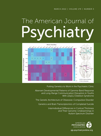
22q11.2 deletion syndrome is due to genetic mutations that involve microdeletions on the long arm of chromosome 22, which include the deletion of genes that are related to neurotransmission. Individuals with 22q11.2 deletions have various symptom profiles that are associated with numerous congenital physical problems as well as with cognitive and neuropsychiatric difficulties (4). It is estimated that 25%–30% of individuals with this genetic mutation will develop psychotic disorders (5). Based on this and considerable research, 22q11.2 deletion carriers have been used in research as a means to understand mechanisms that may more broadly underlie psychotic disorders. Mancini and colleagues (3) used a sample of 58 individuals with 22q11.2 deletions and 48 healthy comparison subjects to understand alterations in oscillatory rhythms that reflect coordinated neuronal activity across brain regions. Participants were 7–30 years old; this age range allowed for an assessment of potential 22q11.2-related developmental alterations in oscillatory rhythms. High-density EEG measures were used to assess responses during a 40-Hz auditory steady-state response (ASSR) task, which allows for the assessment of brain electrical activity in relation to the repeated presentations of auditory stimuli. This paradigm is of considerable interest as ASSRs are abnormal in schizophrenia and are thought to be related to altered gamma oscillatory activity. The researchers focused on gamma-band activity (i.e., 40 Hz; gamma range 25–140 Hz) because it is associated with long-range neuronal coupling, the performance of numerous cognitive functions, and is altered in some neuropsychiatric disorders including schizophrenia. Based on more basic research, it has been hypothesized that reductions in gamma oscillations in schizophrenia are due to alterations in prefrontal cortical inhibitory interneuron activity (6). The current findings demonstrate significantly decreased gamma-band responses in individuals with 22q11.2 deletions that with source location methods were located over the anterior cingulate cortex, posterior cingulate cortex, thalamus, and right primary auditory cortex. Decreases in theta band activity (4–7 Hz) in response to the ASSR were also noted but in this case more generally across the cortex. When data from 22q11.2 deletion individuals with psychotic symptoms were analyzed separately, similar but stronger decreases in gamma power were observed. Other work has demonstrated that gamma power increases with maturation from childhood to adolescence and into early adulthood, and the data from the comparison subjects in the current study confirmed this finding. However, no age-related developmental effect was seen in the 22q11.2 deletion carriers. In addition to assessing gamma oscillatory activity, the investigators also characterized other measures including theta-gamma phase amplitude coupling, which is thought to reflect the coordinated function of local and distant brain networks. The most prominent findings were in the psychotic subset of 22q11.2 deletion individuals, which were characterized by significant reductions in theta-gamma coupling between the anterior cingulate and various other regions. In her editorial(7), Dr. Melissa Larsen from Copenhagen University reviews the results from this study and discusses the value of performing neurobiological studies before the onset of symptoms in samples with known genetic mutations, in this case 22q11.2 deletions.

Insights Into the Genetic Architecture Underlying the Heritability of Obsessive-Compulsive Disorder

OCD occurs in approximately 2% of the population and when severe can result in marked suffering and disability. Mahjani et al. (8) use the largest sample to date to understand the contributions of common and rare genetic variants to the heritability of OCD. Drawn from a sample of Swedish-born individuals, 2,090 OCD patients and 4,567 controls, the investigators used genotyped single-nucleotide polymorphisms (SNPs) to estimate familial relatedness and OCD heritability. Previous work from twin and familial studies reported varying rates for OCD heritability, with an overall estimate thought to be around 40% (9). Additionally, and not entirely consistent across studies, some smaller sample GWAS studies have proposed that the majority of the OCD heritability is conferred by the most common SNP variants, those with a minor allele frequency (MAF) of >0.3, with no significant heritability accounted for by SNPs with a MAF <0.05 (10). MAF is an assessment of the frequency of the presence of the minor, or second most frequent, allele in a SNP in a population. As the authors of this paper point out, this finding would be somewhat unusual when comparing OCD to other psychiatric disorders and has implications for understanding the molecular underpinnings of OCD as well as the evolutionary forces that have shaped its prevalence and expression. By using the SNP data to estimate heritability, the authors found the heritability of OCD in their cohort to be 29%. It is important to note that SNPs with varying frequencies contributed to the heritability and, in contrast to the previous study, lower frequency SNPs (MAF 0.01–0.05) contributed 10% to the overall OCD heritability. These new data are consistent with a polygenic model of heritability, which is thought to underlie the heritability of most psychiatric disorders and also suggests that low-frequency SNPs, or rare genetic variation, could be important in understanding the pathophysiology and treatment of OCD. In his editorial (11), Dr. James Leckman from Yale University discusses the implications of the findings and relevant nonheritable factors that confer OCD risk. He also emphasizes the importance of future genetic work in larger populations that could characterize genetic variation in relation to the heterogeneity of OCD as it often presents with various other comorbid psychiatric illnesses.

Dorsolateral Prefrontal Cortical Gene Expression Alterations Associated with Violent Suicides

Punzi et al. (12) focus on understanding gene expression alterations in the dorsolateral prefrontal cortex of patients that died by suicide with an emphasis on characterizing molecular signatures that distinguish individuals that died by suicide via violent means. The dorsolateral prefrontal cortex is of particular interest as it is a region that broadly mediates executive functions, provides top-down regulation to limbic and subcortical regions involved in emotional and behavioral regulation, and has been implicated in various psychiatric illnesses. The authors point out the importance of aggression in suicidal behavior and posit that there is a different “state of mind” in individuals with high levels of aggression that die by suicide as compared to individuals who kill themselves by less violent means. They also propose that one way to reduce heterogeneity when studying the biology of suicide is to account for the means by which patients kill themselves, in this case nonviolent versus violent suicides. RNA sequencing was performed from dorsolateral prefrontal cortex tissue collected from 329 brains that are part of the Lieber Institute postmortem RNAseq data set (nonsuicide comparison subjects=103; nonsuicide cases [i.e., schizophrenia, major depression, and bipolar depression]=99; nonviolent suicide cases=50; and violent suicide cases=77). The results demonstrated notable differences between gene expression patterns in violent relative to nonviolent suicide cases and in those who did not die by suicide. However, very few differences in transcriptional patterns were observed when violent suicide patients were compared to nonpatient controls. Of interest is the greatest differences in dorsolateral prefrontal cortex gene expression were detected when comparing violent to nonviolent suicide cases. This comparison revealed that nonviolent suicide cases had greater expression of genes related to Huntington’s disease pathways, whereas violent suicide cases had increased expression of genes related to purinergic signaling such as G protein-coupled purinergic nucleotide receptor activity. Weighted Gene Co-Expression Analyses suggested a unique pattern of dorsolateral prefrontal cortex gene co-expression such that in violent suicide cases the expression of purinergic signaling genes appeared to be associated with the expression of genes implicating increased mitochondrial metabolism. The authors also used gene expression data from the fruit fly drosophila, which, consistent with their human findings, identified genes associated with purinergic signaling to be associated with aggressive behavior. Purines such as adenine and guanine are well known as nucleotide bases that contribute to the backbone of DNA, but they also are constituents of other molecules such as ATP and GTP. Additionally, by binding to brain receptors, including those specific for purinergic signaling, purines and related molecules can modulate neuronal and glial function. Other analyses on the human data suggested that some of the gene expression changes followed a linear continuum across the three disorder groups and comparison subjects. For example, gene expression related to G-coupled purinergic signaling showed the following pattern: violent suicides>nonsuicide cases>comparison>nonviolent suicide cases. On the basis of these data, the authors speculate that, regardless of diagnosis, individuals that die by suicide via violent means are distinctly different from other patients, as well as from those that die by suicide via nonviolent means. In their editorial (13), Drs. Daniel Almeida and Gustavo Turecki from McGill University discuss the potential importance of the findings and place them in the context of earlier work related to violent suicidal behavior.

Using Cortical Thickness and Gene Expression Data to Parse the Heterogeneity of Autism Spectrum Disorder

Enormous clinical heterogeneity exists across individuals with the diagnosis of autism spectrum disorder (ASD), with affected individuals having varying admixtures of symptoms that affect social functioning, behavioral regulation, sensory perception, and cognition. To further understand ASD heterogeneity in relation to brain structure, Ecker and colleagues (14) analyzed cortical thickness data obtained from structural MRIs from ASD (N=360) and comparison (N=279) subjects, 6–30 years of age, that were participants in the EU-AIMS Longitudinal European Autism Project. In addition to ascertaining group differences between ASD and comparison subjects, the researchers assessed cortical thickness in brain regions that did not show group differences but in which some ASD individuals were neuroanatomical outliers when compared with controls. In the between-group comparison, the researchers found ASD-related increases in cortical thickness in multiple regions including the anterior cingulate cortices and anterior temporal lobes. In other regions, including the left dorsolateral prefrontal cortex, ASD subjects had reduced cortical thickness. The authors note that the effect sizes for these between-group differences were small. When performing outlier analyses, the researchers found numerous additional regions in which ASD subjects had cortical thickness measures that were outside the 90% prediction interval for normal control subjects. Most of the ASD cortical thickness outlying values were located to the temporal cortical and medial PFC regions. Furthermore, across the entire brain, ASD individuals were found to have more outlying cortical thickness values relative to controls and a metric reflecting an individual’s total outlier values had a positive predictive value for ASD of 78.25%. It should be emphasized that the ASD outlier findings were not specific to ASD, as a high percentage of control subjects that also had intellectual disabilities were found to be neuroanatomical outliers. When using human brain gene expression data from another source, the Allen Brain Atlas, the researchers found that the regions that were identified to contain ASD cortical thickness outliers were also enriched for gene expression patterns known to be associated with ASD. Furthermore, polygenic scores derived from the subjects in this study revealed that they were correlated with an individual’s measure of total cortical surface atypicality as well as with the severity of their symptoms. Taken together, these findings support the strategy of combining neuroanatomical, genetic, and phenotypic data to better understand ASD heterogeneity and associated alterations in underlying neurodevelopmental pathways.

Conclusions

Our field continues to make advances in understanding the heritable and nonheritable factors that confer risk for the development of psychiatric illnesses. We have learned that the genetics of the illnesses that we treat are in general complex and polygenic, involving relatively small contributions of multiple “risk” and “protective” genes. It is also clear that our current understanding of the contributions of specific genes does not account for the total amount of estimated heritability for specific illnesses, and that different psychiatric illnesses, to some degree, share genetic underpinnings. These genomic findings, along with other neuroimaging and neurophysiological data, support transdiagnostic, dimensional approaches to understanding and, perhaps in the future, treating psychopathology. The papers in this issue highlight new findings that add to our understanding of how genes and their expression patterns relate to illness-related neural alterations as well as symptom severity and heterogeneity. The major findings gleaned from these reports include: 1) the value of studying individuals with 22q11.2 deletions to understand neurodevelopmental processes underlying alterations in gamma-band oscillations that are associated with psychosis; 2) how the genetic architecture of OCD is similar to that of other psychiatric disorders involving SNPs that are both common and rare variants; 3) elucidation of a genetic network involving purinergic signaling that is distinctly associated with violent suicides; and 4) expansion of the characterization of regional cortical alterations in individuals with ASD as they relate to polygenic risk scores and symptoms.
As we continue to advance our understanding of how genetic vulnerabilities influence the development and course of psychiatric illnesses, we can expect significant impacts that will be relevant to transforming our approaches to diagnosis and treatment. Specifically, it is anticipated that these data will help to redefine diagnostic classifications incorporating dimensionality and heterogeneity as key concepts, help to uncover new therapeutic targets, guide interventions toward specific illness-related neurodevelopmental processes, and eventually, along with other neural and behavioral data, support personalized treatment approaches.

REFERENCES

1.
McMahon FJ: Putting genetics to work in the psychiatric clinic. Am J Psychiatry 2022; 179:182–188
2.
Jacquemont S, Huguet G, Klein M, et al: Genes to Mental Health (G2MH): a framework to map the combined effects of rare and common variants on dimensions of cognition and psychopathology. Am J Psychiatry 2022; 179:189–203
3.
Mancini V, Rochas V, Seeber M, et al: Aberrant developmental patterns of gamma-band response and long-range communication disruption in youths with 22q11.2 deletion syndrome. Am J Psychiatry 2022; 179:204–215
4.
McDonald-McGinn DM, Sullivan KE, Marino B, et al: 22q11.2 deletion syndrome. Nat Rev Dis Primers 2015; 1:15071
5.
Tang SX, Gur RE: Longitudinal perspectives on the psychosis spectrum in 22q11.2 deletion syndrome. Am J Med Genet A 2018; 176:2192–2202
6.
Chung DW, Fish KN, Lewis DA: Pathological basis for deficient excitatory drive to cortical parvalbumin interneurons in schizophrenia. Am J Psychiatry 2016; 173:1131–1139
7.
Larsen KM: Auditory steady-state responses probe GABA-related oscillatory cortical circuits and their relation to neurodevelopmental mental disorders. Am J Psychiatry 2022; 179:175–176
8.
Mahjani B, Klei L, Mattheisen M, et al: The genetic architecture of obsessive-compulsive disorder: contribution of liability to OCD from alleles across the frequency spectrum. Am J Psychiatry 2022; 179:216–225
9.
Mahjani B, Bey K, Boberg J, et al: Genetics of obsessive-compulsive disorder. Psychol Med 2021; 51:2247–2259
10.
Davis LK, Yu D, Keenan CL, et al: Partitioning the heritability of Tourette syndrome and obsessive compulsive disorder reveals differences in genetic architecture. PLoS Genet 2013; DOI: 10.1371/journal.pgen.1003864. Epub 2013 Oct 24. PMID: 24204291
11.
Leckman JF: New insights into the genetic architecture of obsessive-compulsive disorder: another step along the way. Am J Psychiatry 2022; 179:177–179
12.
Punzi G, Ursini G, Chen Q, et al: Genetics and brain transcriptomics of completed suicide. Am J Psychiatry 2022; 179:226–241
13.
Almeida D, Turecki G: Is violent suicide molecularly distinct? Am J Psychiatry 2022; 179:180–181
14.
Ecker C, Pretzsch CM, Bletsch A, et al: Interindividual differences in cortical thickness and their genomic underpinnings in autism spectrum disorder. Am J Psychiatry 2022; 179:242–254

Information & Authors

Information

Published In

Go to American Journal of Psychiatry
American Journal of Psychiatry
Pages: 171 - 174
PubMed: 35236116

History

Received: 19 January 2022
Accepted: 19 January 2022
Published in print: March 2022
Published online: 3 March 2022

Keywords

  1. Genetics/Genomics
  2. Suicide and Self-Harm
  3. Neurodevelopmental Disorders
  4. Autism Spectrum Disorder
  5. Obsessive-Compulsive Disorder (OCD)

Authors

Details

Ned H. Kalin, M.D. [email protected]
Department of Psychiatry, University of Wisconsin School of Medicine and Public Health, Madison.

Notes

Send correspondence to Dr. Kalin ([email protected]).

Funding Information

Disclosures of Editors’ financial relationships appear in the April 2021 issue of the Journal.

Metrics & Citations

Metrics

Citations

Export Citations

If you have the appropriate software installed, you can download article citation data to the citation manager of your choice. Simply select your manager software from the list below and click Download.

For more information or tips please see 'Downloading to a citation manager' in the Help menu.

Format
Citation style
Style
Copy to clipboard

View Options

View options

PDF/EPUB

View PDF/EPUB

Login options

Already a subscriber? Access your subscription through your login credentials or your institution for full access to this article.

Personal login Institutional Login Open Athens login

Not a subscriber?

Subscribe Now / Learn More

PsychiatryOnline subscription options offer access to the DSM-5-TR® library, books, journals, CME, and patient resources. This all-in-one virtual library provides psychiatrists and mental health professionals with key resources for diagnosis, treatment, research, and professional development.

Need more help? PsychiatryOnline Customer Service may be reached by emailing [email protected] or by calling 800-368-5777 (in the U.S.) or 703-907-7322 (outside the U.S.).

Media

Figures

Other

Tables

Share

Share

Share article link

Share