Full access
Regular Articles
Published Online: 22 February 2016

Apathy Is Associated With Ventral Striatum Volume in Schizophrenia Spectrum Disorder

Publication: The Journal of Neuropsychiatry and Clinical Neurosciences

Abstract

Apathy is prevalent in schizophrenia, but its etiology has received little investigation. The ventral striatum (VS), a key brain region involved in motivated behavior, has been implicated in studies of apathy. We therefore evaluated whether apathy is associated with volume of the VS on MRI in 23 patients with schizophrenia using voxel-based morphometry. Results indicated that greater self-reported apathy severity was associated with smaller volume of the right VS even when controlling for age, gender, depression, and total gray matter volume. The finding suggests that apathy is related to abnormality of brain circuitry subserving motivated behavior in patients with schizophrenia.
Apathy may be described as a syndrome characterized by decreased interest or initiative, poor persistence in activities, restricted engagement in social interactions, and/or emotional indifference.1 Such motivational deficits are common in schizophrenia and have been associated with poor compliance with or inability to benefit from treatment,2 functional outcomes,3,4 and quality of life.5
A growing neuroimaging literature in neurological populations supports an association between apathy and integrity of frontal-subcortical circuitry.68 In patients with schizophrenia, apathy has been reported to be associated with lower resting cerebral blood flow in the frontal lobe,9 smaller volume of the frontal lobes bilaterally,10 as well as abnormalities of the cortical thickness and white matter connections of the left orbitofrontal cortex and left anterior cingulate cortex.11,12 In addition, apathy has been observed to be related to greater impairment on neuropsychological measures sensitive to frontal-subcortical circuitry dysfunction in both first episode13 and chronic10 patients.
Among subcortical regions, the ventral striatum (VS) has been receiving increasing attention as a possible substrate of apathy.14,15 The VS has been reported in human and animal studies to play a role in emotion and goal-directed, motivated behavior including the processing of reward.16,17 Structural MRI studies have observed that smaller volume of the VS is associated with greater apathy in HIV-positive adults,18 patients with frontotemporal dementia,19 and in those with Parkinson’s disease.20 A positron emission tomography study of depressed patients with Parkinson’s disease found that greater apathy was associated with lower dopamine and noradrenergic transporter binding in the VS.21 Furthermore, functional MRI (fMRI) studies have observed that greater apathy, but not other negative symptoms, was associated with reduced activation of the VS during anticipation of monetary reward in patients with schizophrenia.2224
In the present investigation, we used MRI and voxel-based morphometry (VBM)25 to examine the hypothesis that apathy is associated with reduced gray matter volume in the VS of patients with schizophrenia. Such a VBM volume-of-interest approach has been implemented in previous research on schizophrenia.26 A measure of depression was included as a covariate in the analysis given the partial overlap of the symptoms of apathy and depression.27

Methods

Participants

Participants consisted of 23 patients with DSM-IV diagnoses of schizophrenia (N=14) or schizoaffective disorder (N=9), recruited from outpatient community clinics through referral to the study by a treatment provider. The sample had a mean age of 39.43 years (SD=12.28), 52% were men, 87% were right handed, and mean parental education was 13.07 years (SD=3.79). Diagnosis was established using the Structured Clinical Interview for DSM-IV-TR.28
Participants were excluded if they had a history of neurological disorder (e.g., epilepsy, neurodegenerative disorder), mental retardation, or significant systemic medical illness. In addition, patients were excluded if they had a history of substance abuse within the 6 months before participation. All patients were receiving stable doses (at least 12 weeks) of antipsychotic medication at the time of the study (atypical only, N=20; both atypical and typical, N=2; typical only, N=1), with most taking multiple medications. After complete description of the study, written informed consent was obtained following a protocol approved by the Dartmouth College Committee for the Protection of Human Subjects.

Procedure

We assessed severity of clinical symptoms using the Scale for the Assessment of Positive Symptoms (SAPS)29 and Scale for the Assessment of Negative Symptoms (SANS)30 and assessed depression using the Beck Depression Inventory–II (BDI-II).31 The Apathy Evaluation Scale (AES) was used to assess self-reported apathy.32 The AES is an 18-item questionnaire that is rated on a 4-point scale ranging from “not at all” to “a lot,” with scores ranging from 0 to 54, where higher scores indicate greater apathy.

MRI Data Acquisition

Scans were acquired with a GE Signa 1.5-T Horizon LX magnet (General Electric, Fairfield, Conn.) with echo speed gradients using a standard RF head coil. A T1-weighted three-dimensional spoiled gradient echo (SPGR) coronal volume was acquired. Parameters were TR=25 ms, TE=3 ms, flip angle=40 degrees, NEX=1, and slice thickness=1.5 mm (no gap), yielding 124 contiguous slices with a 24-cm field of view, and a 256×256 matrix with 0.9375 mm in-plane resolution. A fast spin echo T2-weighted scan was also acquired to screen for focal lesions or other incidental findings (TR=3000, TE=96, 3-mm contiguous axial slices).

MRI Data Processing

We performed VBM25 using the statistical parametric mapping package (SPM12; Wellcome Department of Imaging Neuroscience, London, UK). This involved automated segmentation of gray matter, white matter, and CSF in the SPGR data, nonlinear registration of all images to the Montreal Neurological Institute (MNI152) template space via a data-derived gray matter/white matter–based template of 1.5 mm3 isotropic resolution created using Diffeomorphic Anatomical Registration Through Exponentiated Lie Algebra (DARTEL),33 with modulation to preserve tissue volumes and spatial smoothing (full width at half maximum=4 mm).
Several steps to ensure accurate segmentation were completed, including visual inspection of raw images, visual inspection of gray matter, white matter, and CSF segmentations, and comparison of the gray matter-to-white matter ratio and total brain volume across participants. One scan was discarded based on poor raw image quality (leaving N=23 for analysis). Smoothed normalized gray matter maps were used for statistical analysis after a final visual inspection of segmentation quality.
To examine gray matter volume in the VS, we performed an analysis using a volume of interest (VOI) specified by a voxel mask from a publication-based probabilistic atlas.34 The probabilistic VOI mask was thresholded at the 0.5 probability level, which had been previously used in fMRI studies of the VS in schizophrenia,35 to create a binary mask. 
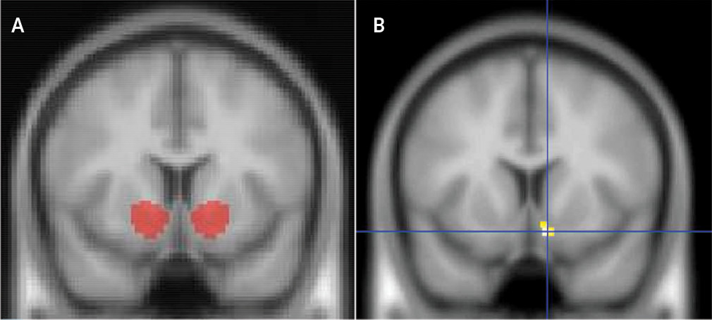
Figure 1 [A] illustrates the anatomic boundary of the VOI on the MNI152 template brain.
FIGURE 1. [A] Ventral Striatum Volume of Interest on the MNI152 Template Brain. [B] More Severe Self-Reported Apathy Associated With Lower Regional Gray Matter Volume in the Right Ventral Striatum in Patients With Schizophrenia.a
aControlling for age, gender, Beck Depression Inventory–II score, and total gray matter volume.

Statistical Analysis

We conducted statistical parametric mapping, restricted to the VOI, on a voxel-by-voxel basis using a general linear model and random-effects analysis as implemented in SPM12. Multiple regression analysis was used to examine the correlation between apathy score and gray matter volume in the VOI controlling for age, gender, BDI-II score, and total gray matter volume. A statistical threshold for significance of p<0.01 was implemented without correction for multiple comparisons given the focused VOI approach, although the PWE-corrected p value is also presented for interested readers. Data are presented in MNI152 atlas coordinates.

Results

The sample had a mean AES score of 14.43 (SD=10.11, range=1–48) indicating a relatively mild level of apathy. SAPS (mean=7.0, SD=3.5) and SANS (mean=10.6, SD=3.8) scores were also generally in the mild range. BDI-II scores were typically in the minimal to mild range (mean=13.3, SD=11.6).
VBM indicated that greater apathy was associated with smaller gray matter volume in the right VS VOI [Z=3.67; Puncorrected=0.001, k=21; peak (xyz) = 9, 10, –14; cluster-level pFWE of 0.051] (Figure 1 [B]). Furthermore, an exploratory whole-brain analysis also produced a significant association of apathy and right VS volume (Z=3.60, p=0.001, k=174), albeit only when uncorrected for multiple comparisons. No significant correlation was observed between apathy and left VS volume. No significant association was observed between VS volume and either BDI-II score or chlorpromazine-equivalent medication dosage.

Discussion

The present investigation revealed an association between greater self-reported apathy and reduced gray matter volume in the right VS, particularly the nucleus accumbens, in patients with schizophrenia. This relationship was observed despite statistically controlling for depression, consistent with other work showing that apathy can be dissociated from depression.3638 Our finding is also in line with investigations in other patient populations that have implicated abnormality of the VS in apathy.18,20,21 Furthermore, we observed that apathy was related specifically to right VS volume. Such an association between apathy and the right VS has previously been observed in studies of schizophrenia22 and frontotemporal dementia.19 The precise functional significance of this lateralization, however, remains to be elucidated.
A number of authors have proposed that the pathophysiology of apathy involves the VS given the region’s involvement in motivation and emotion.14,15 fMRI studies have reported that patients with schizophrenia show reduced responsiveness to behaviorally salient stimuli in brain regions involved in motivation, such as decreased activation of the VS and related basal forebrain regions in response to target stimuli during an auditory oddball task,39 and reduced activation of the VS, including the right VS, to cues signaling the possibility of obtaining a monetary reward.2224,40 These findings along with that of the present study suggest that apathy in schizophrenia may be due in part to deficient processing of stimuli signaling the potential for reward, or a more general dysfunction in processing motivationally significant stimuli, resulting from structural and functional abnormalities of the VS and related circuitry. Involvement of the VS is also consistent with the affective/motivational subtype of apathy proposed by Levy.14 In that model, it is argued that abnormality of basal ganglia subregions such as the VS and the orbital and mesial prefrontal cortices leads to difficulty associating affective/emotional signals with ongoing and future behaviors, consequently resulting in diminishing motivation.
It is important to note that it is unlikely that VS abnormality fully accounts for apathy in schizophrenia. Case studies involving lesions restricted to the VS are rare and have not been associated with the presence of apathy.41 In a similar manner, behavior akin to apathy has not been consistently reported following focal lesions to the VS in primates.4244 Previous research has implicated the frontal lobes in apathy in schizophrenia1012 and in neurological disorders.19,45 It is therefore possible that frontal abnormality is playing a role in driving dysregulation of the VS in schizophrenia. A recent fMRI study that observed normal VS response, but abnormal striatal-cortical connectivity to motivational cues that was related to negative symptom severity in patients with schizophrenia, may support that hypothesis.46 A more complete understanding of the etiology of apathy in schizophrenia will therefore require further investigation of the structural and functional integrity of subregions within the frontal-striatal circuitry, as well as functional connectivity of the circuitry.
The results of the present investigation must be interpreted within the context of its limitations. The sample size was modest; therefore, the findings require replication in a larger sample, which should also evaluate whether they are upheld when considering subtypes of the disorder, gender, and handedness. The relationship observed between apathy and VS volume was significant using a focused volume-of-interest approach and is consistent with previous studies, but it was nonetheless a relatively small effect. This may be due in part to the overall mild severity of apathy in the sample. It is possible that we would observe an even stronger relationship in patients with schizophrenia having more severe levels of apathy. Further study is also needed to determine whether antipsychotic medication could have affected the findings, although the latter seems unlikely given that we found no association between VS volume and medication dosage, and a previous study found a relationship between apathy and functional activation of the VS in patients with schizophrenia taking atypical antipsychotics.22 Finally, although we did not have information pertaining to duration of illness in our patient sample, it is unlikely that this variable can account for our results given that other work has found that duration of illness is unrelated to volume of the accumbens in patients with schizophrenia.47
With those caveats, the present findings provide further support for the hypothesis that apathy in schizophrenia is associated with abnormality of neural circuitry subserving motivated behavior. Such findings could inform the treatment of apathy, as well as raising the possibility of using a neuroimaging marker (i.e., abnormal structure and/or function of the VS) to identify patients who may be at elevated risk of experiencing apathy and thus provide early intervention.

Acknowledgments

We thank Mary Brunette, Doug Noordsy, Chris O’Keefe, and their colleagues for facilitating patient referrals to this study.

References

1.
Marin RS, Wilkosz PA: Disorders of diminished motivation. J Head Trauma Rehabil 2005; 20:377–388
2.
Tattan TM, Creed FH: Negative symptoms of schizophrenia and compliance with medication. Schizophr Bull 2001; 27:149–155
3.
Fervaha G, Foussias G, Agid O, et al: Motivational deficits in early schizophrenia: prevalent, persistent, and key determinants of functional outcome. Schizophr Res 2015; 166:9–16
4.
Konstantakopoulos G, Ploumpidis D, Oulis P, et al: Apathy, cognitive deficits and functional impairment in schizophrenia. Schizophr Res 2011; 133:193–198
5.
Fervaha G, Foussias G, Siddiqui I, et al: Abbreviated quality of life scales for schizophrenia: comparison and utility of two brief community functioning measures. Schizophr Res 2014; 154:89–92
6.
Baggio HC, Segura B, Garrido-Millan JL, et al: Resting-state frontostriatal functional connectivity in Parkinson’s disease-related apathy. Mov Disord 2015; 30:671–679
7.
Tunnard C, Whitehead D, Hurt C, et al: AddNeuroMed Consortium: Apathy and cortical atrophy in Alzheimer’s disease. Int J Geriatr Psychiatry 2011; 26:741–748
8.
Peavy GM, Salmon DP, Edland SD, et al: Neuropsychiatric features of frontal lobe dysfunction in autopsy-confirmed patients with Lewy bodies and “pure” Alzheimer disease. Am J Geriatr Psychiatry 2013; 21:509–519
9.
Suzuki M, Kurachi M, Kawasaki Y, et al: Left hypofrontality correlates with blunted affect in schizophrenia. Jpn J Psychiatry Neurol 1992; 46:653–657
10.
Roth RM, Flashman LA, Saykin AJ, et al: Apathy in schizophrenia: reduced frontal lobe volume and neuropsychological deficits. Am J Psychiatry 2004; 161:157–159
11.
Ohtani T, Bouix S, Hosokawa T, et al: Abnormalities in white matter connections between orbitofrontal cortex and anterior cingulate cortex and their associations with negative symptoms in schizophrenia: a DTI study. Schizophr Res 2014; 157:190–197
12.
Mørch-Johnsen L, Nesvåg R, Faerden A, et al: Brain structure abnormalities in first-episode psychosis patients with persistent apathy. Schizophr Res 2015; 164:59–64
13.
Faerden A, Vaskinn A, Finset A, et al: Apathy is associated with executive functioning in first episode psychosis. BMC Psychiatry 2009; 9:1
14.
Levy R: Apathy: a pathology of goal-directed behaviour: a new concept of the clinic and pathophysiology of apathy. Rev Neurol (Paris) 2012; 168:585–597
15.
Duffy JD: Apathy and related disorders of diminished motivation, in Guide to Neuropsychiatric Therapeutics. Edited by Coffey CE, McAllister TW, Silver JM. Philadelphia, PA, Lippincott Williams & Wilkins, 2007, pp 1–13
16.
Knutson B, Fong GW, Adams CM, et al: Dissociation of reward anticipation and outcome with event-related fMRI. Neuroreport 2001; 12:3683–3687
17.
Robbins TW, Everitt BJ: Neurobehavioural mechanisms of reward and motivation. Curr Opin Neurobiol 1996; 6:228–236
18.
Paul RH, Brickman AM, Navia B, et al: Apathy is associated with volume of the nucleus accumbens in patients infected with HIV. J Neuropsychiatry Clin Neurosci 2005; 17:167–171
19.
Eslinger PJ, Moore P, Antani S, et al: Apathy in frontotemporal dementia: behavioral and neuroimaging correlates. Behav Neurol 2012; 25:127–136
20.
Carriere N, Besson P, Dujardin K, et al: Apathy in Parkinson’s disease is associated with nucleus accumbens atrophy: a magnetic resonance imaging shape analysis. Mov Disord 2014; 29:897–903
21.
Remy P, Doder M, Lees A, et al: Depression in Parkinson’s disease: loss of dopamine and noradrenaline innervation in the limbic system. Brain 2005; 128:1314–1322
22.
Simon JJ, Biller A, Walther S, et al: Neural correlates of reward processing in schizophrenia--relationship to apathy and depression. Schizophr Res 2010; 118:154–161
23.
Kirschner M, Hager OM, Bischof M, et al: Ventral striatal hypoactivation is associated with apathy but not diminished expression in patients with schizophrenia. J Psychiatry Neurosci 2015; 40:140383
24.
Hägele C, Schlagenhauf F, Rapp M, et al: Dimensional psychiatry: reward dysfunction and depressive mood across psychiatric disorders. Psychopharmacology (Berl) 2015; 232:331–341
25.
Ashburner J, Friston KJ: Voxel-based morphometry--the methods. Neuroimage 2000; 11:805–821
26.
Bangalore SS, Prasad KM, Montrose DM, et al: Cannabis use and brain structural alterations in first episode schizophrenia--a region of interest, voxel based morphometric study. Schizophr Res 2008; 99:1–6
27.
Landes AM, Sperry SD, Strauss ME, et al: Apathy in Alzheimer’s disease. J Am Geriatr Soc 2001; 49:1700–1707
28.
First MB, Spitzer RL, Gibbon M, et al: Structured Clinical Interview for DSM-IV-TR Axis I Disorders (SCID-I/P), Patient Edition. Washington, DC, American Psychiatric Publishing, Inc., 2002
29.
Andreasen NC: Scale for the Assessment of Positive Symptoms (SAPS). Iowa City, College of Medicine, University of Iowa, 1984
30.
Andreasen NC: Scale for the Assessment of Negative Symptoms (SANS). Iowa City, College of Medicine, University of Iowa, 1984
31.
Beck AT, Steer RA, Brown GK: Beck Depression Inventory-II (BDI-II). San Antonio, TX, The Psychological Corporation, 1996
32.
Marin RS, Biedrzycki RC, Firinciogullari S: Reliability and validity of the Apathy Evaluation Scale. Psychiatry Res 1991; 38:143–162
33.
Ashburner J: A fast diffeomorphic image registration algorithm. Neuroimage 2007; 38:95–113.
34.
Fox PT, Lancaster JL: Opinion: Mapping context and content: the BrainMap model. Nat Rev Neurosci 2002; 3:319–321
35.
Juckel G, Schlagenhauf F, Koslowski M, et al: Dysfunction of ventral striatal reward prediction in schizophrenia. Neuroimage 2006; 29:409–416
36.
Castellon SA, Hinkin CH, Wood S, et al: Apathy, depression, and cognitive performance in HIV-1 infection. J Neuropsychiatry Clin Neurosci 1998; 10:320–329
37.
Kirsch-Darrow L, Fernandez HH, Marsiske M, et al: Dissociating apathy and depression in Parkinson disease. Neurology 2006; 67:33–38
38.
Onyike CU, Sheppard J-ME, Tschanz JT, et al: Epidemiology of apathy in older adults: the Cache County Study. Am J Geriatr Psychiatry 2007; 15:365–375
39.
Liddle PF, Laurens KR, Kiehl KA, et al: Abnormal function of the brain system supporting motivated attention in medicated patients with schizophrenia: an fMRI study. Psychol Med 2006; 36:1097–1108
40.
Arrondo G, Segarra N, Metastasio A, et al: Reduction in ventral striatal activity when anticipating a reward in depression and schizophrenia: a replicated cross-diagnostic finding. Front Psychol 2015; 6:1280
41.
Levy R, Dubois B: Apathy and the functional anatomy of the prefrontal cortex-basal ganglia circuits. Cereb Cortex 2006; 16:916–928
42.
Brown CA, Campbell MC, Karimi M, et al: Dopamine pathway loss in nucleus accumbens and ventral tegmental area predicts apathetic behavior in MPTP-lesioned monkeys. Exp Neurol 2012; 236:190–197
43.
Butters N, Rosvold HE: Effect of caudate and septal nuclei lesions on resistance to extinction and delayed-alternation. J Comp Physiol Psychol 1968; 65:397–403
44.
Stern CE, Passingham RE: The nucleus accumbens in monkeys (Macaca fascicularis): I. The organization of behaviour. Behav Brain Res 1994; 61:9–21
45.
Marshall GA, Monserratt L, Harwood D, et al: Positron emission tomography metabolic correlates of apathy in Alzheimer disease. Arch Neurol 2007; 64:1015–1020
46.
Reckless GE, Andreassen OA, Server A, et al: Negative symptoms in schizophrenia are associated with aberrant striato-cortical connectivity in a rewarded perceptual decision-making task. Neuroimage Clin 2015; 8:290–297
47.
van Erp TG, Hibar DP, Rasmussen JM, et al: Subcortical brain volume abnormalities in 2028 individuals with schizophrenia and 2540 healthy controls via the ENIGMA consortium. Mol Psychiatry (Epub ahead of print, Aug 18, 2015)

Information & Authors

Information

Published In

Go to The Journal of Neuropsychiatry and Clinical Neurosciences
The Journal of Neuropsychiatry and Clinical Neurosciences
Pages: 191 - 194
PubMed: 26900738

History

Received: 1 October 2015
Revision received: 9 December 2015
Accepted: 17 January 2016
Published online: 22 February 2016
Published in print: Summer 2016

Authors

Affiliations

Robert M. Roth, Ph.D.
From the Brain Imaging Laboratory, Dept. of Psychiatry, Geisel School of Medicine at Dartmouth/DHMC, Lebanon, NH (RMR, MAG, LAF, JCP, JCF); Program in Neuroscience, Dept. of Psychology, Bates College, Lewiston, ME (NSK); Dept. of Psychiatry, Indiana University School of Medicine, Indianapolis, IN (TWM); and the Center for Neuroimaging, Dept. of Radiology, Indiana University School of Medicine, Indianapolis, IN (AJS).
Matthew A. Garlinghouse, Ph.D.
From the Brain Imaging Laboratory, Dept. of Psychiatry, Geisel School of Medicine at Dartmouth/DHMC, Lebanon, NH (RMR, MAG, LAF, JCP, JCF); Program in Neuroscience, Dept. of Psychology, Bates College, Lewiston, ME (NSK); Dept. of Psychiatry, Indiana University School of Medicine, Indianapolis, IN (TWM); and the Center for Neuroimaging, Dept. of Radiology, Indiana University School of Medicine, Indianapolis, IN (AJS).
Laura A. Flashman, Ph.D.
From the Brain Imaging Laboratory, Dept. of Psychiatry, Geisel School of Medicine at Dartmouth/DHMC, Lebanon, NH (RMR, MAG, LAF, JCP, JCF); Program in Neuroscience, Dept. of Psychology, Bates College, Lewiston, ME (NSK); Dept. of Psychiatry, Indiana University School of Medicine, Indianapolis, IN (TWM); and the Center for Neuroimaging, Dept. of Radiology, Indiana University School of Medicine, Indianapolis, IN (AJS).
Nancy S. Koven, Ph.D.
From the Brain Imaging Laboratory, Dept. of Psychiatry, Geisel School of Medicine at Dartmouth/DHMC, Lebanon, NH (RMR, MAG, LAF, JCP, JCF); Program in Neuroscience, Dept. of Psychology, Bates College, Lewiston, ME (NSK); Dept. of Psychiatry, Indiana University School of Medicine, Indianapolis, IN (TWM); and the Center for Neuroimaging, Dept. of Radiology, Indiana University School of Medicine, Indianapolis, IN (AJS).
J. Cara Pendergrass, Ph.D.
From the Brain Imaging Laboratory, Dept. of Psychiatry, Geisel School of Medicine at Dartmouth/DHMC, Lebanon, NH (RMR, MAG, LAF, JCP, JCF); Program in Neuroscience, Dept. of Psychology, Bates College, Lewiston, ME (NSK); Dept. of Psychiatry, Indiana University School of Medicine, Indianapolis, IN (TWM); and the Center for Neuroimaging, Dept. of Radiology, Indiana University School of Medicine, Indianapolis, IN (AJS).
James C. Ford, Ph.D.
From the Brain Imaging Laboratory, Dept. of Psychiatry, Geisel School of Medicine at Dartmouth/DHMC, Lebanon, NH (RMR, MAG, LAF, JCP, JCF); Program in Neuroscience, Dept. of Psychology, Bates College, Lewiston, ME (NSK); Dept. of Psychiatry, Indiana University School of Medicine, Indianapolis, IN (TWM); and the Center for Neuroimaging, Dept. of Radiology, Indiana University School of Medicine, Indianapolis, IN (AJS).
Thomas W. McAllister, M.D.
From the Brain Imaging Laboratory, Dept. of Psychiatry, Geisel School of Medicine at Dartmouth/DHMC, Lebanon, NH (RMR, MAG, LAF, JCP, JCF); Program in Neuroscience, Dept. of Psychology, Bates College, Lewiston, ME (NSK); Dept. of Psychiatry, Indiana University School of Medicine, Indianapolis, IN (TWM); and the Center for Neuroimaging, Dept. of Radiology, Indiana University School of Medicine, Indianapolis, IN (AJS).
Andrew J. Saykin, Psy.D.
From the Brain Imaging Laboratory, Dept. of Psychiatry, Geisel School of Medicine at Dartmouth/DHMC, Lebanon, NH (RMR, MAG, LAF, JCP, JCF); Program in Neuroscience, Dept. of Psychology, Bates College, Lewiston, ME (NSK); Dept. of Psychiatry, Indiana University School of Medicine, Indianapolis, IN (TWM); and the Center for Neuroimaging, Dept. of Radiology, Indiana University School of Medicine, Indianapolis, IN (AJS).

Notes

Send correspondence to Dr. Roth; e-mail: [email protected]

Competing Interests

The authors report no financial relationships with commercial interests.

Funding Information

This work is supported by grants from the National Alliance for Medical Image Computing (NIH U54 EB005149), Stanley Medical Foundation, National Alliance for Research on Schizophrenia and Depression, and the Hitchcock Foundation. Research reported in this study was also supported by The Dartmouth Clinical and Translational Science Institute, under award number UL1TR001086 from the National Center for Advancing Translational Sciences (NCATS) of the National Institutes of Health (NIH). The content is solely the responsibility of the authors and does not necessarily represent the official views of the NIH.

Metrics & Citations

Metrics

Citations

Export Citations

If you have the appropriate software installed, you can download article citation data to the citation manager of your choice. Simply select your manager software from the list below and click Download.

For more information or tips please see 'Downloading to a citation manager' in the Help menu.

Format
Citation style
Style
Copy to clipboard

There are no citations for this item

View Options

View options

PDF/ePub

View PDF/ePub

Get Access

Login options

Already a subscriber? Access your subscription through your login credentials or your institution for full access to this article.

Personal login Institutional Login Open Athens login
Purchase Options

Purchase this article to access the full text.

PPV Articles - Journal of Neuropsychiatry and Clinical Neurosciences

PPV Articles - Journal of Neuropsychiatry and Clinical Neurosciences

Not a subscriber?

Subscribe Now / Learn More

PsychiatryOnline subscription options offer access to the DSM-5-TR® library, books, journals, CME, and patient resources. This all-in-one virtual library provides psychiatrists and mental health professionals with key resources for diagnosis, treatment, research, and professional development.

Need more help? PsychiatryOnline Customer Service may be reached by emailing [email protected] or by calling 800-368-5777 (in the U.S.) or 703-907-7322 (outside the U.S.).

Media

Figures

Other

Tables

Share

Share

Share article link

Share