Full access
Special Articles
Published Online: 25 June 2018

Bridging the Great Divide: What Can Neurology Learn From Psychiatry?

Publication: The Journal of Neuropsychiatry and Clinical Neurosciences

Abstract

Neurology and psychiatry share common historical origins and rely on similar tools to study brain disorders. Yet the practical integration of medical and scientific approaches across these clinical neurosciences remains elusive. Although much has been written about the need to incorporate emerging systems-level, cellular-molecular, and genetic-epigenetic advances into a science of mind for psychiatric disorders, less attention has been given to applying clinical neuroscience principles to conceptualize neurologic conditions with an integrated neurobio-psycho-social approach. In this perspective article, the authors briefly outline the historically interwoven and complicated relationship between neurology and psychiatry. Through a series of vignettes, the authors then illustrate how some traditional psychiatric conditions are being reconceptualized in part as disorders of neurodevelopment and awareness. They emphasize the intersection of neurology and psychiatry by highlighting conditions that cut across traditional diagnostic boundaries. The authors argue that the divide between neurology and psychiatry can be narrowed by moving from lesion-based toward circuit-based understandings of neuropsychiatric disorders, from unidirectional toward bidirectional models of brain-behavior relationships, from exclusive reliance on categorical diagnoses toward transdiagnostic dimensional perspectives, and from silo-based research and treatments toward interdisciplinary approaches. The time is ripe for neurologists and psychiatrists to implement an integrated clinical neuroscience approach to the assessment and management of brain disorders. The subspecialty of behavioral neurology & neuropsychiatry is poised to lead the next generation of clinicians to merge brain science with psychological and social-cultural factors. These efforts will catalyze translational research, revitalize training programs, and advance the development of impactful patient-centered treatments.
In the last analysis, we see only what we are ready to see, what we have been taught to see. We eliminate and ignore everything that is not a part of our practices.
—Jean-Martin Charcot, 1825–1893
Although most organs have one dedicated medical specialty, the brain has been historically divided into two disciplines, neurology and psychiatry.1,2 Neurology and psychiatry have been separated by theory, domains of investigation, vocabulary, and interventions. This separation has led to a lack of a shared nomenclature and, at times, different diagnostic criteria for the same disorder. Traditional terms such as “psychogenic” and “organic” perpetuate an artificial dualism. Despite recent appeals by the Nobel laureate Eric Kandel,3 prominent academics,48 and leaders of the National Institutes of Health in the United States9, “closing the great divide” between neurology and psychiatry remains elusive.10 Furthermore, at times the distinction between neurologic and psychiatric disorders appears arbitrary. Some disorders with well-established neurobiological foundations, including schizophrenia, are almost exclusively reported on in the psychiatric literature, and other conditions with prominent affective and behavioral phenotypes, such as Huntington’s disease and behavioral variant frontotemporal dementia, are predominantly characterized in the neurologic literature.11
Paradigm shifts in science, as well as clinical need for a convergence between neurology and psychiatry, are based on the unprecedented expansion of our emerging neurobiological knowledge of complex mental operations. Increasing evidence blurs the boundaries between neurologic and psychiatric disorders, which includes growth in network science and genomics, as well as the realization that many brain disorders are not due to detectable “lesions” but originate from dysfunction across broadly distributed networks. Furthermore, there is increasing recognition that the causes of many brain disorders are multifactorial and bidirectional in nature. For example, psychosocial (environmental) factors alter neurocircuits, and maladaptive neural function impairs psychosocial behaviors.12 These factors, in addition to an aging population, are driving the need for a renaissance, one that necessitates the development of specialists who can transdiagnostically care for patients with complex brain disorders.13
In this perspective article, we present four vignettes illustrating how advances in clinical neuroscience are eroding the traditional boundaries separating the disciplines of neurology and psychiatry. Schizophrenia is presented as a neurodevelopmental disorder; functional neurological disorder as a disturbance of awareness; Tourette syndrome, obsessive-compulsive disorder (OCD), and attention-deficit hyperactivity disorder (ADHD) as heritable disorders of overlapping, dysregulated circuits with interrelated motor-cognitive-affective components; and poststroke neuropsychiatric symptoms as impairments in distributed networks. These examples illustrate a practical clinical neuroscience approach that incorporates neurobio-psycho-social elements.
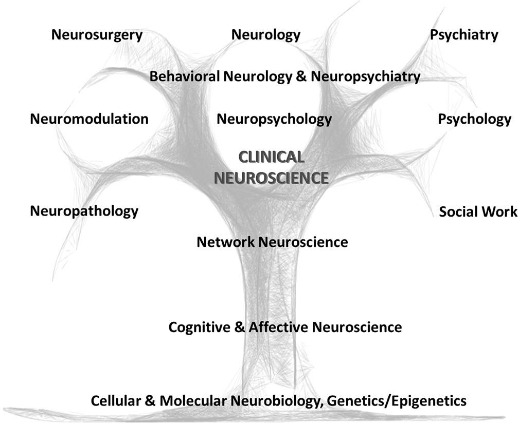
Thereafter, we discuss core themes that can help move neurology and psychiatry toward a more integrated clinical and research approach. Emphasis is given to identifying steps neurologists can take to incorporate psychiatric and psychosocial factors, which will facilitate an interdisciplinary approach to patient care. The subspecialty of behavioral neurology & neuropsychiatry is well-positioned to catalyze these transformations (Figure 1).
FIGURE 1. An Integrated Clinical Neuroscience Approach for the Assessment, Management, and Investigation of Brain Disorders

Schizophrenia

Case Description

A 24-year-old African American woman was brought by her parents to clinic. In childhood, she had concentration difficulties and was diagnosed with ADHD. Two of her maternal aunts had schizophrenia, and her brother had bipolar disorder. Her parents divorced when the patient was a teenager. In junior high school, she began displaying gradual social withdrawal, declining grades, cannabis use, and worries that her classmates might be spreading evil rumors about her homosexuality. She also reported occasional whispers saying such things (although she was uncertain whether these perceptions were real). During her first semester in college, she faced academic pressures and began experiencing “internal chatter” by two voices that kept a running commentary on her actions. She was now convinced that these voices were real and resulted from a chip implanted in her teeth by a classmate. She confronted and assaulted this classmate, which led to an involuntary hospitalization.
Five years later, she continued experiencing auditory hallucinations, was unemployed, and mostly spent time in her basement. She displayed disorganized speech, disinhibited behavior, reduced motivation, poor personal hygiene and remained unconvinced about the need for medications.
This case illustrates the trajectory of schizophrenia with subtle premorbid cognitive impairments in childhood, followed by a prodrome with gradual social and functional decline in adolescence. Psychosis typically sets in during late adolescence or adulthood and follows a chronic or intermittent course. Regarding pathogenesis, it is now widely held that schizophrenia is due to either a failure of optimal brain development, an abnormal refinement of pruning of synapses during adolescence, or both.14 The neuroanatomy of schizophrenia is increasingly well understood. Impaired executive functions, poor insight, and disinhibited behavior point to deficits in dorsolateral and ventromedial prefrontal cortex, respectively, termed previously as “hypofrontality.” Hallucinations and delusions are thought to relate to inappropriate subcortical activations, including in the hippocampus.15 Formal thought disorder is linked to abnormal functioning of language circuits.16 In terms of neurochemical pathophysiology, ventral striatal and mesolimbic dopaminergic abnormalities likely underlie positive psychotic symptoms, whereas recent data implicate mesocortical dopaminergic deficits and altered glutamatergic and GABA function as mediating cognitive and negative symptoms.17 Finally, as to etiology, there is compelling evidence that genetic factors, including genes mediating synapse integrity, ion channels, and immune function, are implicated in the pathophysiology of schizophrenia; environmental and epigenetic risk factors are also involved.18
Clearly, this woman is experiencing a progressive developmental derailment beginning in childhood, perhaps resulting from a combination of genetic and environmental (cannabis) risk factors, which have led to chronic network alterations in several key prefrontal, limbic, and superior temporal “nodes,” and multiple neurotransmitter aberrations. A neurobio-psycho-social formulation in her case would also include roles for parental discord and academic stress framed as predisposing and precipitating factors.19 Such a formulation not only would incorporate a common language for both psychiatrists and neurologists but also would point to actionable treatments and research initiatives. The neurodevelopmental pathogenesis and premorbid/prodromal impairments, including cognitive decline, point to a role for early interventions.
A cognitive test battery and neuroimaging biomarkers may identify a subtype of psychosis characterized primarily by cognitive dysfunction, and one that could point to a role for time-intensive cognitive rehabilitation.20 Delineation of distinct networks underlying negative/depressive symptoms, cognitive deficits, and positive symptoms can point to personalized treatments, such as cognitive remediation,21 cognitive-behavioral therapy (CBT),22 psychopharmacology, and neuromodulation.23

Functional Neurological Disorder

Case Description

A 30-year-old single, unemployed Hispanic woman with a history of medically unexplained neck pain and posttraumatic stress disorder following childhood abuse abruptly developed episodes of decreased arousal with asynchronous side-to-side body movements. She also experienced clouded thinking, difficulty walking, and a sense that her legs collapsed. She was hospitalized, and her examination showed a variable/distractible head tremor and a positive right Hoover’s sign. A typical spell was captured on video-electroencephalography without electrographic correlate. She was referred for physical therapy but developed full-body shaking on attempted gait and elected against additional sessions. She did not show up for her outpatient psychiatry appointment, and the patient and family members had difficulty understanding how this could be “all in the patient’s head.”
On the basis of emerging neuroimaging findings, the patient’s symptoms of nonepileptic seizures, functional limb weakness, chronic pain, affective dysregulation, and cognitive complaints may be partially localized to networks mediating multimodal sensory-motor, affective, cognitive, and perceptual integration, including the anterior insula, dorsal anterior cingulate cortex (ACC), amygdala, and temporoparietal junction.2432 Patients with functional neurological disorder show dorsal ACC structural alterations compared with controls,32,33 and individuals with medically unexplained pain display central pain matrix volumetric reductions.34 Parallel inverse associations between left anterior insular gray matter volume and self-report measures of symptom severity and childhood abuse burden have also been recently reported in functional neurological disorder.29,31 Additionally, resting-state functional connectivity studies of functional neurological disorder display heightened connectivity between limbic/paralimbic areas (ACC, insula, amygdala) and somatomotor/executive control networks.3538 Increased paralimbic-supplementary motor area functional connectivity has also correlated with nonepileptic seizure frequency.36,37 While cingulo-insular abnormalities may pertain to impaired emotional awareness/regulation, aberrant viscero-somatic processing, and heightened affective influence over motor behaviors, right temporo-parietal junction network alterations in functional neurological disorder have been linked to motor intention awareness and action-authorship recognition deficits.26,3941
Although there are multiple predisposing vulnerabilities for functional neurological disorder, over 50% of individuals with nonepileptic seizures endorse traumatic experiences, and childhood abuse is associated with increased symptom severity.42,43 Large-scale studies in healthy subjects identify links between adverse life events and reduced ACC, insular, hippocampal, orbitofrontal cortex, and caudate volumes.4446 Associations between the magnitude of childhood trauma and heightened amygdalar activations during affective processing have also been reported in healthy subjects,44 and task fMRI studies in functional neurological disorder consistently report increased amygdalar activations across several affectively valenced paradigms.27,4749 These findings highlight a potential convergence between trauma-mediated aberrant neuroplasticity and functional neurological disorder.
An understanding of the emerging neurobiology of functional neurological disorder reduces stigma and helps guide delivery of the diagnosis.50,51 Providing a brain-based model of functional neurological disorder, along with presenting the diagnosis as genuine and common, is an effective communication strategy. An appreciation of the neurobiology of functional neurological disorder may, in our opinion, increase the comfort of neurologists who feel that they are placed in the awkward position of sitting in the psychiatrists’ chair.52 Furthermore, effective delivery of the diagnosis comes with the prerequisite of having at least a basic understanding of functional neurological disorder treatments, which include physical/occupational therapy53 and CBT.54 The ability of clinicians to evaluate maladaptive behaviors (e.g., avoidance) and listen for unhelpful thought patterns (e.g., “I will never get better”) are important aspects of the assessment. Thus, clinicians should receive training in the assessment of psychological and psychosocial factors that may be predisposing, precipitating, or perpetuating a patient’s illness.

Tourette Syndrome, OCD, and ADHD

Case Description

An 11-year-old Caucasian girl presented with motor and vocal tics. A few days prior to her 9th birthday, she developed repetitive bursts of eye blinking and sniffing that waxed and waned and were briefly suppressible. She subsequently experienced recurrent facial grimacing, shoulder shrugs, coughing, spitting, and a need to tense her forearm muscles the same number of times on each side of her body. Six months prior to evaluation, she developed forceful backward head jerks and eye rolling to the side that would continue for hours. The tics did not respond to alpha−2 agonists or atypical neuroleptics.
Her birth and developmental history were unremarkable, although at age 6 she began to repeatedly check light switches before leaving the house, which caused her to arrive late for school. In third grade, she struggled with homework and finishing timed tests. Inspection of her schoolwork revealed neatly formed letters and numbers with multiple erasure marks, to the point of leaving holes in the paper. She also had frequent anger outbursts. This patient was followed clinically, and her symptoms improved by early adulthood.
This individual’s symptom complex is characteristic of Tourette syndrome. More than 85% of patients with Tourette syndrome have one or more comorbidities,55,56 most frequently OCD and ADHD. It is notable that these other symptoms are often more impairing than the motor and vocal tics.57,58 Given the significant clinical overlap between these traditional “neurologic” and “psychiatric” disorders, patients can easily “fall through the cracks.” In contrast, genetic,5961 neuroimaging,62 and neurophysiologic studies63 have demonstrated that Tourette syndrome and OCD or ADHD that presents in the context of tics are not distinct disorders but instead arise from common neurodevelopmental abnormalities of parallel cortical-striatal-thalamo-cortical circuits regulating initiation, selection, execution, learning, and reinforcement of intended movements, thoughts, behaviors and moods.64,65
The patient in this case had arm-tensing tics that required her to perform tics a specific number of times on each side of her body. Many complex tics have a compulsive component, in particular a need for ”evening up” as well as a need to repeat tics until they feel “just right.”66 These compulsive tics are often accompanied by additional OCD symptoms of checking, ordering/arranging, and symmetry obsessions/compulsions.67 Biologically, symmetry OCD symptoms correlate more strongly with aggregated Tourette syndrome polygenic risk rather than with OCD polygenic risk.68 One neuroimaging study demonstrated that symmetry OCD symptom provocation among patients with Tourette syndrome resulted in increased cerebral blood flow within the orbitofrontal cortex and supplementary motor area, representing brain regions considered to be part of OCD and Tourette syndrome circuitry, respectively.69 This genetic and neurobiological overlap between tics and OCD is not just of academic interest, since OCD symptoms among those with tics are more likely to be refractory to selective serotonin reuptake inhibitors (SSRIs) and may require neuroleptics.70 Additional questioning about triggers for the patient’s violent head jerks and eye-rolling tics revealed a need to look up and behind her at the corner of the ceiling until her eyes lined up “just right.” SSRI augmentation with risperidone markedly reduced both head jerking and eye rolling, further blurring the distinctions between tics and compulsions.
This individual also had executive dysfunction consistent with ADHD. However, her scholastic difficulties on timed tests proved to be driven by writing/rewriting compulsions that responded to a combination of an SSRI and CBT. Her anger outbursts can be partially attributed to impaired impulse control.58,71 However, functional analysis demonstrated that her frustration tolerance was lowest during periods of severe tics, fatigue, and interruption of her checking compulsions. In addition to behavioral therapy for tics72,73 and CBT for OCD and anger symptoms, psychoeducation of her siblings and family therapy was a key component of treatment. Last, longitudinal care and prognostic discussions require consideration of developmental trajectories, given that this patient improved as she neared adulthood, which is common in Tourette syndrome.74

Poststroke Neuropsychiatric Symptoms

Case Description

A 17-year-old Caucasian male with migraine headaches was driving when he perceived his eyes acting “like a zoom lens, going in and out of focus.” He then experienced the roadside scenery as if it had been “drawn in with crayon,” with cartoon figures on the sidewalk. After parking his car and reaching for his jacket, he saw flowers sprout from the jacket and topple over as he picked it up. He retained insight throughout that the visual imagery was hallucinatory in nature, and he was brought to an emergency room, where a head computed tomography and toxicology screen were normal. A brain MRI revealed a small acute central thalamic infarct in the left centromedian/parafascicular region, and several risk factors for stroke were discovered.75
This individual had peduncular hallucinosis, which involves visual hallucinations, often cartoonlike, experienced in the setting of focal thalamic or brainstem lesions.76 Insight is typically preserved, which differs from hallucinations in idiopathic psychotic disorders. The causative lesions in peduncular hallucinosis are varied but localize to a common intrinsically correlated functional network that is positively connected with the lateral geniculate nucleus and negatively associated with the extrastriate visual cortex.75 Lesions that cause peduncular hallucinosis alter the network dynamics at these remote sites77 and may increase the probability of experiencing visual hallucinations.
Cases in which a specific neuropsychiatric symptom is linked to a focal brain lesion offer a promising avenue of research for gaining neurobiological insights. While there is a long tradition of mapping lesion location relative to cognitive and behavioral deficits, there are a variety of recent advances in lesion analysis78,79 that incorporate a network perspective to consider the lesion’s functional consequences.75,80,81 These modern approaches for drawing brain-symptom inferences build upon the strong tradition of lesion mapping and complement other brain mapping initiatives, such as the Human Connectome Project.82 Neurologists and psychiatrists should now discuss neuropsychiatric symptoms in relation to discrete network localizations rather than a primary discussion on focal brain lesions. A better network understanding of brain dysfunction in neuropsychiatric presentations may inform innovative treatments, such as imaging-guided neuromodulation.23
Neuropsychiatric symptoms following focal brain lesions are common, underrecognized, and undertreated.83 These patients tend to receive care from neurologists, whose clinical focus is often on identifying the underlying cause of the lesion, addressing modifiable stroke risk factors, and treating the underlying disease process (e.g., hypercoagulability). However, affective, perceptual, behavioral, and cognitive symptoms that manifest following an acquired brain lesion may be intimately tied to one’s health-related quality of life. Treating these symptoms can profoundly affect rehabilitation success and life satisfaction. As such, optimal neurologic care for these patients necessitates a comprehensive evaluation beyond sensory-motor and language functions and symptomatic treatments when indicated,84 including pharmacotherapy, CBT, neuromodulation, and psychosocial support.

Discussion

We discuss below themes that can help move neurology and psychiatry toward a more integrated clinical and research approach. Emphasis is given to outlining how neurologists can better assess and treat patients with complex brain disorders by using a neurobio-psycho-social perspective.

From Lesion-Based Toward Circuit-Based Conceptualizations of Neurobehavioral and Neuropsychiatric Symptoms

The absence of clear-cut “lesions” in psychiatric disorders such as major depression has frequently led neurologists to ignore that affective and other psychiatric disturbances are brain (neurologic) symptoms. The traditional neurologic domains include sensory-motor, visual-spatial, attentional, language, behavioral, memory, and executive functions. Lacking is the parallel consideration of affect, mood, and thought and the ways affectively valenced elements of patients’ presentations interact with other neurologic domains of inquiry. The lack of an integration of emotional and thought disturbances in the assessment and management of neurologic disorders is striking given the prevalence of these neuropsychiatric symptoms in cerebrovascular disease,84 multiple sclerosis,85 epilepsy,86 and neurodegenerative disorders, among other conditions. For example, in a memory clinic, depression, anxiety, and psychosis are all mimics of mild cognitive impairment87 and potential markers of impending dementia.88 Furthermore, affective and perceptual impairments in neurologic conditions may become the focus of treatment; for instance, one-third of poststroke patients develop depression, anxiety, or apathy, and these symptoms reduce quality of life independent of physical limitations.83
The circuit-level understanding of the pathophysiology of depression, anxiety, and psychosis, although requiring additional clarification, is sufficiently advanced that dominant neurobiological paradigms have been developed.14,17,8991 Furthermore, neurocircuits implicated in cognition overlap with those mediating emotion regulation. It is important to note that circuit-level formulations of brain-symptom relationships on the basis of emerging neuroscience require continual refinement, given a rapidly evolving scientific literature.

From Unidirectional Toward Bidirectional Models of Brain-Behavior Relationships

Psychosocial (environmental) factors change the brain and warrant assessment among neurology patients. In our experience supervising neurology residents, we increasingly appreciate that psychosocial factors are given limited importance. Apart from an understanding of interpersonal relationships, educational background, and employment status, a developmental and trauma history may elucidate predisposing, precipitating, or perpetuating factors for a patient’s symptom complex. For example, adverse early-life events have been linked to increased symptom severity in functional neurological disorder29,42,43 and can predict comorbid depression in epilepsy.42 A longitudinal study also identified that chronic stress accelerates cognitive decline among patients with mild cognitive impairment.92 Thus, an advanced understanding of brain-symptom and brain-prognosis relationships requires a sophisticated understanding of psychosocial factors at the individual level.

From Categories Toward Dimensions

A transdiagnostic, dimensional approach to brain-symptom relationships should be emphasized to create a common language of clinical neuroscience.19 In our opinion, one of the major barriers to increased integration across neurology and psychiatry, and a move toward improved interdisciplinary care, is that neurologists and psychiatrists do not speak a common clinical language. The National Institute of Mental Health has recently adopted the Research Domain Criteria (RDoC), with a major focus on the linking of syndromes and specific symptom dimensions to the underlying pathophysiology93 while de-emphasizing categorical distinctions. Although the current RDoC system (negative valence, positive valence, cognitive, social, and arousal/regulatory) would require additional domains to comprehensively consider the full spectrum of brain disorders (i.e., viscerosomatic and motor execution/control systems), we believe that when neurologists are discussing lesion localization, the conversation should shift toward localizing the implicated neurocircuits and critical nodes within a circuit that relate to a patient’s symptoms. This would require dialogues focused on deficits in impulse control rather than “personality change” that map more specifically onto discrete neural systems. This is similar to discussions in which psychiatrists are now engaged, focusing not on the biology of major depression but rather on the pathophysiology of anhedonia, mood modulation, and attentional bias that map onto nodes of the ACC-subcortical-limbic circuit, including the ventral striatum, subgenual ACC, and amygdala.90

From Silos Toward Interdisciplinary Patient Care

Our discussion of the need for a common language in the formulation and assessment of neurologic and psychiatric symptoms does not imply that we are arguing for a complete overlap of the specialized skill sets across the clinical neurosciences. Although we feel it is important for neurologists and psychiatrists to preserve their identity, we argue that each group must have a deeper understanding of the clinical aspects performed by the other. This requires, for example, psychiatrists becoming comfortable performing neurologic examinations as part of diagnostic assessments (particularly because diagnostic criteria for some disorders, including functional neurological disorder, require identification of “rule-in” positive examination findings) and to accurately screen for medication side effects (e.g., medication-induced parkinsonism). Similarly, psychotherapy interventions are increasingly utilized among neurologic populations for behavioral, cognitive, affective, and social impairments as well as to aid an individual’s ability to effectively cope with chronic illnesses. This requires neurologists to have a working knowledge of CBT principles. This is important to facilitate patient engagement, especially because neurologists will most frequently field questions such as, “How will psychotherapy help me with my (neurologic) symptoms?”

Implementing Change and the Path Forward: Behavioral Neurology & Neuropsychiatry Should Lead

In the United States, the fellowship training program for behavioral neurology & neuropsychiatry is an integrated subspecialty certification program sponsored by the United Council for Neurological Subspecialities.94 Although there are many strengths to combined training (which includes dual neurology-psychiatry training as an alternative), practically, this integration allows psychiatrists to develop proficiency in the neurologic examination and use of diagnostic biomarkers while, in parallel, allowing neurologists to gain a sophisticated understanding of typical and atypical psychiatric presentations as well as the use of pharmacologic, psychological, and behavioral/psychosocial interventions.
In the context of arguing that behavioral neurologists and neuropsychiatrists should play significant clinical teaching and mentoring roles for trainees, we suggest the following initiatives:
1.
Embedding subspecialists in behavioral neurology & neuropsychiatry into subspecialty clinics (e.g., movement disorders, epilepsy, cerebrovascular, chronic pain, and neuromuscular units) as well as in general psychiatry clinics would facilitate the implementation of an interdisciplinary clinical neuroscience approach.
2.
The assessment of mood and affect should become an emphasized part of clinical encounters for all neurologists, alongside the elemental neurological examination and cognitive testing. Furthermore, the burden is on educators and senior clinicians (including department chairs) to lead by example.
3.
For interdisciplinary models of care to develop, neurologists can no longer be taught only by other neurologists but rather require hands-on training from specialists in other allied disciplines, including behavioral neurology & neuropsychiatry, psychology, neuropsychology, and social work, among others.
4.
Neurologists should discuss network localizations for mood, affective, and perceptual disturbances and not limit themselves to sensory-motor, cognitive, and behavioral domains. This requires neurologists to receive teaching on the neurobiology of arousal, negative emotional processing/regulation, reward processing, salience, and social functioning, among other dimensions.
5.
Neurologists need an expanded and more nuanced “toolbox.” Just as a neurologist may choose to selectively perform a Dix-Hallpike maneuver from his or her diagnostic repertoire only with patients complaining of positional vertigo, clinicians across the aisle should have the ability to perform neuropsychiatric and psychosocial assessments when indicated. Examples include conducting a sensitive exploration of psychosocial trauma; carrying out a careful mental status examination to evaluate thought versus language disorder; and using standardized, valid, and reliable cognitive screening tools followed by tests with greater cognitive domain specificity.
6.
Case formulation using a neurobio-psych-social model should be encouraged where appropriate,19 particularly emphasizing the identification of predisposing vulnerabilities, acute precipitants, and predisposing factors.
The implementation of an interdisciplinary approach to the assessment and management of the full spectrum of brain disorders will decrease the stigma faced by many with mental illness. Furthermore, important barriers need to be addressed in parallel, such as lack of parity in the insurance coverage of mental health care and the separate handling of psychiatric medical records that impede collaborations.8 Last, we need to incentivize trainees in neurology and psychiatry to pursue advanced training in behavioral neurology & neuropsychiatry in order to develop the next generation of forward-thinking clinical neuroscientists.
Expanding knowledge of the neurobiology of psychiatric disorders and the increased appreciation of affective, behavioral, and psychosocial consequences of neurological disorders are eroding boundaries between neurology and psychiatry. Interdisciplinary training between neurology and psychiatry is needed now more than ever before. Embracing a clinical neuroscience approach to patient care and research will revitalize training programs and advance the implementation of collaborative, patient-centered treatments.

References

1.
Price BH, Adams RD, Coyle JT: Neurology and psychiatry: closing the great divide. Neurology 2000; 54:8–14
2.
Cunningham MG, Goldstein M, Katz D, et al: Coalescence of psychiatry, neurology, and neuropsychology: from theory to practice. Harv Rev Psychiatry 2006; 14:127–140
3.
Cowan WM, Kandel ER: Prospects for neurology and psychiatry. JAMA 2001; 285:594–600
4.
Silbersweig D: Integrating models of neurologic and psychiatric disease. JAMA Neurol 2017; 74:759–760
5.
Heckers S: Project for a scientific psychiatry: neuroscience literacy. JAMA Psychiatry 2017; 74:315
6.
Yudofsky SC, Hales RE: Neuropsychiatry and the future of psychiatry and neurology. Am J Psychiatry 2002; 159:1261–1264
7.
Yudofsky SC, Hales RE: Neuropsychiatry: back to the future. J Nerv Ment Dis 2012; 200:193–196
8.
Schildkrout B, Benjamin S, Lauterbach MD: Integrating neuroscience knowledge and neuropsychiatric skills into psychiatry: the way forward. Acad Med 2016; 91:650–656
9.
Insel TR, Landis SC: Twenty-five years of progress: the view from NIMH and NINDS. Neuron 2013; 80:561–567
10.
Martin JB: The integration of neurology, psychiatry, and neuroscience in the 21st century. Am J Psychiatry 2002; 159:695–704
11.
Downar J, Blumberger DM, Daskalakis ZJ: The neural crossroads of psychiatric illness: an emerging target for brain stimulation. Trends Cogn Sci 2016; 20:107–120
12.
Nemeroff CB: Paradise lost: the neurobiological and clinical consequences of child abuse and neglect. Neuron 2016; 89:892–909
13.
Arambepola NM, Rickards H, Cavanna AE: The evolving discipline and services of neuropsychiatry in the United Kingdom. Acta Neuropsychiatr 2012; 24:191–198
14.
Keshavan MS, Giedd J, Lau JY, et al: Changes in the adolescent brain and the pathophysiology of psychotic disorders. Lancet Psychiatry 2014; 1:549–558
15.
Tamminga CA, Stan AD, Wagner AD: The hippocampal formation in schizophrenia. Am J Psychiatry 2010; 167:1178–1193
16.
Shenton ME, Kikinis R, Jolesz FA, et al: Abnormalities of the left temporal lobe and thought disorder in schizophrenia. A quantitative magnetic resonance imaging study. N Engl J Med 1992; 327:604–612
17.
Lewis DA, Moghaddam B: Cognitive dysfunction in schizophrenia: convergence of gamma-aminobutyric acid and glutamate alterations. Arch Neurol 2006; 63:1372–1376
18.
Schizophrenia Working Group of the Psychiatric Genomics Consortium: Biological insights from 108 schizophrenia-associated genetic loci. Nature 2014; 511:421–427
19.
Torous J, Stern AP, Padmanabhan JL, et al: A proposed solution to integrating cognitive-affective neuroscience and neuropsychiatry in psychiatry residency training: the time is now. Asian J Psychiatr 2015; 17:116–121
20.
Clementz BA, Sweeney JA, Hamm JP, et al: Identification of distinct psychosis biotypes using brain-based biomarkers. Am J Psychiatry 2016; 173:373–384
21.
Keshavan MS, Vinogradov S, Rumsey J, et al: Cognitive training in mental disorders: update and future directions. Am J Psychiatry 2014; 171:510–522
22.
Rector NA, Beck AT: Cognitive behavioral therapy for schizophrenia: an empirical review. J Nerv Ment Dis 2012; 200:832–839
23.
Fox MD, Buckner RL, Liu H, et al: Resting-state networks link invasive and noninvasive brain stimulation across diverse psychiatric and neurological diseases. Proc Natl Acad Sci USA 2014; 111:E4367–E4375
24.
Shackman AJ, Salomons TV, Slagter HA, et al: The integration of negative affect, pain and cognitive control in the cingulate cortex. Nat Rev Neurosci 2011; 12:154–167
25.
Craig AD: How do you feel—now? The anterior insula and human awareness. Nat Rev Neurosci 2009; 10:59–70
26.
Maurer CW, LaFaver K, Ameli R, et al: Impaired self-agency in functional movement disorders: a resting-state fMRI study. Neurology 2016; 87:564–570
27.
Aybek S, Nicholson TR, O’Daly O, et al: Emotion-motion interactions in conversion disorder: an FMRI study. PLoS One 2015; 10:e0123273
28.
Aybek S, Nicholson TR, Zelaya F, et al: Neural correlates of recall of life events in conversion disorder. JAMA Psychiatry 2014; 71:52–60
29.
Perez DL, Matin N, Barsky A, et al: Cingulo-insular structural alterations associated with psychogenic symptoms, childhood abuse and PTSD in functional neurological disorders. J Neurol Neurosurg Psychiatry 2017; 88:491–497
30.
Voon V, Cavanna AE, Coburn K, et al: Functional neuroanatomy and neurophysiology of functional neurological disorders (conversion disorder). J Neuropsychiatry Clin Neurosci 2016; 28:168–190
31.
Perez DL, Williams B, Matin N, et al: Corticolimbic structural alterations linked to health status and trait anxiety in functional neurological disorder. J Neurol Neurosurg Psychiatry 2017; 88:1052–1059
32.
Perez DL, Matin N, Williams B, et al: Cortical thickness alterations linked to somatoform and psychological dissociation in functional neurological disorders. Hum Brain Mapp 2018; 39:428–439
33.
Labate A, Cerasa A, Mula M, et al: Neuroanatomic correlates of psychogenic nonepileptic seizures: a cortical thickness and VBM study. Epilepsia 2012; 53:377–385
34.
Perez DL, Barsky AJ, Vago DR, et al: A neural circuit framework for somatosensory amplification in somatoform disorders. J Neuropsychiatry Clin Neurosci 2015; 27:e40–e50
35.
Van der Kruijs SJ, Bodde NM, Vaessen MJ, et al: Functional connectivity of dissociation in patients with psychogenic non-epileptic seizures. J Neurol Neurosurg Psychiatry 2012; 83:239–247
36.
Li R, Li Y, An D, et al: Altered regional activity and inter-regional functional connectivity in psychogenic non-epileptic seizures. Sci Rep 2015; 5:11635
37.
Li R, Liu K, Ma X, et al: Altered functional connectivity patterns of the insular subregions in psychogenic nonepileptic seizures. Brain Topogr 2015; 28:636–645
38.
Wegrzyk J, Kebets V, Richiardi J, et al: Identifying motor functional neurological disorder using resting-state functional connectivity. Neuroimage Clin 2017; 17:163–168
39.
Voon V, Gallea C, Hattori N, et al: The involuntary nature of conversion disorder. Neurology 2010; 74:223–228
40.
Baek K, Doñamayor N, Morris LS, et al: Impaired awareness of motor intention in functional neurological disorder: implications for voluntary and functional movement. Psychol Med 2017; 47:1624–1636
41.
Arthuis M, Micoulaud-Franchi JA, Bartolomei F, et al: Resting cortical PET metabolic changes in psychogenic non-epileptic seizures (PNES). J Neurol Neurosurg Psychiatry 2015; 86:1106–1112
42.
Selkirk M, Duncan R, Oto M, et al: Clinical differences between patients with nonepileptic seizures who report antecedent sexual abuse and those who do not. Epilepsia 2008; 49:1446–1450
43.
Roelofs K, Keijsers GP, Hoogduin KA, et al: Childhood abuse in patients with conversion disorder. Am J Psychiatry 2002; 159:1908–1913
44.
Dannlowski U, Stuhrmann A, Beutelmann V, et al: Limbic scars: long-term consequences of childhood maltreatment revealed by functional and structural magnetic resonance imaging. Biol Psychiatry 2012; 71:286–293
45.
Ansell EB, Rando K, Tuit K, et al: Cumulative adversity and smaller gray matter volume in medial prefrontal, anterior cingulate, and insula regions. Biol Psychiatry 2012; 72:57–64
46.
Cohen RA, Grieve S, Hoth KF, et al: Early life stress and morphometry of the adult anterior cingulate cortex and caudate nuclei. Biol Psychiatry 2006; 59:975–982
47.
Voon V, Brezing C, Gallea C, et al: Emotional stimuli and motor conversion disorder. Brain 2010; 133:1526–1536
48.
Morris LS, To B, Baek K, et al: Disrupted avoidance learning in functional neurological disorder: implications for harm avoidance theories. Neuroimage Clin 2017; 16:286–294
49.
Hassa T, Sebastian A, Liepert J, et al: Symptom-specific amygdala hyperactivity modulates motor control network in conversion disorder. Neuroimage Clin 2017; 15:143–150
50.
LaFrance WC Jr, Reuber M, Goldstein LH: Management of psychogenic nonepileptic seizures. Epilepsia 2013; 54(Suppl 1):53–67
51.
Carson A, Lehn A, Ludwig L, et al: Explaining functional disorders in the neurology clinic: a photo story. Pract Neurol 2016; 16:56–61
52.
Kanaan R, Armstrong D, Barnes P, et al: In the psychiatrist’s chair: how neurologists understand conversion disorder. Brain 2009; 132:2889–2896
53.
Nielsen G, Stone J, Matthews A, et al: Physiotherapy for functional motor disorders: a consensus recommendation. J Neurol Neurosurg Psychiatry 2015; 86:1113–1119
54.
LaFrance WC Jr, Baird GL, Barry JJ, et al: Multicenter pilot treatment trial for psychogenic nonepileptic seizures: a randomized clinical trial. JAMA Psychiatry 2014; 71:997–1005
55.
Hirschtritt ME, Lee PC, Pauls DL, et al: Lifetime prevalence, age of risk, and genetic relationships of comorbid psychiatric disorders in Tourette syndrome. JAMA Psychiatry 2015; 72:325–333
56.
Freeman RD, Fast DK, Burd L, et al: An international perspective on Tourette syndrome: selected findings from 3,500 individuals in 22 countries. Dev Med Child Neurol 2000; 42:436–447
57.
Gorman DA, Thompson N, Plessen KJ, et al: Psychosocial outcome and psychiatric comorbidity in older adolescents with Tourette syndrome: controlled study. Br J Psychiatry 2010; 197:36–44
58.
Sukhodolsky DG, Scahill L, Zhang H, et al: Disruptive behavior in children with Tourette’s syndrome: association with ADHD comorbidity, tic severity, and functional impairment. J Am Acad Child Adolesc Psychiatry 2003; 42:98–105
59.
Yu D, Mathews CA, Scharf JM, et al: Cross-disorder genome-wide analyses suggest a complex genetic relationship between Tourette’s syndrome and OCD. Am J Psychiatry 2015; 172:82–93
60.
Davis LK, Yu D, Keenan CL, et al: Partitioning the heritability of Tourette syndrome and obsessive compulsive disorder reveals differences in genetic architecture. PLoS Genet 2013; 9:e1003864
61.
Anttila V, Bulik-Sullivan B, Finucane HK, et al: Analysis of shared heritability in common disorders of the brain. bioRxiv 2016 (Epub ahead of print, Sept 6, 2017)
62.
Worbe Y, Gerardin E, Hartmann A, et al: Distinct structural changes underpin clinical phenotypes in patients with Gilles de la Tourette syndrome. Brain 2010; 133:3649–3660
63.
Gilbert DL, Bansal AS, Sethuraman G, et al: Association of cortical disinhibition with tic, ADHD, and OCD severity in Tourette syndrome. Mov Disord 2004; 19:416–425
64.
Alexander GE, DeLong MR, Strick PL: Parallel organization of functionally segregated circuits linking basal ganglia and cortex. Annu Rev Neurosci 1986; 9:357–381
65.
Jahanshahi M, Obeso I, Rothwell JC, et al: A fronto-striato-subthalamic-pallidal network for goal-directed and habitual inhibition. Nat Rev Neurosci 2015; 16:719–732
66.
Leckman JF, Walker DE, Goodman WK, et al: “Just right” perceptions associated with compulsive behavior in Tourette’s syndrome. Am J Psychiatry 1994; 151:675–680
67.
Worbe Y, Mallet L, Golmard JL, et al: Repetitive behaviours in patients with Gilles de la Tourette syndrome: tics, compulsions, or both? PLoS One 2010; 5:e12959
68.
Darrow SM, Hirschtritt ME, Davis LK, et al: Identification of two heritable cross-disorder endophenotypes for Tourette syndrome. Am J Psychiatry 2017; 174:387–396
69.
De Vries FE, Van den Heuvel OA, Cath DC, et al: Limbic and motor circuits involved in symmetry behavior in Tourette’s syndrome. CNS Spectr 2013; 18:34–42
70.
Bloch MH, Landeros-Weisenberger A, Kelmendi B, et al: A systematic review: antipsychotic augmentation with treatment refractory obsessive-compulsive disorder. Mol Psychiatry 2006; 11:622–632
71.
Budman CL, Bruun RD, Park KS, et al: Explosive outbursts in children with Tourette’s disorder. J Am Acad Child Adolesc Psychiatry 2000; 39:1270–1276
72.
Piacentini J, Woods DW, Scahill L, et al: Behavior therapy for children with Tourette disorder: a randomized controlled trial. JAMA 2010; 303:1929–1937
73.
Wilhelm S, Peterson AL, Piacentini J, et al: Randomized trial of behavior therapy for adults with Tourette syndrome. Arch Gen Psychiatry 2012; 69:795–803
74.
Hassan N, Cavanna AE: The prognosis of Tourette syndrome: implications for clinical practice. Funct Neurol 2012; 27:23–27
75.
Boes AD, Prasad S, Liu H, et al: Network localization of neurological symptoms from focal brain lesions. Brain 2015; 138:3061–3075
76.
Manford M, Andermann F: Complex visual hallucinations. Clinical and neurobiological insights. Brain 1998; 121:1819–1840
77.
Geddes MR, Tie Y, Gabrieli JD, et al: Altered functional connectivity in lesional peduncular hallucinosis with REM sleep behavior disorder. Cortex 2016; 74:96–106
78.
Bates E, Wilson SM, Saygin AP, et al: Voxel-based lesion-symptom mapping. Nat Neurosci 2003; 6:448–450
79.
Mah YH, Husain M, Rees G, et al: Human brain lesion-deficit inference remapped. Brain 2014; 137:2522–2531
80.
Carrera E, Tononi G: Diaschisis: past, present, future. Brain 2014; 137:2408–2422
81.
Fornito A, Zalesky A, Breakspear M: The connectomics of brain disorders. Nat Rev Neurosci 2015; 16:159–172
82.
Van Essen DC, Smith SM, Barch DM, et al: The WU-Minn Human Connectome Project: an overview. Neuroimage 2013; 80:62–79
83.
Ferro JM, Caeiro L, Figueira ML: Neuropsychiatric sequelae of stroke. Nat Rev Neurol 2016; 12:269–280
84.
Robinson RG, Jorge RE: Post-stroke depression: a review. Am J Psychiatry 2016; 173:221–231
85.
Murphy R, O’Donoghue S, Counihan T, et al: Neuropsychiatric syndromes of multiple sclerosis. J Neurol Neurosurg Psychiatry 2017; 88:697–708
86.
Kanner AM: Management of psychiatric and neurological comorbidities in epilepsy. Nat Rev Neurol 2016; 12:106–116
87.
Stone J, Pal S, Blackburn D, et al: Functional (psychogenic) cognitive disorders: a perspective from the neurology clinic. J Alzheimers Dis 2015; 48(Suppl 1):S5–S17
88.
Panza F, Frisardi V, Capurso C, et al: Late-life depression, mild cognitive impairment, and dementia: possible continuum? Am J Geriatr Psychiatry 2010; 18:98–116
89.
Hamani C, Mayberg H, Stone S, et al: The subcallosal cingulate gyrus in the context of major depression. Biol Psychiatry 2011; 69:301–308
90.
Price JL, Drevets WC: Neurocircuitry of mood disorders. Neuropsychopharmacology 2010; 35:192–216
91.
Etkin A: Functional neuroanatomy of anxiety: a neural circuit perspective. Curr Top Behav Neurosci 2010; 2:251–277
92.
Peavy GM, Salmon DP, Jacobson MW, et al: Effects of chronic stress on memory decline in cognitively normal and mildly impaired older adults. Am J Psychiatry 2009; 166:1384–1391
93.
Insel T, Cuthbert B, Garvey M, et al: Research domain criteria (RDoC): toward a new classification framework for research on mental disorders. Am J Psychiatry 2010; 167:748–751
94.
Arciniegas DB, Kaufer DI: Core curriculum for training in behavioral neurology and neuropsychiatry. J Neuropsychiatry Clin Neurosci 2006; 18:6–13

Information & Authors

Information

Published In

Go to The Journal of Neuropsychiatry and Clinical Neurosciences
The Journal of Neuropsychiatry and Clinical Neurosciences
Pages: 271 - 278
PubMed: 29939105

History

Received: 10 October 2017
Revision received: 15 February 2018
Accepted: 16 March 2018
Published online: 25 June 2018
Published in print: Fall 2018

Keywords

  1. Neuropsychiatry
  2. Behavioral Neurology
  3. Clinical Neuroscience
  4. Education
  5. Brain-Behavior Relationships

Authors

Details

David L. Perez, M.D., M.M.Sc. [email protected]
From the Department of Neurology, Behavioral Neurology Unit, Massachusetts General Hospital, Harvard Medical School, Boston (DLP, BHP); the Department of Psychiatry, Neuropsychiatry Unit, Massachusetts General Hospital, Harvard Medical School, Boston (DLP); the Department of Psychiatry, Beth Israel Deaconess Medical Center, Harvard Medical School, Boston (MSK); the Department of Neurology, Movement Disorders Unit, Massachusetts General Hospital, Harvard Medical School, Boston (JMS); the Department of Neurology, Division of Cognitive and Behavioral Neurology, Brigham and Women’s Hospital, Boston (JMS); the Departments of Pediatrics, Neurology, and Psychiatry, University of Iowa Health Care, Carver College of Medicine, Iowa City, Iowa (ADB); and the Department of Neurology, McLean Hospital, Harvard Medical School, Belmont, Mass. (BHP).
Matcheri S. Keshavan, M.D.
From the Department of Neurology, Behavioral Neurology Unit, Massachusetts General Hospital, Harvard Medical School, Boston (DLP, BHP); the Department of Psychiatry, Neuropsychiatry Unit, Massachusetts General Hospital, Harvard Medical School, Boston (DLP); the Department of Psychiatry, Beth Israel Deaconess Medical Center, Harvard Medical School, Boston (MSK); the Department of Neurology, Movement Disorders Unit, Massachusetts General Hospital, Harvard Medical School, Boston (JMS); the Department of Neurology, Division of Cognitive and Behavioral Neurology, Brigham and Women’s Hospital, Boston (JMS); the Departments of Pediatrics, Neurology, and Psychiatry, University of Iowa Health Care, Carver College of Medicine, Iowa City, Iowa (ADB); and the Department of Neurology, McLean Hospital, Harvard Medical School, Belmont, Mass. (BHP).
Jeremiah M. Scharf, M.D., Ph.D.
From the Department of Neurology, Behavioral Neurology Unit, Massachusetts General Hospital, Harvard Medical School, Boston (DLP, BHP); the Department of Psychiatry, Neuropsychiatry Unit, Massachusetts General Hospital, Harvard Medical School, Boston (DLP); the Department of Psychiatry, Beth Israel Deaconess Medical Center, Harvard Medical School, Boston (MSK); the Department of Neurology, Movement Disorders Unit, Massachusetts General Hospital, Harvard Medical School, Boston (JMS); the Department of Neurology, Division of Cognitive and Behavioral Neurology, Brigham and Women’s Hospital, Boston (JMS); the Departments of Pediatrics, Neurology, and Psychiatry, University of Iowa Health Care, Carver College of Medicine, Iowa City, Iowa (ADB); and the Department of Neurology, McLean Hospital, Harvard Medical School, Belmont, Mass. (BHP).
Aaron D. Boes, M.D., Ph.D.
From the Department of Neurology, Behavioral Neurology Unit, Massachusetts General Hospital, Harvard Medical School, Boston (DLP, BHP); the Department of Psychiatry, Neuropsychiatry Unit, Massachusetts General Hospital, Harvard Medical School, Boston (DLP); the Department of Psychiatry, Beth Israel Deaconess Medical Center, Harvard Medical School, Boston (MSK); the Department of Neurology, Movement Disorders Unit, Massachusetts General Hospital, Harvard Medical School, Boston (JMS); the Department of Neurology, Division of Cognitive and Behavioral Neurology, Brigham and Women’s Hospital, Boston (JMS); the Departments of Pediatrics, Neurology, and Psychiatry, University of Iowa Health Care, Carver College of Medicine, Iowa City, Iowa (ADB); and the Department of Neurology, McLean Hospital, Harvard Medical School, Belmont, Mass. (BHP).
Bruce H. Price, M.D.
From the Department of Neurology, Behavioral Neurology Unit, Massachusetts General Hospital, Harvard Medical School, Boston (DLP, BHP); the Department of Psychiatry, Neuropsychiatry Unit, Massachusetts General Hospital, Harvard Medical School, Boston (DLP); the Department of Psychiatry, Beth Israel Deaconess Medical Center, Harvard Medical School, Boston (MSK); the Department of Neurology, Movement Disorders Unit, Massachusetts General Hospital, Harvard Medical School, Boston (JMS); the Department of Neurology, Division of Cognitive and Behavioral Neurology, Brigham and Women’s Hospital, Boston (JMS); the Departments of Pediatrics, Neurology, and Psychiatry, University of Iowa Health Care, Carver College of Medicine, Iowa City, Iowa (ADB); and the Department of Neurology, McLean Hospital, Harvard Medical School, Belmont, Mass. (BHP).

Notes

Send correspondence to Dr. Perez; e-mail: [email protected]

Competing Interests

Dr. Keshavan has served as a consultant to Forum Pharmaceuticals; and he is the Editor of Schizophrenia Research. All other authors report no financial relationships with commercial interests.

Funding Information

Sidney R. Baer, Jr. Foundation10.13039/100001479:
National Institute of Mental Health10.13039/100000025: 1K23MH111983- 01A1
Supported by NIMH grant 1K23MH111983-01A1 and Massachusetts General Hospital Physician-Scientist Career Development Award (to Dr. Perez); also supported by the Sidney R. Baer Jr. Foundation (to Drs. Perez and Price).

Metrics & Citations

Metrics

Citations

Export Citations

If you have the appropriate software installed, you can download article citation data to the citation manager of your choice. Simply select your manager software from the list below and click Download.

For more information or tips please see 'Downloading to a citation manager' in the Help menu.

Format
Citation style
Style
Copy to clipboard

View Options

View options

PDF/EPUB

View PDF/EPUB

Login options

Already a subscriber? Access your subscription through your login credentials or your institution for full access to this article.

Personal login Institutional Login Open Athens login
Purchase Options

Purchase this article to access the full text.

PPV Articles - Journal of Neuropsychiatry and Clinical Neurosciences

PPV Articles - Journal of Neuropsychiatry and Clinical Neurosciences

Not a subscriber?

Subscribe Now / Learn More

PsychiatryOnline subscription options offer access to the DSM-5-TR® library, books, journals, CME, and patient resources. This all-in-one virtual library provides psychiatrists and mental health professionals with key resources for diagnosis, treatment, research, and professional development.

Need more help? PsychiatryOnline Customer Service may be reached by emailing [email protected] or by calling 800-368-5777 (in the U.S.) or 703-907-7322 (outside the U.S.).

Media

Figures

Other

Tables

Share

Share

Share article link

Share