Full access
Special Articles
Published Online: 10 August 2023

Poststroke Depression: An Update

Publication: The Journal of Neuropsychiatry and Clinical Neurosciences

Abstract

Tweet: Antidepressants may decrease the incidence and severity of poststroke depression, enhance recovery, and increase years of survival.

Abstract

The presence of neuropsychiatric disorders after stroke has been recognized for more than 100 years, but controlled systematic studies did not begin until the 1970s. The most clinically important advances, however, have been in the treatment and prevention of poststroke depression (PSD). Recent meta-analyses of randomized controlled trials (RCTs) for the treatment of PSD have demonstrated the efficacy of antidepressants. Similarly, RCTs for the prevention of PSD have shown that antidepressants significantly decrease the incidence of PSD compared with placebo. Early treatment of PSD with antidepressants also appears to enhance both physical and cognitive recovery from stroke and may increase survival up to 10 years. Genetic and epigenetic variations, white matter disease, cerebrovascular deregulation, altered neuroplasticity, and changes in glutamate neurotransmission may be relevant etiological factors.
Robert Gaupp, a disciple of Emil Kraepelin, was among the first to systematically describe forms of depression related to “arteriosclerotic brain disease,” such as mood disorders with cognitive and motor deficits caused by cortical strokes (for further details, see supplementary reference [SR] 1 in the online supplement). Gaupp subsequently described subtypes of cerebrovascular emotional disorders, such as anxious depression progressing onto “mental weakness, affective lability, sentimentalism and euphoria”; short-lived anxious agitation with… “shouting and crying [leading] to arteriosclerotic lethargy”; aphasia comorbid with “anxious delusional depressive agitation”; “melancholia-like depression with delusions of unworthiness and self-reproach”; dysphoric hypochondriasis with anxiety and preserved insight; and melancholia followed by nocturnal anxiety and panic attacks (SR1, online supplement). Later, Sir Martin Roth suggested an association between atherosclerotic disease and depression, and Folstein et al. demonstrated that depression was significantly more common among patients with stroke than among patients with comparable physical impairments caused by orthopedic injuries (1) (SR2, online supplement).
The literature on poststroke depression (PSD) has grown exponentially over the past four decades. In the present review, we address relevant conceptual issues in the diagnosis of PSD, as well as its prevalence, risk factors, relationship to physical and cognitive impairment, and association with mortality. We also review treatment and prevention of PSD, its etiology, and suggestions for future research.

Diagnosis of PSD

DSM-5 stresses the difficulty of diagnosing major depression among individuals with a general medical condition and suggests that depressive symptoms should count toward a diagnosis of major depression “except when they are clearly and fully attributable to a general medical condition” (2). Thus, the question arises of how to diagnose depression when stroke symptoms overlap with the criteria for major depression (e.g., weight loss, fatigue, and sleep changes). One option is to use “nonvegetative symptoms,” such as dysphoria, anhedonia, guilt or worthlessness, impaired concentration or indecision, and suicidal thoughts; however, DSM-5 states that use of the full criteria for major depression and the use of only nonvegetative symptoms may identify the same individuals (2).
One strategy to diagnose PSD is to divide symptoms of depression into somatic (vegetative) and psychological categories. Whereas this classification may have heuristic value, the delimitation of these putative domains lacks consistency and validity, and symptoms may overlap (e.g., fatigue may be considered either a somatic or psychological symptom). Cohen-Cole and Stoudemire suggested four different strategies to diagnose depression among the physically ill (3). The “inclusive” strategy counts depressive symptoms regardless of whether they may have been produced by the physical illness. The “etiological” strategy counts symptoms only if the examiner considers them causally related to the disease. The “substitutive” strategy replaces somatic symptoms of depression with psychological ones, and the “exclusive” strategy removes symptoms from the diagnostic criteria if they are not more frequent among patients with depression than among those without depression. We will discuss the abundant empirical evidence that supports the inclusive strategy. De Coster et al. reported good sensitivity for the diagnosis of PSD with somatic symptoms, such as decreased appetite, psychomotor retardation, and fatigue, thus arguing against the substitutive or exclusive diagnostic approaches (4). Several studies have compared the phenomenology of depression in stroke to the phenomenology of depression associated with other acute medical conditions. Lipsey et al. and Andersen found no significant differences in symptoms of depression between patients with PSD compared with patients with primary or functional (i.e., no known brain injury) depression (5) (SR3, online supplement). Paradiso and Robinson found a significant increase in both somatic and psychological symptoms of depression among patients with PSD compared with a control group of stroke patients without depression (6). When somatic symptoms of depression were excluded, the frequency of a diagnosis of PSD remained unchanged. Fedoroff et al. reported similar findings, concluding that both somatic and psychological symptoms of depression are strongly associated with sad mood after stroke (7). Cumming et al. examined whether somatic symptoms are overreported in PSD compared with primary depression (8). A factor analysis on the Montgomery-Åsberg Depression Rating Scale revealed two factors that are considered to represent somatic and psychological symptoms of depression (SR4, online supplement). There were no significant differences in these factors between patients with PSD and individuals with primary depression or between stroke patients and control participants without depression. The authors concluded that somatic symptoms should be considered in the diagnosis of PSD and that PSD is as valid a syndrome as primary depression (8).
In contrast, Gainotti et al. compared the phenomenology of depression in stroke and primary depression and found more severe anxiety and greater frequencies of catastrophic reaction, hyperemotionalism, and diurnal mood variation in PSD and more suicidal ideation and anhedonia in primary depression (9). These authors concluded that PSD includes more “motivated” symptoms (e.g., apathy), whereas primary depression includes more “unmotivated” symptoms (e.g., anhedonia). This proposal has several important limitations. First, the division of depressive symptoms into motivated and unmotivated categories is conceptually unclear and empirically unsubstantiated. Second, these authors used an ad hoc scale that included the symptoms of “catastrophic reaction” and “emotionalism,” which are not included in primary depression. Thus, the finding of higher frequencies of these symptoms in PSD compared with primary depression is a creation of the rating instrument. Third, anxiety and apathy are considered specifiers of major depression in DSM-5 but are not included as symptoms (2). Thus, the finding of a different syndromic profile for PSD compared with primary depression is a result of an arbitrary nosological modification. Finally, the study by Gainotti et al. was flawed by making phenomenological considerations in the absence of statistical analyses (9). The same research group proposed that the diagnosis of PSD relies upon the subjective report of specific symptoms, the external observation of the patient’s behavior, and the information supplied by depression inventories and scales (10). This proposal is conceptually unclear: first, symptoms considered in isolation may lack adequate diagnostic specificity and sensitivity; second, the unspecified technique of “external” observation of putative “inner” feelings not only falls into Cartesian dualism but also has no empirical validation; and third, the validity of diagnosing depression with rating scale severity scores depends on the psychometric attributes of each instrument. Whereas Quaranta et al. criticized the use of instruments validated in primary depression for use in stroke (10), most phenomenological studies in PSD have failed to find differences between primary depression and PSD. Furthermore, the use of generic psychiatric instruments and standardized diagnostic criteria for the diagnosis of PSD has relevant external validation, such as a significant improvement of PSD following treatment with antidepressants (10, 11). Finally, the diagnostic accuracy of the Post-Stroke Depression Rating Scale used in the Quaranta et al. study was assessed against the Hamilton Depression Rating Scale (HAM-D), an instrument designed to rate the severity of primary depression (10).
In DSM-5, PSD is listed as a depressive disorder caused by another medical condition, which is defined as a “prominent and persistent period of depressed mood or markedly diminished interest and pleasure in all, or almost all, activities that predominates in the clinical picture,” with the depressive disorder being “the direct pathophysiological consequence of another medical condition” (2). Robinson and his research group have diagnosed PSD by using DSM-III and DSM-IV criteria for major and minor depression (11). In DSM-5, the criteria for major depression were essentially unchanged, whereas minor depression was replaced by a “depressive episode with insufficient symptoms,” including depressed affect and at least one of the eight symptoms for a major depressive episode, that has a significant clinical impact and lasts for at least 2 weeks in the absence of a personal history of affective disorders. A mild type of depression has been frequently reported among stroke patients. Variously labeled minor, subsyndromal, or subthreshold depression, this entity is at the lower end of a spectrum of depression and depends on the quantity, quality, severity, and duration of depressive symptoms (12). “Dysphoria” is another construct used in stroke and is conceptualized as ≥2 weeks of sad mood with loss of interest and anhedonia. The DSM-III and DSM-IV categories of minor depression include sad mood, anhedonia, or dysphoria and two additional symptoms of depression (13).
In conclusion, our analysis suggests that PSD should be diagnosed by using the inclusive strategy and the DSM-5 criteria for major depression, whereas the DSM-5 criteria for depressive disorder with insufficient symptoms require validation.

Frequency of PSD

Before we examine the frequency of PSD, relevant diagnostic confounds, such as lesion type, age, aphasia, and recruitment characteristics, need to be discussed. Stroke may occur after thrombosis or thromboembolism of proximal arteries resulting in large strokes in the middle, anterior, or posterior cerebral arteries, which subsequently affect distal arterioles and result in lacunar strokes or ischemic lesions in watershed regions. Some strokes may present as sudden events with defined clinical manifestations or as covert or silent events with subtle or no clinical expression. Major depression, as defined by DSM-5, is frequently identified after strokes caused by large vessel occlusion and after silent lacunar infarctions (14). Covert or silent strokes include small ischemic lesions or microbleeds that do not produce sensorimotor deficits (14). Given that silent strokes occur five times more frequently than overt strokes, vascular depression (related to silent strokes) may occur more frequently than PSD (related to overt strokes) (14). Moreover, the incidence of silent strokes among elderly depressed individuals is as high as 46% (14). The type of stroke may also influence the phenomenology of depression because vascular depression is associated with more severe cognitive deficits, psychomotor retardation, apathy, guilt, loss of insight, and anhedonia than stroke without depression (13).
Several studies have examined whether the phenomenology of depression is age related. Alexopoulos et al. found that older individuals may report more somatic and cognitive symptoms of depression than emotional symptoms (15). Blazer and Williams reported a higher frequency of sad mood, loss of concentration, and psychomotor retardation, as well as decreased awareness of health problems, among older individuals with depression than among young individuals, a syndrome that, in the investigators’ opinion, did not fit any DSM category (16). Finally, Amore et al. reported that elderly individuals with depression showed a high frequency of anxiety, nervousness, irritability, and importuning behavior (13). Given that stroke is most frequent among elderly individuals, it is possible that the phenomenology of PSD is influenced by age, but it remains unclear whether diagnostic criteria should be modified.
Aphasia occurs in about one-third of stroke patients, which imposes a major challenge to the diagnosis of depression and thus prevents a more accurate estimation of prevalence. Several instruments have been adapted or designed to diagnose depression among individuals with aphasia, but most of these instruments have poor internal consistency, poor reliability, and unclear validity. The Stroke Aphasic Depression Questionnaire–Hospital Version (SADQ-H) is a 21-item instrument that was found to be valid and reliable in the diagnosis of depression among individuals with aphasia (SR5, online supplement). One limitation of this instrument is that its validity was calculated in comparison with the Hospital Anxiety and Depression Scale, an instrument that is not valid or reliable among patients with severe comprehension deficits or global aphasia. Cobley et al. selected 10 items of the SADQ-H and found this abridged instrument to be reliable for PSD diagnosis, but its validity was not examined (SR5, online supplement). Ashaie et al. used the Center for Epidemiologic Studies Depression Scale (CES-D) to diagnose “clinical” and subthreshold depression in a series of 144 stroke patients with aphasia and found that most patients with depression had Broca’s aphasia (65%), followed by anomic aphasia (13%), Wernicke’s aphasia (11%), and conduction aphasia (11%). There were no significant differences in the severity of aphasia among patients with major depression, subthreshold depression, or no depression (17). Major depression was diagnosed among 19% of patients in the study sample, and subthreshold depression was diagnosed among 22%. Given the need for a basic level of verbal comprehension to answer questions for a reliable diagnosis of depression, the validity of the diagnosis of depression among individuals with severe aphasia cannot be determined.
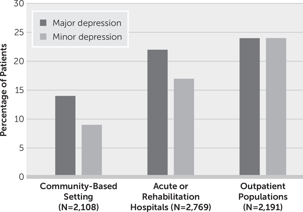
Several studies have examined the epidemiology of PSD. Robinson performed a pooled analysis of studies published prior to 2003 and included only studies that used structured interviews and diagnostic criteria to identify major and minor depression in community-based settings, acute or rehabilitation hospitals, or outpatient clinics (18) (Figure 1). The analysis demonstrated that the frequency of PSD is influenced by clinical setting and stroke severity. The frequencies for major and minor depression were 14% and 9%, respectively; in acute or rehabilitation hospitals, the frequencies were 22% and 17%; and in outpatient clinics, the frequencies were 24% and 24%.
FIGURE 1. Major and minor depression among stroke patients across different settingsa
aPatients were examined with a standardized mental status examination and were assessed with DSM-IV diagnostic criteria for depression following stroke. Minor depression was defined as more than two but fewer than five symptoms of major depression. Pooled analyses revealed that the prevalence of poststroke depression is 31%. Data from Robinson (18).
A systematic review and meta-analysis that included data from 51 studies and 25,488 stroke patients provided a pooled frequency estimate for depression of 31% (95% CI=28–35) at any time point up to 5 years after stroke (19). In this study, PSD was variously defined as depressive disorder, depressive symptoms, or psychological distress. On the basis of DSM-5 classifications, the disorders of major depression, minor depression, or dysthymia were diagnosed by using structured or semistructured psychiatric interviews (studies without categorical diagnoses were excluded). There was marked heterogeneity in the cutoff points for particular depression rating scales: for example, four different cutoff points were used with the Geriatric Depression Scale, the Beck Depression Inventory, and the Hospital Anxiety and Depression Scale, and three different cutoff points were used with the HAM-D (SR6–SR9, online supplement). Similar pooled estimates of depression were reported for people with a history of depression before stroke and for individuals with aphasia.
Another analysis by Ayerbe et al. included 20,293 patients within 15 years after a stroke (20). The study found a pooled prevalence of PSD of 29% (95% CI=25–32) at any time point; the prevalence of PSD was 22% in population studies, 30% in hospital studies and 30% in rehabilitation settings. The 5-year cumulative incidence of PSD was 42%, with a pooled prevalence of 29% that remained stable in the first 10 years after stroke (20). The authors suggested that the source of variability in estimates of PSD prevalence mostly related to different diagnostic methods, source of patient recruitment, and time of assessment since stroke.
Hornsten et al. carried out a postal inquiry for people ≥65 years old in northern Sweden and Finland (SR10, online supplement). The prevalence of stroke was 7%, and the prevalence of depression was 13%. The prevalence of PSD ranged from 11% among 65-year-old individuals to 18% among 80-year-old individuals, demonstrating that PSD increases with age. The prevalence ratio of depression in stroke was 1.77, but the estimate varied greatly depending on the definition of depression. Mitchell et al. carried out a meta-analysis on the frequency of PSD on the basis of ICD or DSM criteria and reported an estimate for major depression of 18%, minor depression of 13%, dysthymia of 3%, adjustment disorder of 7%, and any depressive disorder of 33% (21). The frequency of PSD ranged from 16% in the community to 20% in rehabilitation settings, with a point prevalence of 18%. Cassidy et al. estimated the frequency of PSD by using the CES-D, the HAM-D, and a DSM-IV checklist (SR11, online supplement). The frequency of major depression was 20% among patients undergoing inpatient rehabilitation, with a point prevalence of 3% in the general population (SR11, online supplement). Taylor-Rowan et al. carried out a systematic review and meta-analysis on the prevalence of prestroke depression and PSD (22). Prestroke depression was diagnosed by using the Structured Clinical Interview for DSM-IV, a specific cutoff score on valid depression scales, self-report, or methods of unclear validity (22). A potential recall bias on the basis of the presence of PSD was not explored. The pooled prevalence of prestroke depression was 12%, which was associated with increased odds of PSD (odds ratio=3.0, 95% CI=2.3–4.0) (22). Finally, Wu et al. examined the frequency of depression (as determined with the nine-item Patient Health Questionnaire) among 243 patients with silent lacunar infarction and found a frequency of PSD of 25% (SR12 and SR13, online supplement).
In conclusion, the epidemiology of PSD depends on diagnostic instruments and use of diagnostic criteria, and it is influenced by the presence of aphasia, age, and type of stroke. Regardless of these confounders, most epidemiological studies and meta-analyses suggest a prevalence of PSD of approximately 30%.

Variables Associated With PSD

Demographic, Clinical, and Functional Status

The poststroke variable most strongly associated with increased rate of PSD is severity of impairment in activities of daily living (ADLs). A review of all studies of PSD found a significant correlation between severity of PSD and severity of impairment in ADLs in 83% of studies (18). The mean correlation, however, was r=0.31, indicating that impairment in ADLs explained only about 10% of the variance in severity of PSD.
Both premorbid and poststroke-related variables, however, have also been associated with increased and decreased likelihood of developing PSD. The premorbid variables associated with increased rates of PSD include family history of depression and personal history of depression or anxiety disorder (23, 24).
Cerebral small vessel vascular disease, characterized by white matter hyperintensities, lacunes, enlarged perivascular spaces, and cerebral atrophy, prior to stroke increases the rate of incident stroke, dementia, PSD, and all-cause mortality. Another prestroke variable related to an increased incidence of PSD is subcortical atrophy of the lateral and third ventricles (25). In a study of 13 pairs of stroke patients matched for size and location of stroke lesion, one patient from each pair had major depression, while the other did not. The patients with depression had larger lateral and third ventricles than the patients without depression, suggesting prestroke atrophy was a contributing factor (25).
A systematic review of 24 studies of stroke patients reported that gender was not a significant risk factor for PSD in 13 out of 21 studies. However, in one-third of these studies, female sex was identified as a risk factor for PSD (24). Age was not associated with PSD in 16 of these 21 studies. These findings were replicated in a systematic review of 23 studies that included 18,374 stroke patients (26).

Lesion Location

Our initial studies of lesion location were not based on any hypothesis except the findings of Folstein et al. that stroke patients were more depressed than nonstroke patients with a similar severity of physical impairment (1). We therefore surmised that ischemic infarction of the brain might lead to depression, similar to the lateralized effect of middle cerebral infarction in rats that causes depletion of brain catecholamines in our rat model of stroke (see Robinson [27]).
Our first longitudinal study of PSD and lesion location utilized a conservative series of 184 patients admitted to the University of Maryland Hospital with acute stroke (28). Of these patients, 53 were excluded due to severe comprehension deficits, prior history of alcohol or drug abuse or psychiatric treatment, family history of psychiatric disorder, or more than one ischemic lesion on computed tomography (CT) scan. A diagnosis of depression was based on findings from the structured mental status examination (Present State Examination) and the Research Diagnostic Criteria for major or minor depression (SR14 and SR15, online supplement). Analysis of specific CT scan measurements was performed with chi-square analyses and correlation coefficients.
Among the 18 patients with unilateral stroke in the left hemisphere, 11 had major or minor depression, compared with two of 12 patients with unilateral stroke in the right hemisphere (χ2=4.4, df=30, p<0.05). CT scan measurements revealed that the correlation between the distance of the left hemisphere lesion from the frontal pole and severity of depression was r=−0.92 (p<0.001), while among patients with right hemisphere lesions, the correlation was r=0.76 (p<0.01).
These findings of increased frequency and severity of PSD among patients with left anterior stroke compared with patients with right hemisphere stroke led to a follow-up study of cortical versus subcortical stroke and PSD by using the same entry criteria, diagnostic criteria, depression severity measurements, and CT scan measurements (29). Among 16 patients with left cortical lesions, four had major depression, and three had minor depression (total of 44%), while among 13 patients with left subcortical lesions, four had major depression, and one had minor depression (total of 38%). Among nine patients with right cortical lesions, none had major depression, and one had minor depression (total of 11%), while among seven patients with right subcortical lesions, one had major depression, and none had minor depression (total of 14%). An analysis of the frequency of depression among patients with left versus right hemisphere stroke found significant differences (p<0.01). On the basis of these studies revealing that left frontal lesions were associated with higher rates of PSD, subsequent studies of stroke patients almost always examined the rates of PSD among patients with stroke in the left or right hemisphere.
The stroke factor associated with increases in the rate of PSD that has received the most study is lesion location. Our initial studies of stroke, on the basis of CT imaging, found that left frontal lesions were associated with higher rates of PSD than any other lesion location (28). Two subsequent studies found a significantly higher frequency of major or minor depression among patients with left frontal lesions or left basal ganglia lesions when compared with patients with lesions in other locations (28, 29). Both studies also found significant correlations between the distance of the anterior border of the ischemic lesion from the left frontal pole and severity of depression for both cortical and subcortical regions (28, 29). A meta-analysis conducted by our group more than 10 years after these lesion location studies found that the association of PSD with left frontal lesions and left basal ganglia lesions was a transient phenomenon restricted to the first 2 months following stroke (30). Meta-analyses of data from stroke patients with either acute or chronic manifestations and one or more stroke lesions revealed no significant association with lesion location (20, 26), while another meta-analysis that included only patients diagnosed with a structured mental status examination found a highly significant correlation between the anterior border of the lesion in the left hemisphere and severity of depression (30). The largest meta-analysis analyzed 43 studies involving 5,507 stroke patients and reported an odds ratio of 0.99 (95% CI=0.88–1.11) for the association of stroke location and depression risk (31). However, this meta-analysis included studies that failed to use a standardized mental status examination for the diagnosis of PSD, similar time points after stroke, consistent definitions of lesion location (e.g., left frontal cortical vs. left anterior), and standardized neuroimaging analyses to determine lesion location. As previously mentioned, the well-established method for diagnosing mental disorders is to use structured mental status interviews and established diagnostic criteria. Cutoff scores on rating scales are not adequate for diagnosis.
There is an association between PSD and left frontal lesions or left basal ganglia lesions within 2 months following a first clinical stroke, and there is strong scientific evidence of brain lateralization of emotion and evidence that focal brain stimulation with repetitive transcranial magnetic stimulation is only effective when it is administered to the left dorsolateral prefrontal cortex among patients with vascular depression (32, 33). However, identifying the role that lesion location plays in PSD requires the formulation of a new pathophysiological model of the disorder that integrates these disparate findings.

Genetic Factors

Several genes have been examined as risk factors for PSD. The 5-HTTLPR and the STin2 variable number tandem repeat polymorphisms of the serotonin transporter gene (SERT) have been associated with PSD among stroke survivors (34). Epigenetic modifications of 5-HTTLPR have also been implicated in the onset and severity of PSD. In a study of 286 stroke patients, increased methylation status was independently associated with PSD at 2 weeks and at 1 year following stroke, a finding that was only observed in the presence of the 5-HTTLPR s/s genotype (35). In addition, among the same group of stroke patients, higher brain-derived neurotrophic factor (BDNF) gene methylation status was associated with incident PSD and more severe symptoms at the 12-month follow-up (36).

PSD and Social Support

The variable associated with the greatest decrease in the incidence of PSD is social support. In a study of poststroke patients divided into those with good social support (N=55) and those without social support (N=88), on the basis of their the Social Functioning Examination score (see Robinson et al. [37]), the investigators found that patients with high social support during the 3–6 months after hospital admission had a lower incidence of depression and significantly greater improvement in ADLs than patients with low social support (group-by-time interaction: F=11.0, df=1 and 56, p=0.002) (37) (SR16, online supplement).
Another study of 96 Australian patients with stroke found that only 30% of patients with high spousal support had PSD, while 70% of those with low spousal support had depression (SR17, online supplement). A recent prospective study also found that lack of social support at admission was associated with an increased rate of PSD at the 3-month follow-up (38).

PSD, Cognitive Function, and Cognitive Impairment

Although it has long been recognized that stroke may affect cognitive function, the severity of poststroke cognitive impairment can range from mild with recovery to moderate with partial recovery to poststroke dementia with progressive deterioration (39).
The study of poststroke dementia has led to intriguing recent investigations of the mechanism by which stroke often leads to dementia and progressive deterioration. A recent comprehensive review of poststroke dementia found that the prevalence of dementia 1 year after stroke ranged from 7% to 41% (40, 41). Another study found that the factors most strongly associated with poststroke dementia were poststroke major depression, left hemisphere stroke, and history of prior stroke (SR18 and SR19, online supplement). Another recent study found that among 245 stroke patients, progressive cognitive impairment was significantly associated with a chronic course of PSD, executive dysfunction, white matter hyperintensities, prior stroke, and left hemisphere lesions (41).
There are no known biomarkers that discriminate individuals at risk for Alzheimer’s disease and those at risk for poststroke dementia. Stroke, however, has been shown to produce brain inflammation, which is associated with progressive cognitive decline similar to that found in Alzheimer’s disease. Thus, stroke may be related to an inflammatory mechanism that leads to progressive dementia in both Alzheimer’s disease and poststroke dementia (40) (SR19, online supplement). In addition, another study of stroke laterality found that among 100 patients, 43% with left hemisphere stroke had progressive dementia at 6 months following stroke, and 53% had progressive dementia at 2 years following stroke (42). This suggests that disrupted networks in the left hemisphere, but not the right hemisphere, may contribute to progressive dementia.
The relationship between PSD and cognitive function (especially executive dysfunction) has been well established (18). Initial studies found significantly lower Mini-Mental State Examination scores, indicating greater impairment, among stroke patients with major depression following left hemisphere stroke than among nondepressed patients or patients with minor depression and right hemisphere stroke (SR19, online supplement). A previously mentioned study of 13 pairs of stroke patients matched for size and location of stroke lesion—one of each pair had major depression, while the other did not—found that stroke patients with major depression had significantly greater cognitive impairment than patients without major depression (25).

Interactions Between Premorbid Neuropsychiatric Symptoms and Long-Term Stroke Outcome

Another intriguing new area of neuropsychiatric research related to stroke examines interactions between neuropsychiatric disorders prior to stroke and long-term stroke outcome. A systematic review and meta-analysis was conducted on data from 47,625 poststroke patients who were over age 65 and had been followed for a median of 9 years (43). In this study, the investigators found a 21% higher rate of myocardial infarction (hazard ratio=1.21, 95% CI=1.18–1.59) among patients with apathy symptoms, defined as patients having the three apathy-related subitems on the Geriatric Depression Scale (GDS) at initial evaluation, than among patients without apathy symptoms. In addition, patients with apathy symptoms had a 36% higher rate of stroke (hazard ratio=1.36, 95% CI=1.18–1.56) and a 44% higher rate of all-cause mortality (hazard ratio=1.44, 95% CI=1.35–1.53) than patients without apathy symptoms. Patients with depressive symptoms, defined as having three symptoms of depression on the GDS at initial evaluation, had a 37% higher risk of stroke (hazard ratio=1.36, 95% CI=1.18–1.56) and a 47% higher risk of all-cause mortality (hazard ratio=1.44, 95% CI=1.35–1.53), but there was no differential risk of myocardial infarction. This study demonstrated that neuropsychiatric symptoms, such as depression and apathy, are not only a consequence of stroke, but their presence prior to stroke increases the risk of stroke and mortality. The mechanism for this phenomenon is another area deserving further research (43).

Effect of PSD on Stroke Recovery and Mortality

Numerous studies have examined the relationship between depression at initial examination and functional and motor outcomes (44) (SR20 and SR21, online supplement). Five of six studies that examined the relationship between depression at baseline and ADLs at 1 or more years of follow-up found that depression severity was an independent predictor of severity of ADL impairment.
Consistent with these findings, patients with PSD who responded to treatment with nortriptyline or fluoxetine showed significantly better improvement in ADLs than patients with PSD given placebo (45). Furthermore, response to treatment of PSD with nortriptyline or fluoxetine was associated with greater cognitive recovery over 2 years compared with changes associated with placebo administration (SR22, online supplement).
PSD also leads to increased mortality. The first study to report this phenomenon was published in 1993 (46). In this study, 103 patients were followed up at 10 years after their index stroke to determine mortality rates. Patients who developed PSD immediately after stroke had higher mortality rates than equally impaired stroke patients with no in-hospital depression (odds ratio=3.4, 95% CI=1.4–8.4, p=0.007) (46). A similar finding was reported by House et al. who showed that even mild severity of PSD was associated with increased mortality as early as 1 year after stroke (SR23, online supplement). Furthermore, in a cohort of 51,119 veterans with ischemic stroke, patients who developed PSD had a higher 3-year mortality risk than those without a mental health diagnosis (47).
A recent study of 1,354 patients that used the Hospital Anxiety and Depression Scale (HADS; higher scores indicate greater impairment) to assess PSD found increased mortality at the 5-year follow-up among patients with HADS scores ≥7 at 3 months after stroke compared with patients with scores <7 at 3 months (hazard ratio=1.41, 95% CI=1.13–1.77, p=0.02) (48) (SR24, online supplement). The investigators also reported increased mortality among patients who were started on selective serotonin reuptake inhibitors (SSRIs) in the 3 months after stroke. However, this was not a randomized trial, and the data analysis on which this assertion was based did not control for all the relevant confounders, such as level of disability, depression severity, and the effects of comorbid medical conditions (48).
The association of PSD with mortality appears to be the result of an increase in cardiovascular mortality (48). We reported that decreased heart rate variability, which has been demonstrated to play an etiological role in mortality associated with depression and myocardial infarction, was also associated with PSD (49, 50). Thus, disruption of autonomic system function among patients with PSD may contribute to cardiovascular mortality.
Antidepressant use may influence long-term survival after stroke (51). A 9-year follow-up study of patients with or without PSD who had been treated with nortriptyline (100 mg/day) or fluoxetine (40 mg/day) early after an index stroke showed that patients receiving active treatment (N=53) had an increased probability of survival at the 9-year follow-up compared with similar patients given placebo (N=28) (i.e., a 59% 9-year survival rate among patients treated with nortriptyline or fluoxetine versus a 35% rate among patients given a placebo; adjusted odds ratio=3.7, 95% CI=1.1–12.2, p=0.03) (51). A beneficial effect of antidepressants on long-term survival after stroke was also observed in a group of 790 veterans with stroke who were followed over a 7-year period (SR25, online supplement).

Effect of Antidepressants on Stroke Recovery

Many basic and animal studies have shown that SSRIs have consistent effects on neural plasticity, including neurogenesis, synaptic remodeling, and network reorganization (52) (SR26 and SR27, online supplement). These effects are mediated, at least in part, by the activation of neurotrophic-signaling pathways (e.g., BDNF, tropomyosin receptor kinase B receptors, and translation factors such as cAMP response element-binding protein), as well as reductions in oxidative stress and neuroinflammation (53). Furthermore, SSRIs and other small molecules were shown to induce, in adults, a form of plasticity that is typically observed during developmental critical periods (54).
A recent systematic review and meta-analysis of data from studies examining the effects of antidepressants in animal models of stroke (44 publications and 22 medications) found that antidepressants reduced infarct volume by 27% (95% CI=21–34) and enhanced motor and behavioral outcomes by 53% (95% CI=46–61). SSRIs, the most frequently studied antidepressants, improved neurobehavioral outcomes by 52% (95% CI=39–65) (55) (SR28 and SR29, online supplement).
In humans, antidepressants have been reported to improve motor, cognitive, and functional outcomes. For example, the Fluoxetine for Motor Recovery After Acute Ischemic Stroke trial examined the efficacy of fluoxetine to enhance motor recovery following ischemic stroke (56). In this double-blind, placebo-controlled trial, 118 stroke patients undergoing physical rehabilitation were randomly assigned to receive fluoxetine (20 mg/day) or placebo for 3 months starting 5–10 days after the onset of stroke. The primary outcome measure was the change in Fugl-Meyer Motor Scale scores (FMMS; lower scores indicate more severe impairment) following the intervention. The improvement in FMMS score at day 90 was significantly greater in the fluoxetine group than in the placebo group. Furthermore, the number of patients that achieved independence (defined as a score of 0–2 on the modified Rankin Scale [mRS]) was higher in the fluoxetine group (56).
Our group examined the efficacy of escitalopram to improve cognitive outcomes following stroke. The 12-month trial included 129 stroke patients without depression who were randomly assigned to three arms: a double-blind, placebo-controlled comparison of escitalopram (N=43) with placebo (N=45) and a nonblinded arm engaged in problem-solving therapy, a form of psychotherapy (N=41). The primary outcome measure was change in score on the Repeatable Battery for the Assessment of Neuropsychological Status (RBANS; with lower scores indicating greater impairment) (57).
When compared with patients who received placebo or underwent problem-solving therapy, stroke patients who received escitalopram improved in global cognitive functioning, immediate memory, and delayed memory (57). These beneficial effects of escitalopram were independent of escitalopram’s effect on depression. Furthermore, there was a positive, but not statistically significant, correlation between change in RBANS total score and change in Functional Independence Measure total score (Spearman’s correlation=0.25, p=0.20). Thus, the reported changes in neuropsychological test scores were associated with a measurable, albeit not statistically significant, improvement in ADLs.
A recent meta-analysis of 52 trials examined the efficacy of SSRIs to reduce disability among 4,060 participants with a recent stroke. The authors concluded that SSRIs may improve recovery after stroke. However, larger rigorous trials are needed to replicate these findings, as well as to determine the safety of SSRIs in the context of stroke (58).
The cumulative evidence suggesting a beneficial effect of fluoxetine on stroke recovery led to three large multicenter randomized controlled trials (RCTs) in the United Kingdom (Fluoxetine or Control Under Supervision [FOCUS]), Sweden (Efficacy of Fluoxetine–a Randomised Controlled Trial in Stroke [EFFECTS]), and Australasia (Assessment of Fluoxetine in Stroke Recovery [AFFINITY]) (SR30, online supplement). These trials share essential methodological aspects and provide individualized and combined results on the efficacy of fluoxetine to reduce dependency and disability among patients with recent stroke. The targeted enrollment across the three studies was 6,000 participants. The actual enrollment amounted to 4,344 participants.
In the FOCUS trial, 1,564 patients with a recent stroke and focal neurological deficits were allocated to the fluoxetine (20 mg/day) group, and 1,563 patients were allocated to a matching placebo group (59). The primary outcome was functional status, measured with the mRS, at 6 months. Fluoxetine was not better than placebo in reducing disability following stroke, although it effectively prevented depressive disorders (59).
The AFFINITY trial was conducted in hospitals in Australia, New Zealand, and Vietnam. A total of 1,280 patients with recent stroke (ischemic or hemorrhagic) were randomly assigned to fluoxetine (20 mg/day) or placebo groups for 6 months. There were no significant differences in disability outcomes measured with the mRS. On the other hand, the patients receiving fluoxetine had a higher risk of falls, bone fractures, and epileptic seizures than patients receiving placebo (60) (SR31, online supplement).
The EFFECTS trial was conducted in 35 stroke and rehabilitation centers in Sweden. A total of 1,500 patients were allocated to receive fluoxetine (20 mg/day) or placebo and followed for 6 months. Although fluoxetine was efficacious in preventing depression, there were no significant effects on disability outcomes. In addition, patients receiving fluoxetine had a higher frequency of bone fractures and hyponatremia (61).
The findings of these trials provide consistent evidence that fluoxetine is not better than placebo in improving disability outcomes measured with the mRS. However, we need to consider certain limitations that temper the clinical implications of these findings. First, the three trials enrolled patients with milder forms of stroke (the median National Institutes of Health Stroke Scale scores were 3, 6, and 6 for the EFFECTS, AFFINITY, and FOCUS trials, respectively), making them more vulnerable to ceiling effects. Second, although the mRS is an established measure of stroke-related disability, the scale is not sensitive to the occurrence of more subtle changes (e.g., cognitive, motivational, and autonomic changes) that may have an effect on long-term outcomes. Finally, it is important to note that these pragmatic trials did not require participants to be involved in rehabilitation therapy, nor was the intensity of rehabilitation procedures recorded. This is a drawback because the mechanisms of action of SSRIs in stroke recovery involve activity-dependent plasticity, and thus, the network reorganization associated with fluoxetine depends on the concurrent performance of rehabilitation practices.

Etiological Mechanisms of PSD

There is consensus that psychological, social, and biological factors contribute to the mechanisms underlying PSD (SR32, online supplement). The occurrence of PSD has been consistently associated with stroke severity and the degree of functional physical and cognitive impairment (26).
Biological factors contributing to PSD include alterations in ascending monoaminergic systems, hypothalamic-pituitary-adrenal (HPA) axis abnormalities, disruption of prefrontal-subcortical circuits, alterations in neuroplasticity and glutamate neurotransmission, and excessive levels of proinflammatory cytokines (6264) (SR33–SR35, online supplement). However, a pathophysiological hypothesis of PSD that can integrate these changes into a coherent explanatory model has yet to be formulated.
From a translational approach, there have been several attempts to model PSD in experiments with rodents. We conducted the earliest studies using a rat model of middle cerebral artery (MCA) ligation. Right MCA ligation produced significant bilateral depletion of norepinephrine and dopamine in both the cortex and brainstem, as well as hyperactivity and other behavioral changes not seen in rats given sham ligations (65). Subsequent experiments using this animal model demonstrated that the effects on biogenic amines and behavior were lateralized—they did not occur after left MCA ligation—and lasted up to 4 weeks (SR36, online supplement).
In recent years, there has been renewed interest in modeling the neuropsychiatric consequences of stroke. Kronenberg et al. used a mouse model of mild stroke (i.e., a 30-minute MCA occlusion that limits the ischemic damage to the basal ganglia) (66). A subgroup of these mice was given citalopram, starting 1 week after MCA occlusion. Left, but not right, MCA occlusion produced delayed degeneration of dopaminergic neurons in the left ventral tegmental area, which resulted in reduced dopamine concentrations in the striatum. The behavioral correlates of these neurodegenerative and neurochemical changes were anhedonia and behavioral despair assessed with the sucrose consumption test and forced swim test, respectively. Importantly, chronic citalopram treatment initiated 7 days after stroke prevented the degeneration of dopaminergic neurons and reversed the behavioral phenotype (66).
There have also been animal studies that combined experimental ischemic lesions with social isolation or chronic mild stress protocols implemented during the stroke recovery process (67) (SR37, online supplement). Permanent left MCA occlusion coupled with a 14-day chronic mild stress protocol produced a depressive phenotype characterized by decreased exploratory behavior and sucrose consumption These behavioral effects were reversed by the administration of citalopram and a serotonin-1A antagonist, and there was evidence of increased neurogenesis in the hippocampus (68).
In a rodent model of PSD, electrical stimulation of the cerebellar fastigial nucleus decreased depression-like symptoms and increased regional cerebral blood flow (SR38, online supplement). Fastigial nucleus stimulation was associated with a reduction in the expression of inflammatory cytokines (SR39, online supplement).
Unfortunately, there have been relatively few studies on the mechanisms underlying PSD in clinical populations. However, one study of PSD conducted by our group showed decreased cerebral spinal fluid levels of serotonin or norepinephrine metabolites that were significantly associated with severity of PSD (69).
There is a growing consensus that ischemic lesions (single or multiple) of the neural circuits that connect the prefrontal cortex, basal ganglia, thalamus, and amygdala (independent of their lateralization) may disrupt mood regulation and executive function, leading to similar clinical presentations. Furthermore, there appears to be a threshold by which the confluence of multiple etiological factors or further damage to specific white matter tracts, such as the cingulate bundle, the uncinate fasciculus, and the superior longitudinal fasciculus, triggers the onset of clinical depression. Thus, acute ischemia can reveal the presence of vascular depression (SR33, online supplement).
From a network standpoint, resting connectivity patterns revealed by functional MRI appear to be similar in vascular depression and PSD, with increased activation of the default mode network and limbic structures and decreased activation of task-related networks and the dorsolateral aspects of the prefrontal cortex (70).
Earlier studies also examined dysregulation of the HPA axis in PSD, particularly the association of PSD with elevated cortisol levels and abnormal negative feedback control of cortisol secretion (63). More recently, several studies emphasized the role of proinflammatory cytokines in the development of PSD (62) (SR35 and SR40–SR44, online supplement). Increased serum concentrations of interleukin-6 were associated with increased severity of somatic symptoms of PSD, and increased levels of inflammatory cytokines reduce the synthesis and availability of serotonin through their enhancing effect on the activity of the enzyme indolamine 2,3-dioxygenase. A high neutrophil-to-lymphocyte ratio on admission, measured systemically, predicted PSD after 1 month and 6 months (SR45 and SR46, online supplement).
In summary, although there are numerous possible physiological mechanisms related to PSD, many investigators have concluded that this complex disorder, like most major psychiatric disorders not associated with stroke, may best be described as a biopsychosocial disorder. However, this general theoretical framework does little to help us elucidate pathophysiological mechanisms leading to specific symptoms. The different etiological factors described above may have more salient roles in some forms or symptoms of PSD, and their effects may vary at different times after stroke. We believe future studies should attempt to identify the mechanisms underlying specific symptoms or clinical characteristics of PSD rather than the whole syndrome.

Treatment of PSD

Selected RCTs of PSD and vascular depression are shown in Table 1 (33, 7183). The first randomized double-blind treatment trial was reported in 1984 by Lipsey et al. (71). Patients randomly assigned to the nortriptyline group (50–100 mg/day) had a significantly greater reduction in HAM-D scores (indicating lower severity) over 6 weeks of treatment compared with patients in the placebo group. The first double-blind controlled trial to examine the efficacy of SSRIs was reported by Andersen et al. in 1994 (73). Among 33 poststroke patients given citalopram (10–20 mg/day), there was a significantly greater reduction in HAM-D scores over 6 weeks among 33 poststroke patients given citalopram (10–20 mg/day) than among 33 patients with similar levels of depression given placebo.
TABLE 1. Selected randomized controlled trials of poststroke depression and vascular depressiona
StudyNMedication, maximum daily dose, and sample sizeDuration (weeks)Evaluation methodResults (response rates)b
Lipsey et al. (71)34Nortriptyline, 100 mg, N=14; placebo, N=206HAM-D,
ZSDS
Nortriptyline (79%) more effective than placebo (25%)
Reding et al. (72)16Trazodone, 200 mg, N=7; placebo, N=932±6 days, mean±SDZSDSTrazodone more effective than placebo
Andersen et al. (73)66Citalopram, 20 mg or 10 mg (participants ≥65 years), N=33; placebo, N=336HAM-D,
MES
Citalopram (49%) more effective than placebo (29%)
Grade et al. (74)21Methylphenidate, 30 mg, N=10; placebo, N=113HAM-D,
ZSDS
Methylphenidate more effective than placebo
Robinson et al. (75)56Fluoxetine, 40 mg, N=23; nortriptyline, 100 mg, N=16; placebo, N=1712HAM-DNortriptyline (77%) more effective than placebo (31%); fluoxetine (14%) not better than placebo (31%)
Wiart et al. (76)31Fluoxetine, 20 mg, N=16; placebo, N=156MADRSFluoxetine (63%) more effective than placebo (33%)
Fruehwald et al. (77)54Fluoxetine, 20 mg, N=28; placebo, N=2612BDI,
HAM-D
Fluoxetine (69%) not better than placebo (75%)
Rampello et al. (78)31Reboxetine, 4 mg, N=16; placebo, N=1516BDI,
HAM-D
Reboxetine more effective than placebo for psychomotor retardation
Choi-Kwon et al. (79)51Fluoxetine, 20 mg, N=19; placebo, N=3212BDIFluoxetine not better than placebo
Rampello et al. (80)74Citalopram, 20 mg, N=37; reboxetine, 4 mg, N=3717BDI,
HAM-D
Anxious depression reduced with citalopram; depression with psychomotor retardation reduced with reboxetine
Zanardi et al. (81)93Sertraline and tDCS daily, N=31; sertraline and tDCS twice daily, N=31; sertraline and sham tDCS, N=312HAM-D,
MODA
Augmentation with tDCS daily (42%) and twice daily (68%) reduced both depressive symptoms and cognitive impairment compared with sham (no response); remission rates were 29%, 58%, and 0%, respectively
Zhu et al. (82)100tDCS, N=50; tDCS and citalopram, N=508HAM-D,
MMSE
Significant decrease in HAM-D score in TMS and citalopram group compared with TMS alone group
Jorge et al. (33)92Experiment 1: rTMS (12,000 pulses), N=15; sham, N=15; Experiment 2: rTMS (18,000 pulses), N=33; sham, N=293HAM-DExperiment 1: rTMS: 33% decrease in HAM-D score, 33% response rate, 13% remission rate. Sham: 14% decrease in HAM-D score, 7% response rate, 7% remission rate. Experiment 2: rTMS: 42% decrease in HAM-D score, 39% response rate, 27% remission rate. Sham: 18% decrease in HAM-D score, 7% response rate, 4% remission rate
Gu and Chang (83)24rTMS, N=12; sham, N=124 (plus 1 day)BDI,
HAM-D
BDI and HAM-D scores significantly decreased in rTMS group compared with sham group
a
BDI=Beck Depression Inventory; HAM-D=Hamilton Depression Rating Scale; MADRS=Montgomery-Åsberg Depression Rating Scale; MES=Bech-Rafaelsen Melancholia Scale; MMSE=Mini-Mental State Examination; MODA=Milan Overall Dementia Assessment; rTMS=repetitive transcranial magnetic stimulation; tDCS=transcranial direct current stimulation; ZSDS=Zung Self-Rating Depression Scale.
b
Lower scores on HAM-D and BDI indicate lower severity.
A recent meta-analysis of 16 RCTs (with 12 trials using antidepressants and four evaluating the efficacy of psychotherapy) that included 1,655 patients found a significant beneficial effect of antidepressant medication, whereas psychotherapy was not more effective than a control intervention (84). Brief psychosocial therapies, however, that place an emphasis on care management, psychoeducation, and family support can be beneficial when administered in combination with antidepressants to treat or prevent PSD (SR47, online supplement).
Recent small RCTs of escitalopram and citalopram also showed beneficial effects on depressive symptoms and endothelial function (85) (SR48, online supplement). In addition, a clinical trial examining the efficacy of intravenous alteplase on PSD showed a significant reduction in the severity of depressive symptoms 3 months after an index stroke (SR49, online supplement). Similarly, pioglitazone (a peroxisome proliferator-activated receptor-gamma agonist) decreased the severity of PSD symptoms among stroke patients with diabetes mellitus type 2 (SR50, online supplement).
It should be acknowledged, however, that treatment with antidepressants is not without risk. For example, SSRI use has been associated with an increased risk of hemorrhagic complications and an increased risk of falls among the elderly population (86). Furthermore, other epidemiological studies have reported that SSRIs are associated with increased risk of stroke, myocardial infarction, and all-cause mortality (87) (SR51, online supplement). The effects of these antidepressants may be due to interactions with other variables, such as depression, disability, and comorbid medical conditions, that require further elucidation.
There have been recent small studies of nonpharmacological interventions to treat PSD. Nguyen et al. used cognitive-behavioral therapy and reported a significant decrease in depressive symptoms, along with improvements in fatigue and sleep quality (88). Environmental enrichment and gamified training with virtual reality and other advanced technological tools demonstrated beneficial effects for patients with PSD (89) (SR52, online supplement). Behavioral activation therapy, which aims to prolong the frequency of pleasant or enjoyable events, also showed some promising results (SR53, online supplement). Preliminary data from group-based acceptance and commitment therapy and self-help relaxation seem to confer certain positive benefits in terms of PSD resolution (90) (SR54, online supplement). A social support and health education program using a two-pronged approach of providing rehabilitation-related information support and emotional support appears to help when provided over a minimum of 8 weeks of therapy (SR55, online supplement). Reminiscence therapy—aimed at recalling important life events to improve pleasure and belonging—also appears to help in reducing the PSD burden (SR56, online supplement). We have previously shown that repetitive transcranial magnetic stimulation of the left dorsolateral prefrontal cortex may be efficacious in treating PSD (91). These findings were replicated in a recent independent study (83). In addition, transcranial direct current stimulation of the dorsolateral prefrontal cortex was shown to be safe and efficacious in reducing the severity of PSD (92). Studies of transcranial alternate current stimulation are also under way (SR57, online supplement). Finally, both aerobic exercise and aquatic therapy have been employed to decrease the burden of PSD (93) (SR58 and SR59, online supplement).
Prevention of PSD

Based on prevalence rates of approximately 30% for PSD during the first year following acute stroke, it is logical to target PSD with preventive treatment. Rasmussen et al. reported the first double-blind trial of sertraline to prevent PSD over the first year following acute stroke (94). The rate of depression in the sertraline group was 8%, while the rate in the placebo group was 23% (although the difference was not statistically significant). Our group published the first RCT in which 58 acute stroke patients without depression were treated with escitalopram (5 mg/day for those ≥65 years old, 10 mg/day for those <65 years old) over 1 year and had an incidence rate of PSD of 9%. In comparison, a rate of 12% was observed among 59 patients receiving problem-solving therapy, and a rate of 22% was observed among 59 patients receiving placebo (95). Controlling for age, gender, severity of stroke, and severity of impairment, the risk of onset of depression among patients receiving placebo was more than four times greater than that for patients treated with escitalopram (adjusted hazard ratio=4.5, 95% CI=2.4–8.2, p<0.001) (95).
A meta-analysis of prevention trials summarized the findings of eight RCTs assessing the efficacy of preventive interventions among 776 initially nondepressed stroke patients (96). Pooled analyses revealed that the likelihood of developing PSD was significantly reduced among patients receiving active pharmacological treatment, among those receiving treatment for 1 year, and among those receiving an SSRI (96).

Future Directions

Since the 1970s, substantial progress has been made in establishing methods for diagnosis of PSD, as well as in identifying stroke-related correlates of PSD; risk factors for PSD; the effects of PSD on physical recovery, cognitive recovery, and mortality; and strategies for the treatment and prevention of PSD. Thus, it seems to us that identifying additional aspects of the mechanisms underlying PSD is the most urgent need for future research because it may lead to more specific therapeutic interventions. For example, findings on the association of interleukin-6 with somatic symptoms of depression and the association of low levels of serum BDNF within hours after stroke with the development of depression after 3 months are intriguing findings that may lead to the identification of how specific symptoms of PSD may be mediated and may also suggest that neurotrophic and anti-inflammatory agents may be effective in treating or preventing not only PSD but also other neuropsychiatric disorders resulting from stroke (97) (SR60–SR65, online supplement). Research is urgently needed to determine the mechanisms by which PSD leads to the 50% increase in death rate that has been observed 7 years following stroke. Elucidating the effects of interleukin-6 and heart rate variability on poststroke mortality could substantially increase survival rate following stroke. Finally, identifying the mechanisms by which antidepressants enhance physical and cognitive recovery after stroke, for example, through neurogenesis induced by SSRIs, could lead to improved poststroke recovery.

Supplementary Material

File (appi.neuropsych.21090231.ds001.pdf)

References

1.
Folstein MF, Maiberger R, McHugh PR: Mood disorder as a specific complication of stroke. J Neurol Neurosurg Psychiatry 1977; 40:1018–1020
2.
Diagnostic and Statistical Manual of Mental Disorders, 5th ed. Washington, DC, American Psychiatric Association, 2013
3.
Cohen-Cole SA, Stoudemire A: Major depression and physical illness: special considerations in diagnosis and biologic treatment. Psychiatr Clin North Am 1987; 10:1–17
4.
de Coster L, Leentjens AFG, Lodder J, et al: The sensitivity of somatic symptoms in post-stroke depression: a discriminant analytic approach. Int J Geriatr Psychiatry 2005; 20:358–362
5.
Lipsey JR, Spencer WC, Rabins PV, et al: Phenomenological comparison of poststroke depression and functional depression. Am J Psychiatry 1986; 143:527–529
6.
Paradiso S, Robinson RG: Minor depression after stroke: an initial validation of the DSM-IV construct. Am J Geriatr Psychiatry 1999; 7:244–251
7.
Fedoroff JP, Lipsey JR, Starkstein SE, et al: Phenomenological comparisons of major depression following stroke, myocardial infarction or spinal cord lesions. J Affect Disord 1991; 22:83–89
8.
Cumming TB, Churilov L, Skoog I, et al: Little evidence for different phenomenology in poststroke depression. Acta Psychiatr Scand 2010; 121:424–430
9.
Gainotti G, Azzoni A, Razzano C, et al: The Post-Stroke Depression Rating Scale: a test specifically devised to investigate affective disorders of stroke patients. J Clin Exp Neuropsychol 1997; 19:340–356
10.
Quaranta D, Marra C, Gainotti G: Mood disorders after stroke: diagnostic validation of the Poststroke Depression Rating Scale. Cerebrovasc Dis 2008; 26:237–243
11.
Robinson RG, Jorge RE: Post-stroke depression: a review. Am J Psychiatry 2016; 173:221–231
12.
Lyness JM, Heo M, Datto CJ, et al: Outcomes of minor and subsyndromal depression among elderly patients in primary care settings. Ann Intern Med 2006; 144:496–504
13.
Amore M, Tagariello P, Laterza C, et al: Beyond nosography of depression in elderly. Arch Gerontol Geriatr 2007; 44:13–22
14.
Vermeer SE, Longstreth WT, Jr., Koudstaal PJ: Silent brain infarcts: a systematic review. Lancet Neurol 2007; 6:611–619
15.
Alexopoulos GS, Borson S, Cuthbert BN, et al: Assessment of late life depression. Biol Psychiatry 2002; 52:164–174
16.
Blazer D, Williams CD: Epidemiology of dysphoria and depression in an elderly population. Am J Psychiatry 1980; 137:439–444
17.
Ashaie SA, Hurwitz R, Cherney LR: Depression and subthreshold depression in stroke-related aphasia. Arch Phys Med Rehabil 2019; 100:1294–1299
18.
Robinson RG: The Clinical Neuropsychiatry of Stroke, 2nd ed. Cambridge, Cambridge University Press, 2006
19.
Hackett ML, Pickles K: Part I: Frequency of depression after stroke: an updated systematic review and meta-analysis of observational studies. Int J Stroke 2014; 9:1017–1025
20.
Ayerbe L, Ayis S, Crichton S, et al: The natural history of depression up to 15 years after stroke: the South London Stroke Register. Stroke 2013; 44:1105–1110
21.
Mitchell AJ, Sheth B, Gill J, et al: Prevalence and predictors of post-stroke mood disorders: a meta-analysis and meta-regression of depression, anxiety and adjustment disorder. Gen Hosp Psychiatry 2017; 47:48–60
22.
Taylor-Rowan M, Momoh O, Ayerbe L, et al: Prevalence of pre-stroke depression and its association with post-stroke depression: a systematic review and meta-analysis. Psychol Med 2019; 49:685–696
23.
Morris PL, Robinson RG, Raphael B: Prevalence and course of depressive disorders in hospitalized stroke patients. Int J Psychiatry Med 1990; 20:349–364
24.
De Ryck A, Brouns R, Geurden M, et al: Risk factors for poststroke depression: identification of inconsistencies based on a systematic review. J Geriatr Psychiatry Neurol 2014; 27:147–158
25.
Starkstein SE, Robinson RG, Price TR: Comparison of patients with and without poststroke major depression matched for size and location of lesion. Arch Gen Psychiatry 1988; 45:247–252
26.
Kutlubaev MA, Hackett ML: Part II: Predictors of depression after stroke and impact of depression on stroke outcome: an updated systematic review of observational studies. Int J Stroke 2014; 9:1026–1036
27.
Robinson RG: Differential behavioral and biochemical effects of right and left hemispheric cerebral infarction in the rat. Science 1979; 205:707–710
28.
Robinson RG, Kubos KL, Starr LB, et al: Mood disorders in stroke patients: importance of location of lesion. Brain 1984; 107:81–93
29.
Starkstein SE, Robinson RG, Price TR: Comparison of cortical and subcortical lesions in the production of poststroke mood disorders. Brain 1987; 110:1045–1059
30.
Narushima K, Kosier JT, Robinson RG: A reappraisal of poststroke depression, intra- and inter-hemispheric lesion location using meta-analysis. J Neuropsychiatry Clin Neurosci 2003; 15:422–430
31.
Wei N, Yong W, Li X, et al: Post-stroke depression and lesion location: a systematic review. J Neurol 2015; 262:81–90
32.
Davidson RJ, Irwin W: The functional neuroanatomy of emotion and affective style. Trends Cogn Sci 1999; 3:11–21
33.
Jorge RE, Moser DJ, Acion L, et al: Treatment of vascular depression using repetitive transcranial magnetic stimulation. Arch Gen Psychiatry 2008; 65:268–276
34.
Kohen R, Cain KC, Mitchell PH, et al: Association of serotonin transporter gene polymorphisms with poststroke depression. Arch Gen Psychiatry 2008; 65:1296–1302
35.
Kim JM, Stewart R, Kang HJ, et al: A longitudinal study of SLC6A4 DNA promoter methylation and poststroke depression. J Psychiatr Res 2013; 47:1222–1227
36.
Kim JM, Stewart R, Kang HJ, et al: A longitudinal study of BDNF promoter methylation and genotype with poststroke depression. J Affect Disord 2013; 149:93–99
37.
Robinson RG, Bolduc PL, Kubos KL, et al: Social functioning assessment in stroke patients. Arch Phys Med Rehabil 1985; 66:496–500
38.
Herrmann N, Black SE, Lawrence J, et al: The Sunnybrook Stroke Study: a prospective study of depressive symptoms and functional outcome. Stroke 1998; 29:618–624
39.
Tang EY, Amiesimaka O, Harrison SL, et al: Longitudinal effect of stroke on cognition: a systematic review. J Am Heart Assoc 2018; 7:e006443
40.
Pendlebury ST, Rothwell PM: Prevalence, incidence, and factors associated with pre-stroke and post-stroke dementia: a systematic review and meta-analysis. Lancet Neurol 2009; 8:1006–1018
41.
Douven E, Aalten P, Staals J, et al: Co-occurrence of depressive symptoms and executive dysfunction after stroke: associations with brain pathology and prognosis. J Neurol Neurosurg Psychiatry 2018; 89:859–865
42.
Baccaro A, Wang YP, Brunoni AR, et al: Does stroke laterality predict major depression and cognitive impairment after stroke? Two-year prospective evaluation in the EMMA study. Prog Neuropsychopharmacol Biol Psychiatry 2019; 94:109639
43.
Eurelings LS, van Dalen JW, Ter Riet G, et al: Apathy and depressive symptoms in older people and incident myocardial infarction, stroke, and mortality: a systematic review and meta-analysis of individual participant data. Clin Epidemiol 2018; 10:363–379
44.
Parikh RM, Robinson RG, Lipsey JR, et al: The impact of poststroke depression on recovery in activities of daily living over a 2-year follow-up. Arch Neurol 1990; 47:785–789
45.
Chemerinski E, Robinson RG, Arndt S, et al: The effect of remission of poststroke depression on activities of daily living in a double-blind randomized treatment study. J Nerv Ment Dis 2001; 189:421–425
46.
Morris PL, Robinson RG, Andrzejewski P, et al: Association of depression with 10-year poststroke mortality. Am J Psychiatry 1993; 150:124–129
47.
Williams LS, Ghose SS, Swindle RW: Depression and other mental health diagnoses increase mortality risk after ischemic stroke. Am J Psychiatry 2004; 161:1090–1095
48.
Ayerbe L, Ayis S, Crichton SL, et al: Explanatory factors for the increased mortality of stroke patients with depression. Neurology 2014; 83:2007–2012
49.
Carney RM, Blumenthal JA, Freedland KE, et al: Low heart rate variability and the effect of depression on post-myocardial infarction mortality. Arch Intern Med 2005; 165:1486–1491
50.
Robinson RG, Spalletta G, Jorge RE, et al: Decreased heart rate variability is associated with poststroke depression. Am J Geriatr Psychiatry 2008; 16:867–873
51.
Jorge RE, Robinson RG, Arndt S, et al: Mortality and poststroke depression: a placebo-controlled trial of antidepressants. Am J Psychiatry 2003; 160:1823–1829
52.
Santarelli L, Saxe M, Gross C, et al: Requirement of hippocampal neurogenesis for the behavioral effects of antidepressants. Science 2003; 301:805–809
53.
Autry AE, Monteggia LM: Brain-derived neurotrophic factor and neuropsychiatric disorders. Pharmacol Rev 2012; 64:238–258
54.
Castrén E, Antila H: Neuronal plasticity and neurotrophic factors in drug responses. Mol Psychiatry 2017; 22:1085–1095
55.
McCann SK, Irvine C, Mead GE, et al: Efficacy of antidepressants in animal models of ischemic stroke: a systematic review and meta-analysis. Stroke 2014; 45:3055–3063
56.
Chollet F, Tardy J, Albucher JF, et al: Fluoxetine for Motor Recovery After Acute Ischaemic Stroke (FLAME): a randomised placebo-controlled trial. Lancet Neurol 2011; 10:123–130
57.
Jorge RE, Acion L, Moser D, et al: Escitalopram and enhancement of cognitive recovery following stroke. Arch Gen Psychiatry 2010; 67:187–196
58.
Mead GE, Hsieh CF, Lee R, et al: Selective serotonin reuptake inhibitors (SSRIs) for stroke recovery. Cochrane Database Syst Rev 2012; 11:CD009286
59.
Dennis M, Mead G, Forbes J, et al: Effects of fluoxetine on functional outcomes after acute stroke (FOCUS): a pragmatic, double-blind, randomised, controlled trial. Lancet 2019; 393:265–274
60.
Hankey GJ, Hackett ML, Almeida OP, et al: Twelve-month outcomes of the AFFINITY trial of fluoxetine for functional recovery after acute stroke. Stroke 2021; 52:2502–2509
61.
Lundstrӧm E, Isaksson E, Näsman P, et al: Safety and efficacy of fluoxetine on functional recovery after acute stroke (EFFECTS): a randomised, double-blind, placebo-controlled trial. Lancet Neurol 2020; 19:661–669
62.
Li W, Ling S, Yang Y, et al: Systematic hypothesis for post-stroke depression caused inflammation and neurotransmission and resultant on possible treatments. Neuro Endocrinol Lett 2014; 35:104–109
63.
Aström M, Olsson T, Asplund K: Different linkage of depression to hypercortisolism early versus late after stroke: a 3-year longitudinal study. Stroke 1993; 24:52–57
64.
Glodzik-Sobanska L, Slowik A, McHugh P, et al: Single voxel proton magnetic resonance spectroscopy in post-stroke depression. Psychiatry Res 2006; 148:111–120
65.
Robinson RG, Shoemaker WJ, Schlumpf M, et al: Effect of experimental cerebral infarction in rat brain on catecholamines and behaviour. Nature 1975; 255:332–334
66.
Kronenberg G, Balkaya M, Prinz V, et al: Exofocal dopaminergic degeneration as antidepressant target in mouse model of poststroke depression. Biol Psychiatry 2012; 72:273–281
67.
Holmes A, Xu Y, Lee J, et al: Post-stroke social isolation reduces cell proliferation in the dentate gyrus and alters miRNA profiles in the aged female mice brain. Int J Mol Sci 2020; 22:99
68.
Wang SH, Zhang ZJ, Guo YJ, et al: Involvement of serotonin neurotransmission in hippocampal neurogenesis and behavioral responses in a rat model of post-stroke depression. Pharmacol Biochem Behav 2010; 95:129–137
69.
Bryer JB, Starkstein SE, Votypka V, et al: Reduction of CSF monoamine metabolites in poststroke depression: a preliminary report. J Neuropsychiatry Clin Neurosci 1992; 4:440–442
70.
Lassalle-Lagadec S, Sibon I, Dilharreguy B, et al: Subacute default mode network dysfunction in the prediction of post-stroke depression severity. Radiology 2012; 264:218–224
71.
Lipsey JR, Robinson RG, Pearlson GD, et al: Nortriptyline treatment of post-stroke depression: a double-blind study. Lancet 1984; 1:297–300
72.
Reding MJ, Orto LA, Winter SW, et al: Antidepressant therapy after stroke: a double-blind trial. Arch Neurol 1986; 43:763–765
73.
Andersen G, Vestergaard K, Lauritzen L: Effective treatment of poststroke depression with the selective serotonin reuptake inhibitor citalopram. Stroke 1994; 25:1099–1104
74.
Grade C, Redford B, Chrostowski J, et al: Methylphenidate in early poststroke recovery: a double-blind, placebo-controlled study. Arch Phys Med Rehabil 1998; 79:1047–1050
75.
Robinson RG, Schultz SK, Castillo C, et al: Nortriptyline versus fluoxetine in the treatment of depression and in short-term recovery after stroke: a placebo-controlled, double-blind study. Am J Psychiatry 2000; 157:351–359
76.
Wiart L, Petit H, Joseph PA, et al: Fluoxetine in early poststroke depression: a double-blind placebo-controlled study. Stroke 2000; 31:1829–1832
77.
Fruehwald S, Gatterbauer E, Rehak P, et al: Early fluoxetine treatment of post-stroke depression: a three-month double-blind placebo-controlled study with an open-label long-term follow up. J Neurol 2003; 250:347–351
78.
Rampello L, Alvano A, Chiechio S, et al: An evaluation of efficacy and safety of reboxetine in elderly patients affected by “retarded” post-stroke depression: a random, placebo-controlled study. Arch Gerontol Geriatr 2005; 40:275–285
79.
Choi-Kwon S, Han SW, Kwon SU, et al: Fluoxetine treatment in poststroke depression, emotional incontinence, and anger proneness: a double-blind, placebo-controlled study. Stroke 2006; 37:156–161
80.
Rampello L, Chiechio S, Nicoletti G, et al: Prediction of the response to citalopram and reboxetine in post-stroke depressed patients. Psychopharmacology 2004; 173:73–78
81.
Zanardi R, Poletti S, Prestifilippo D, et al: Transcranial direct current stimulation: a novel approach in the treatment of vascular depression. Brain Stimul 2020; 13:1559–1565
82.
Zhu Z, Zhu HX, Jing SW, et al: Effect of transcranial magnetic stimulation in combination with citalopram on patients with post-stroke depression. Front Hum Neurosci 2022; 16:962231
83.
Gu SY, Chang MC: The effects of 10-Hz repetitive transcranial magnetic stimulation on depression in chronic stroke patients. Brain Stimul 2017; 10:270–274
84.
Hackett ML, Anderson CS, House A, et al: Interventions for treating depression after stroke. Cochrane Database Syst Rev 2008; 4:CD003437
85.
Cao JX, Liu L, Sun YT, et al: Escitalopram improves neural functional prognosis and endothelial dysfunction in patients with acute cerebral infarction. Restor Neurol Neurosci 2020; 38:385–393
86.
Coupland C, Dhiman P, Morriss R, et al: Antidepressant use and risk of adverse outcomes in older people: population based cohort study. BMJ 2011; 343:d4551
87.
Smoller JW, Allison M, Cochrane BB, et al: Antidepressant use and risk of incident cardiovascular morbidity and mortality among postmenopausal women in the Women’s Health Initiative study. Arch Intern Med 2009; 169:2128–2139
88.
Nguyen S, Wong D, McKay A, et al: Cognitive behavioural therapy for post-stroke fatigue and sleep disturbance: a pilot randomised controlled trial with blind assessment. Neuropsychol Rehabil 2019; 29:723–738
89.
Khan F, Amatya B, Elmalik A, et al: An enriched environmental programme during inpatient neuro-rehabilitation: a randomized controlled trial. J Rehabil Med 2016; 48:417–425
90.
Majumdar S, Morris R: Brief group-based acceptance and commitment therapy for stroke survivors. Br J Clin Psychol 2019; 58:70–90
91.
Jorge RE, Robinson RG, Tateno A, et al: Repetitive transcranial magnetic stimulation as treatment of poststroke depression: a preliminary study. Biol Psychiatry 2004; 55:398–405
92.
Valiengo LC, Goulart AC, de Oliveira JF, et al: Transcranial direct current stimulation for the treatment of post-stroke depression: results from a randomised, sham-controlled, double-blinded trial. J Neurol Neurosurg Psychiatry 2017; 88:170–175
93.
Pérez-de la Cruz S: Influence of an aquatic therapy program on perceived pain, stress, and quality of life in chronic stroke patients: a randomized trial. Int J Environ Res Public Health 2020; 17:4796
94.
Rasmussen A, Lunde M, Poulsen DL, et al: A double-blind, placebo-controlled study of sertraline in the prevention of depression in stroke patients. Psychosomatics 2003; 44:216–221
95.
Robinson RG, Jorge RE, Moser DJ, et al: Escitalopram and problem-solving therapy for prevention of poststroke depression: a randomized controlled trial. JAMA 2008; 299:2391–2400
96.
Salter KL, Foley NC, Zhu L, et al: Prevention of poststroke depression: does prophylactic pharmacotherapy work? J Stroke Cerebrovasc Dis 2013; 22:1243–1251
97.
Mikami K, Jorge RE, Moser DJ, et al: Prevention of post-stroke generalized anxiety disorder, using escitalopram or problem-solving therapy. J Neuropsychiatry Clin Neurosci 2014; 26:323–328

Information & Authors

Information

Published In

Go to The Journal of Neuropsychiatry and Clinical Neurosciences
The Journal of Neuropsychiatry and Clinical Neurosciences
Pages: 22 - 35
PubMed: 37559511

History

Received: 14 September 2021
Revision received: 14 July 2022
Accepted: 9 May 2023
Published online: 10 August 2023
Published in print: Winter 2024

Keywords

  1. Antidepressants
  2. Poststroke Depression
  3. Depression
  4. Selective Serotonin Reuptake Inhibitors
  5. Cerebral Vascular Disease
  6. Stroke Recovery

Authors

Details

Robert G. Robinson, M.D. [email protected]
Department of Psychiatry, Carver College of Medicine, University of Iowa, Iowa City (Robinson); Mental Health Service Line, Michael E. DeBakey Veterans Affairs Medical Center, and Division of Neuropsychiatry, Department of Psychiatry and Behavioral Sciences, Baylor College of Medicine, Houston (Jorge); Department of Psychiatry, University of Western Australia, Perth, Australia (Starkstein).
Ricardo E. Jorge, M.D.
Department of Psychiatry, Carver College of Medicine, University of Iowa, Iowa City (Robinson); Mental Health Service Line, Michael E. DeBakey Veterans Affairs Medical Center, and Division of Neuropsychiatry, Department of Psychiatry and Behavioral Sciences, Baylor College of Medicine, Houston (Jorge); Department of Psychiatry, University of Western Australia, Perth, Australia (Starkstein).
Sergio E. Starkstein, M.D.
Department of Psychiatry, Carver College of Medicine, University of Iowa, Iowa City (Robinson); Mental Health Service Line, Michael E. DeBakey Veterans Affairs Medical Center, and Division of Neuropsychiatry, Department of Psychiatry and Behavioral Sciences, Baylor College of Medicine, Houston (Jorge); Department of Psychiatry, University of Western Australia, Perth, Australia (Starkstein).

Notes

Send correspondence to Dr. Robinson ([email protected]).

Competing Interests

The authors report no financial relationships with commercial interests.

Metrics & Citations

Metrics

Citations

Export Citations

If you have the appropriate software installed, you can download article citation data to the citation manager of your choice. Simply select your manager software from the list below and click Download.

For more information or tips please see 'Downloading to a citation manager' in the Help menu.

Format
Citation style
Style
Copy to clipboard

View Options

View options

PDF/EPUB

View PDF/EPUB

Login options

Already a subscriber? Access your subscription through your login credentials or your institution for full access to this article.

Personal login Institutional Login Open Athens login
Purchase Options

Purchase this article to access the full text.

PPV Articles - Journal of Neuropsychiatry and Clinical Neurosciences

PPV Articles - Journal of Neuropsychiatry and Clinical Neurosciences

Not a subscriber?

Subscribe Now / Learn More

PsychiatryOnline subscription options offer access to the DSM-5-TR® library, books, journals, CME, and patient resources. This all-in-one virtual library provides psychiatrists and mental health professionals with key resources for diagnosis, treatment, research, and professional development.

Need more help? PsychiatryOnline Customer Service may be reached by emailing [email protected] or by calling 800-368-5777 (in the U.S.) or 703-907-7322 (outside the U.S.).

Media

Figures

Other

Tables

Share

Share

Share article link

Share