Full access
Special Article
Published Online: 1 November 2002

Executive Control Function : A Review of Its Promise and Challenges for Clinical Research. A Report From the Committee on Research of the American Neuropsychiatric Association

Publication: The Journal of Neuropsychiatry and Clinical Neurosciences

Abstract

This report reviews the state of the literature and opportunities for research related to “executive control function” (ECF). ECF has recently been separated from the specific cognitive domains (memory, language, and praxis) traditionally used to assess patients. ECF impairment has been associated with lesions to the frontal cortex and its basal ganglia–thalamic connections. No single putative ECF measure can yet serve as a “gold standard.” This and other obstacles to assessment of ECF are reviewed. ECF impairment and related frontal system lesions and metabolic disturbances have been detected in many psychiatric and medical disorders and are strongly associated with functional outcomes, disability, and specific problem behaviors. The prevalence and severity of ECF deficits in many disorders remain to be determined, and treatment has been attempted in only a few disorders. Much more research in these areas is necessary.
The Research Committee of the American Neuropsychiatric Association has chosen the subject of executive control function (ECF) for this report because of its impression that ECF is vital to human autonomy and a major determinant of problem behavior and disability in neuropsychiatric disorders. The core of this review is based on a literature search conducted in the spring of 1998. It was the Committee's intention to examine factor analyses of putative executive measures, community-based epidemiological studies of the prevalence of ECF impairment, and placebo-controlled clinical trials with executive outcome measures. All English-language articles and reviews published after 1966 that contained the keywords “frontal” or “executive” and were listed in the MEDLINE, EMBASE, PsychLit, or PsycINFO databases were considered. These articles were then separately cross-indexed with the keywords “controlled” (including both “placebo controlled” and “controlled clinical trial” subheadings), “prevalence,” and “factors.” Broad terms were used because of our impression that few data would be available at this stage in the literature's development. Peer-reviewed articles were retained. As we expected, very few relevant articles were identified. However, the original search was then further supplemented by backtracking to original sources and scholarly reviews of related topics. In addition, the original computer search strategy was repeated in January 2001 to take advantage of the exponentially increasing volume of research in this area.
In this review, we hope to provide a comprehensive, albeit still superficial, overview of the progress in ECF assessment. This concept is rapidly evolving across a wide range of disciplines. We first discuss the history of ECF and review its anatomical substrates. Then we address the obstacles to defining an executive “gold standard.” Next we examine recent functional neuroimaging studies. These have raised important questions about the localization of executive processes. We explore the relevance of ECF to various neuropsychiatric disorders. ECF may be particularly relevant to disability and problem behavior. Finally, we examine the possibilities for treatment of ECF impairment and suggest an agenda for future research.

HISTORICAL BACKGROUND

The “executive functions” broadly encompass a set of cognitive skills that are responsible for the planning, initiation, sequencing, and monitoring of complex goal-directed behavior. Although a coherent framework of executive control has yet to be developed, two central themes are emerging.
The first theme associates ECF with specific higher cognitive functions such as insight, will, abstraction, and judgment, which are mostly dependent on the frontal lobes. 1,2 This view implies that, like memory or language, the executive cognitive functions are acquired skills that can be directly measured. ECF impairment results in the loss of these capacities.
The second theme emphasizes the cybernetic (from the Greek kybernetes, meaning “pilot”) aspects of executive function. Executive functions control the execution of complex activities. This view implies first that ECF interacts with nonexecutive processes, and second that ECF impairment is made visible only via the disorganized operations of nonexecutive domains. The cybernetic view of frontal function is not necessarily incompatible with the older emphasis on higher cognitive abilities, but it does bring a new emphasis on the dynamic interactions between frontal control systems and the processes they interact with.
The frontal lobes have been associated with the “higher” cognitive functions since at least the famous case of Phineas Gage. 3 However, the more limited sense of executive control has only recently emerged. This concept follows efforts to apply cybernetic principals to human behavior. For example, Miller et al. in 1960 4 applied the systems engineering concept of “TOTE” (Test Operate Test Exit) procedures to human cognition. Luria in 1969 5 initiated the modern era of clinical executive function assessment with his careful descriptive study of frontal head injuries among World War II veterans. In his book The Working Brain (1973), 6 he described the clinical manifestations of disruption to a functional system for the “programming, regulation, and verification” of behavior. As early as 1977, Butterfield and Belmont 7 described executive function as the faculty in use “[when] a subject spontaneously changes a control process…as a reasonable response to an objective change in an information processing task” (p. 244). Norman and Shallice developed the concept of a “supervisory attentional system” in 1980. 8 This idea has been further refined into the “central executive,” 9,10 although the nature and functions of the central executive are still a matter of debate. 1113
Clinicians soon associated frontal lobe injuries with the loss of behavioral regulation predicted by Shallice, Norman, 14,15 and Duncan. 16 Meanwhile, Marsden in 1982 pointed to the notable role of the basal ganglia in organizing and controlling motor actions. 17 Major advances followed the work of Alexander and colleagues. 18,19 Working with primates, they demonstrated that the frontal lobes were associated with distinct basal ganglia–thalamocortical circuits. Lesions to these circuits produce “frontal lobe” behavior and personality changes. Moreover, Goldman-Rakic and colleagues demonstrated that the effects of frontal cortical lesions can be reproduced all along the related circuit. 2023 This research explained the appearance of “frontal” syndromes following subcortical lesions and greatly expanded the list of conditions that could potentially affect executive control.
In 1990, DeKosky and Scheff 24 identified mesiofrontal synaptic density as the strongest pathological determinant of dementia severity ratings that has yet been reported in Alzheimer's disease (AD). This finding opens up the possibility that frontal pathology, and by extension ECF impairment, may be the essential feature of dementia. Later studies have shown that only pathology in the frontal cortex (or select afferents) is both necessary and sufficient to explain the clinically recognized dementia in AD 25 and non-AD dementias. 26
Concurrent with these developments, researchers using functional imaging began to identify frontal metabolic deficits and correlate them with clinical pathology in conditions as diverse as schizophrenia, major depression, and attention-deficit/hyperactivity disorder (ADHD). These and other clinical correlations led, in 1994, to the inclusion of ECF in the American Psychiatric Association's definition of dementia. 27
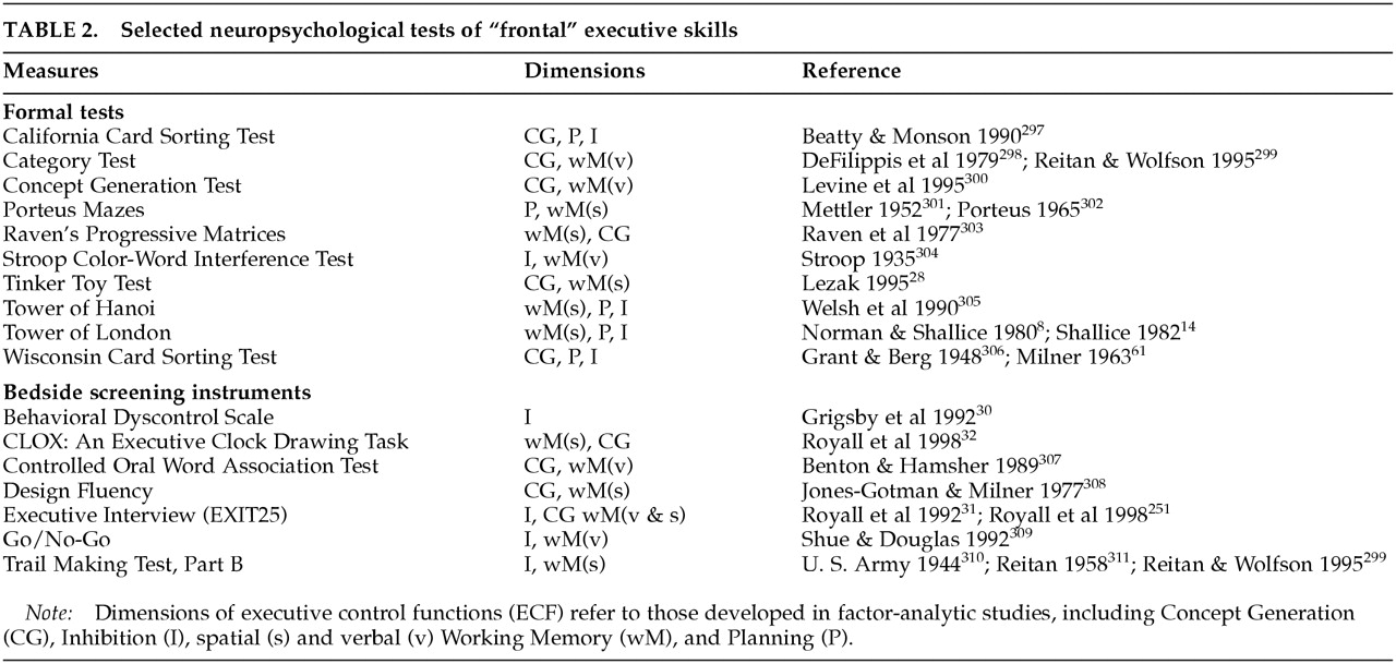
However, the clinical assessment of executive function has lagged behind these advances. This is partly because of the lack of suitable measures. The Stroop Color/Word Interference Test (Stroop), the Trail Making Test Part B (Trails B) of the Halstead-Reitan battery, the Conceptualization Task of the Dementia Rating Scale, and a variety of other tests of abstraction and mental control have been offered as putative ECF measures. 28 The Wisconsin Card Sorting Test (WCST) is perhaps the best described ECF test (see box, p. 391), but these and other formal executive measures are often impractical for widespread use outside of academic settings.
In 1990, Kaye et al. introduced the Behavioral Dyscontrol Scale (BDS), a brief compilation of clinical items adapted from the work of Luria. 29,30 In 1992, Royall et al. introduced the Executive Interview (EXIT25), 31 followed in 1998 by CLOX: An Executive Clock Drawing Task. 32 Most recently, the Frontal Assessment Battery (FAB) 33 has been introduced. This instrument is similar to the BDS and the EXIT25 in that it is a compilation of simple clinical ECF assessments. However, the FAB differs from earlier measures in that its item set was designed to elicit several distinct executive tasks, each of which can be significantly correlated with frontal metabolic changes.
Another approach to ECF assessment has been to identify the behavioral sequelae of executive dyscontrol and to measure these. Behavior rating scales, such as the Neuropsychiatric Inventory (NPI), 34 contain subtests for behaviors that have been specifically associated with frontal lesions. The Behavioural Assessment of the Dysexecutive Syndrome (BADS) 35 and the Frontal Lobe Personality Scale (FLOPS) 36 have been explicitly developed to measure “dysexecutive” behavior syndromes.
This new generation of ECF instruments can be administered by clinicians in almost any setting. Consequently, executive impairment has been demonstrated in almost every major neuropsychiatric disorder (reviewed below). In many of these conditions, measures of executive function are more strongly associated with functional status, level of care, and need for services than are either syndrome-specific positive symptoms (e.g., psychosis, mood disturbance, or memory loss) or nonexecutive cognitive domains.

ANATOMICAL SUBSTRATES OF ECF

The Prefrontal Cortex

The role of the prefrontal cortex in executive function is suggested by its unique structure and pattern of connectivity. 37 The prefrontal cortex (Brodmann areas [BA] 8–11, 24, 25, 32, 45–47) comprises more than 30% of the brain's weight and surface area. It is a phylogenetically recent structure, representing only 10% to 20% of the primate brain. 38
The frontal cortex can be grossly divided into two cytoarchitectural regions. The posterior portion is “agranular” in nature. This term refers to the minimal representation of the internal granular layer IV in posterior frontal cortical sections. In contrast, the regions that are most closely associated with executive function (e.g., the anterior [“prefrontal”] portion of the frontal lobes, which comprises the dorsolateral and orbital/medial regions) consist of “granular cortex.” This term refers to a cortical architecture in which layer IV is distinct and well developed. Layer IV is most developed in BA 46 and becomes progressively less distinct as one moves ventrally and posteriorly from there.
Cortical layer IV is rich in inhibitory GABAergic interneurons. These interneurons receive input from bioaminergic nuclei in the brainstem and “feed forward” to provide inhibition to local pyramidal cells in cortical layers III and V. GABAergic interneurons have been implicated in the executive impairments of schizophrenia 39 and may represent one of the principal targets of atypical neuroleptics.
Several unique aspects of the prefrontal cortex suggest that it mediates ECF. First, the prefrontal cortex is connected to more brain regions than any other cortical region. Only the primary sensorimotor cortices and subcortical sensorimotor relay nuclei do not have direct or simple indirect connections to the prefrontal cortex. Second, the frontal cortices are “metamodal”: they receive direct cortical input only from other heteromodal association areas. Thus, they are positioned to act on information that has already been processed at lower levels. The integrative nature of prefrontal regions is reflected even at the cellular level. Many frontal neurons increase their firing rate in response to the combined activity of sensory and motor regions. Additionally, frontal firing patterns may be altered by manipulating the motivational importance of environmental stimuli. Third, the prefrontal cortex is the major neocortical target for information processed in the limbic circuits. It is the only cortical region positioned to integrate cognitive and sensorimotor information with emotional valences and internal motivations. Fourth, although wide areas of the cortex project into the basal ganglia–thalamocortical circuits, the prefrontal cortex is that system's major target. Thus, the frontal lobe is the only cortical region capable of integrating motivational, mnemonic, emotional, somatosensory, and external sensory information into unified, goal-directed action.
In addition, the prefrontal cortex has bilateral connections to the basal ganglia–thalamocortical circuits' targets in the thalamus. Similarly, the prefrontal cortex has bilateral connections to its afferents in the parietal, temporal, and occipital association cortices, the limbic circuits, and the major brainstem biogenic aminergic nuclei, as well as to the cholinergic neurons of the nucleus basalis of Meynert. These connections put the prefrontal cortex in a unique position to modify the information it acts on. Moreover, in the case of the major brainstem bioaminergic nuclei, which project diffusely to the cortex, the prefrontal cortex is positioned to indirectly influence the activity of the nonfrontal cortex as well.

Frontal Basal Ganglia–Thalamocortical Circuits

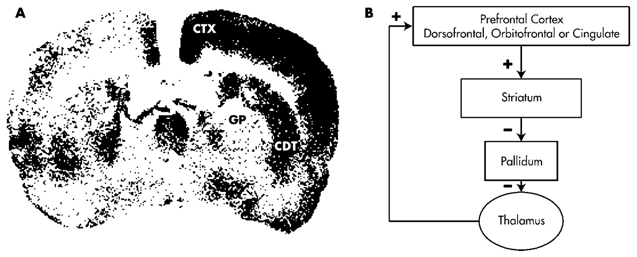
Certain subcortical lesions can affect ECF either directly or indirectly via frontal cortical metabolic changes (e.g., by diaschisis). The caudate, putamen, pallidum, nucleus accumbens, and thalamus are related to the frontal cortex through basal ganglia–thalamocortical behavioral control “circuits” (Figure 1A). 19,40,41 Although each of these circuits passes through different structures, all of the frontal circuits are similar in design.
The neurochemistry of these circuits' connections is known. 42 Excitatory glutamatergic fibers from the cortex project to the neostriatum (caudate, putamen); then inhibitory GABAergic fibers project to the globus pallidus/substantia nigra and from there to specific targets in the thalamus. These connections form dynamically balanced direct and indirect circuits connecting the prefrontal cortex to the thalamus. The thalamus closes the circuit by projecting back to prefrontal cortical regions via stimulatory glutamatergic fibers. Cholinergic projections to the frontal cortex facilitate thalamic activation of that structure. Dopamine (DA) projections from the ventral tegmentum also innervate the cortex. DA projections from the nigra innervate the striatum.
In each circuit, the corresponding frontal cortical region and striatum receives inputs from cortical regions that are more posterior. 4346 These inputs provide insights into each circuit's functional role by revealing the processes with which it interacts. The dorsofrontal circuit receives information from the parietal and temporal cortex. These regions provide access to complex spatial and temporal information. The orbitofrontal circuit receives input from visual and auditory processing areas in the occipital and temporal lobes, as well as limbic centers in the amygdala and temporal poles. The anterior cingulate/mesiofrontal cortex receives input from the hippocampus, amygdala, and paralimbic cortex. Some authors have labeled the anterior cingulate circuit “paralimbic” for this reason.
Several aspects of this circuitry also deserve special mention. First, these circuits funnel information from widespread cortical areas into relatively small thalamocortical targets. These targets are all in the prefrontal cortex, consistent with the role of these circuits in behavioral/cognitive control. Second, the behaviors that mark each circuit can be reproduced by lesions at various points along their path. For example, the ability to perform certain visuospatial “working memory” tasks (which involve the short-term maintenance of information during its manipulation) is dependent on the integrity of the dorsolateral prefrontal cortex. 23 However, the same tasks are disrupted by lesions to the caudate 20 and to the mediodorsal thalamic nucleus 21,22 in the dorsofrontal circuit. This association suggests that frontal cortical damage is a sufficient but not a necessary cause of executive dyscontrol. Finally, the circuits appear to be discrete (i.e., nonoverlapping) and spatially constrained. At the level of the cortex, they are widely separated. Cortical lesions can divorce the behaviors associated with one circuit from another. Subcortically, however, the circuits are in much closer proximity. This anatomy suggests that subcortical pathology is likely to lesion multiple circuits simultaneously, mixing the syndromes together.
Three frontal circuits are particularly relevant to executive control: the dorsolateral prefrontal circuit, the lateral orbitofrontal circuit, and the anterior cingulate circuit. 18,47

Dorsolateral Prefrontal Circuit:

The dorsolateral convexities of the frontal lobes consist of BA 8–12, 46, and 47. The blood supply for these regions is from the middle cerebral artery. In the dorsolateral circuit, corticofugal pathways project to the dorsolateral caudate nucleus, which also receives input from the posterior parietal cortex and the premotor area. The circuit then connects to the dorsolateral portion of the globus pallidus and the rostral substantia nigra reticulata and continues to the parvocellular region of the medial dorsal and ventral anterior thalamic nuclei. The circuit is closed via thalamic projections back to the frontal dorsolateral convexity. Lesions to this circuit have been implicated in a variety of higher cognitive functions, including goal selection, planning, sequencing, response set formation, set shifting, verbal and spatial working memory, self-monitoring, and self-awareness (metacognition). 38,4852 The WCST consistently activates dorsolateral frontal regions.

Lateral Orbitofrontal Circuit:

The “orbit” of the frontal lobes refers to a continuous region including ventral anterior and inferior lateral regions (BA 10–15 and 47). Medial regions are vascularly supplied by the anterior cerebral artery, and lateral regions lie in the territory of the middle cerebral artery. Cortical projections terminate on the ventromedial caudate nucleus, which also receives input from other cortical association areas, including the superior temporal gyrus (auditory) and inferior temporal gyrus (visual), as well as brainstem regions (e.g., the reticular formation). Projections continue to the dorsomedial aspect of the internal globus pallidus and to the rostromedial portion of the substantia nigra reticulata. Pathways continue to the magnocellular region of the medial dorsal and ventral anterior thalamic nuclei, and then return to the lateral orbitofrontal region.
The orbitofrontal circuit appears to be involved in the initiation of social and internally driven behaviors and the inhibition of inappropriate behavioral responses. 48,52 Orbitofrontal function may be particularly relevant to risk assessment. Choosing between small but likely rewards and large yet unlikely rewards activates inferior and orbitofrontal regions. 53 Impairment on the “go/no-go” task has been associated with orbitofrontal lesions in animals 54 and humans. 55 Orbitofrontal lesions also lead to clinical features such as environmental dependency and utilization behavior. 5658

Anterior Cingulate Circuit:

Frontal regions involved in this circuit are medially located (BA medial 9–13, 24, and 32), and receive their blood supply from the anterior cerebral artery. The circuit connects to the ventral striatum (nucleus accumbens and olfactory tubercle), which receives additional input from “paralimbic association” cortex, including anterior temporal pole, amygdala, inferior hippocampus, and entorhinal cortex. The circuit continues to the ventral pallidum and rostrodorsal substantia nigra, and then to the medial dorsal thalamic nucleus. It terminates at the anterior cingulate, completing the circuit.
The anterior cingulate is important in monitoring behavior and error correction. The Stroop activates the anterior cingulate and its mesiofrontal extensions. 59 The EXIT25 31 has also been specifically associated with left mesiofrontal cerebral blood flow by single-photon emission computed tomography (SPECT). 60

OBSTACLES TO DEFINING AN EXECUTIVE “GOLD STANDARD”

One of the obstacles to ECF research has been the lack of a clear “gold standard” measure against which putative ECF measures can be compared. This measure would presumably call upon specific frontal functions and be selectively vulnerable to frontal pathologies. However, this may not be an achievable goal for three reasons. First, since the frontal lobe represents so much of the brain's weight and surface area, it seems unlikely that any one measure could assess its functions comprehensively. We may be searching for a frontal-executive battery, not an executive measure. Second, the anatomy of frontal systems suggests that specific subcortical pathologies are also relevant to ECF. Thus, we may not even be looking for a frontal battery so much as a frontal system battery. Finally, the cybernetic character of ECF implies an intimate relationship between ECF and its associated targets. We will need to qualitatively distinguish between the loss of executive control over a nonexecutive domain and a primary disruption of the domain itself.
For example, although some tasks (e.g., the WCST, the Stroop, the Category Test, the EXIT25, and Trails B) have been specifically associated with frontal structural or metabolic changes, 6166 they can also be affected by more posterior lesions. 6770 WCST performance is not specific for frontal lobe damage unless deficits in comprehension or visual search are controlled. 71 Furthermore, both the WCST and the Stroop measure multiple dimensions of executive control in factor-analytic studies. These dimensions may not be localizable to the frontal lobes even if frontal systems are a major determinant of their variance.
Peterson et al. 59 provide an example of this problem for the Stroop. This measure activated multiple nonfrontal cortical regions, which in turn resolved themselves into seven discriminable factors. These factors were interpreted as representing distributed neuronal networks supporting error monitoring, working memory, selective attention, and motor planning (among others). Although several Stroop factors shared the anterior cingulate, cingulate activation does not uniquely explain Stroop variance, and many nonfrontal lesions have the potential to affect Stroop performance. Nevertheless, activation studies have been criticized for their sensitivity to “subclinical” differences in performance. 72 Frontal lesions selectively affect the Stroop in actual patients. 73 Thus, poor Stroop performance may yet be indicative of frontal pathology, despite the complexity of activation studies.
It appears that neither the measures used to assess ECF nor the biological substrates they activate are easily localizable. Four important dichotomies need to be addressed before these apparent discrepancies can be resolved. Each will be discussed in turn.
1.
Frontal Lobe vs. Frontal System: Frontal cortical lesions may be sufficient, but are not necessary causes of executive impairment.
2.
Structure vs. Function: Frontal cortical function may be compromised by subcortical lesions (i.e., vascular disease) in the absence of demonstrable local cortical pathology.
3.
Control vs. Process: Executive functions control performance in other neuropsychological domains. Some tasks that were previously ascribed to nonexecutive domains may be sensitive to frontal system pathology because they require executive control. Conversely, lesions outside the frontal systems may undermine ECF test performance, in the absence of executive dyscontrol, by disrupting the processes being controlled during the task.
4.
Executive Function vs. Executive Function(s): Some measures may be sensitive to only a subset of executive functions.

Frontal Lobe vs. Frontal System

It has proven difficult to localize specific executive operations to specific prefrontal regions. Rather, ECF may depend on the integrity of frontal systems. For example, L'Hermitte et al. 57 have described the phenomenon of “utilization behavior” (in which a patient automatically utilizes a familiar object in a habitual way, regardless of its appropriateness to the current context) following orbitofrontal lesions. The same behavior has been described following massive bilateral frontal lesions 74 and mesiofrontal lesions, 75 both of which might involve orbitofrontal regions. However, utilization behavior has also been reported following lesions to other frontal system structures, including the caudate 76 and thalamus. 77 The unity of frontal circuit activity can be deduced from factor analyses of regional brain metabolism: 70% of regional variance in total cerebral glucose utilization can be explained by a single factor that contains the frontal circuits (e.g., the frontal cortex, cingulate gyrus, caudate nucleus, putamen, and thalamus) and temporal cortex. 78
There may be several reasons for the difficulty in making clinicopathological correlations between ECF and frontal lesions: 1) the taxonomy of executive impairments has not been adequately developed—many authors may not be comparing identical phenomena; 2) although discrete prefrontal pathways have been partially established, precise anatomical boundaries are not well defined, especially at the cortical level, and certain frontal functions are limited to subregions of traditional BA regions of interest; 79 3) lesions to the frontal lobes are often not well defined or do not follow clear and reproducible boundaries across subjects (e.g., most frontal strokes cause additional damage to subcortical or posterior regions); 4) frontal lobe pathology, such as tumors, stroke, or trauma, frequently results in remote effects secondary to vascular changes, pressure effects, and disconnection of neural pathways. Data from psychosurgery (tumor evacuation or frontal leukotomy) can be especially difficult to interpret for several reasons: a) these studies often use abnormal patients to begin with, b) cognitive outcome assessment is often rudimentary, and c) follow-up is typically short term (i.e., months rather than years).
Nonetheless, it now appears that there are regional differences in behavioral sequelae of frontal cortical lesions. 5,38,8082 Damage to the dorsolateral prefrontal cortex impairs planning, hypothesis generation, and behavioral control. Episodic memory encoding and retrieval is affected by ventrolateral lesions. Working memory is affected by more dorsal pathology. Orbitofrontal lesions lead to impaired insight, judgment, and impulse control. These traits were part of Phineas Gage's deterioration. Mesiofrontal/anterior cingulate lesions lead to indifference and attentional dyscontrol. Patients generate little speech or behavior spontaneously, yet may respond correctly if prompted.
Moreover, the dysexecutive neuropsychological profile of prefrontal cortical disorders such as frontotemporal dementia can also be observed in subcortical frontal system disorders such as Parkinson's disease (PD), Huntington's disease (HD), progressive supranuclear palsy, 83 or subcortical vasculopathy. 84 Even neuropsychiatric disorders such as major depression and schizophrenia are associated with a similar pattern on psychometric testing, suggesting that they too may involve frontal system pathology. 85,86
In summary, executive functions have been difficult to localize within the frontal cortex. This situation might be improved with more careful attention to lesion location and a formal approach to frontobehavioral nomenclature. Nonetheless, the logic of frontal basal ganglia–thalamocortical networks suggests that frontal system lesions are both sufficient and necessary to executive impairments.

Structure vs. Function

Another dichotomy that deserves attention is that between frontal structure and function. Executive control can be compromised without a frontal cortical lesion. Frontal function can be indirectly affected by lesions to frontal lobe afferents or related frontal system circuit structures. Conversely, lesions to corticofugal tracts can disconnect the frontal operations from the processes they control.
Human and animal studies suggest that subcortical lesions to frontal system networks may remotely affect frontal cortical metabolism (e.g., by diaschisis), either increasing or decreasing frontal metabolism. Figure 1A presents the results of a study by Kelly and McCulloch 87 in which rats received a 500-ng injection of muscimol (a GABAergic agonist) to the left caudate nucleus. This lesion resulted in a functional caudate lesion on that side. The effects of this lesion were studied using [14C]2-deoxyglucose autoradiography. Brain regions that were metabolically active at the time of injection took up this radioligand. Regions that were metabolically inactive, including the left caudate, did not take up the tracer. In each section, the right (unaffected) side served as the left's control.
Figure 1B demonstrates that the caudate lesion resulted in disinhibition of the ipsilateral globus pallidus, leading to increased inhibition of the ipsilateral medial thalamic nucleus, resulting in reduced activation of the ipsilateral cortex. In short, a discrete subcortical lesion in frontal networks may lead to remote changes in frontal cortical metabolic function. This finding can be understood in the context of frontal circuit anatomy (Figure 1A) and may help to explain the finding of frontal behavioral syndromes and ECF impairment in subcortical dementias, 88 as well as the specific association between subcortical vasculopathy and frontal hypometabolism in vascular dementia (VaD) and late-onset major depression. 8991
Patients with PD, major depression, and schizophrenia often appear “hypofrontal” by functional neuroimaging. 9295 In PD and major depression, this may be related to cortical deafferentation of medial nigral or ventral tegmental DA inputs. 96 The hypofrontality of both disorders is associated with tests that are linked to DA physiology. 97,98 Alternatively, these deficits might be related to cortical deafferentation of the thalamic inputs. 19,90,99 Medial thalamic infarction results in frontal cortical hypometabolism by positron emission tomography (PET) and SPECT. 100,101 Thalamic outputs to the frontal cortex can be disrupted indirectly after globus pallidus lesions. 102
However, executive impairments are not only associated with frontal hypometabolism. In obsessive-compulsive disorder (OCD), cortical hypermetabolism 103 is associated with poor performance on ECF measures. 104 Similarly, in HD the degree of prefrontal activation during the WCST is inversely proportional to the subject's performance, yet is statistically associated with the amount of caudate atrophy. 105
These seemingly paradoxical findings may be understood from the point of view of frontal systems physiology. OCD has been associated with hypometabolism in the globus pallidus and thalamic disinhibition. Thalamic disinhibition might result in increased thalamocortical glutamatergic tone (Figure 1A). Thalamocortical glutamatergic inputs co-localize with inhibitory dopamine D1 receptors on pyramidal cell dendrites in the prefrontal cortex. 40 The balance between these opposing influences affects prefrontal signal-to-noise processing. 106 Either increasing glutamatergic excitation or diminishing dopaminergic pyramidal cell inhibition should lead to increased pyramidal cell activity, at the expense of signal specificity. A precise range of DA receptor activity within the prefrontal cortex must be maintained for optimal function. 107108 In the case of OCD, DA's inhibitory effects may be overwhelmed by increased glutamatergic tone.
In summary, executive control depends on the integrity of frontal systems. Executive impairment may follow disruption of frontal system information processing, regardless of the location of the lesion within the system or the direction of the perturbation. In some cases, remote lesions can affect processing within the frontal circuits.

Control vs. Process

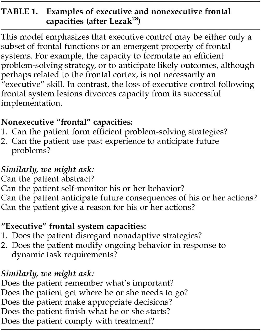
Lezak 28 has offered a simple test for defining what constitutes an “executive” measure. Questions about executive functions explain “how or whether a person goes about doing something…questions about [traditional] cognitive functions are generally phrased in terms of what or how much.” (p. 42). This simple dichotomy cleaves the vast array of frontal functions into control functions and their target processes. Either may be dependent on frontal activities; however, only the control functions are “executive” in a cybernetic sense. The subset of frontal functions that are “executive” depends on how the question is asked (Table 1). 28
This distinction can be addressed experimentally. For example, there is an extensive literature associating schizophrenia with deficits on the WCST. However, patients with schizophrenia benefit from cueing during the WCST test procedure. 109 In other words, they can generate the abstract concepts demanded by the task, but they do not apply them unless prompted. Thus, although the abstract concept formation demanded by the WCST may in fact be localizable to the frontal lobes, it is not necessarily an executive control function in the limited sense required by Lezak because it merely addresses what patients can do and not whether they do it when needed. In contrast, the failure of patients with schizophrenia to inhibit automatic but inappropriate verbal responses on tests such as the Stroop 110 would be more consistent with Lezak's view of executive control.
Authors who emphasize a cybernetic view of ECF point to the potential to observe executive dyscontrol in performance on many seemingly “nonexecutive” tasks. 12,111 By analogy, at least some variance in all neuropsychometric tests may be specifically attributable to the executive control demanded by the testing paradigm (see “g” below). We will examine the executive control of clock drawing, memory, and language.
The clock-drawing task (CDT) has traditionally been viewed as a visuospatial task, sensitive to right hemisphere pathology. 112 However, frontal leukotomy selectively affects CDT performance relative to age, disease, and education-matched control subjects. 113 CDT failures among frontally impaired subjects challenge a chiefly visuospatial conceptualization of the CDT and suggest the need for a separate analysis of the executive control demanded by the testing paradigm.
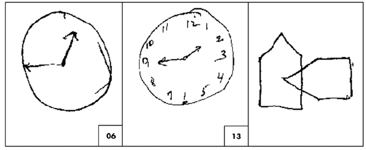
Figure 2 presents a patient's performances on CLOX, an executive CDT. 32 CLOX1 is an unprompted task. CLOX2 is a copied version. The visuospatial components of these tasks are similar. However, CLOX1 entails executive control because it requires the subject to generate a figure in the absence of relevant visual cues. The validity of CLOX1 as an executive paradigm is suggested by the fact that, in elderly retirees, both CLOX1 and the EXIT25, but neither CLOX2 nor the Mini-Mental State Examination (MMSE) makes significant independent contributions to the number of categories achieved on the WCST. 114 Figure 2 presents the pattern of CLOX performance expected in a frontal system disorder. Executive measures (the unprompted CLOX1 and the EXIT25) are impaired. CLOX2 (copied) and the MMSE are not.
The same qualitative dissociation between control and process can be elicited in other domains, such as memory. Memory tasks can be affected by frontal, parietal, and mesiotemporal cortical lesions. However, the pattern of memory loss that follows frontal system lesions is discriminable from traditional limbic amnesia. 115119 The ability of a “memory” task to activate dorsofrontal systems depends greatly on the structure provided to the subject during memory testing. 120,121 For example, the intentional, goal-directed retrieval of information results in frontal activation relative to incidental cued recall. 122 Patients with frontal lesions are unimpaired in their ability to recall cued information, but have difficulty with tasks that require them to organize, sequence, or monitor the information themselves. Thus, they have trouble with free recall, temporal order, and source memory. Similarly, confabulation among amnestic subjects appears to reflect mesiofrontal/anterior cingulate impairment, resulting in a failure to ignore active but currently irrelevant memory traces. 123
Not all memory tasks that activate frontal regions are necessarily “executive.” In neuroimaging studies, tasks that call for relatively simple episodic or semantic encoding tend to activate the left ventrolateral prefrontal cortex. 124 Those that call for retrieval activate the right ventrolateral prefrontal cortex. However, if the subject is asked to manipulate the information while encoding or retrieving it, the focus of activation shifts toward more dorsolateral regions. 125
Language skills are also affected by ECF impairment. Arbuckle and Gold 126 have associated disorganized and hyperverbose speech, but not language impairment per se, with impaired working memory and executive control. Similarly, only a small amount (25%) of variance in verbal fluency scores can be explained in multivariate regression models by tests of verbal memory, verbal attention, and vocabulary. 127
The idea that ECF may explain some variance in most cognitive measures, regardless of the domains they purport to measure, is similar to Spearman's concept of “general intelligence” or “g.” 128 “g” represents the shared variance across domains and has been repeatedly observed in batteries of multiple cognitive measures. For example, in normal aging there are significant declines in cognitive test performance across several domains. Salthouse et al. 129 found moderate age-related declines on a battery of tests that included the WCST, Trail Making, Wechsler Adult Intelligence Scale–Revised (WAIS-R) Block Design, and Digit Symbol Substitution (DSS).
However, correlation-based analyses revealed that the age-related effects on different measures were not independent. Instead, the effect of age was observed specifically in the fraction of variance (averaging 58%) shared across all measures (i.e., “g”); “g” has been localized to dorsolateral prefrontal cortex by PET 130 and associated with working memory (also associated with dorsolateral prefrontal cortex; see below) 131,132 and with formal executive measures. 133
In summary, there is no established framework for interpretation of the executive functions. Some authors emphasize the frontal lobes and their importance in planning, hypothesis generation, and abstraction. Others, however, work within a more limited subset of frontal functions. These authors see ECF as a specific subset of frontal lobe activities, revealed by the examination of how the frontal systems interact with other systems to produce and control complex goal-directed activities.

Executive Function vs. Executive “Function”

Another dichotomy that has yet to be resolved is whether there is a single executive control, as opposed to multiple controls for discrete operations. The idea of a single executive is implied in the concept of the “central executive” and the multimodal nature of the frontal lobe's anatomy and functional connections. Researchers have developed computer models of subject task performance on putative “frontal” measures that successfully model patient task performance on four frontal tasks (the WCST, the Stroop task, motor sequencing, and a context-dependent memory task). 134 Frontal-type errors on all tasks can be observed after degrading a single domain (working memory).
However, patients with frontal lesions often display disassociations in their performance on select frontal tasks. This effect might be due to regional differences in the types of processes to which frontal mechanisms are applied. 135 Although the frontal lobes appear to be less functionally committed than more posterior cortical regions, 136 their functions can be roughly divided along four spatial dimensions: left–verbal/right–nonverbal, anterior–cognitive/posterior–motor, ventral–perception/dorsal–action, and medial–internal focus/lateral–external focus. Thus, the verbal aspects of working memory tasks may activate the left dorsolateral prefrontal cortex and nonverbal aspects may activate the right. 137139 Even within the domain of nonverbal working memory, recall of faces activates more ventral regions of the right dorsolateral frontal cortex than does recall of spatial location. 140 This functional specificity may go all the way down to the cellular level. 141
Goldman-Rakic has suggested that different prefrontal areas may perform the same operation on different inputs. 23 This hypothesis is consistent with the functional segregation of the basal ganglia–thalamocortical circuits. Support for a modular organization of frontal function has been developed in humans. 142 Cognitive test performance is most closely related to dorsofrontal cerebral glucose metabolism, whereas social behavior and disturbances of comportment are related to mesio-/orbitofrontal metabolism. Similarly, dorsal regions of the anterior cingulate are activated by attention-demanding Stroop-like interference tasks, whereas ventral regions of the anterior cingulate respond when similar tasks are applied to emotionally laden content. 143

Dimensions of Executive Control:

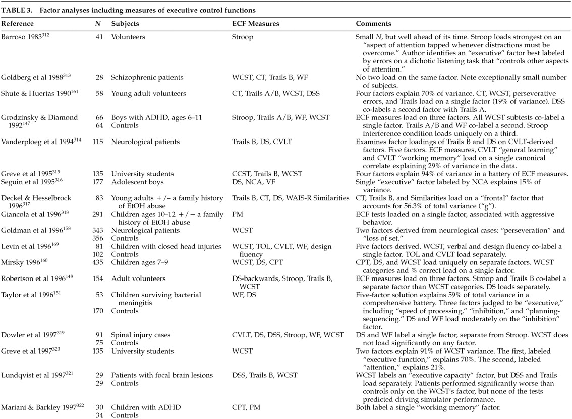
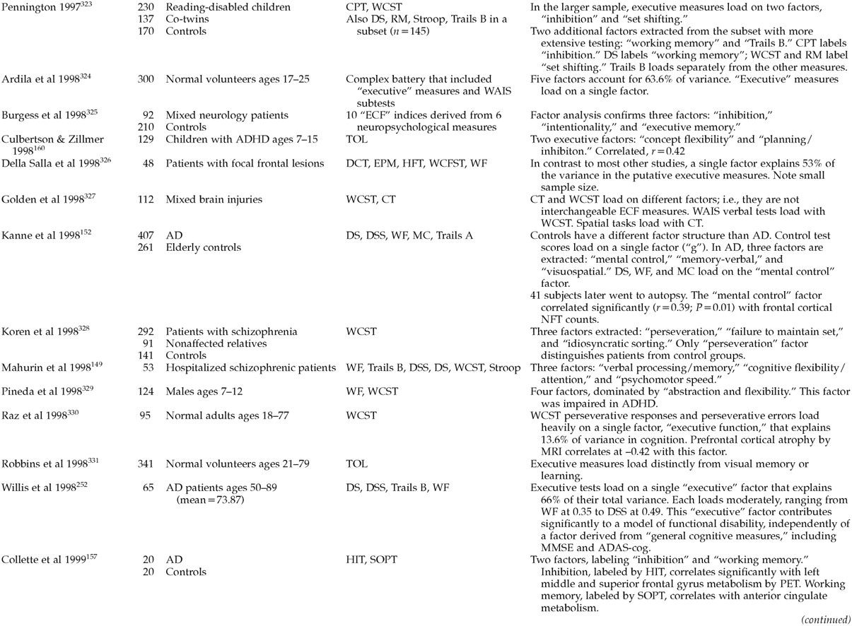
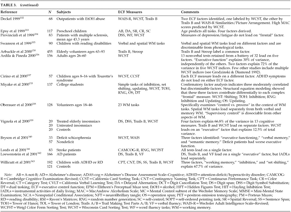
There are many putative ECF measures 144 (Table 2). However, it is not at all clear that these all test the same dimensions of executive control. Our literature review identified several studies containing factor analyses of putative ECF measures (Table 3A, Table 3B, and Table 3C). Interpreting these studies can be difficult. 145 Few have been intentionally designed to address ECF. Prior to about 1998, most authors interpreted their results without regard to ECF or frontal function. Instead, factors with strong loadings by ECF measures were thought to represent “vigilance” or “attention.” The differences between ECF and simple attention have been extensively studied. 146 It is relevant to the cybernetic formulation of ECF that “judgment,” “concept formation,” “problem solving,” and “decision making” are seldom mentioned in factor analyses of ECF measures.
Putative ECF measures do not load onto a single, overarching executive construct. Most studies find multiple dimensions of executive control. The available studies tend to confirm a rule discovery factor labeled by tests such as the WCST categories; a working memory factor labeled by tests such as the California Verbal Learning Test, the Wechsler Intelligence Scale for Children–Revised (WISC-R), Digit Span (verbal), and the Tower of London (nonverbal); an attentional control factor labeled by tests such as the Continuous Performance Task or Digit Cancellation; and a response inhibition factor labeled by tests such as the WISC-R Digit Span Backwards, Trails B, or the Stroop. Rule discovery and working memory are most closely related to dorsolateral cortical function. Attentional control and response inhibition depend more on ventromedial regions.
These domains are fairly robust. Different authors have found the same instruments to load together in different samples. For example, Trails B and the Stroop co-label a single factor (response inhibition) in Grodzinsky and Diamond's study of boys with ADHD, 147 Robertson et al.'s study of normal adults, 148 Mahurin et al.'s study of schizophrenic patients, 149 and Arbuckle et al.'s study of elderly adults. 150 In addition, there is limited evidence that ECF factors are multimodal. For example, Taylor et al. 151 found that both verbal and design fluency tasks loaded on the same factor. This finding suggests that the executive control identified in this paradigm may be equally applicable to both verbal and constructional processes, presumably mediated by different cerebral hemispheres.
Unfortunately, most of the available ECF factor-analytic studies have methodological flaws. Large sample sizes are needed before stable factor structures can emerge. Executive and nonexecutive measures need to be included, and key reference measures should be used across samples to facilitate comparisons.
Two recent studies can serve as models for future work. 152,153 Kanne et al. 152 examined the factor structure of a comprehensive battery of neuropsychological measures, including several ECF measures, among 407 AD patients and 261 elderly control subjects. Control data exhibited a different factor structure than that found in data for AD patients. Control test scores loaded on a single factor (i.e., they showed high “g”). In contrast, the data from AD cases was best represented by a three-factor model. The authors labeled these factors Mental Control, Memory-Verbal, and Visuospatial. Digit Span, verbal fluency, and the Mental Control subtest of the Wechsler Memory Scale loaded on the Mental Control factor. This factor explained most of the variance in both early AD and moderately advanced AD subgroups. Autopsies were later performed on 41 AD subjects. Each factor was significantly correlated with the severity of AD pathology in a different cortical region. The “Mental Control” factor correlated significantly (r=0.39, P=0.01) with frontal cortical neurofibrillary tangle counts. Digit Symbol Substitution, a test that is often purported to measure ECF, did not load on the Mental Control factor, nor was it correlated with frontal pathology.
Miyake et al. 153 examined putative ECF measures, including the WCST, the Tower of Hanoi (TOH), random number generation (RNG), operation span, and dual tasking in a moderately large sample of college students (N=137). A confirmatory factor analysis of these measures indicated three moderately correlated but discriminable factors, which they labeled Set Shifting, Inhibition, and Updating. Structural equation models showed that these three factors contribute differentially to each of the “complex” ECF measures. The Set Shifting factor contributed most to WCST performance, the Inhibition factor contributed most to TOH, and both Inhibition and Updating contributed to RNG. The Updating factor also contributed to operation span scores. This type of analysis reveals that 1) classical “ECF” measures are often multidimensional; 2) no single measure comprehensively assesses all ECF domains; and 3) specific combinations of ECF measures may compliment each other, while others may be redundant.
For a discussion of the Wisconsin Card Sorting Test as a possible gold-standard ECF measure, see box (p. 391).

FUNCTIONAL IMAGING AND EXECUTIVE CONTROL

Lesion studies associate response inhibition with the orbitofrontal region, attentional control with the mesiofrontal region, and working memory (verbal and nonverbal) and rule discovery with the dorsolateral region. 162 These observations are generally supported by neuroimaging. Bench et al. 65 studied the associations between a modified Stroop and regional cortical metabolism PET. During the Stroop's interference condition, the right orbitofrontal cortex and posterior parietal cortex were both activated (i.e., control and process). However, these regions may both be under the control of the anterior cingulate. The anterior cingulate is thought to be important in error detection and sequencing of ongoing action plans. 143 It has been shown to be activated by stimuli that are incongruent with expectation and that may need correction. Liotti et al. 163 have studied the temporal sequencing of cortical activity during the Stroop's interference condition, using event-related potentials (ERPs). Differences in ERP between Incongruent compared with Congruent trials first appear in the anterior cingulate (peaking at 410 ms), then in the temporoparietal cortex (500–800 ms post stimulus).
Working memory tasks activate dorsolateral prefrontal regions. The left hemisphere may mediate verbal working memory. The right may mediate nonverbal working memory. 138 There is some overlap between these regions and other executive tasks. Verbal fluency tests tend to activate the left dorsofrontal cortex, 164,165 although in one study a test of category fluency activated the right dorsolateral prefrontal cortex relative to a baseline reading task. 166 Tasks requiring sustained attention have also been found to activate the right dorsolateral prefrontal cortex. 167
However, the factor-analytic studies reviewed above suggest that most ECF measures are complicated tasks that may draw on several executive domains simultaneously. The Tower of London, for example, loads on two factors in Culbertson and Zillmer's study of boys with attention-deficit/hyperactivity disorder (ADHD) 168 and on three separate factors in Levin et al.'s study of head-injured children, 169 and it has been reported to activate the left dorsolateral prefrontal cortex 170 and mesiofrontal/anterior cingulate. 171 In a functional MRI (fMRI) study by Peterson et al., 59 seven factors were derived from the brain regions activated by the Stroop. The anterior cingulate (mesiofrontal system) loaded significantly on each of these seven factors (see Liotti et al. 163 ).
The nonspecificity of putative ECF clinical measures is in sharp contrast to the relatively discrete frontal activations associated with certain tasks in neuroimaging studies. The “delayed response,” “A-not-B,” “go/no-go,” “n-back,” and “object retrieval” paradigms all reproducibly activate very specific frontal regions. However, it should be kept in mind that the skills represented by these tasks are achieved by human beings very early in development, long before clinically relevant executive skills have developed. The A-not-B, delayed response, and object retrieval paradigms are essentially in place in human infants by the age of 12 months. 172174 Thus, these easily localized tasks, while clearly dependent on frontal functions, may be merely the heteromodal processes on which truly cybernetic “executive” functions operate.

APPLICABILITY TO NEUROPSYCHIATRIC DISORDERS

Assuming that the obstacles to ECF assessment can be overcome, what is the promise of this domain? First, it is important to realize that ECF impairment, frontal system lesions, and frontal metabolic deficits have been detected in a wide variety of both neuropsychiatric and medical disorders. This commonality offers the possibility of unified disability and behavioral outcomes assessments that could be validly applied across a wide variety of conditions. 175,176 Moreover, treatment and assessment strategies that are developed in one condition may be relevant to many others as well. Second, ECF may predict disability more accurately than tests based on other cognitive domains. And third, certain behavioral features may serve as indices of ECF impairment. These could have prognostic and treatment significance.

ECF Deficits Are Common

Our literature review identified only a single community-based study of the prevalence of ECF impairment. Presumably, such studies have been limited by the dearth of reliable, valid ECF measures that could be suitable for use in epidemiological or clinical trials. Grigsby et al. 177 used a brief ECF measure, the Behavioral Dyscontrol Scale (BDS), that is essentially a compilation of items based on the work of Luria. They examined the prevalence of BDS failure in a community sample (N=1,145; mean age[±SD]=72.9±7.2 years) of community-dwelling elderly persons residing in southwestern Colorado. The mean level of education in this sample was 10.5±3.7 years. Many subjects were Hispanic.
The authors found a high prevalence of ECF impairments: 25.5% of their subjects showed impairment on the BDS. Half of these had normal Mini-Mental State Examination scores. 178 The MMSE has been criticized for poor sensitivity to early cognitive decline in older persons and for poor specificity for dementia in minority and undereducated samples. 179 However, the BDS was a stronger predictor of impaired functional status than the MMSE, suggesting that this sample's ECF impairment was already functionally significant.
This study is notable for several reasons. First, it points out how little is actually known about the community prevalence of ECF impairment. Second, it illustrates how traditional measures tend to underestimate the severity of cognitive impairment in ECF-impaired subjects. These issues are relevant to both case definition and disability assessment. 180 The American Psychiatric Association in 1994 added ECF impairment to its list of the domains that should be considered when making a diagnosis of dementia. 27 Nonetheless, there are no large dementia studies that use ECF-sensitive measures in their case definitions. The frequency of ECF impairment reported by Grigsby et al. 177 is almost twice the rate of dementia reported by most studies. Royall et al. 181 have reported similar results among well elderly retirees with advanced education and excellent health (N=561; mean age=78.1 years). Although 86% pass the MMSE at 24/30 (mean 27.7), 32% fail the EXIT25 and 42% fail CLOX1 at the 5th percentile for young adults. The EXIT25 and CLOX 1, but neither the MMSE nor CLOX2, distinguish level of care in fully adjusted models. The advent of bedside ECF measures such as the BDS, CLOX, EXIT25, and FAB now makes it feasible to explore the epidemiology of this domain.
The need for this work is suggested by the previous documentation of ECF deficits in a wide range of neuropsychiatric disorders. Some “functional” disorders, such as schizophrenia, major depression, alcoholism, and certain personality disorders, have been found to be associated with regionally specific frontal atrophy and cytoarchitectural disorganization. 182184 ECF is affected by both cortical and subcortical structural disorders.

Schizophrenia:

A well-developed literature links the functional, behavioral, and cognitive deficits of schizophrenia with frontal system impairment. 185,186 Schizophrenia is associated with diminished frontal gray and total white matter volumes 187 without clear cell loss. 188 These changes disproportionately affect frontal, particularly inferior ventrolateral and orbitofrontal, regions of interest. 187 The severity of orbitofrontal atrophy is correlated with negative symptoms. 189 There are dorsolateral prefrontal metabolic and regional cerebral blood flow reductions at rest 95,190 and during activation by executive tasks. 97,191 Executive deficits are present from the beginning of the disorder, even among drug-naïve, first-episode cases. 192 It may be interesting to note that only measures related to rule discovery and working memory are initially affected (see Dimensions of Executive Control, pp. 386–387 above). Attentional control and response inhibition impairments appear later. 193

Major Depression:

There is also evidence of frontal system pathology in major depression. 194 Major depression is associated with reduced frontal metabolism in both unipolar and bipolar presentations. 94 There is also evidence of selective cortical atrophy 195 and widespread alterations in frontal cortical architecture in depressed patients. 183,196,197 Frontal stroke is strongly associated with poststroke depressive syndromes. 198 Frank major depression may also follow basal ganglia lesions. 91,102,199 The executive impairments of depression improve with resolution of its symptoms. 200

Structural Brain Disease:

Frontal system pathology is common in AD, 201 VaD, and traumatic brain injury. 202 In addition, ECF impairments have been reported in a wide variety of neurodegenerative disorders, including amyotrophic lateral sclerosis, frontotemporal dementia, HD, Lewy body dementia, PD, and progressive supranuclear palsy. 203207
In AD, frontal lobe pathology generally correlates better with dementia severity than hippocampal or temporal cortical AD pathology. 25,208 In fact, frontal cortical synaptic density is the strongest reported pathological correlate of dementia severity (r=0.79 vs. the MMSE). 24,209 This pathology is associated with reduced cerebral blood flow by SPECT and is associated with an early decline in ECF measures. 210 ECF impairment is correlated with functional status in AD 211 and is present relative to age-matched control subjects in preclinical cases of age-associated memory impairment. 212
VaD disproportionately affects frontal systems. 213,214 Subcortical lesions indirectly affect frontal cortical metabolism, particularly if they include lacunar infarctions of the basal ganglia and thalamus, or anterior periventricular hyperintensities. 90 White matter lesions are specifically associated with poor performance on tests of frontal function. 215,216 Aneurysm of the anterior communicating artery is another common cause of ECF impairment. 217

Diabetes Mellitus:

Patients with diabetes mellitus show impairment on ECF measures. These tests include the DSS, 218,219 verbal fluency 220 (not found by Perlmuter et al. 218 ), abstract reasoning, 220,221 Grooved Pegboard, 222 Trail Making, 222,223 Stroop-Word Naming, 222 Picture Arrangement, 222 CLOX1, and the EXIT25. 223 Keymeulen et al. 224 have documented regionally specific frontotemporal hypoperfusion by SPECT in chronic type 1 (insulin-dependent) diabetic patients, but not recent-onset cases or age-matched normal control subjects. The potential causes of ECF impairment among diabetic patients might include subcortical vascular disease, polypharmacy, iatrogenic hypoglycemia, and/or concurrent major depression.

Normal Aging:

Old age may be associated with frontal system deficits even in the absence of AD or ischemic vascular disease. 225228 Reduced executive control can be detected in healthy adults as young as age 45 to 65 years relative to education- and gender-matched 20- to 35-year-olds. 229 In longitudinal studies, ECF deteriorates at an exponential rate. 230 Interestingly, the pattern of age-related cognitive decline in “nonexecutive” domains is most consistent with the loss of executive control over intact processes (see Control vs. Process, pp. 384–385 above). 115,116,231,235
Disproportionately frontal age-associated metabolic deficits have been observed by functional neuroimaging in healthy volunteers ranging in age from 18 to 78 years. 236 In animals, age-related frontal task performance has been associated with diminished dopaminergic (D2) and alpha-2-adrenergic (α2) activity in the prefrontal cortex. 107,237,238 In humans, the age-associated decline in regional D2 receptor density is linearly related to frontal cortical and anterior cingulate metabolism by PET and associated with diminished WCST and Stroop performance. 239,240
There is also structural age-associated frontal system pathology. Coffey et al., examining the MRIs of healthy elders free from vascular disease or hypertension, reported an age-related cortical atrophy that disproportionately affected frontal relative to temporal, parietal, and hippocampal regions. 241 Recent studies suggest that age-related atrophy disproportionately affects mesiofrontal and dorsofrontal more than orbitofrontal regions. There are also age-related increases in caudate and putamen hyperintensities. These lesions occur in many apparently healthy elderly persons and can produce executive impairment that is comparable in severity to frontal lobe degeneration. 242

ECF Impairment and Disability

Because functional outcomes, medication compliance, cooking, housekeeping, and working are all examples of goal-directed activities, they are inherently vulnerable to executive dyscontrol. 243 Thus, frontal system pathology/metabolic impairment and psychometric ECF measures are emerging as robust predictors of disease severity and functional disability across diagnoses.
For example, the effects of ECF impairment have been described in rehabilitation settings. 244 Allen 245,246 has developed the Allen Cognitive Levels (ACL) Assessment. The ACL is essentially a functional status measure that provides information on a variety of executive tasks. Scores on this version correlate moderately to strongly with performance on activities of daily living in subjects with dementia (Feeding, r=0.83; Toileting, r=0.75; Grooming, r=0.74; Dressing, r=0.74; Housekeeping, r=0.68; Ambulation, r=0.67; Bathing, r=0.65; Cooking, r=0.65; Shopping, r=0.64; Laundry, r=0.60; Telephone, r=0.58; Finances, r=0.52; Transportation, r=0.32; Medication, r=0.32; all significant at P<0.05). Velligan et al. 247 have associated the majority of variance in ACL scores with neuropsychological measures of working memory and response inhibition (e.g., ECF factors derived from factor analyses). In schizophrenia, frontal neuroimaging and ECF performance are better predictors of long-term functional outcomes than is successful treatment of psychosis. 248 Roughly 25% of community-dwelling schizophrenic adults can be expected to have ECF impairment. 249 This is comparable to the prevalence of ECF impairment in well elderly persons. 177 Elderly retirees are as executively impaired as schizophrenic patients when the two groups are matched to the services they receive from their respective institutions. 250
The association of ECF with level of care has been specifically examined in the context of retirement communities. 31,181 Retirement communities are essentially closed systems in which a resident's living setting may change in proportion to the services and supervision he or she requires. Royall et al. 251 examined the relative ability of the EXIT25, the MMSE, demographic variables, physical health status (age, Cumulative Illness Rating Scale score, and number of medications prescribed), and behavior problems (Nursing Home Behavior Problem Scale score) to predict level of care in one such facility. In a stepwise linear regression model, five variables made significant independent contributions. Together, these variables accounted for 57% of the total variance in level of care (R2=0.57; F=29.2, df=7,154, P<0.0001). The EXIT25 loaded first (R2=0.43; F=119.52, df=7,154, P<0.0001).
In a second retirement community, Royall et al. 181 found that ECF measures distinguish levels of care even among noninstitutionalized retirees with normal MMSE scores. Moreover, the use of prosthetic devices declined with ECF impairment, even as level of care increased. This finding suggests that ECF impairment may undermine a disabled person's capacity to adopt assistive devices and that this impairment develops before cognitive impairment is detected in nonexecutive domains. In elderly community residents, executive measures explain independent variance in functional status, beyond that explained by specific cognitive domains 29,252 and may be the strongest determinant of functional status. 253 The effect of longitudinal change in ECF on functional status is comparable to that of age and more important than that of comorbid medical conditions. 230 These results suggest that ECF is a major determinant of level of care in elderly populations.
Finally, we note that ECF may explain significant variance in more specialized functional capacities, such as financial and medical decision-making. 254,255 Dymek et al. 256 found that the EXIT25 explained 45% of the variance in a “rational reasons” standard of decision-making and 56% of the variance in “understanding treatment,” independently of a comprehensive battery of cognitive measures, including verbal fluency. This is remarkable because the EXIT25 has little face validity as a measure of verbal reasoning or abstraction. Instead, like the Stroop, it appears to invoke response inhibition or attentional control factors. These may be more relevant to complex decision-making than is widely appreciated (see Control vs. Process, pp. 384–385 above, and Table 1).

ECF Impairment and Problem Behavior

Certain problem behaviors can be specifically associated with frontal system dysfunction in general, and with ECF impairment in particular. 257 Informant-based ratings of these behaviors may be useful as indicators for damage to these networks. 82 The FLOPS attempts to isolate frontal-type behaviors into three domains: apathy, disinhibition, and “executive dysfunction” (i.e., impaired abstraction), each theoretically linked to a different frontal circuit (mesiofrontal, orbitofrontal, and dorsolateral, respectively). 36
The latter construct may be misleading. First, it implies that apathy and disinhibition are not “dysexecutive” behaviors, when in fact they fit the cybernetic definition of ECF. Second, it implies that impairments in judgment and abstraction are indicative of executive impairment, when in fact they may not be (see Dimensions of Executive Control, pp. 386–387 above). Items addressing working memory might have been a better behavioral indicator of dorsolateral executive dysfunction.
Frith has described the behavioral deficits of schizophrenia in terms of three similar factors based on factor-analytic models derived from clinical ratings, 258,259 namely negative symptoms (apathy), positive symptoms (hallucinations and delusions), and disorganization. Negative symptoms arise from the inability to generate plans, goals, or intentions. Patients cannot spontaneously initiate complex behaviors. Martinot et al. 260 have specifically correlated negative symptom indices to central D2 receptors in schizophrenia. In contrast, disorganization arises from the failure to control automatic attentional resources. Patients do not inhibit their attention from wandering to irrelevant cues in the environment. Negative symptoms and disorganization, but not positive symptoms, are associated with poor cognitive test performance in schizophrenia, 149,261,262 particularly on tests of ECF, including the WCST, the Stroop test, and Trail Making. 263,264 It is important to note that these symptom clusters are not unique to schizophrenia. Apathy and negative symptoms can be recognized across the full spectrum of conditions that affect ECF. 265267
Attempts have been made to fit Liddle's system of three frontal syndromes into the neuroanatomical model of cortical-striatal-thalamic circuits described by Cummings. 82 Mahurin et al. 149 have associated negative symptoms and disorganization with poor performance on specific, but not overlapping, ECF measures. Negative symptoms were associated with tests of verbal fluency, cognitive flexibility, and working memory (all associated with dorsolateral prefrontal cortex, especially left). Disorganization was associated with attentional ECF measures such as Trails B and the Stroop (associated with dorsolateral and orbitofrontal cortices, respectively, especially right). Similarly, Liddle and Morris 264 associated Psychomotor Poverty with verbal fluency, the Stroop, and Trails A. Disorganization was associated with Trails B and WCST perseverative errors. These same instruments label the first extracted factor Cognitive Flexibility in Shute and Huertas's study of normal young adults. 174 Berman et al. 268 have associated reality distortion and positive symptoms with deficits on measures of working memory.

TREATMENT OF ECF IMPAIRMENT

ECF offers a new perspective from which to study the pharmacotherapy of major neuropsychiatric disorders. Moreover, there may be regionally specific differences in ECF treatment response. Dopamine D1 receptor agonists improve performance on working memory–related tasks that are thought to be dependent on dorsolateral prefrontal activity. The response is nonlinear (an inverted ∪ shape). Too much or too little DA activity can adversely affect function. 269 The response to DA can be predicted by performance on working memory–sensitive tasks such as Digit Span. Normal aging is associated with both diminished dopaminergic function and impaired Digit Span performance, suggesting one possible and potentially reversible explanation for age-associated cognitive decline. Norepinephrine α2 agonists also improve working memory–related tasks, 107 whereas α1 agonists impair working memory. 270 In contrast, serotonin deficiency impairs function on tasks that have been related to orbitofrontal activity. 271
Nevertheless, our literature review did not identify any clinical trials of ECF impairment. However, our collateral review identified many studies that could be interpreted from this perspective. Much depends on one's definition of an “executive measure.” DSS or WAIS-R subtests such as Category Formation are sometimes suggested to invoke ECF. However, they seldom co-label factors with other ECF measures. On the other hand, the recognition that frontal lesions lead to reproducible patterns of behavioral disorganization suggests that even behavioral outcomes may be sensitive to ECF-related change. We have chosen to limit our discussion to the few studies with less ambiguous executive outcomes. Initial results look promising. ECF psychometric and frontal system neuroimaging deficits have been found to respond to treatment in ADHD, major depression, and schizophrenia. Each has well-documented frontal/ECF deficits and a well-developed ECF literature.
In ADHD, studies have repeatedly demonstrated the effect of stimulants such as methylphenidate (Ritalin, Focalin), d-amphetamine (Dexedrine) or pemoline (Cylert) on impulsive behavior and response inhibition tests. 154,272 Stimulants also appear to improve verbal and spatial working memory. 273,274 These drugs have mixed agonist effects at postsynaptic dopaminergic D1 and noradrenergic α2 receptors.
An emerging literature suggests that selective serotonin reuptake inhibitors (SSRIs) may have efficacy against the cognitive impairments of depression 275,276 However, all SSRIs may not be equally effective. The apathetic behavior profile of depression, along with the relationship of apathy to hypodopaminergic states, provides a rationale for the specific use of sertraline against depression-associated ECF impairment 277 (see ECF Impairment and Problem Behavior, pp. 394–395). Ventral tegmental DA inputs are directed largely toward the mesiofrontal cortex and nucleus accumbens (in the mesiofrontal circuit). Wolfe et al. 98 have associated a dysexecutive pattern of neuropsychological test scores with low cerebrospinal fluid (CSF) homovanillic acid levels in patients with Parkinson's disease, major depression, and an apathetic subset of AD cases. Similarly, negative symptoms in schizophrenia have been associated with low levels of CSF DA metabolites. 278 Sertraline, unlike the other available SSRIs, is a potent DA reuptake inhibitor. 279 It is roughly half as potent as amphetamine, 280 although this effect is not likely to be seen at usual therapeutic doses.
Keefe et al. 281 have published a meta-analysis of the effects of atypical antipsychotics on cognitive function among patients with schizophrenia. Fifteen studies were reviewed (including three double-blind controlled trials). After correction for multiple comparisons, significant improvement was found in the DSS, verbal fluency, and “executive function.” Three double-blind trials have compared the effects of atypical antipsychotics to haloperidol on cognition, using ECF measures. Schizophrenic subjects treated with risperidone have been found to perform better than those treated with haloperidol on Trails B (but not Trails A) 282 and tests of verbal working memory. 283 Schizophrenic subjects treated with clozapine have been found to perform better than those treated with haloperidol on tests of verbal fluency. 284 And schizophrenic subjects treated with quetiapine have been found to perform better than those treated with haloperidol on tests of verbal and design fluency. 285 The ability of atypical agents to improve ECF specifically would be important. Frontal metabolic function and performance on ECF measures are better indications of long-term functional outcomes than is the successful reduction of psychotic symptoms. 248
It is important to note that antipsychotic medications may have differential effects on ECF-related symptom clusters. Positive symptoms remit with traditional antipsychotic treatment, but cognitive impairment does not. 286,287 Both positive symptoms and cognition improve with the atypical antipsychotic clozapine. These differential effects may reflect frontal cortical DA receptor distributions. 288 Prefrontal cortical GABAergic interneurons (in layer IV) express dopamine D2 289 and D4 290 receptors that may mediate the antipsychotic effects of neuroleptics. In contrast, pyramidal cells in layers III and V express high densities of D1 receptors. D1 receptors mediate WCST performance in humans. 108 These receptors are downregulated in schizophrenia 291 and by conventional antipsychotic agents. 269 D1 receptor blockade can lead to worsened performance on putative executive measures. 292294

RESEARCH AGENDA

Much more research is needed with regard to ECF. First, there needs to be a definitive taxonomy, both of the different dimensions of executive control and of the clinical phenomena associated with them. Both questions can be approached through latent class analyses, which are useful in the absence of a gold standard. 295 This taxonomy should be independent of the features of any single disorder. “Negative symptoms,” for example, are no more specific to schizophrenia than “apathy” is to depression.
Second, neuroimaging and advanced statistical techniques are pushing us toward the limits of a localization model of executive control. Neither the executive functions themselves nor the instruments that purport to measure them map reliably into specific regions of interest. However, once factor analyses, cluster analyses, grade of membership, or discriminant modeling studies have defined the major frontal syndromes and their associated psychometric characteristics, it will be possible to map them to specific (yet distributed) neural networks. Notable advances in this regard have already taken place (e.g., Liddle and Morris's 264 approach to the neurobehavioral symptoms of schizophrenia, Mahurin and colleagues' 149 efforts to co-localize psychometric factors with distributed networks of cortical regions derived from functional neuroimaging, Kanne and coworkers' 152 pathological correlations with “frontal” factor scores, and Peterson's recent fMRI study of the Stroop 59 ).
Third, ECF needs to be incorporated into routine clinical assessment. The prevalence and severity of ECF impairment in most disorders is still unknown, but ECF impairment is likely to be common and also to predict behavioral/functional disability independently of impairment in traditional cognitive domains. Clinicians may not be appropriately trained to recognize ECF impairment at the bedside and distinguish it from affective or behavioral impairments. Similarly, cognitive assessments and screening batteries are increasingly being recognized as deficient in their ability to sensitively detect ECF impairment. This can lead to the underdetection or underestimation of cognitive impairment, particularly in those disorders that disproportionately affect frontal system function.
Fourth, the risk factors for ECF impairment need to be understood. Little is known about ECF-specific risk factors. Both genetic and environmental factors need to be considered.
Fifth, there is a pressing need for pharmacological treatment trials directed at specific ECF domains. It may be that we have already been seeing treatment-related improvement in ECF, but, without ECF-specific outcome measures, this effect is likely to be misattributed to change in “depressive symptoms,” “noncognitive behaviors,” “functional status,” nonspecific attentional factors, or other cognitive domains.
CONCLUSION

“Executive control functions” can be separated from the specific cognitive domains (memory, language, and praxis) that are traditionally used to assess patients. ECF impairment has been associated with lesions to the frontal cortex and its basal ganglia–thalamic connections. Although there is no “gold standard” ECF assessment, many measures are available for each executive domain. Newer instruments can facilitate widespread clinical assessment of executive control. Attention to this domain promises major rewards. ECF impairment, frontal system lesions, and frontal metabolic deficits have been detected in a wide variety of both psychiatric and medical disorders and are strongly associated with functional outcomes, disability, and specific problem behaviors. Although treatment of ECF impairment has been attempted in only a few disorders, initial results look promising. Much more research is needed.
FIGURE 1. Functional sequelae of caudate impairment (adapted from Kelly & McCulloch87). A: A functional lesion to the left caudate–putamen (CDT) results in disinhibition of the ipsilateral globus pallidus (GP), with resultant inhibition of the thalamus (Thal) and loss of cortical tone (CTX). B: These relationships are predicted by the basal ganglia–thalamocortical circuit anatomy of Alexander et al.19 and can explain the loss of executive control following subcortical lesions in such circuits.
FIGURE 2.
CLOX performance in subcortical frontal system vasculopathy. Results shown are for a 77-year-old right-handed male with type 2 frontal system vascular dementia. CLOX1 is represented at left, CLOX2 at center; at right, the patient's MMSE pentagon item is provided for comparison. Total CLOX scores appear in the box below each drawing. CLOX is scored on a 15-point metric; lower scores indicate impairment.
The CLOX has been normed to young adult control subjects. A CLOX1 score of 10/15 or a CLOX2 score of 12/15 represents the 5th percentile for young adults. The pattern of CLOX scores obtained by this patient suggests the loss of executive control over intact constructional skills. An isolated impairment in ECF is supported by his other test scores: EXIT25, 19/50 (scores>15/50 impaired; 18/50 is the mean for elderly retirees living in assisted living settings),251 and MMSE, 29/30 (scores <24/30 impaired). His ECF impairment affects memory functions as well. He freely recalls only 2 of 4 words after distraction on the Memory Impairment Scale,296 but recalls 4 of 4 with cues (total MIS score 6/8).
TABLE 1. Examples of executive and nonexecutive frontal capacities (after Lezak28)
TABLE 2. Selected neuropsychological tests of “frontal” executive skills
TABLE 3A. Factor analyses including measures of executive control functions
TABLE 3B. Factor analyses including measures of executive control functions (continued)
TABLE 3C. Factor analyses including measures of executive control functions (continued)
THE WISCONSIN CARD SORTING TEST AS A “GOLD STANDARD” ECF MEASURE

References

1.
Tranel D, Anderson SW, Benton A: Development of the concept of “executive function” and its relationship to the frontal lobes, in Handbook of Neuropsychology, vol 9, edited by Boller F, Grafman J. Amsterdam, Elsevier Science, 1994, pp 125-148
2.
Goel V, Gold B, Kapur S, et al: Neuroanatomical correlates of human reasoning. J Cogn Neurosci 1998; 10:293-302
3.
Harlow JM: Recovery from the passage of an iron bar through the head. Publ Mass Med Soc 1868; 2:327-347
4.
Miller GA, Galanter EH, Pribram KH: Plans and the Structure of Behavior. New York, Holt, Rinehart and Winston, 1960
5.
Luria AR: Frontal lobe syndromes, in Handbook of Clinical Neurology, vol 2, edited by Vinken PJ, Bruyn GW. Amsterdam, North-Holland, 1969, pp 725-757
6.
Luria AR: The Working Brain: An Introduction to Neuropsychology. New York, Basic Books, 1973
7.
Butterfield EC, Belmont JM: Assessing and improving executive cognitive functions of mentally retarded people, in Psychological Issues in Mental Retardation, edited by Bialar I, Sternlicht M. New York, Psychological Dimensions, 1977, pp 277-318
8.
Norman DA, Shallice T: Attention to action: willed and automatic control of behavior. CHIP Report 99. San Diego, CA, University of California at San Diego, 1980
9.
Goldman-Rakic PS: Architecture of the prefrontal cortex and the central executive, in Structure and Functions of the Human Prefrontal Cortex, vol 769, edited by Grafman J, Holyoak KK, Boller F. New York, Ann NY Acad Sci, 1995, pp 71-83
10.
Pennington BF, Bennetto L, McAleer O, et al: Executive functions and working memory: theoretical and measurement issues, in Attention, Memory and Executive Function, edited by Lyon GR, Krasnegor N. Baltimore, Paul H. Brooks, 1996, pp 327-348
11.
Badderly A: Exploring the central executive. Q J Exp Psychol A 1996; 49:5-28
12.
Denkla MB: Research on executive function in a neurodevelopmental context: application of clinical measures. Dev Neuropsychology 1996; 12:5-15
13.
Parkin AJ: The central executive does not exist. JINS 1998; 4:518-522
14.
Shallice T: Specific impairments of planning. Philos Trans R Soc Lond B Biol Sci 1982; 298:199-209
15.
Norman DA, Shallice T: Attention to action: willed and automatic control of behavior, in Consciousness and Self-Regulation, vol 4, edited by Davidson RJ, Schwartz GE, Shapiro D. New York, Plenum, 1986, pp 1-18
16.
Duncan J: Disorganization of behavior after frontal lobe damage. Cogn Neuropsychol 1986; 3:271-290
17.
Marsden CD: The mysterious motor function of the basal ganglia: the Robert Wartenberg lecture. Neurology 1982; 32:514-539
18.
Alexander GE, DeLong MR, Strick P: Parallel organization of functionally segregated circuits linking basal ganglia and cortex. Annu Rev Neurosci 1986; 9:357-381
19.
Alexander GE, Crutcher MD, DeLong MR: Basal ganglia-thalamocortical circuits: parallel substrates for motor, oculomotor, “prefrontal” and “limbic” functions, in Progress in Brain Research, vol 85, edited by Uylings HBM, Van Eden CG, De Bruin JPC, et al. London, Elsevier, 1990, pp 119-146
20.
Goldman PS, Rosvold HE: The effects of selective caudate lesions in infant and juvenile rhesus monkeys. Brain Res 1972; 43:53-66
21.
Goldman PS: An alternative to developmental plasticity: heterology of CNS structures in infants and adults, in CNS Plasticity and Recovery of Function, edited by Stein DG, Rosen J, Butters N. New York, Academic Press, 1974, pp 149-174
22.
Alexander GE, Goldman PS: Functional development of the dorsolateral prefrontal cortex: an analysis utilizing reversible cryogenic depression. Brain Res 1978; 143:233-249
23.
Goldman-Rakic PS: Circuitry of primate prefrontal cortex and regulation of behavior by representational knowledge, in Handbook of Physiology, vol 5, edited by Plum F, Mountcastle V. Bethesda, MD, American Physiological Society, 1987, pp 373-417
24.
DeKosky ST, Scheff SW: Synapse loss in frontal cortex biopsies in Alzheimer's disease: correlation with cognitive severity. Ann Neurol 1990; 27:457-464
25.
Royall DR, Palmer R, Mulroy A, et al: Pathological determinants of clinical dementia in Alzheimer's disease. Exp Aging Res 2002; 28:143-162
26.
Natté R, Maat-Schieman MLC, Haan J, et al: Dementia in hereditary cerebral hemorrhage with amyloidosis-Dutch type is associated with cerebral amyloid angiopathy but is independent of plaques and neurofibrillary tangles. Ann Neurol 2001; 50:765-772
27.
American Psychiatric Association: Diagnostic and Statistical Manual of Mental Disorders, 4th edition. Washington, DC, American Psychiatric Association, 1994
28.
Lezak MD: Neuropsychological assessment, 3rd edition. New York, Oxford University Press, 1995, pp 602-649
29.
Kaye K, Grigsby J, Robbins LJ, et al: Prediction of independent functioning and behavior problems in geriatric patients. J Am Geriatr Soc 1990; 38:1304-1310
30.
Grigsby J, Kaye K, Robbins LJ: Reliabilities, norms and factor structure of the Behavioral Dyscontrol Scale. Percept Mot Skills 1992; 74:833-892
31.
Royall DR, Mahurin RK, Gray K: Bedside assessment of executive impairment: the Executive Interview (EXIT). J Am Geriatr Soc 1992; 40:1221-1226
32.
Royall DR, Cordes JA, Polk M: CLOX: an executive clock drawing task. J Neurol Neurosurg Psychiatry 1998; 64:588-594
33.
Dubois B, Slachevsky A, Litvan I, et al: The FAB: a frontal assessment battery at the bedside. Neurology 2000; 55:1621-1626
34.
Cummings JL, Mega M, Gray K, et al: The Neuropsychiatric Inventory: comprehensive assessment of psychopathology in dementia. Neurology 1994; 44:2308-2314
35.
Wilson BA, Alderman N, Burgess PW, et al: The Behavioural Assessment of the Dysexecutive Syndrome. Flempton, Bury St Edmunds, UK, Thames Valley Test Company, 1996
36.
Paulsen JS, Stout JC, De La Pena J, et al: Frontal behavioral syndromes in cortical and subcortical dementia. Assessment 1996; 3:327-337
37.
Weinberger DR: A connectionist approach to the prefrontal cortex. J Neuropsychiatry Clin Neurosci 1993; 5:241-253
38.
Fuster JM: The Prefrontal Cortex. New York, Raven, 1980
39.
Lewis DA: Chandler cells: shedding light on altered cortical circuitry in schizophrenia. Mol Psychiatry 1998; 3:468-471
40.
Goldman-Rakic PS, Selemon LD: New frontiers in basal ganglia research. Trends Neurosci 1990; 7:241-244
41.
Goldman-Rakic PS, Selemon LD: Functional and anatomical aspects of prefrontal pathology in schizophrenia. Schizophr Bull 1997; 23:437-458
42.
Graybiel AM: The basal ganglia and the initiation of movement. Rev Neurol (Paris) 1990; 146:540-544
43.
Petrides M, Pandya DN: Projections to the frontal cortex from the posterior parietal region in the rhesus monkey. J Comp Neurol 1984; 228:105-116
44.
Vogt BA, Pandya DN: Cingulate cortex of the rhesus monkey, II: cortical afferents. J Comp Neurol 1987; 262:271-289
45.
Petrides M, Pandya DN: Association fiber pathways to the frontal cortex from the superior temporal region in the rhesus monkey. J Comp Neurol 1988; 273:52-66
46.
Seltzer B, Pandya D: Frontal lobe connections of the superior temporal sulcus in the rhesus monkey. J Comp Neurol 1989; 281:97-113
47.
Mega MS, Cummings JL: Frontal-subcortical circuits in neuropsychiatric disorders. J Neuropsychiatry Clin Neurosci 1994; 6:358-370
48.
Cummings JL: Anatomic and behavioral aspects of frontal-subcortical circuits, in Structure and Functions of the Human Prefrontal Cortex, vol 769, edited by Grafman J, Holyoak KJ, Boller F. Ann NY Acad Sci, 1995, pp 1-13
49.
Fuster JM: Memory and planning: two temporal perspectives of frontal lobe function, in Epilepsy and the Functional Anatomy of the Frontal Lobe, edited by Jasper HH, Riggio S, Goldman-Rakic PS. New York, Raven, 1995, pp 9-18
50.
Goldman-Rakic PS: Anatomical and functional circuits in prefrontal cortex of non-human primates: relevance to epilepsy, in Epilepsy and the Functional Anatomy of the Frontal Lobe, edited by Jasper HH, Riggio S, Goldman-Racik PS. New York, Raven, 1995, pp 85-96
51.
Milner B: Aspects of human frontal lobe function, in Epilepsy and the Functional Anatomy of the Frontal Lobe, edited by Jasper HH, Riggio S, Goldman-Racik PS. New York, Raven, 1995, pp 67-81
52.
Truelle JL, Le Gall D, Joseph PA, et al: Movement disturbances following frontal lobe lesions: qualitative analysis of gesture and motor programming. Neuropsychiatry Neuropsychol Behav Neurol 1995; 8:14-19
53.
Rogers RD, Own AM, Middleton HC, et al: Choosing between small, likely rewards and large, unlikely rewards activates inferior and orbital prefrontal cortex. J Neurosci 1999; 19:9029-9038
54.
Thorpe SJ, Rolls ET, Maddison S: The orbitofrontal cortex: neuronal activity in the behaving monkey. Exp Brain Res 1983; 49:93-115
55.
Drewe EA: Go no-go learning after frontal lobe lesions in humans. Cortex 1975; 11:8-16
56.
L'Hermitte F: “Utilization behavior” and its relation to lesions of the frontal lobes. Brain 1983; 106 (part 2):237-255
57.
L'Hermitte F, Pillon B, Serdaru M: Human autonomy and the frontal lobes, part 1: imitation and utilization behavior, a neuropsychological study of 75 patients. Ann Neurol 1986; 19:326-334
58.
L'Hermitte F: Human autonomy and the frontal lobes, part II: patient behavior in complex and social situations: the environmental dependency syndrome. Ann Neurol 1986; 19:335-343
59.
Peterson BS, Skudlarski P, Getenby JC, et al: An fMRI study of Stroop Word-Color Interference: evidence for anterior cingulate subregions subserving multiple distributed attentional systems. Biol Psychiatry 1999; 45:1237-1258
60.
Gaviria M: Brain imaging: frontal lobe function and neurosurgical outcomes. Paper presented at the annual meeting of the American Psychiatric Association, May 17, 1996, New York, NY. Proceedings, p 103
61.
Milner B: Effects of different brain lesions on card sorting. Arch Neurol 1963; 9:90-100
62.
Drewe EA: The effect of type and area of brain lesion on Wisconsin Card Sorting Test performance. Cortex 1974; 10:159-170
63.
Malloy P, Rasmussen S, Braden W, et al: Topographic evoked potential mapping in obsessive-compulsive disorder: evidence of frontal lobe dysfunction. Psychiatry Res 1989; 28:63-71
64.
Marenco S, Coppola R, Daniel DG, et al: Regional cerebral blood flow during Wisconsin Card Sorting Test in normal subjects studied by xenon-133 dynamic SPECT: comparison of absolute values, percent distribution values, and covariance analysis. Psychiatry Res 1993; 50:177-192
65.
Bench CJ, Frith CD, Grasby PM, et al: Investigations of the functional anatomy of attention using the Stroop test. Neuropsychologia 1993; 31:907-922
66.
Royall DR, Rauch R, Román GC, et al: MRI findings associated with impairment on the Executive Interview (EXIT25). Exp Aging Res 2001; 27:293-308
67.
Klove H: Validation studies in adult clinical neuropsychology, in Clinical Neuropsychology: Current Status and Applications, edited by Reitan RM, Davidson LA. Washington, DC, VH Winston and Sons, 1974, pp 211-227
68.
Pendleton MG, Heaton RK: A comparison of the Wisconsin Card Sorting Test and the Category Test. J Clin Psychology 1982; 38:392-396
69.
Van den Broek MD, Bradshaw CM, Szabadi E: Utility of the Modified Wisconsin Card Sorting Test in neuropsychological assessment. Br J Clin Psychology 1993; 32:333-343
70.
Anderson SW, Damasio H, Jones RD, et al: Wisconsin Card Sorting Test performance as a measure of frontal lobe damage. Clin Exp Neuropsychol 1991; 13:909-922
71.
Stuss DT, Levine B, Alexander MP, et al: Wisconsin Card Sorting Test performance in patients with focal frontal and posterior brain damage: effects of lesion location and test structure on separable cognitive processes. Neuropsychologia 2000; 38:388-402
72.
Price CJ, Friston KJ: Scanning patients with tasks they can perform. Hum Brain Mapp 1999; 8:102-108
73.
Stuss DT, Folden D, Alexander MP, et al: Stroop performance in focal lesion patients: dissociation of processes and frontal lobe lesion location. Neuropsychologia 2001; 39:771-786
74.
Hoffmann MW, Bill PL: The environmental dependency syndrome, imitation behavior and utilization behavior as presenting symptoms of bilateral frontal lobe infarction due to moyamoya disease. S Afr Med J 1992; 81:227-273
75.
Shallice T, Burgess PW, Schon F, et al: The origins of utilization behaviour. Brain 1989; 112:1587-1598
76.
Degos JD, da Fonesca N, Gray F, et al: Severe frontal syndrome associated with infarcts of the left anterior cingulate gyrus and the head of the right caudate nucleus: a clinico-pathological case. Brain 1993; 116:1541-1548
77.
Eslinger PJ, Warner GC, Grattan LM, et al: “Frontal lobe” utilization behavior associated with paramedian thalamic infarction. Neurology 1991; 41:450-452
78.
Szabo Z, Camargo EE, Sostre S, et al: Factor analysis of regional cerebral rates in healthy men. Eur J Nucl Med 1992; 19:469-475
79.
Goldman-Rakic PS: Localization of function all over again. Neuroimage 2000; 11:451-457
80.
Blumer D, Benson DF: Personality changes with frontal lobe lesions, in Psychiatric Aspects of Neurological Disease, edited by Blumer D, Benson DF. New York, Grune and Stratton, 1975, pp 151-170
81.
Stuss DT, Benson DF: The Frontal Lobes. New York, Raven, 1986
82.
Cummings JL: Frontal-subcortical circuits and human behavior. Arch Neurol 1993; 50:585-589
83.
Pillon B, DuBois B, Ploska A, et al: Severity and specificity of cognitive impairment in Alzheimer's, Huntington's, Parkinson's diseases and progressive supranuclear palsy. Neurology 1991; 41:634-643
84.
Boone KB, Miller BL, Lesser I, et al: Neuropsychological correlates of white-matter lesions in healthy elderly subjects: a threshold effect. Arch Neurol 1992; 49:549-554
85.
Readings PJ: Frontal lobe dysfunction in schizophrenia and Parkinson's disease: a meeting point for neurology, psychology and psychiatry. J R Soc Med 1991; 84:349-353
86.
Pantelis C, Barnes RE, Nelson HE: Is the concept of frontal-subcortical dementia relevant to schizophrenia? Br J Psychiatry 1992; 160:442-460
87.
Kelly PA, McCulloch J: Extrastriatal circuits activated by intrastriatal muscimol: a [14C]2-deoxyglucose investigation. Brain Res 1984; 292:357-366
88.
Lauterbach EC, Cummings JL, Duffy J, et al : Neuropsychiatric correlates and treatment of lenticulostriatal diseases: a review of the literature and overview of research opportunities in Huntington's, Wilson's, and Fahr's diseases. A report from the Committee on Research of the American Neuropsychiatric Association. J Neuropsychiatry Clin Neurosci 1998; 10:249-266
89.
Starkstein SE, Robinson RG, Price TR: Comparison of patients with and without poststroke major depression matched for size and location of lesion. Arch Gen Psychiatry 1988; 45:247-252
90.
Sultzer DL, Mahler M, Cummings JL, et al: Cortical abnormalities associated with subcortical lesions in vascular dementia. Arch Neurol 1995; 52:773-780
91.
Lauterbach EC, Jackson JG, Price ST, et al: Clinical, motor, and biological correlates of depressive disorders after focal subcortical lesions. J Neuropsychiatry Clin Neurosci 1997; 9:259-266
92.
Perlmutter JS, Raichle ME: Regional blood flow in hemiparkinsonism. Neurology 1985; 35:1127-1134
93.
Wolfson LI, Leenders KL, Brown LL, et al: Alterations of regional cerebral blood flow and oxygen metabolism in Parkinson's disease: Neurology 1985; 35:1399-1405
94.
Baxter LR Jr, Schwartz JM, Phelps ME, et al: Reduction of prefrontal cortex glucose metabolism common to three types of depression. Arch Gen Psychiatry 1989; 46:243-250
95.
Weinberger DR, Berman KF, Zec RF: Physiological dysfunction of the dorsolateral prefrontal cortex in schizophrenia, I: regional cerebral blood flow evidence. Arch Gen Psychiatry 1986; 43:114-124
96.
Rinne JO, Rummukainen J, Paljarvi L, et al: Dementia in Parkinson's disease is related to neuronal loss in the medial substantia nigra. Ann Neurol 1989; 26:47-50
97.
Weinberger DR, Berman KF, Illowsky BP: Physiological dysfunction of dorsolateral prefrontal cortex in schizophrenia, II: a new cohort and evidence for a monoaminergic mechanism. Arch Gen Psychiatry 1988; 45:609-615
98.
Wolfe N, Katz DI, Albert ML, et al: Neuropsychological profile linked to low dopamine, in Alzheimer's disease, major depression and Parkinson's disease. J Neurol Neurosurg Psychiatry 1990; 53:915-917
99.
Krishnan KRR: Neuroanatomic substrates of depression in the elderly. J Geriatr Psychiatry Neurol 1993; 6:39-58
100.
Szelies B, Herholz K, Pawlik G, et al: Clinic and Policlinic for Neurology Psychiatry, Cologne, Federal Republic of Germany. Arch Neurol 1991; 48:178-182
101.
Sandson TA, Daffner KR, Carvalho PA, et al: Frontal lobe dysfunction following infarction of the left-sided medial thalamus: Arch Neurol 1991; 48:1300-1303
102.
Lauterbach EC, Jackson JG, Price ST, et al: Major depression after left posterior globus pallidus lesions. Neuropsychiatry Neuropsychol Behav Neurol 1997; 10:9-16
103.
Baxter LR, Phelps ME, Mazziotta JC, et al: Local cerebral glucose metabolic rates in obsessive compulsive disorder. Arch Gen Psychiatry 1987; 44:211-218
104.
Malloy P: Frontal lobe dysfunction in obsessive-compulsive disorder, in The Frontal Lobes Revisited, edited by Perecman E. New York, IRBN Press, 1987, pp 207-223
105.
Weinberger DR, Berman KF, Chase TN: Mesiocortical dopamine and human cognition. Ann NY Acad Sci 1988; 537:330-338
106.
Sawaguchi T, Matsumura M, Kubota K: Effects of dopamine antagonists on neuronal activity related to a delayed response task in monkey prefrontal cortex. J Neuophysiol 1990; 63:1401-1412
107.
Arnsten AFT, Cai JX, Steere JC, et al: Dopamine D2 receptor mechanisms contribute to age-related cognitive decline: the effects of quinpirole on memory and motor performance in monkeys. J Neurosci 1995; 15:3429-3439
108.
Williams GV, Goldman-Rakic PS: Modulation of memory fields by dopamine D1 receptors in prefrontal cortex. Nature 1995; 376:572-575
109.
Goldman RS, Axelrod BN, Tomkins LM: Effect of instructional cues on schizophrenic patients' performance on the Wisconsin Card Sorting Test. Am J Psychiatr 1992; 149:1718-1722
110.
Trenerry MR, Crosson B, DeBoe J, et al: Stroop Neuropsychological Screening Test. Odessa, FL, Psychological Assessment Resources, 1989
111.
Royall DR, Mahurin RK: Executive cognitive functions: neuroanatomy, measurement and clinical significance, in Review of Psychiatry, edited by Dickstein LJ, Oldham JM, Riba MB. Washington, DC, American Psychiatric Press, 1996, pp 175-204
112.
Freedman M, Leach L, Kaplan E, et al: Clock Drawing: A Neuropsychological Analysis. New York, Oxford University Press, 1994
113.
Black DN, Stip E, Bedard M, et al: Leukotomy revisited: late cognitive and behavioral effects in chronic institutionalized schizophrenics. Schizophr Res 2000; 43:57-64
114.
Royall DR, Chiodo LK, Polk MJ: Bedside measures as proxies for Wisconsin Card Sorting Test performance in old age (abstract). J Neuropsychiatry Clin Neurosci 1997; 9:684-685
115.
Schacter DL: Memory, amnesia, and frontal lobe dysfunction. Psychobiology 1987; 15:21-36
116.
Shimamura AP, Janowsky JS, Squire LR: Memory for the temporal order of events in patients with frontal lobe lesions and amnesic patients: Neuropsychologia 1990; 28:803-813
117.
Massman PJ, Delis DC, Butters N, et al: The subcortical dysfunction hypothesis of memory deficits in depression: neuropsychological validation of a subgroup of patients. J Clin Exp Neuropsychol 1993; 14:687-706
118.
Reed BR, Eberling JL, Mungas D, et al: Memory failure has different mechanisms in subcortical stroke and Alzheimer's disease. Ann Neurol 2000; 48:275-284
119.
Van der Werf YD, Witter M, Uylings HB, et al: Neuropsychology of infarctions in the thalamus: a review. Neuropsychologia 2000; 38:613-627
120.
Fletcher PC, Shallice T, Dolan RJ: The functional roles of prefrontal cortex in episodic memory, I: encoding. Brain 1998; 121:1239-1248
121.
Fletcher PC, Shallice T, Frith CD, et al: The functional roles of prefrontal cortex in episodic memory, II: retrieval. Brain 1998; 121:1249-1256
122.
Rugg MD, Fletcher PC, Frith CD, et al: Brain regions supporting intentional and incidental memory: a PET study. Neuroreport 1997; 8:1283-1287
123.
Schnider A, Ptak R: Spontaneous confabulators fail to suppress currently irrelevant memory traces. Nat Neurosci 1999; 2:677-681
124.
Tulving E, Markowitsch HJ, Craik FIM, et al: Novelty and familiarity activations in PET studies of memory encoding and retrieval. Cereb Cortex 1996; 6:71-79
125.
Fletcher PC, Henson RN: Frontal lobes and human memory. Brain 2001; 124:849-881
126.
Arbuckle TY, Gold DP: Aging, inhibition and verbosity. J Gerontol Psychol Sci 1993; 48:225-232
127.
Ruff RM, Light RH, Parker B, et al: The psychological construct of word fluency. Brain Lang 1997; 57:394-405
128.
Spearman C: The Abilities of Man. New York, Macmillan, 1927
129.
Salthouse TA, Hancock HE, Meinz EJ, et al: Interrelations of age, visual acuity, and cognitive functioning: J Gerontol B Psychol Sci 1996; 51:P317-330
130.
Duncan J, Seitz RJ, Kolodny J, et al: A neural basis for general intelligence. Science 2000; 289:457-460
131.
Kyllonen PC, Christal RE: Reasoning ability is (little more than) working memory capacity?! Intelligence 1990; 14:389-433
132.
Jensen AR: Spearman's g: links between psychometrics and biology. Ann NY Acad Sci 1993; 702:103-129
133.
Duncan J, Johnson R, Swales M, et al: Frontal lobe deficits after head injury: unity and diversity of function. Cogn Neuropsychol 1997; 14:713-741
134.
Kimberg DY, Farah MJ: A unified account of cognitive impairments following frontal lobe damage: the role of working memory in complex, organized behavior. J Exp Psychol 1993; 122:411-428
135.
Eslinger PJ, Grattan LM: Frontal lobe and fronto-striatal substrates for different forms of human cognitive flexibility. Neuropsychologia 1993; 31:17-23
136.
Miller EK: The prefrontal cortex: no simple matter. Neuroimage 2000; 11:447-450
137.
McCarthy G, Puce A, Constable RT, et al: Activation of human prefrontal cortex during spatial and nonspatial working memory tasks measured by functional MRI. Cereb Cortex 1996; 6:600-611
138.
Smith EE, Jonides J, Koeppe RA: Dissociating verbal and spatial working memory using PET. Cereb Cortex 1996; 11:11-20
139.
Nystrom LE, Braver TS, Saab FW, et al: Working memory for letters, shapes, and locations: fMRI evidence against stimulus-based regional organization in human prefrontal cortex. Neuroimage 2000; 11:424-446
140.
Courtney SM, Ungerleider LG, Keil K, et al: Object and spatial visual working memory activate separate neural systems in human cortex. Cereb Cortex 1996; 6:39-49
141.
Goldman-Rakic PS: Cellular basis of working memory. Neuron 1995; 14:477-485
142.
Sarazin M, Pillon B, Giannakopoulos P, et al: Clinicometabolic dissociation of cognitive functions and social behavior in frontal lobe lesions. Neurology 1998; 51:142-148
143.
Bush G, Luu P, Posner MI: Cognitive and emotional influences in anterior cingulate cortex. Trends Cogn Sci 2000; 4:215-222
144.
Malloy PF, Richardson ED: Assessment of frontal lobe functions. J Neuropsychiatry Clin Neurosci 1994; 6:399-410
145.
Barkley RA: ADHD and the Nature of Self-Control. New York, Guilford, 1997, pp 108-153
146.
Lyon GR, Krasnegor N: Attention, Memory and Executive Function. Baltimore, Paul H. Brookes, 1996
147.
Grodzinsky GM, Diamond R: Frontal lobe functioning in boys with attention-deficit hyperactivity disorder. Dev Neuropsychology 1992; 8:427-445
148.
Robertson IH, Ward T, Ridgeway V, et al: The structure of human attention: the test of everyday attention. J Int Neuropsychol Soc 1996; 2:525-534
149.
Mahurin RK, Velligan DI, Miller AL: Executive-frontal lobe cognitive dysfunction in schizophrenia: a symptom subtype analysis. Psychiatry Res 1998; 79:139-149
150.
Arbuckle TY, Nohara-LeClair M, Gold D: Effect of off-target verbosity on communication efficiency in a referential communication task. Psychol Aging 2000; 15:65-77
151.
Taylor HG, Schatschneider C, Petrill S, et al: Executive dysfunction in children with early brain disease: outcomes post Haemophilus influenzae meningitis. Dev Neuropsychol 1996; 12:35-51
152.
Kanne SM, Balota DA, Storandt M, et al: Relating anatomy to function in Alzheimer's disease: neuropsychological profiles predict regional neuropathology 5 years later. Neurology 1998; 50:979-985
153.
Miyake A, Friedman NP, Emerson MJ, et al: The unity and diversity of executive functions and their contributions to complex “frontal lobe” tasks: a latent variable analysis. Cogn Psychol 2000; 41:49-100
154.
Berman KF, Randolf C, Gold J, et al: Physiological activation of a cortical network during performance of the Wisconsin Card Sorting Test: a positron emission tomography study. Neuropsychologia 1995; 33:1027-1046
155.
Daniel DG, Weinberger DR, Jones DW, et al: The effect of amphetamine on regional cerebral blood flow during cognitive activation in schizophrenia. J Neurosci 1991; 11:1907-1917
156.
Dias R, Robbins TW, Roberts AC: Dissociable forms of inhibitory control within prefrontal cortex with an analog of the Wisconsin Card Sort Test: restriction to novel situations and independence from “on-line” processing. J Neurosci 1997; 17:9285-9297
157.
Fisher NJ, DeLuca JW: Developmental factor structure of the Wisconsin Card Sorting Task. Paper presented at the 26th Annual Meeting of the International Neuropsychological Society, Honolulu, HI, February, 1998
158.
Goldman RS, Axelrod BN, Heaton RK, et al: Latent structure of the WCST with the standardization samples. Assessment 1996; 3:73-78
159.
Sullivan EV, Mathalon DH, Zipursky RB, et al: Factors of the Wisconsin Card Sorting Test as measures of frontal-lobe function in schizophrenia and in chronic alcoholism. Psychiatry Res 1993; 46:175-199
160.
Mirsky AF: Disorders of attention: a neuropsychological perspective, in Attention, Memory and Executive Function, edited by Lyon RG, Kasnegor NA. Baltimore, Paul H. Brooks, 1996, pp 71-96
161.
Shute GE, Huertas V: Developmental variability in frontal lobe function. Dev Neuropsychol 1990; 6:1-11
162.
Barkley RA: Behavioral inhibition, sustained attention, and executive functions: constructing a unifying theory of ADHD. Psychol Bull 1997; 121:65-94
163.
Liotti M, Woldorff MG, Perez R, et al: An ERP study of the temporal course of the Stroop color-word interference effect. Neuropsychologia 2000; 38:701-711
164.
Frith CD, Friston KJ, Liddle PF, et al: Willed action and the prefrontal cortex in man: a study with PET. Proc R Soc London 1991; 244:241-246
165.
Collette F, Van der Linden M, Salmon E: Executive dysfunction in Alzheimer's disease. Cortex 1999; 35:57-72
166.
Cardebat D, Demonet JF, Viallard G, et al: Brain functional profiles in formal and semantic fluency tasks: a SPECT study in normals. Brain Lang 1996; 52:305-313
167.
Pardo JV, Fox PT, Raichle ME: Localization of a human system for sustained attention by positron emission tomography. Nature 1991; 349:61-64
168.
Culbertson WC, Zillmer EA: The construct validity of the Tower of London DX as a measure of executive functioning of ADHD children. Assessment 1998; 5:215-226
169.
Levin HS, Fletcher JM, Kufera JM, et al: Dimensions of cognition measured by the Tower of London and other cognitive tasks. Dev Neuropsychol 1996; 12:17-34
170.
Baker SC, Rogers RD, Owen AM, et al: Neural systems engaged by planning: a PET study of the Tower of London task. Neuropsychologia 1996; 34:515-526
171.
Rezai K, Andreason NC, Alliger R, et al: The neuropsychology of the prefrontal cortex. Arch Neurol 1993; 50:636-642
172.
Diamond A: Development of the ability to use recall to guide action, as indicated by infants' performance on AB. Child Dev 1985; 56:868-883
173.
Diamond A, Doar B: The performance of human infants on a measure of frontal cortex function, the Delayed Response Task. Dev Psychobiol 1989; 22:271-294
174.
Diamond A: Frontal lobe involvement in cognitive changes during the first year of life, in Brain Maturation and Cognitive Development: Comparative and Cross-cultural Perspectives, edited by Gibson KR, Petersen AC. New York, Aldine and Gruyter, 1991, pp 127-180
175.
Swerdlow NR, Koob GF: Dopamine, schizophrenia, mania and depression: toward a unified hypothesis of cortico-striato-pallido-thalamic function. Behav Brain Sci; 1987; 10:197-245
176.
Fogel BS: The significance of frontal system disorders for medical practice and health policy. J Neuropsychiatry Clin Neurosci 1994; 6:343-347
177.
Grigsby J, Kaye K, Baxter J, et al: Executive cognitive abilities and functional status among community-dwelling older persons in the San Luis Valley Health and Aging Study. J Am Geriatr Soc 1998; 46:590-596
178.
Folstein M, Folstein S, McHugh P: “Mini-Mental State”: a practical method for grading the cognitive state of patients for the clinician. J Psychiatr Res 1975; 12:189-198
179.
Coffey CE, Cummings JL, Duffy JD, et al: Cognitive screening instruments in neuropsychiatry: a report from the Committee on Research of the American Neuropsychiatric Association. J Neuropsychiatry Clin Neurosci 1997; 9:189-197
180.
Royall DR: Executive cognitive impairment: a novel perspective on dementia. Neuroepidemiology 2000; 19:293-299
181.
Royall DR, Chiodo LK, Polk MJ: Correlates of disability among elderly retirees with “sub-clinical” cognitive impairment. J Gerontol Med Sci 2000; 55A:M541-M546
182.
Lewis DA, Anderson SA: The functional architecture of the prefrontal cortex and schizophrenia. Psychol Med 1995; 25:887-894
183.
Rajkowska G, Miguel-Hidalgo JJ, Wei J, et al: Morphometric evidence for neuronal and glial prefrontal cell pathology in major depression. Biol Psychiatry 1999; 45:1085-1098
184.
Nicolás JN, Estruch R, Salamero M, et al: Brain impairment in well-nourished chronic alcoholics is related to ethanol intake. Ann Neurol 1997; 41:590-598
185.
Raine A, Lencz T, Reynolds GP, et al: An evaluation of structural and functional prefrontal deficits in schizophrenia: MRI and neuropsychological measures. Psychiatry Res: Neuroimaging 1992; 45:123-137
186.
Weinberger DR, Aloia MS, Goldberg TE, et al: The frontal lobes and schizophrenia. J Neuropsychiatry Clin Neurosci 1994; 6:419-427
187.
Buchanan RW, Vladar K, Barta PE, et al: Structural evaluation of the prefrontal cortex in schizophrenia. Am J Psychiatry 1998; 155:1049-1055
188.
Heckers S: Neuropathology of schizophrenia: cortex, thalamus, basal ganglia, and neurotransmitter-specific projection systems. Schizophr Bull 1997; 23:403-421
189.
Sanfilipo M, Lafargue T, Rusinek H, et al: Volumetric measure of the frontal and temporal lobe regions in schizophrenia: relationship to negative symptoms. Arch Gen Psychiatry 2000; 57:471-480
190.
Paulman RG, DeVous MD, Gregory RR, et al: Hypofrontality and cognitive impairment in schizophrenia: single-photon tomography and neuropsychological assessment of schizophrenic brain function. Biol Psychiatry 1990; 27:377-399
191.
Curtis VA, Bullmore ET, Brammer MJ, et al: Attenuated frontal activation during a verbal fluency task in patients with schizophrenia. Am J Psychiatry 1998; 155:1056-1063
192.
Hutton SB, Puri BK, Duncan LJ, et al: Executive function in first-episode schizophrenia. Psychol Med 1998; 28:463-473
193.
Scully PJ, Coakley G, Kinsella A, et al: Psychopathology, executive (frontal) and general cognitive impairment in relation to duration of initially untreated versus subsequently treated psychosis in chronic schizophrenia. Psychol Med 1997; 27:1303-1310
194.
Royall DR: Frontal systems impairment in major depression, in Executive Functions, edited by Tucker GJ, Mahurin RK (guest editor). Semin Clin Neuropsychiatry 1999; 4:13-23
195.
Coffey CE, Wilkinson WE, Weiner RD, et al: Quantitative cerebral anatomy in depression: a controlled magnetic resonance imaging study. Arch Gen Psychiatry 1993; 50:7-15
196.
Ongur D, An X, Price JL: Prefrontal cortical projections to the hypothalamus in macaque monkeys: J Comp Neurol 1998; 401:480-505
197.
Cotter D, Mackay D, Landau S, et al: Reduced glial cell density and neuronal size in the anterior cingulate cortex in major depressive disorder. Arch Gen Psychiatry 2001; 58:545-553
198.
Robinson RG, Kubos KL, Starr LB, et al: Mood disorders in stroke patients: importance of location of the lesion. Brain 1984; 109:537-546
199.
Krishnan KRR, Hays JC, Blazer DG: MRI-defined vascular depression. Am J Psychiatry 1997; 154:497-501
200.
Beats B, Sahakian BJ, Levy R: Memory, planning and executive function depression in old age. Psychol Med 1996; 26:591-603
201.
Braak H, Braak E: Evolution of neuronal changes in the course of Alzheimer's disease. J Neural Transm 1998; 53:127-140
202.
Mattson AJ, Levin HS: Frontal lobe dysfunction following closed head injury: a review of the literature. J Nerv Ment Dis 1990; 178:282-291
203.
Taylor AE, Saint-Cyr JA, Lange AE: Frontal lobe dysfunction in Parkinson's disease: the cortical focus of neostriatal outflow. Brain 1986; 109:845-883
204.
Brandt J, Folstein SE, Folstein MF: Differential cognitive impairment in Alzheimer's disease and Huntington's disease. Ann Neurol 1988; 23:555-561
205.
Gotham AM, Brown RG, Marsdan CD: “Frontal” cognitive function in patients with Parkinson's disease “on” and “off” levadopa. Brain 1988; 111:299-231
206.
Lange KW, Shahakian BJ, Quinn NP, Marsden TW: Comparison of executive and visuospatial memory function in Huntington's disease and dementia of the Alzheimer type matched for degree of dementia. J Neurol Neurosurg Psychiatry 1995; 58:598-606
207.
Abe K, Fujimura H, Toyooka K, et al: Cognitive function in amyotrophic lateral sclerosis. J Neurol Sci 1997; 148:95-100
208.
Neary D, Snowden JS, Mann DMA, et al: Alzheimer's disease: a correlative study. J Neurol Neurosurg Psychiatry 1986; 49:229-237
209.
Terry RD, Masliah E, Salmon DP, et al: Physical basis of cognitive alterations in Alzheimer's disease: synapse loss is the major correlate of cognitive impairment. Ann Neurol 1991; 30:572-580
210.
Binetti G, Magni E, Cappa SF, et al: Executive dysfunction in early Alzheimer's disease. J Neurol Neurosurg Psychiatry 1996; 60:91-93
211.
Chen ST, Sultzer DL, Hinkin CH, et al: Executive dysfunction in Alzheimer's disease: association with neuropsychiatric symptoms and functional impairment. J Neuropsychiatry Clin Neurosci 1998; 10:426-432
212.
Hanninen T, Hallikainen M, Koivisto K, et al: Decline in frontal lobe functions in subjects with age-associated memory impairment. Neurology 1997; 48:148-153
213.
Ishii N, Nishihara Y, Imamura T: Why do frontal lobe symptoms predominate in vascular dementia with lacunes? Neurology 1986; 36:340-345
214.
Wolfe N, Linn R, Babikian VL, et al: Frontal systems impairment following multiple lacunar infarcts. Arch Neurol 1990; 47:129-132
215.
Steingart A, Hachinski VC, Lau C, et al: Cognitive and neurologic findings in demented patients with diffuse white matter lucencies on computed tomographic scan (leuko-ariosis). Arch Neurol 1987; 44:32-35
216.
Kramer JH, Reed BR, Mungas D, et al: Executive dysfunction in subcortical ischaemic vascular disease. J Neurol Neurosurg Psychiatry 2002; 72:217-220
217.
DeLuca J, Diamond BJ: Aneurysm of the anterior communicating artery: a review of the neuroanatomical and neuropsychological sequelae. J Clin Exp Neuropsychol 1995; 17:100-121
218.
Perlmuter LC, Tun P, Sizer N, et al: Age and diabetes related changes in verbal fluency. Exp Aging Res 1987; 13:9-14
219.
Tun PA, Perlmuter LC, Russo P, et al: Memory self-assessment and performance in aged diabetics and non-diabetics. Exp Aging Res 1987; 13:151-157
220.
Reaven GM, Thompson LW, Nahum D, et al: Relationship between hyperglycemia and cognitive function in older NIDDM patients. Diabetes Care 1990; 13:16-21
221.
Desmond DW, Tatemichi TK, Paik M, et al: Risk factors for cerebrovascular disease as correlates of cognitive function in a stroke free cohort. Arch Neurol 1993; 50:162-166
222.
Meneilly GS, Cheung E, Tessier D, et al: The effects of improved glycemic control on cognitive functions in the elderly patient with diabetes. J Gerontol 1993; 48:M117-M121
223.
Royall DR, Chiodo LK, Polk MJ: Prevalence and severity of executive cognitive impairment among diabetic outpatients (abstract). Gerontologist 1999; 39(suppl 1):470
224.
Keymeulen B, Jacobs A, deMetz K, et al: Regional cereberal hypoperfusion in long-term type-1 (insulin dependent) diabetic patients: relation to hypoglycaemic events. Nucl Med Commun 1995; 16:10-16
225.
Rinn WE: Mental decline in normal aging: a review. J Geriatr Psychiatry Neurol 1988; 1:144-158
226.
Boone KB, Miller BL, Lesser IM, et al: Performance on frontal lobe tests in healthy older individuals. Dev Neuropsychol 1990; 6:215-223
227.
Hinkin C, Cummings JL, Van Gorp WG, et al: Frontal/subcortical features of normal aging: an empirical analysis. Can J Aging 1990; 9:104-111
228.
Van Gorp WG, Mahler ME: Subcortical features of normal aging, in Subcortical Dementia, edited by Cummings JL. Oxford, UK, Oxford University Press, 1990, pp 231-250
229.
Daigneault S, Braun CMJ, Whitaker HA: Early effects of normal aging on perseverative and non-perseverative prefrontal measures. Dev Neuropsychol 1992; 8:99-114
230.
Palmer R, Royall DR, Chiodo LK, et al: Growth curve models of longitudinal change in ECF: relationship to functional status (abstract). Gerontology 2001; 47(suppl 1):50
231.
Baddeley A, Della Sala S, Spinnler H: The two-component hypothesis of memory deficit in Alzheimer's disease. J Clin Exp Neuropsychol 1991; 13:372-380
232.
Litvan I, Mohr E, Williams J, et al: Differential memory and executive functions in demented patients with Parkinson's and Alzheimer's disease. J Neurol Neurosurg Psychiatry 1991; 54:25-29
233.
Becker JT, Bajulaiye O, Smith C: Longitudinal analysis of a two-component model of the memory deficit in Alzheimer's disease. Psychol Med 1992; 22:437-445
234.
Massman PJ, Butters NM, Delis DC: Some comparisons of the verbal learning deficits in Alzheimer dementia, Huntington disease, and depression, in Dementia: Presentation, Differential Diagnosis and Nosology, edited by Emery VOB, Oxman T. Baltimore, Johns Hopkins University Press, 1994, pp 232-248
235.
Parkin AJ, Yeomans J, Bindschaedler C: Further characterization of the executive memory impairment following frontal lobe lesions. Brain Cogn 1994; 26:23-42
236.
Kuhl DE: The effects of normal aging on patterns of local cerebral glucose utilization. Ann Neurol 1984; 15:S133-S137
237.
Arnsten AFT, Goldman-Racik PS: α2 Adrenergic mechanisms in prefrontal cortex associated with cognitive decline in aged nonhuman primates. Science 1985; 230:1273-1276
238.
Arnsten AFT, Steere JC, Hunt RD: The contribution of α2 noradrenergic mechanisms to prefrontal cortical cognitive function. Arch Gen Psychiatry 1996; 53:448-455
239.
Volkow ND, Gur RC, Wang G-J, et al: Association between decline in brain dopamine activity with age and cognitive and motor impairment in healthy individuals. Am J Psychiatry 1998; 155:344-349
240.
Volkow ND, Logan J, Fowler JS, et al: Association between age-related decline in brain dopamine activity and impairment in frontal and cingulate metabolism. Am J Psychiatry 2000; 157:75-80
241.
Coffey CE, Wilkinson WE, Parashos IA, et al: Quantitative cerebral anatomy of the aging human brain: a cross-sectional study using magnetic resonance imaging. Neurology 1992; 42:527-536
242.
Boone KB, Miller BL, Lesser IM: Frontal lobe cognitive functions in aging: methodological considerations. Dementia 1993; 4:2332-2336
243.
Fogel BS, Brock D, Goldscheider F, et al: Cognitive dysfunction and the need for long-term care: implications for public policy. Public Policy Issue Paper. Washington, DC, American Association of Retired Persons (AARP), 1994
244.
Eslinger PJ, Grattan LM, Geder L: Impact of frontal lobe lesions on rehabilitation and recovery from acute brain injury. Neuropsychological Rehabilitation 1995; 5:161-182
245.
Allen CK: Allen Cognitive Level (ACL) Test. Colchester, CT, S&S/Worldwide, 1990
246.
Allen CK, Allen RE: Cognitive disabilities: measuring the social consequences of mental disorders. J Clin Psychiatry 1987; 48:185-190
247.
Velligan DI, Bow-Thomas CC, Mahurin R, et al: Concurrent and predictive validity of the Allen Cognitive Levels Assessment. Psychiatry Res 1998; 80:287-298
248.
Breier A, Schreiber JL, Dyer J, et al: National Institute of Mental Health Longitudinal Study of Chronic Schizophrenia: prognosis and predictors of outcome. Arch Gen Psychiatry 1991; 48:239-246
249.
Kelly C, Sharkey V, Morrison G, et al: Cognitive function in a catchment-area-based population of patients with schizophrenia. Br J Psychiatry 2000; 177:348-353
250.
Royall DR, Mahurin RK, True J, et al: Executive impairment among the functionally dependent: comparisons between schizophrenic and elderly subjects. Am J Psychiatry 1993; 150:1813-1819
251.
Royall DR, Cabello M, Polk M: Executive dyscontrol: an important factor affecting the level of care received by elderly retirees. J Am Geriatr Soc 1998; 46:1519-1524
252.
Willis SL, Allen-Burge R, Dolan MM, et al: Everyday problem solving among individuals with Alzheimer's disease. Gerontologist 1998; 38:569-577
253.
Cahn-Weiner DA, Malloy PF, Boyle PA, et al: Prediction of functional status from neuropsychological tests in community-dwelling elderly individuals. Clin Neuropsychol 2000; 14:187-195
254.
Holzer JC, Gansler DA, Moczynski NP, et al: Cognitive functions in the informed consent evaluation process. J Am Acad Psychiatry Law 1997; 25:531-540
255.
Royall DR, Cordes J, Polk MJ: Executive control and the comprehension of medical information by elderly retirees. Exp Aging Res 1997; 23:301-313
256.
Dymek MP, Atchison P, Harrell L, et al: Competency to consent to medical treatment in cognitively impaired patients with Parkinson's. Neurology 2001; 56:17-24
257.
Royall DR: Précis of executive dyscontrol as a cause of problem behavior in dementia. Exp Aging Res 1994; 20:73-94
258.
Frith CD: The Cognitive Neuropsychology of Schizophrenia. Hove, UK; Hillside, NJ, Lawrence Erlbaum, 1992
259.
Johnstone EC, Frith CD: Validation of three dimensions of schizophrenic symptoms in a large sample of patients. Psychol Med 1996; 26:669-680
260.
Martinot JLZ, Paillere-Martinot ML, Loc'h C, et al: Central D2 receptors and negative symptoms of schizophrenia. Br J Psychiatry 1994; 164:27-34
261.
Addington J, Addington D, Maticka-Tyndale E: Cognitive functioning and positive and negative symptoms in schizophrenia. Schizophr Res 1991; 4:123-134
262.
Censitis DM, Ragland JD, Gur RC, et al: Neuropsychological evidence supporting a neurodevelopmental model of schizophrenia: a longitudinal study. Schizophr Res 1997; 24:289-298
263.
Liddle PF: Schizophrenic syndromes, cognitive performance and neurological dysfunction. Psychol Med 1987; 17:49-57
264.
Liddle PF, Morris DL: Schizophrenic syndromes and frontal lobe performance. Br J Psychiatry 1991; 158:340-345
265.
Mundt CH, Kasper S, Huerkamp M: The diagnostic specificity of negative symptoms and their psychopathological context. Br J Psychiatr 1989; 155(suppl):32-36
266.
Sandyk R, Kay SR: The relationship of negative schizophrenia to parkinsonism. Int J Neurosci 1990; 55:1-59
267.
Mahurin RK, Feher EP, Dody RS, et al: Positive and negative psychiatric symptoms in Alzheimer's disease (abstract). Ann Neurol 1991; 30:239
268.
Berman I, Viegner D, Merson A, et al: Differential relationship between positive and negative symptoms and neuropsychological deficits in schizophrenia. Schizophr Res 1997; 25:1-10
269.
Lidow MS, Williams GV, Goldman-Rakic PS: The cerebral cortex: a case for a common site of action of antipsychotics. Trends Pharmacol Sci 1998; 19:136-140
270.
Arnsten FT, Mathew R, Urbiani R, et al: α-1 Noradrenergic receptor stimulation impairs prefrontal cortical cognitive function. Biol Psychiatry 1999; 45:26-31
271.
Dias R, Robbins TW, Roberts AC: Disassociation in prefrontal cortex of attentional and affective shifts. Nature 1996; 380:69-72
272.
Trommer BL, Hoeppner J, Zecker SG: The GO-No Go Test in attention deficit disorder is sensitive to methylphenidate. J Child Neurol 1991; 6:126-129
273.
Barkley RA: The ecological validity of laboratory and analogue assessments of ADHD symptoms. J Abnorm Child Psychol 1991; 19:149-178
274.
Rapport MD, Kelley KL: Psychostimulant effects on learning and cognitive function, in Handbook of Hyperactivity in Children, edited by Matson JL. Boston, Allyn and Bacon, 1993, pp 97-135
275.
Jenkyn LR, Coffey DJ, Coffey AK, et al: Effects of antidepressants on cognitive and motor functions in the elderly. Paper presented at the 49th annual meeting of the American Geriatric Society, Washington DC, 1992
276.
Tollefson GD, Holman SL: Analysis of the Hamilton Depression Rating Scale factors from a double-blind, placebo-controlled trial of fluoxetine in geriatric major depression. Int Clin Psychopharmacol 1993; 8:253-259
277.
McEntee W, Oxman T, Ko G, et al: The effects of sertraline on cognition in depressed geriatric patients. Paper presented at NCDEU (New Clinical Drug Evaluation Unit), National Institute of Mental Health, Washington, DC, 1992
278.
Van Kammen DP, Van Kammen WB, Mann LS, et al: Dopamine metabolism in the cerebrospinal fluid of drug-free schizophrenic patients with and without cortical atrophy. Arch Gen Psychiatry 1986; 43:978-983
279.
Preskorn SH: Clinical Pharmacology of Selective Serotonin Reuptake Inhibitors. Caddo, OK, Professional Communications Inc., 1996
280.
Richelson E: Pharmacology of antidepressants: characteristics of an ideal drug. Mayo Clin Proc 1994; 69:1069-1081
281.
Keefe RSE, Silva SG, Perkins DO, et al: The effects of atypical antipsychotics on neurocognitive impairment in schizophrenia: a review and meta-analysis. Schizophr Bull 1999; 25:201-222
282.
McGurk SR, Green MF, Wirshing WC, et al: The effects of risperidone versus haloperidol on cognitive functioning in treatment-resistant schizophrenia: the Trail Making Test. CNS Spectrums 1997; 2:60-64
283.
Green MF, Marshall BD, Wirshing WC, et al: Does risperidone improve verbal working memory in treatment-resistant schizophrenia? Am J Psychiatry 1997; 31:159-165
284.
Buchanan RW, Holstein C, Brier A: The comparative efficacy and long-term effect of clozapine treatment on neuropsychological test performance. Biol Psychiatry 1994; 6:717-725
285.
Purdon S, Malla A, Labelle A, et al: Long-term treatment with quetiapine improves cognitive function in schizophrenia: a double-blind study. Poster presented at the annual meeting of the American College of Neuropsychopharmacology, Acapulco, Mexico, December 12-16, 1999
286.
Medalia A, Gold J, Merriam A: The effects of neuroleptics on neuropsychological test results of schizophrenics. Arch Clin Neuropsychol 1988; 3:249-271
287.
Nuechterlein KH, Dawson ME, Gitlin M, et al: Developmental processes in schizophrenic disorders: longitudinal studies of vulnerability and stress. Schizophr Bull 1992; 18:387-425
288.
Goldman-Rakic PS: The physiological approach: functional architecture of working memory and disordered cognition in schizophrenia. Biol Psychiatry 1999; 46:650-661
289.
Grobin AC, Deutch AY: Dopaminergic regulation of extracellular gamma-aminobutyric acid levels in the prefrontal cortex of the rat. J Pharmacol Exp Ther 1998; 285:350-357
290.
Mrzljak L, Bergson C, Pappy M, et al: Localization of dopamine D4 receptors on GABAergic neurons of the primate brain. Nature 1996; 381:245-248
291.
Okubo Y, Suhara T, Suzuki K, e al: Decreased prefrontal dopamine D1 receptors in schizophrenia revealed by PET. Nature 1997; 385:634-636
292.
Gilbertson MM, van Kammen DP: Recent and remote memory dissociation: medication effect and hippocampal function in schizophrenia. Biol Psychiatry 1997; 42:585-595
293.
Peretti CS, Danion JM, Kauffmann-Muller F, et al: Effects of haloperidol and amisulpride on motor and cognitive skill learning in healthy volunteers. Psychopharmacol 1997; 131:329-338
294.
Vitiello B, Martin A, Hill J, et al: Cognitive and behavioral effects of cholinergic, dopaminergic and serotonergic blockade in humans. Neuropsychopharmacology 1997; 16:15-24
295.
Faraone SV, Tsuang MT: Measuring diagnostic accuracy in the absence of a “gold standard.” Am J Psychiatry 1994; 151:650-657
296.
Buschke H, Kuslansky G, Katz M, et al: Screening for dementia with Memory Impairment Screen. Neurology 1999; 52:231-238
297.
Beatty WW, Monson N: Problem solving in Parkinson's disease: comparison of performance on the Wisconsin and California card sorting tests. J Geriatr Psychiatry Neurol 1990; 3:163-171
298.
DeFilippis NA, McCampbell E, Rogers P: Development of a booklet form of the Category Test: normative and validity data. J Clin Neuropsychol 1979; 1:339-342
299.
Reitan RM, Wolfson D: Category-Test and Trail Making Test as measures of frontal lobe functions. Clin Neuropsychol 1995; 9:50-56
300.
Levine B, Stuss DT, Milberg WP: Concept Generation: validation of a test of executive functioning in a normal aging population. J Clin Exp Neuropsychol 1995; 17:740-758
301.
Mettler FA: Psychosurgical Problems. New York, Blakiston, 1952
302.
Porteus SD: Porteus Maze Test: Fifty Years' Application. Palo Alto, CA, Pacific Books, 1965
303.
Raven JC, Court JH, Raven J: Manual for Raven's Progressive Matrices and Vocabulary Scales. London, H.K. Lewis, 1977
304.
Stroop RJ: Studies on interference in serial verbal reactions. J Exp Psychol 1935; 18:643-662
305.
Welsh MC, Pennington BF, Ozonoff S, et al: Neuropsychology of early treated phenylketonuria: specific executive functions deficits. Child Dev 1990; 61:1697-1713
306.
Grant DA, Berg EA: A behavioral analysis of the degree of reinforcement and ease of shifting to new responses in a Weigl-type card sorting problem. J Exp Psychology 1948; 38:404-411
307.
Benton AL, Hamsher KS: Multilingual Aphasia Examination. Iowa City, IA, AJA Associates, 1989
308.
Jones-Gotman M, Milner B: Design Fluency: the invention of nonsense drawings after focal cortical lesions. Neuropsychologia 1977, 15:653-674
309.
Shue KL, Douglas VI: Attention deficit hyperactivity disorder and the frontal lobe brain syndrome. Brain Cogn 1992; 20:104-124
310.
U.S. Army: Army Individual Test Battery, 1944: Manual of Directions and Scoring. Washington, DC, War Department, Adjutant General's Office, 1944
311.
Reitan RM: Validity of the Trail Making Test as an indicator of organic brain damage. Percept Mot Skills 1958; 8:271-276
312.
Barroso F: An approach to the study of attentional components in auditory tasks. Journal of Auditory Research 1983; 23:157-180
313.
Goldberg TE, Kelsoe JR, Weinberger DR, et al: Performance of schizophrenic patients on putative neuropsychological tests of frontal lobe function. Int J Neurosci 1988; 42:51-58
314.
Vanderploeg RD, Schinka JA, Retzlaff P: Relationships between measures of auditory verbal learning and executive functioning. J Clin Exp Neuropsychol 1994; 16:243-252
315.
Greve KW, Farrell JF, Besson PS, et al: A psychometric analysis of the California Card Sorting Test. Arch Clin Neuropsychol 1995; 10:265-278
316.
Seguin JR, Pihl RO, Harden PW, et al: Cognitive and neuropsychological characteristics of physically aggressive boys: J Abnorm Psychol 1995; 104:614-624
317.
Deckel AW, Hesselbrock V: Behavioral and cognitive measurements predict scores on the MAST: a 3-year prospective study. Alcohol Clin Exp Res 1996; 20:1173-1178
318.
Giancola PR, Martin CS, Tarter RE, et al: Executive cognitive functioning and aggressive behavior in preadolescent boys at high risk for substance abuse/dependence. J Stud Alcohol 1996; 57:352-359
319.
Dowler RN, Harrington DL, Haaland KY, et al: Profiles of cognitive functioning in chronic spinal cord injury and the role of moderating variables. J Int Neuropsychol Soc 1997; 3:464-472
320.
Greve KW, Brooks J, Crouch JA, et al: Factorial structure of the Wisconsin Card Sorting Test. Br J Clin Psychol 1997; 36:283-285
321.
Lundqvist A, Alinder J, Alm H, et al: Neuropsychological aspects of driving after brain lesion: simulator study and on-road driving. Appl Neuropsychol 1997; 4:220-230
322.
Mariani M, Barkley RA: Neuropsychological and academic functioning in preschool children with attention deficit hyperactivity disorder. Dev Neuropsychol 1997; 13:111-129
323.
Pennington BF: Dimensions of executive functions in normal and abnormal development, in Development of the Prefrontal Cortex: Evolution, Neurobiology, and Behavior, edited by Krasnegor N, Lyon GR, Goldman-Rakic PC. Baltimore, Paul H. Brooks, 1997, pp 265-281
324.
Ardila A, Galeano LM, Rosselli M: Towards a model of neuropsychological activity. Neuropsychol Rev 1998; 8:171-190
325.
Burgess PW, Alderman N, Evans J, et al: The ecological validity of tests of executive function. J Int Neuropsychol Soc 1998; 4:547-558
326.
Della Salla S, Gray C, Spinnler H, et al: Frontal lobe functioning in man: the riddle revisited. Arch Clin Neuropsychol 1998; 13:663-682
327.
Golden CJ, Kushner T, Lee B, et al: Searching for the meaning of the Category Test and the Wisconsin Card Sort Test: a comparative analysis. Int J Neurosci 1998; 93:141-150
328.
Koren D, Seidman LJ, Harrison RH, et al: Factor structure of the Wisconsin Card Sorting Test: dimensions of deficit in schizophrenia. Neuropsychology 1998; 12:289-302
329.
Pineda D, Ardila A, Rosselli M, et al: Executive dysfunctions in children with attention deficit hyperactivity disorder. Int J Neurosci 1998; 96:177-196
330.
Raz N, Gunning-Dixon FM, Head D, et al: Neuroanatomical correlates of cognitive aging: evidence from structural magnetic resonance imaging. Neuropsychology 1998; 12:95-114
331.
Robbins TW, James M, Owen AM, et al: A study of performance on tests from the CANTAB battery sensitive to frontal lobe dysfunction in a large sample of normal volunteers: implications for theories of executive functioning and cognitive aging: Cambridge Neuropsychological Test Automated Battery. J Int Neuropsychol Soc 1998; 4:474-490
332.
Deckel AW: Tests of executive functioning predict scores on the MacAndrew Alcoholism Scale. Prog Neuropsychopharmacol Biol Psychiatry 1999; 23:209-223
333.
Epsy KA, Kaufmann PM, McDiarmid MD, et al: Executive functioning in preschool children: performance on A-not-B and other delayed response format tasks. Brain Cogn 1999; 41:178-199
334.
Provinciali L, Ceravolo MG, Bartolini M, et al: A multidimensional assessment of multiple sclerosis: relationships between disability domains. Acta Neurol Scand 1999; 100:156-162
335.
Swanson HL, Mink J, Bocian KM: Cognitive processing deficits in poor readers with symptoms of reading disabilities and ADHD: more alike than different? J Ed Psychol 1999; 91:321-333
336.
Ardila A, Pineda DA: Factor structure of non-verbal cognition. Int J Neurosci 2000; 104:125-144
337.
Cirino PT, Chapieski LM, Massman PJ: Card sorting performance and ADHD symptomatology in children and adolescents with Tourette syndrome. J Clin Exp Neuropsychol 2000; 22:245-256
338.
Oberauer K, Suess H-M, Schultz R, et al: Working memory capacity: facets of a cognitive ability construct. Pers Individ Dif 2000; 29:1017-1045
339.
Vignola A, Lamoureux C, Bastein CH, et al: Effects of chronic insomnia and use of benzodiazepines on daytime performance in older adults. J Gerontol Psychol Sci 2000; 55B:P54-P62
340.
Bryson G, Whelahan HA, Bell M: Memory and executive function impairments in deficit syndrome schizophrenia. Psychiatry Res 2001; 102:29-37
341.
Leeds L, Meara RJ, Woods R, et al: A comparison of the new executive functioning domains of the CAMCOG-R with existing tests of executive function in elderly stroke survivors. Age Aging 2001; 30:251-254
342.
Loewenstein DA, Ownby R, Schram L, et al: An evaluation of the NINCDS-ADRDA neuropsychological criteria for the assessment of Alzheimer's disease: a confirmatory factor analysis of single verses multi-factor models. J Clin Exp Neuropsychol 2001; 23:274-284
343.
Willicutt EG, Pennington BF, Boada R, et al: A comparison of the cognitive deficits in reading disability and attention-deficit/hyperactivity disorder. J Abnorm Psychol 2001; 110:157-172

Information & Authors

Information

Published In

Go to The Journal of Neuropsychiatry and Clinical Neurosciences
The Journal of Neuropsychiatry and Clinical Neurosciences
Pages: 377 - 405
PubMed: 12426407

History

Published online: 1 November 2002
Published in print: November 2002

Authors

Details

Donald R. Royall, M.D.
Received August 20, 2002. From the Committee on Research of the American Neuropsychiatric Association. Address correspondence to Dr. Royall, The University of Texas Health Science Center, 7703 Floyd Curl Drive, San Antonio, TX 78229-3900. E-mail: [email protected].
Edward C. Lauterbach, M.D.
Received August 20, 2002. From the Committee on Research of the American Neuropsychiatric Association. Address correspondence to Dr. Royall, The University of Texas Health Science Center, 7703 Floyd Curl Drive, San Antonio, TX 78229-3900. E-mail: [email protected].
Jeffrey L. Cummings, M.D.
Received August 20, 2002. From the Committee on Research of the American Neuropsychiatric Association. Address correspondence to Dr. Royall, The University of Texas Health Science Center, 7703 Floyd Curl Drive, San Antonio, TX 78229-3900. E-mail: [email protected].
Allison Reeve, M.D.
Received August 20, 2002. From the Committee on Research of the American Neuropsychiatric Association. Address correspondence to Dr. Royall, The University of Texas Health Science Center, 7703 Floyd Curl Drive, San Antonio, TX 78229-3900. E-mail: [email protected].
Teresa A. Rummans, M.D.
Received August 20, 2002. From the Committee on Research of the American Neuropsychiatric Association. Address correspondence to Dr. Royall, The University of Texas Health Science Center, 7703 Floyd Curl Drive, San Antonio, TX 78229-3900. E-mail: [email protected].
Daniel I. Kaufer, M.D.
Received August 20, 2002. From the Committee on Research of the American Neuropsychiatric Association. Address correspondence to Dr. Royall, The University of Texas Health Science Center, 7703 Floyd Curl Drive, San Antonio, TX 78229-3900. E-mail: [email protected].
W. Curt LaFrance, Jr., M.D.
Received August 20, 2002. From the Committee on Research of the American Neuropsychiatric Association. Address correspondence to Dr. Royall, The University of Texas Health Science Center, 7703 Floyd Curl Drive, San Antonio, TX 78229-3900. E-mail: [email protected].
C. Edward Coffey, M.D.
Received August 20, 2002. From the Committee on Research of the American Neuropsychiatric Association. Address correspondence to Dr. Royall, The University of Texas Health Science Center, 7703 Floyd Curl Drive, San Antonio, TX 78229-3900. E-mail: [email protected].

Metrics & Citations

Metrics

Citations

Export Citations

If you have the appropriate software installed, you can download article citation data to the citation manager of your choice. Simply select your manager software from the list below and click Download.

For more information or tips please see 'Downloading to a citation manager' in the Help menu.

Format
Citation style
Style
Copy to clipboard

View Options

View options

PDF/EPUB

View PDF/EPUB

Get Access

Login options

Already a subscriber? Access your subscription through your login credentials or your institution for full access to this article.

Personal login Institutional Login Open Athens login
Purchase Options

Purchase this article to access the full text.

PPV Articles - Journal of Neuropsychiatry and Clinical Neurosciences

PPV Articles - Journal of Neuropsychiatry and Clinical Neurosciences

Not a subscriber?

Subscribe Now / Learn More

PsychiatryOnline subscription options offer access to the DSM-5-TR® library, books, journals, CME, and patient resources. This all-in-one virtual library provides psychiatrists and mental health professionals with key resources for diagnosis, treatment, research, and professional development.

Need more help? PsychiatryOnline Customer Service may be reached by emailing [email protected] or by calling 800-368-5777 (in the U.S.) or 703-907-7322 (outside the U.S.).

Media

Figures

Other

Tables

Share

Share

Share article link

Share