Full access
REGULAR
Published Online: 1 October 2006

Aggression and Quantitative MRI Measures of Caudate in Patients With Chronic Schizophrenia or Schizoaffective Disorder

Publication: The Journal of Neuropsychiatry and Clinical Neurosciences
A large and rapidly growing literature in magnetic resonance imaging has shown that several regional brain volumes differ between schizophrenia patients and healthy comparison subjects. 1 Many of these studies have found that regional parenchymal volumes, such as medial temporal and inferior parietal regions, are larger in healthy subjects than in schizophrenia patients. However, the reverse is also found, especially in the basal ganglia.
The meaning of larger regional brain volumes in schizophrenia is unclear. Larger volumes in patients may reflect larger numbers of neurons, increased neuronal size, reduced neuronal density with an equivalent number of neurons, edema, or other pathological processes.
Studies of the neurobiology of aggression suggest that, in general, higher levels of aggression are associated with abnormal brain structure 2 and function. 3
The relationship between caudate nuclei and aggression has apparently not been studied systematically, but several lines of evidence imply that abnormal function in basal ganglia regions may be associated with aggression and psychopathy. 410
However, no prior study addresses the specific relationship between caudate volumes and aggression in treatment-resistant schizophrenia patients. Given the findings described above, variations in caudate volumes might have implications for the pathophysiology of aggression. We chose to examine these relationships in exploratory analyses.

METHOD

Participants

Treatment-resistant patients with schizophrenia or schizoaffective disorder (N=157) participated in a 14-week study comparing clozapine, olanzapine, risperidone, and haloperidol in treatment-resistant schizophrenia 11 at four participating sites. A subset of 62 patients received a magnetic resonance imaging (MRI) scan at baseline. Thirteen scans were unusable due to poor scan quality or other technical problems. Thus, the final sample consisted of 49 patients (six women). These participants were an unselected subset of study participants who consented to and could tolerate MRIs. The MRI portion of the study started after the beginning of the larger project, and patients were enrolled in this substudy using a separate consent form. All eligible patients at sites with access to the MRI (Dorothea Dix Hospital at the University of North Carolina [UNC], Nathan Kline Institute on the grounds of Rockland Psychiatric Center [NKI], Manhattan Psychiatric Center [MPC]) were invited to participate in this substudy. None was excluded due to aggressivity.
Diagnoses were obtained using the Structured Clinical Interview for DSM-IV Axis I Disorders–Clinician Version (SCID-CV). 12 Of the 49 patients in the study, 25 (51%) had a comorbid alcohol use diagnosis (dependence or abuse), and 29 (59%) had a comorbid substance use diagnosis. All patients were hospitalized and were presumed to be free of current alcohol and substance abuse.
Included subjects had a mean age of 41.5 (SD=8.2) years. Mean age of onset, conservatively defined as age at first psychiatric hospitalization, was 22.8 (SD=5.11) years, and patients had a mean duration of illness of 18.6 (SD=7.7) years. Of the 49 patients, 10 were assigned to treatment with clozapine, eight with haloperidol, 18 with olanzapine, and 13 with risperidone. All subjects gave written informed consent to participate in the study.

MRI

Acquisition

Patients were scanned with the 1.5T Siemens Vision system (Erlangen, Germany) at NKI’s Center for Advanced Brain Imaging and with the 1.5T GE Signa system (Milwaukee) located at UNC. For each subject a magnetization prepared rapidly acquired gradient echo (MPRAGE; on Siemens scanners) or spoiled gradient recall (SPGR; on GE scanners) scan was collected in the axial plane parallel to the plane connecting the anterior and posterior commissures (AC-PC plane). These scans were used for anatomical measurements. For scans collected at NKI, the scan parameters were repetition time (TR)=13.5 msec, echo time (TE)=7 msec, Field of View (FOV)=240 mm, matrix=256x256, 124 slices, slice thickness=1.5 mm, no gap. For scans collected at UNC, the scan parameters were TR=15 msec, TE=2.2 msec, FOV=240 mm, matrix=256x256, 124 slices; slice thickness=1.5 mm, no gap, 1 NEX.
A dual echo scan was also acquired for use in segmentation; this scan provided T2- and proton density (PD)-weighted scans. The UNC sequence had TR=4000ms, TE=20/100ms, FOV=20 mm, matrix=256x256, slice thickness=3 mm, no gap. The NKI sequence had TR=6250ms, TE=16/98ms, FOV=240 mm, matrix=256x256, slice thickness=3 mm, no gap.
Although imaging parameters varied by site, volumes are reported in cubic centimeters, which allows direct comparisons of data across sites. No site differences were found when they were explicitly examined in an analysis of variance (ANOVA), in which each volumetric measurement of interest was a dependent variable and study site (UNC, NKI) was a between-subjects factor.

Image Processing

Images were registered to the Talairach space using the mutual information method MIRIT. 13 We used EMS software (Expectation Maximization Segmentation) developed by Van Leemput et al. 14 to create a segmentation file of brain gray matter, white matter, and cerebrospinal fluid (CSF). For the segmentation, the T1 was downward-interpolated to the FSE dimensions. The three scans, T1 (down sampled), PD, and T2 weighted were used as three separate input channels, and segmented using the EMS processing package. A single composite color map file of brain tissue and CSF was produced as output. The T2 gray level image was then used in conjunction with the segmentation file to complete segmentation of subcortical structures.
The caudate nucleus segmentation was performed using IRIS software (available at http://midag.cs.unc.edu) by a single rater who was blind to patient identity, medical history, and patient aggression data. The colored segmentation file was loaded over the grayscale in IRIS. Caudate masking was accomplished by selecting the gray matter label for overpainting in the IRIS window. The gray matter of the caudate on the segmentation file was then masked with a new color label. The head of the caudate nucleus was segmented in the axial view. Segmentation started at the most superior aspect of the lateral ventricle where gray matter of the caudate was found. The ventricle was the medial boundary. The lateral boundary was determined by the edge of the gray matter of the caudate head and white matter of the internal capsule. The volumes of new color labels for right and left caudate were obtained through IRIS.
All segmentation procedures at the UNC Neuroimaging lab are validated with a standardized intra- and interrater reliability. For interrater reliability, five cases replicated three times and blind to raters are segmented by at least two raters to validate the protocol. These protocols are used in many different studies which include segmentation by multiple raters and of left and right structures. The interrater reliability for left and right caudate was above 0.95. The intrarater reliability was performed on five cases measured twice and exceeded 0.99 for both left and right caudate. The display, along with rendered caudate volumes, is shown in Figure 1 . Separate gray matter, white matter, and cerebrospinal fluid hemispheric volumes were generated automatically using AVS software.
FIGURE 1. (Left) Axial (upper left), Sagittal (upper right), and Coronal (bottom right) Views of the Segmented Brain. (Right) Enlargement of Rendered Caudate Volumes
Rendered caudate volumes (blue and green) are shown in the lower left part of the panel. Left side of image is right side of brain, superior is up. Gray matter is in yellow, white matter is in purple, CSF is in cyan color.

Exposure to Typical Antipsychotic Drugs

We did not have access to the lifetime history of antipsychotic drug treatment. As a proxy, we used duration of illness, defined as the period since the age at first hospitalization, to estimate the number of years patients had been treated with typical antipsychotic drugs. We think that this measure provides a reasonable estimate of exposure to typical antipsychotic drugs. Among the atypical antipsychotic drugs, only clozapine and risperidone were commercially available prior to the start of this study, whereas olanzapine received FDA approval only during the course of this study. Thus, it is unlikely that patients in this study received substantial exposure to atypical antipsychotic drugs.

Antipsychotic Efficacy and Safety

We rated psychiatric symptomatology at baseline and throughout the study using the Positive and Negative Syndrome Scale (PANSS). 15 Raters were trained to an interrater reliability of ICC ≥0.93 with the criterion rater (J-PL). (For more details on PANSS administration and ratings, see Volavka et al. 11 )
To assess extrapyramidal symptoms, we administered the Extrapyramidal Symptom Rating Scale (ESRS). 16 The mean total score and akathisia rating was used in covariate analyses.

Aggression Data

Individual incidents of aggression were recorded using the Overt Aggression Scale (OAS). 17 Twenty-one patients (43%) exhibited at least one aggressive incident (verbal or physical) during the 14-week study period, whereas the remaining patients had no incidents. Accordingly, we created a binary variable labeled “incident status” (no incidents=0, one or more incidents=1). In addition, we obtained the log-transformed Total Aggression Severity (TAS) score, which is derived from the OAS, during the 14-week double-blind study.
The method by which TAS was computed is described elsewhere. 18 Briefly, for each aggressive incident, a severity score was computed by summing weighted scores of all OAS subscales that applied (e.g., verbal aggression and physical aggression against others frequently coincided in the same incident, and thus the scores on these subscales were summed). The weights were assigned using an algorithm developed by the OAS authors (e.g., verbal aggression has a lower weight than physical assault). The weighted scores for individual subscales range from 1 (lowest severity of verbal aggression like angry shouting) to 6 (physical aggression against self or other people, resulting in serious injuries). The Total Aggression Severity score (TAS) was obtained by summing these weighted scores for individual incidents over time (if there were multiple incidents) for each individual patient. Thus, the TAS reflects the seriousness and the number of incidents during the period of randomized treatment.
The PANSS Hostility item was also used as a measure of aggression. We have used this measure in other studies to examine relationships between medication effects and aggression. 19, 20 We also used the Poor Impulse Control item of the PANSS as a secondary measure, because it is specifically related to disordered regulation and control of action on inner urges. Thus, four variables were used to describe aggressive behavior: incident status, TAS, PANSS Hostility, and PANSS Poor Impulse Control. Because of a skewed and restricted range, the latter two variables were dichotomized, with scores of 1 (minimum score) being coded as 1 and scores of greater than 1 being coded as 2.

Statistical Analysis

The p=0.05 level (two-sided, comparisonwise) was adopted for all statistical analyses. No correction for test multiplicity was used because of the exploratory nature of the analyses. Aggression measures served as dependent variables in separate general linear model (GLM) analysis (for continuous measures [TAS and PANSS Hostility score]) or logistic regression analysis (for dichotomous measures [incident status and PANSS Impulse Control]). Caudate volumes were used as independent variables. Intracranial volume and age were applied as covariates. In addition to these principal analyses, associations of regional caudate morphometric measures with potentially important demographic variables (age, duration of illness, total head size) and clinical symptoms reflecting acute and chronic exposure to antipsychotic drug treatment (total ESRS scores and akathisia) were investigated with bivariate correlation analysis.

RESULTS

As noted above, 43% of patients had at least one aggressive incident during the 14-week study period. The subjects had a mean TAS score of 6.83 (SD=17.6), a mean PANSS Impulse Control score of 1.67 (SD=1.04), and a mean PANSS Hostility score of 2.05 (SD=1.28).
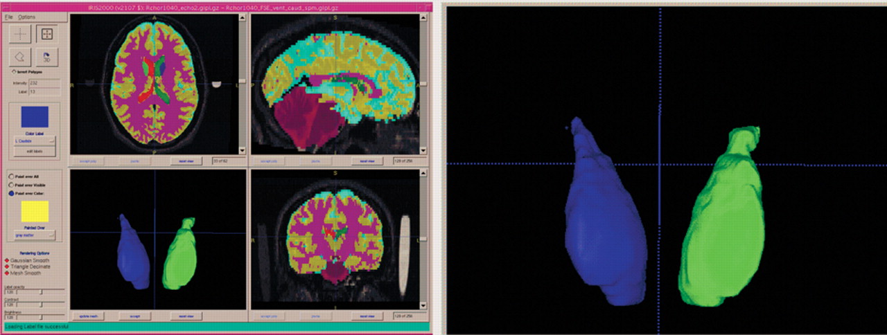
Patients had a mean brain volume of 1,204.04 (SD=113.88) cc, with a mean white matter volume of 429.25 (SD=39.7) cc, a mean gray matter volume of 608.2 (SD=62.6) cc, and a mean CSF volume of 167.5 (SD=27.8) cc. Patients had a left caudate volume of 4.0 (SD=0.6) cc, a right caudate volume of 4.2 (SD=0.6) cc, and a total caudate volume of 8.2 (SD=1.1) cc.
Relationships between caudate volumes and aggression are shown in Table 1 . Larger left caudate volumes were associated with higher TAS scores during the study period. Moreover, regarding the incident status, patients who had any aggressive incident had larger caudate volumes than did those who had no such incidents. This pattern seemed particularly prevalent on the left. Finally, left caudate volumes were larger in patients with lesser degrees of impulse control, with a similar trend for total caudate volumes.
TABLE 1. Relationships Between Aggression and Caudate Volumes 1
Left caudate volumes were inversely correlated with age (r=−0.30, p=0.04). However, caudate volumes did not correlate significantly with the duration of illness. Finally, controlling for age and intracranial volume, ESRS scores did not correlate with caudate volumes.
We conducted additional analyses with gender, substance use disorder, and alcohol use disorder as covariates. The results using these covariates were essentially unchanged from those discussed above.

DISCUSSION

The primary finding of this study was that larger caudate volumes were associated with aggressive behavior. These results were independent of effects due to age, alcohol use disorders, or substance use disorders.
Previous work suggested that treatment with typical antipsychotic drugs increases caudate volumes. Basal ganglia volumes are increased in patients with schizophrenia in vivo (using MRI) 21, 22 and post-mortem. 23 This increase may vary with treatment history. Schizophrenia patients who had been treated with typical antipsychotic drugs showed increases in the volume of the caudate. 24, 25 In a post-mortem study in rats, chronic treatment with haloperidol was associated with increased striatal volumes. 26 Thus, the effect of haloperidol on striatal volumes in patients may be related to treatment rather than to the pathophysiology of schizophrenia. In further support of this argument, when patients were switched to clozapine, an atypical antipsychotic drug, caudate volumes were reduced. 27, 28 Interestingly, clozapine has well-known antiaggressive effects in schizophrenia. 3, 29
The duration of typical antipsychotic drug treatment may be related to increases in volume of the basal ganglia, 30 suggesting that the effects of such treatment may be cumulative. However, in the current study, duration of illness did not account for the relationships between caudate and ventricular volumes and outcome. It is possible that long-term treatment with typical antipsychotic drugs had a nonlinear relationship with the volume increase we measured. The effects may plateau after a certain period of exposure to typical neuroleptics, with no further progression.
The mean duration of patients’ treatment with typical antipsychotic drugs (estimated by the time elapsed since first hospitalization) was 18.6 years. Treatment resistance to antipsychotic drugs was an eligibility criterion for participation in this study. In the past, treatment resistant patients, particularly those showing persistent aggressive behavior, were treated with ever increasing doses of typical antipsychotic drugs. Clinically unsuccessful as it was, such high-dose treatment may have resulted in cumulative effects on the caudate. Thus, the relation between caudate size and aggression we observed could have arisen as an epiphenomenon: patients failed to respond, were aggressive, therefore received high-dose long-term treatment with antipsychotic drugs, and that treatment coincidentally resulted in caudate volume increase. If this was true, the caudate volume increase was a consequence (rather than an antecedent) of aggression, and it was mediated by treatment with antipsychotic drugs.
However, this iatrogenic mechanism may not fully explain the observed relation between caudate size and aggression. Long-term treatment of nonresponding aggressive patients is not necessary for a caudate volume increase: the caudate volume was reported to increase early in the first year of treatment even in first-episode patients responding to pharmacotherapy. 24 Consistent with that observation, 24 an increase in striatal blood flow was reported in first-episode, neuroleptic-naïve schizophrenia patients after the initiation of pharmacotherapy. 31 Our lack of finding a significant relationship between caudate volume increase and duration of illness also points to other contributing factors. Striatal blood flow may be related to antipsychotic 31 treatment as well as to aggression. 9
Caudate dysfunction, perhaps independently of its origin (pharmacological or otherwise), may interfere with the normal functioning of the frontal-subcortical circuitry. Cummings 5 proposed a prefrontal-subcortical circuit, including the caudate, corresponding to the orbitofrontal lobe neurobehavioral syndromes. Empirical observations indicate that penetrating brain wounds causing frontal medial lesions were associated with more aggressive behavior than lesions to other brain areas. 32 Consistent with these ideas, Hoptman et al. 33, 34 found significant negative correlations between white matter integrity, as measured using diffusion tensor imaging, and both impulsivity and aggression in a variety of areas, including the caudate. Because white matter tracts play an important role in the connectivity of the brain, the disruptions of white matter integrity in the regions mentioned may well have implications for the orbitofrontal syndrome.
The current findings have implications for the interpretation of studies in which the relationship between functional outcome and brain volumes is studied. Thus, for volumetric studies of schizophrenia, it is important to know the patients’ treatment history.
The study has some important limitations. The subjects who received the MRI may not be representative of treatment-resistant schizophrenia patients, or even of the small sample from which they were selected. In addition, the sample was not specifically sampled for a high rate of aggressive incidents. Finally, we did not have information on head injury or criminal history in these patients.
In conclusion, in this sample of patients with treatment-resistant schizophrenia, larger caudate volumes were associated with aggression. This association may be due to treatment with antipsychotic drug, to the pathophysiology of schizophrenia, or to a combination of these two mechanisms.

Acknowledgments

This study was supported by NIMH grant (R10 MH53550). Additional support was provided by the UNC-Mental Health and Neuroscience Clinical Research Center (MH MH33127). The authors also acknowledge the Stanley Medical Research Institute, Grant # 01-109, for supporting the development of improved methods and application to this clinical study. The authors thank Janssen Pharmaceutica Research Foundation, Eli Lilly and Company, Novartis Pharmaceuticals Corporation, and Merck and Co., Inc., for their generous gifts of medications. Eli Lilly and Company contributed supplemental funding. However, overall experimental design, data acquisition, statistical analyses, and interpretation of the results were implemented without any input from any of the pharmaceutical companies. MJH received additional support from R01 MH064783 and from a NARSAD Young Investigator Award. JAL is now the Director of New York State Psychiatric Institute, NY, NY and Chairman, Department of Psychiatry, College of Physicians and Surgeons, Columbia University, NY, NY

Footnote

Drs. Hoptman, Volavka, and Lindenmayer are affiliated with the Nathan S. Kline Institute for Psychiatric Research, Orangeburg, New York, and the Department of Psychiatry, New York University School of Medicine, New York, New York. Dr. Lindenmayer is also affiliated with the Manhattan Psychiatric Center, New York, New York. Drs. Czobor and Citrome are affiliated with the Nathan S. Kline Institute for Psychiatric Research and Dov Pharmaceuticals, Hackensack, New Jersey. Drs. Gerig and Lieberman are affiliated with the University of North Carolina, Chapel Hill, North Carolina. Dr. Chakos is affiliated with SUNY Downstate Medical Center, Brooklyn, New York. Dr. Bilder is affiliated with the Department of Psychiatry, University of California at Los Angeles, Los Angeles, California. Address correspondence to Dr. Hoptman, 140 Old Orangeburg Rd., Building 35, Orangeburg, NY 10962; [email protected] (E-mail).
Copyright © 2006 American Psychiatric Publishing, Inc.

References

1.
Niznikiewicz MA, Kubicki M, Shenton ME: Recent structural and functional imaging findings in schizophrenia. Curr Opin in Psychiatry 2003; 16:123–147
2.
Raine A, Lencz T, Bihrle S, et al: Reduced prefrontal gray matter volume and reduced autonomic activity in antisocial personality disorder. Arch Gen Psychiat 2000; 57:119–127
3.
Volavka J: Neurobiology of Violence, 2nd ed. Washington, DC, APA Publishing, 2002
4.
Folstein SE: Huntington’s Disease: A Disorder of families. Baltimore, Johns Hopkins University Press, 1989
5.
Cummings JL: Frontal-subcortical circuits and human behavior. Arch Neurol 1993; 50:873–880
6.
Richfield EK, Twyman R, Berent S: Neurological syndrome following bilateral damage to the head of the caudate nuclei. Ann Neurol 1987; 22:768–771
7.
Mendez MF, Adams NL, Lewandowski KS: Neurobehavioral changes associated with caudate lesions. Neurology 1989; 39:349–354
8.
Soderstrom H, Hultin L, Tullberg M, et al: Reduced frontotemporal perfusion in psychopathic personality. Psychiatry Res: Neuroimaging 2002; 114:81–94
9.
Amen DG, Stubblefield M, Carmichael B, et al: Brain SPECT findings and aggressiveness. Annals of Clin Psychiatry 1996; 8:129–139
10.
Kiehl KA, Smith AM, Hare RD, et al: Limbic abnormalities in affective processing by criminal psychopaths as revealed by functional magnetic resonance imaging. Biol Psychiatry 2001; 50:677–684
11.
Volavka J, Czobor P, Sheitman B, et al: Clozapine, olanzapine, risperidone, and haloperidol in patients with chronic schizophrenia and schizoaffective disorder. Am J Psychiatry 2002; 159:255–262
12.
First MB, Spitzer RL, Gibbon M, et al: Structured Clinical Interview for DSM-IV Axis-I Disorders: SCID Clinician Version. New York, Biometrics Research Department, New York State Psychiatric Institute, Department of Psychiatry, Columbia University, 1995
13.
Maes F, Collignon A, Vandermeulen D, et al: Multi-modality image registration by maximization of mutual information. IEEE Transactions on medical imaging 1997; 16:187–198
14.
Van Leemput K, Maes F, Vandermeulen D, et al: Automated model-based tissue classification of MR images of the brain. IEEE Trans Med Imaging 1999; 18:897–908
15.
Kay SR, Opler LA, Fiszbein A: Positive and Negative Syndrome Scale (PANSS). New York, Bronx Psychiatric Center, 1986
16.
Chouinard G, Ross-Chouinard A, Annable L, et al: Extrapyramidal Symptom Rating Scale. Can J Neurol Sci 1980; 7:233
17.
Yudofsky SC, Silver JM, Jackson W, et al: The overt aggression scale for the objective rating of verbal and physical aggression. Am J Psychiatry 1986; 143:35–39
18.
Volavka J, Czobor P, Nolan K, et al: Overt aggression and psychotic symptoms in patients with schizophrenia treated with clozapine, olanzapine, risperidone, or haloperidol. J Clin Psychopharmacol (Berl) 2004; 24:225–228
19.
Czobor P, Volavka J, Meibach RC: Effect of risperidone on hostility in schizophrenia. J Clin Psychopharmacol (Berl) 1995; 15:243–249
20.
Citrome L, Casey DE, Daniel DG, et al: Adjunctive divalproex and hostility among patients with schizophrenia receiving olanzapine or risperidone. Psychiatr Services 2004; 55:290–294
21.
DeLisi LE, Hoff AL, Schwartz JE, et al: Brain morphology in first-episode schizophrenia-like psychotic patients: a quantitative magnetic resonance imaging study. Biol Psychiatry 1991; 28:159–175
22.
Jernigan TL, Zisook S, Heaton RK, et al: Magnetic resonance imaging abnormalities in lenticular nuclei and cerebral cortex in schizophrenia. Arch Gen Psychiatry 1991; 48:881–890
23.
Heckers S, Heinsen H, Heinsen Y, et al: Cortex, white matter, and basal ganglia in schizophrenia: A volumetric postmortem study. Biol Psychiatry 1991; 29:556–566
24.
Chakos MH, Lieberman JA, Bilder RM, et al: Increase in caudate nuclei volumes of first-episode schizophrenic patients taking antipsychotic drugs. Am J Psychiatry 1994; 151:1430–1436
25.
Keshavan MS, Bagwell WW, Haas GL, et al: Changes in caudate volume with neuroleptic treatment. Lancet 1994; 344:1434
26.
Chakos M, Shirakawa O, Lieberman J, et al: Striatal enlargement in rats chronically treated with neuroleptic. Biol Psychiatry 1998; 44:675–684
27.
Chakos MH, Lieberman JA, Alvir J, et al: Caudate nuclei volumes in schizophrenic patients treated with typical antipsychotics or clozapine. Lancet 1995; 345:456–457
28.
Corson PW, Nopoulos P, Miller DD, et al: Change in basal ganglia volume over 2 years in patients with schizophrenia: typical versus atypical neuroleptics. Am J Psychiatry 1999; 156:1200–1204
29.
Citrome L, Volavka J, Czobor P, et al: Effects of clozapine, olanzapine, risperidone, and haloperidol on hostility among patients with schizophrenia. Psychiatr Services 2001; 52:1510–1514
30.
Lang DJ, Kopala LC, Vandrope RA, et al: An MRI study of basal ganglia volumes in first-episode schizophrenia patients treated with risperidone. Am J Psychiatry 2001; 158:625–631
31.
Corson PW, O’Leary DS, Miller DD, et al: The effects of neuroleptic medications on basal ganglia blood flow in schizophreniform disorders: a comparison between the neuroleptic-naive and medicated states. Biol Psychiatry 2002; 52:855–862
32.
Grafman J, Schwab K, Warden D, et al: Frontal lobe injuries, violence, and aggression: a report of the vietnam head injury study. Neurology 1996; 46:1231–1238
33.
Hoptman MJ, Volavka J, Johnson G, et al: Frontal white matter, aggression and impulsivity in men with schizophrenia. Biol Psychiatry 2002; 52:9–14
34.
Hoptman MJ, Ardekani BA, Butler PD, et al: DTI and impulsivity in schizophrenia: A first voxelwise correlational study. Biol Psychiatry 2004; 15:2467–2470

Information & Authors

Information

Published In

Go to The Journal of Neuropsychiatry and Clinical Neurosciences
The Journal of Neuropsychiatry and Clinical Neurosciences
Pages: 509 - 515
PubMed: 17135376

History

Published online: 1 October 2006
Published in print: Fall, 2006

Authors

Affiliations

Matthew J. Hoptman, Ph.D.
Jan Volavka, M.D., Ph.D.
Leslie L. Citrome, M.D., M.P.H.
Jean-Pierre Lindenmayer, M.D.
Jeffrey A. Lieberman, M.D.
Robert M. Bilder, Ph.D.

Metrics & Citations

Metrics

Citations

Export Citations

If you have the appropriate software installed, you can download article citation data to the citation manager of your choice. Simply select your manager software from the list below and click Download.

For more information or tips please see 'Downloading to a citation manager' in the Help menu.

Format
Citation style
Style
Copy to clipboard

There are no citations for this item

View Options

View options

PDF/ePub

View PDF/ePub

Get Access

Login options

Already a subscriber? Access your subscription through your login credentials or your institution for full access to this article.

Personal login Institutional Login Open Athens login
Purchase Options

Purchase this article to access the full text.

PPV Articles - Journal of Neuropsychiatry and Clinical Neurosciences

PPV Articles - Journal of Neuropsychiatry and Clinical Neurosciences

Not a subscriber?

Subscribe Now / Learn More

PsychiatryOnline subscription options offer access to the DSM-5-TR® library, books, journals, CME, and patient resources. This all-in-one virtual library provides psychiatrists and mental health professionals with key resources for diagnosis, treatment, research, and professional development.

Need more help? PsychiatryOnline Customer Service may be reached by emailing [email protected] or by calling 800-368-5777 (in the U.S.) or 703-907-7322 (outside the U.S.).

Media

Figures

Other

Tables

Share

Share

Share article link

Share