Full access
Article
Published Online: 1 September 2006

Increased 5-HT 2A Receptor Binding in Euthymic, Medication-Free Patients Recovered From Depression: A Positron Emission Study With [ 11 C]MDL 100,907

Publication: American Journal of Psychiatry

Abstract

Objective: A previous positron emission tomography (PET) study reported increased serotonin 5-HT 2A receptor binding in unmedicated depressed patients with high scores on the Dysfunctional Attitudes Scale. The purpose of the present study was to use the highly selective 5-HT 2A receptor ligand [ 11 C]MDL 100,907 in a PET imaging paradigm to assess 1) 5-HT 2A receptor binding potential in euthymic subjects with a history of recurrent depression and 2) the relationship between receptor binding and scores on the Dysfunctional Attitudes Scale. Method: Cortical 5-HT 2A receptor binding was measured in 20 unmedicated, fully recovered unipolar depressed patients and 20 age- and gender-matched comparison subjects. Regional estimates of binding potential were obtained using a reversible plasma input function compartmental model and the cerebellum as a reference region to estimate the free and non-specifically bound [ 11 C]MDL 100,907 in brain tissue. Results: Relative to the comparison subjects, the recovered depressed patients demonstrated significantly higher 5-HT 2A receptor binding potential in the frontal cortex (mean increase: 19%), parietal cortex (mean increase: 25%), and occipital cortex (mean increase: 19%). 5-HT 2A receptor binding potential correlated negatively with age in both patients and comparison subjects and positively with the Dysfunctional Attitudes Scale in the recovered patients. Conclusions: These findings should be considered preliminary but suggest that recovered subjects with a history of recurrent major depression have elevated binding potential of cortical 5-HT 2A receptors. The correlation of increased 5-HT 2A receptor binding potential with increased scores on Dysfunctional Attitudes Scale supports earlier work suggesting that increased 5-HT 2A receptor availability characterizes a group of depressed patients with high levels of dysfunctional attitudes.
Postsynaptic serotonin 5-HT 2A receptors play an important role in mediating the effects of serotonin on local cortical circuitry and are targets for certain antidepressant medications (1) . The 5-HT 2A receptor is found predominantly in cortical areas and is located on both glutamatergic pyramidal neurons and γ-aminobutyric acid (GABA) interneurons (2, 3) . The binding density of 5-HT 2A receptors has been investigated extensively in postmortem studies, and evidence suggests an increase in 5-HT 2A receptor numbers in the prefrontal cortex in suicide victims (4, 5), although there are some notable inconsistencies (6, 7) . Similarly, there has been some limited evidence for an increase in cortical 5-HT 2A receptor binding in dying patients with depression (8, 9) .
The difficulties involved in postmortem ligand binding work (10) have stimulated studies of 5-HT 2A receptors in living human brain through positron emission tomography (PET) and single photon emission tomography (SPET). In contrast to the postmortem work, most imaging studies in depressed patients have found either no change or a decrease in 5-HT 2A receptor binding in the cerebral cortex (reviewed by Stockmeier [11] ). However, methodological problems related to the available radioligands (12, 13) as well as to the use of psychotropic medications prior to scanning (11) limit the conclusions that can be drawn from these studies. In support of this notion, a previous study of acutely depressed patients who were drug free for more than 3 months found increased 5-HT 2A receptor binding in the frontal cortex of subjects with high scores on the Dysfunctional Attitudes Scale (14) .
The development of [ 11 C]MDL 100,907 as a PET radioligand offers potential advantages in terms of receptor selectivity because [ 11 C]MDL 100,907 binds with a high and selective affinity to 5-HT 2A receptors and, when used in conjunction with PET, produces high-quality images that correspond well with known 5-HT 2A binding distribution derived from postmortem studies (1518) . Furthermore, [ 11 C]MDL 100,907 does not give rise to radiolabeled metabolites that are likely to cross the blood-brain barrier, and radioligand binding can be reliably quantified (16, 19, 20) .
In the present study we used [ 11 C]MDL 100,907 to measure brain 5-HT 2A receptor binding in subjects recovered from depression and matched healthy comparison subjects. We studied recovered subjects because this allows measurement of 5-HT 2A receptor binding free from the confounding effects of acute illness and recent antidepressant treatment. On the basis of postmortem data and the previous demonstration of increased 5-HT 2A receptor binding in unmedicated depressed subjects with high levels of dysfunctional attitudes, we predicted that euthymic, medication-free subjects recovered from depression but with recurrent illness would have higher 5-HT 2A receptor binding potential in cortical regions relative to healthy comparison subjects.

Method

Participants

A total of 40 subjects were recruited for the study, which was approved by the Research Ethics Committee at Hammersmith Hospital, London; the Oxfordshire Psychiatric Research Ethics Committee, Oxford, U.K.; and the U.K. Administration of Radioactive Substances Advisory Committee. All subjects gave written informed consent for the study.
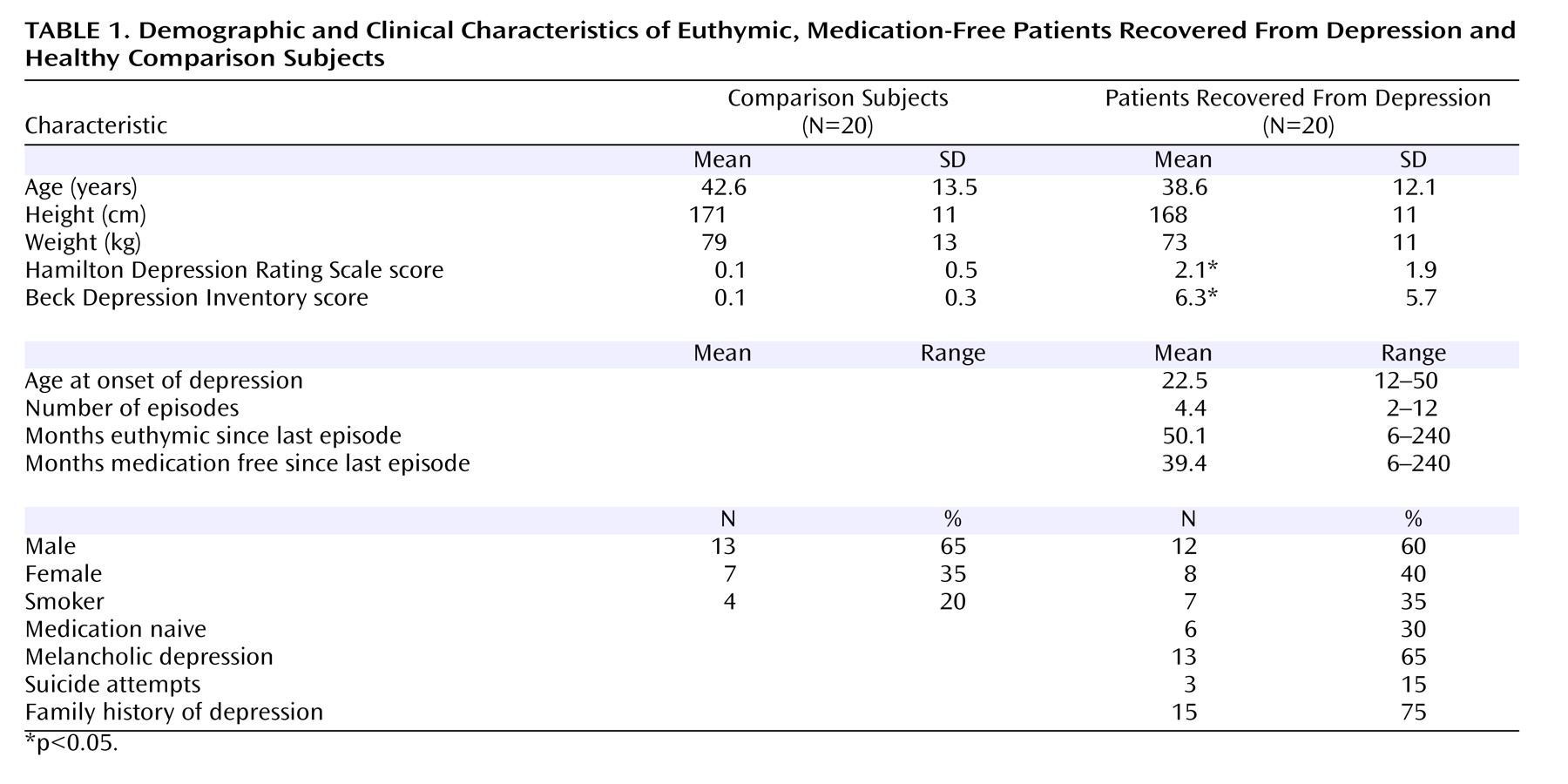
Demographic and clinical characteristics of all subjects are presented in Table 1 . The study group consisted of 20 healthy subjects with no current or past psychiatric history and 20 patients who had experienced at least two episodes of unipolar major depression in the past but at the time of the study were medication free and had been euthymic for a minimum of 6 months. All subjects were screened using the Structured Clinical Interview for DSM-IV Axis I Disorders (SCID). Exclusion criteria for both groups were current or past serious medical or neurological illness, alcohol or illicit substance dependence, or other axis I or II disorders. All subjects were administered the 17-item Hamilton Depression Rating Scale and the Beck Depression Inventory. Subjects were classified as having recovered from depression on the basis of four criteria: self-reported euthymia following the last episode of major depression, clinician-rated euthymia per clinical interview with an experienced psychiatrist, absence of major depressive episode criteria as judged by the SCID, and a Hamilton depression scale score <7 (21) . Only subjects who met all four criteria for recovery in addition to being euthymic and medication free for more than 6 months were included in the study and were subsequently scanned. Because it has previously been reported that ratings on a scale measuring dysfunctional emotional attitudes correlated positively with frontal 5-HT transporter (22) and 5-HT 2A receptor binding in depressed patients (14), all subjects were also asked to complete the 40-item Dysfunctional Attitudes Scale (23) .

PET Scanning Protocol

All subjects had one PET scan with [ 11 C]MDL 100,907 performed on a high-sensitivity Siemens/CTI scanner ECAT EXACT3D with an axial field of view of 23.4 cm and 95 reconstructed transaxial image planes (24) . A 5-minute transmission scan using a 137 Cs point source was carried out prior to each study for subsequent attenuation and scatter correction. The 95-minute three-dimensional dynamic emission scan was acquired in list mode. In the postacquisition frame rebinning, 28 time frames of increasing length were generated (30-second background frame prior to the injection, three 10-second frames, three 20-second frames, three 30-second frames, three 60-second frames, four 120-second frames, six 300-second frames, and five 600-second frames). The spatial resolution of the images reconstructed using filtered back projection is close to isotropic: 5.1 mm full width at half maximum transaxially and 5.9 mm full width at half maximum axially averaged over a radius of 10 cm from the center of the field of view (24) .
The radiotracer [ 11 C]MDL 100,907 was prepared as described previously (15) and was injected into an antecubital vein as a smooth bolus over 30 seconds. There were no significant differences between comparison subjects and patients in terms of mean injected radioactivity dose (359.2 MBq [SD=11.1] and 352.6 MBq [SD=20.7], respectively), radiochemical purity of the injected [ 11 C]MDL 100,907 (98.9% [SD=1.2] and 99.4% [SD=1.1]), injected mass of MDL 100,907 (4.3 μg [SD=2.9] and 3.5 μg [SD=2.2]), or specific activity (47 GBq/μmol [SD=23] and 35 GBq/μmol [SD=17]).
Arterial whole blood activity was monitored continuously for the first 15 minutes of the scan with a bismuth germanate coincidence detector (25) . A total of 10 discrete arterial blood samples were manually sampled at 5, 10, 15, 20, 30, 40, 50, 60, 75, and 95 minutes following injection of the radioligand. The activity concentration of the whole blood was measured. Plasma was removed by centrifugation at 2000 g for 2 minutes and was also measured for radioactivity concentration. Eight plasma samples per scan (sampled at 5, 15, 30, 40, 50, 60, 75, and 95 minutes after the injection of the radioligand) were analyzed for metabolites by using a semiautomated system with online solid-phase extraction followed by reverse-phase chromatography.

Region of Interest Definition

Regions of interest were defined using a modification of a previously described procedure (26) . Briefly, a radioligand-specific [ 11 C]MDL 100,907 PET template was created in standard stereological space (Montreal Neurological Institute [MNI]) stereological space. This template was spatially normalized to each individual’s PET image using standard software (Statistical Parametric Mapping-2 [SPM2], online at http://www.fil.ion.ucl.ac.uk/spm). The deformation parameters used in the transformation were applied to a probabilistic atlas of brain regions of interest also in standard MNI stereological space to provide an individual region of interest atlas for each subject. Regional time activity curves were then generated by applying the individual region of interest atlas on each [ 11 C]MDL 100,907 dynamic image using the medical imaging software ANALYZE (27) . Using a combination of a well-defined region of interest probabilistic atlas and a [ 11 C]MDL 100,907 PET template eliminates observer bias for region of interest definitions that could confound results. While our main region of interest was the frontal cortex, we also examined the parietal, occipital, and temporal cortices in order to establish whether any increase in frontal 5-HT 2A receptor binding would generalize across other cortical regions. Left and right regions were combined.

Data Analysis

Estimates of the microparameters of the reversible two-tissue, four-rate-constant compartment model with free blood volume term were obtained from fits to the measured time activity curves (no decay correction applied), minimizing the weighted sum of squares of the differences between the data and the model by the simplex method (28) .
The total volume of distribution (V D ) was calculated as
Using the definition of binding potential (BP) as introduced by Mintun et al. (29) and with f 2 as the free fraction of radioligand in tissue, an estimate of the binding potential in a target region with specific binding can be obtained from the estimated V D in that region and the estimated V D in a reference region without specific binding, provided that the volumes of distribution of the free and nonspecifically bound [ 11 C]MDL 100,907 are the same throughout the brain.
As with other imaging studies of 5-HT 2A receptor binding in humans, we used the cerebellum as the reference region (14, 30) . The use of the cerebellum as a reference region with a negligible concentration of 5-HT 2A receptors is based on combined evidence from postmortem studies using autoradiography, protein expression, and mRNA expression (18, 31) . We have previously examined the use of the cerebellum as a reference region for the quantification of [ 11 C]MDL 100,907 binding in humans by administering a single oral, 30-mg dose of mirtazapine, a novel 5-HT 2A antagonist antidepressant, to healthy human subjects. Although we observed an occupancy effect of approximately 70% in most brain regions, mirtazapine did not alter cerebellar V D compared with the baseline condition (20) .

Statistical Analysis

Statistical analyses were performed using Statistical Package for Social Sciences (SPSS Inc., Chicago) version 12. We tested our hypothesis that there would be increased 5-HT 2A receptor binding in cortical regions in patients recovered from depression using a repeated measures analysis of covariance (ANCOVA) with “diagnosis” (patients versus healthy subjects) as a between-subjects factor and “region” (cortical brain region) as a within-subjects factor; a Huynh-Feldt correction was applied as appropriate where the assumptions of sphericity were violated. Because of the well-described effect of age on 5-HT 2A binding, age was added as a covariate for all analyses. Significant interactions or main effects were explored using post hoc independent two-tailed t tests. Correlations were analyzed using Pearson’s correlation coefficient, and significance was set at p<0.05.

Results

Preliminary analysis showed no main or interactive effect of gender on 5-HT 2A binding potential; gender was therefore omitted from subsequent analyses. There was no statistically significant difference in cerebellar V D between healthy subjects (19.4 ml plasma/ml tissue [SD=1.4]) and patients (19.2 ml plasma/ml tissue [SD=2.8]) (F=0.07, df=1, 37, p<0.79). There was no effect of age on cerebellar V D values (F=1.48, df=1, 37, p=0.23).
Three outcome measures of PET neuroreceptor quantification are commonly considered, all of which are proportional to the availability of receptors (32, 33) . In this study, we report our results as f 2 ·BP (i.e., the product of the free fraction in tissue and binding potential) because this outcome parameter has been shown in several neuroreceptor PET studies (33) to be the most reproducible and least susceptible to experimental error. However, the use of this parameter is based on the assumption of uniformity of the nondisplaceable compartment across subjects and groups. In our study, there was no significant difference in the V D of the reference region. Therefore, f 2 would appear to be homogeneous across our study group, and results are reported as f 2 ·BP.
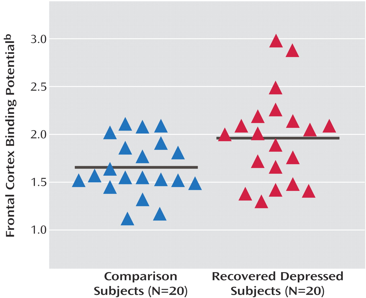
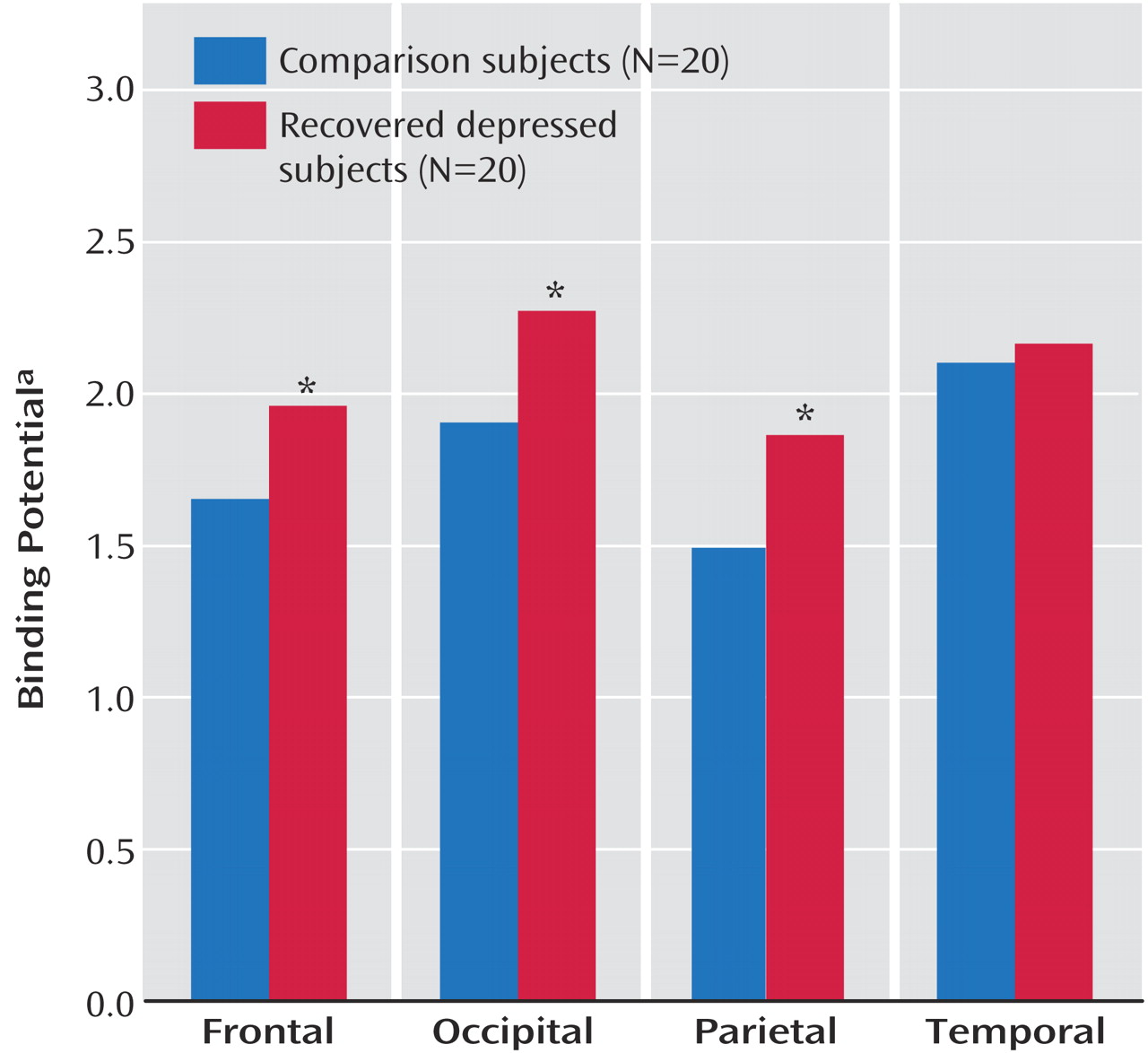
An ANCOVA of regional cortical 5-HT 2A binding potential values with age as a covariate showed significant main effects of region (F=9.40, df=3, 102, p<0.001) and age (F=9.1, df=1, 34, p=0.005) but not diagnosis (F=1.63, df=1, 34, p=0.21). There was, however, a significant diagnosis-by-region interaction (F=4.69, df=3, 102, p<0.02). Post hoc analysis showed that patients had significantly increased 5-HT 2A binding potential in the frontal cortex, occipital cortex, and parietal cortex but not in the temporal cortex ( Figure 1 and Figure 2 ). Due to the effect of age on 5-HT 2A binding, we further examined a subset of 16 patients and 16 comparison subjects who were individually matched for age to within 1 year (healthy subjects: mean=39.6 years [SD=13.3], patients: mean=39.6 years [SD=13.2]; p=0.99). An ANCOVA (with age as a covariate) also revealed a significant region-by-diagnosis interaction (F=4.26, df=3, 78, p<0.02) and a significant main effect of age (F=9.51, df=1, 26, p=0.005).
Figure 1. Frontal Cortex Binding Potential in Euthymic, Medication-Free Patients Recovered From Depression and Healthy Comparison Subjects a
a Solid lines are group means. Post hoc analysis revealed significantly greater 5-HT 2A binding potential in the recovered depressed patients (p<0.05).
b Product of free fraction of radioligand in tissue and binding potential.
Figure 2. Binding Potential in Frontal, Occipital, Parietal, and Temporal Cortices in Euthymic, Medication-Free Patients Recovered From Depression Relative to Healthy Comparison Subjects
a Product of free fraction of radioligand in tissue and binding potential.
* p<0.05.
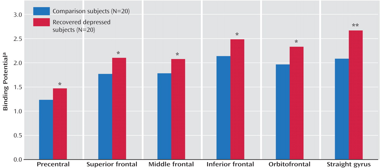
Most of the postmortem work on 5-HT 2A receptor binding in suicide victims has focused on various subregions of the frontal cortex. To explore the increase in 5-HT 2A binding potential in the frontal cortex in more detail, we performed a further analysis of frontal cortical regions examining the superior, middle, and inferior frontal gyri, the orbitofrontal cortex, the straight gyrus, and the precentral gyrus. An ANCOVA of these regions with age as a covariate showed a main effect of diagnosis (F=4.75, df=1, 36, p<0.04) and age (F=6.43, df=1, 36, p<0.02) but no significant interactions between diagnosis and region (F=2.01, df=5, 180, p<0.13). Post hoc analysis of 5-HT 2A binding potential showed that patients had statistically significant increases in binding potential values in all six frontal regions ( Figure 3 ).
Figure 3. Frontal Region Binding Potential in Euthymic, Medication-Free Patients Recovered From Depression and Healthy Comparison Subjects
a Product of free fraction of radioligand in tissue and binding potential.
* p<0.05. ** p=0.005.
In the combined population of patients and comparison subjects, age correlated negatively with 5-HT 2A binding potential in all four cortical regions studied (frontal: r=–0.39, p<0.02; temporal: r=–0.59, p<0.001; parietal: r=–0.36, p<0.03; occipital: r=–0.43, p=0.006). Global 5-HT 2A binding potential in the frontal cortex did not correlate significantly with any of the measured clinical characteristics in the patient group such as age at illness onset, lifetime duration of illness, number of episodes, duration of euthymia, or duration medication free. Notwithstanding the small numbers, there were no statistically significant differences within the patient group in 5-HT 2A receptor binding potential between patients with versus without melancholia, with versus without a family history of depression, or with versus without a history of self-harm.
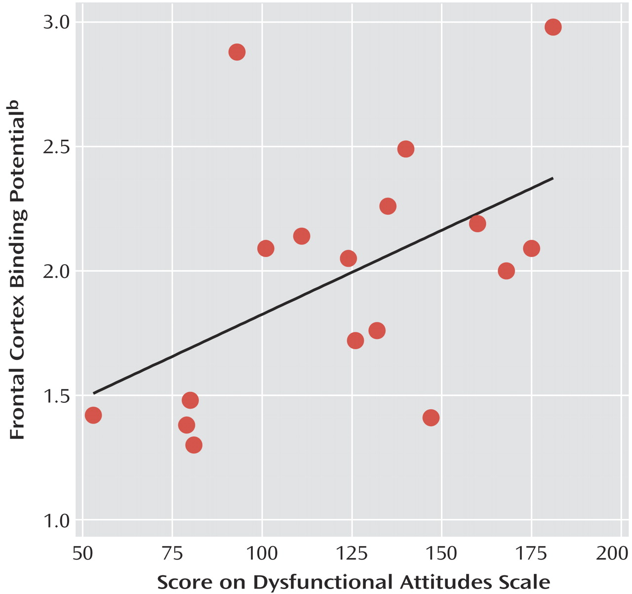
Three of the patients did not complete the Dysfunctional Attitudes Scale and were not included in the analysis. In the 17 remaining subjects, Dysfunctional Attitudes Scale scores were significantly higher (p=0.02) in patients (mean=123 [SD=38]) relative to comparison subjects (mean=97 [SD=19]). In keeping with previous findings (14), an ANCOVA with age and Dysfunctional Attitudes Scale scores as covariates showed significant main effects of Dysfunctional Attitudes Scale score (F=6.24, df=1, 27, p<0.02) and age (F=6.22, df=1, 27, p<0.02) on frontal 5-HT 2A binding potential. Dysfunctional Attitudes Scale scores also correlated significantly with frontal 5-HT 2A binding potential in the patients ( Figure 4 ) but not in healthy subjects (r=–0.31, df=18, p=0.28). There was no significant correlation between age and frontal binding potential in the patients considered alone (r=–0.37, df=15, p=0.11). However, the correlation of Dysfunctional Attitudes Scale scores and frontal 5-HT 2A binding potential in the patients remained significant after partial correlation that controlled for the effect of age ( Figure 4 ).
Figure 4. Correlation of Dysfunctional Attitudes Scale Scores With Frontal Binding Potential in Euthymic, Medication-Free Patients Recovered From Depression a
a The significant correlation (r=0.50, df=15, p=0.04) remained after partial correlation that controlled for the effect of age (r=0.54, p=0.03). Linear R 2 =0.253.
b Product of free fraction of radioligand in tissue and binding potential.

Discussion

Our findings indicate that recovered depressed patients have higher [ 11 C]MDL 100,907 binding potential in several cortical regions compared with healthy subjects. This is consistent with a greater availability of 5-HT 2A receptors. As reported by others, 5-HT 2A receptor binding showed an inverse correlation with age (30, 34) . The mean age of our patient group was slightly but nonsignificantly lower than the comparison group, but recovered depressed subjects had significantly higher 5-HT 2A binding potential even after we adjusted for age with a covariance analysis. In addition, individually matching subjects for age also revealed a significant increase in [ 11 C]MDL 100,907 binding. Finally, Dysfunctional Attitudes Scale scores were significantly higher in the recovered patients relative to healthy comparison subjects and as reported by Meyer and colleagues (14) . 5-HT 2A binding potential was significantly and positively correlated with Dysfunctional Attitudes Scale scores in the depressed subjects but not in the comparison subjects. While the effect size (1.06) of the difference in binding potential between comparison and recovered subjects would not be considered trivial (35), our findings should be considered preliminary until replicated.
In PET neuroreceptor imaging, differences in binding potential do not distinguish between changes in the concentration of binding sites or changes in apparent receptor affinity or the concentration of competing endogenous ligands. There is currently no evidence from postmortem data to suggest that depression is associated with a change in the affinity of 5-HT for the 5-HT 2A receptor. Theoretically, increased 5-HT 2A receptor availability might also be caused by decreased concentrations of synaptic 5-HT (1) . However, neither human nor animal studies suggest that [ 11 C]MDL 100,907 binding is modified by acute increases or decreases in the availability of endogenous 5-HT (36, 37) . Therefore, the increase in 5-HT 2A receptor binding potential observed in the subjects recovered from depression is likely to represent an increase in the density of 5-HT 2A receptor binding sites.
As noted here, the status of cortical 5-HT 2A receptors has been investigated extensively in depressed patients with varying results. Data from postmortem studies of suicide victims suggest increased 5-HT 2A receptor density in the frontal cortex (4, 5), but in vivo imaging studies in acutely depressed subjects have found mostly either a reduction or no change in 5-HT 2A receptor availability (11) . The reason for this apparent discrepancy may relate to particular clinical characteristics associated with suicidal behavior, such as diagnostic comorbidity, substance use, agitation, and insomnia as well as the methodological difficulties involved in carrying out postmortem studies (10) . In vivo imaging studies allow investigation of prospectively well-characterized subjects, but interpretation of results can be complicated by factors such as previous antidepressant treatment and issues relating to ligand specificity and modeling (11) .
Our finding of a significant positive correlation between 5-HT 2A binding potential and Dysfunctional Attitudes Scale scores in recovered depressed subjects is in keeping with previous studies in which relative to healthy subjects depressed subjects with higher Dysfunctional Attitudes Scale scores had increased 5-HT 2A receptor binding (as measured by [ 18 F]setoperone) (14) and 5-HTT binding (measured with [ 11 C]DASB) (22) . While Dysfunctional Attitudes Scale scores tend to normalize with clinical improvement, recovered depressed subjects can still demonstrate higher levels of dysfunctional attitudes than never depressed healthy subjects (38, 39) . Moreover, there are strong correlations between Dysfunctional Attitudes Scale scores of depressed individuals before and after treatment (39, 40) . This raises the possibility that patients with high Dysfunctional Attitudes Scale scores during recovery would also be the individuals with higher Dysfunctional Attitudes Scale scores when depressed. Taken together, the data suggest that increased cortical 5-HT 2A receptor binding identifies a group of depressed patients with higher levels of dysfunctional attitudes both during depression and after clinical recovery.
If 5-HT 2A receptor binding is increased in depressed subjects with high Dysfunctional Attitudes Scale scores, what might be the mechanism? There is much evidence that major depression is associated with decreased brain 5-HT neurotransmission (1), and Meyer et al. (14) suggested that increased 5-HT 2A receptor binding might reflect an up-regulation of cortical 5-HT 2A receptors in response to chronically impaired 5-HT release, particularly in subjects with high Dysfunctional Attitudes Scale scores. Our findings suggest the possibility that this might continue to be the case after clinical recovery and withdrawal of antidepressant treatment. Consistent with this, evidence from 5-HT neuroendocrine challenge tests suggests impaired presynaptic 5-HT function may persist into clinical recovery (41) . It must be noted, however, that 5-HT 2A receptors do not always appear to be regulated classically in terms of neurotransmitter availability, and changes in local receptor trafficking involving protein kinases and arrestins might play a part in the increased 5-HT 2A receptor availability seen in the present study (42) .
Recent animal experimental studies have demonstrated strong reciprocal connections between cortical glutamatergic output neurons and ascending 5-HT pathways from the raphe nuclei (43) . 5-HT 2A receptors are located on glutamatergic cell bodies, postsynaptic to the 5-HT neurons that terminate in frontal cortex (3) . These receptors are therefore ideally placed to regulate the activity of cortical-raphe pathways, one of the functions of which is to provide the raphe with information coding for the controllability of stress (44) . The latter formulation is particularly intriguing, given the correlation between increased cortical 5-HT 2A receptor binding and the Dysfunctional Attitudes Scale. Taken together, these preclinical and clinical data suggest that 5-HT 2A receptors, through their effect on the excitability of glutamatergic neurons, form part of a neurobiological substrate underpinning the dysfunctional beliefs that put individuals at risk of depressive episodes.

Footnote

Received May 8, 2005; revisions received June 10 and Aug. 2, 2005; accepted Aug. 18, 2005. From the University Department of Psychiatry, Warneford Hospital, Oxford, U.K.; Cyclotron Unit, MRC Clinical Sciences Centre, Hammersmith Hospital, London; Department of Psychiatry, Yale University, New Haven, Conn.; and Hammersmith Imanet Ltd., London. Address correspondence and reprint requests to Dr. Bhagwagar, Clinical Neuroscience Research Unit, Abraham Ribicoff Research Facilities, 34 Park St., New Haven, CT 06519-1187; [email protected] (e-mail).Supported by the Medical Research Council. Dr. Cowen is an MRC Clinical Scientist.The authors thank the staff at Hammersmith Imanet (including Andrew Blyth, Hope McDevitt, Stella Ahier, Andreanna Williams, Safiye Osman, Jasbir Grewal, Noora Ali, Vijay Vaja, Sam Tagoe, Deborah Richards, Shaun Creasey, Leonard Schnorr, and many others) for the technical expertise they provided.

References

1.
Duman RS: The neurochemistry of depressive disorders: preclinical studies, in Neurobiology of Mental Illness. Edited by Charney D, Nestler EJ. New York, Oxford University Press, 2004, pp 421–439
2.
Jakab RL, Goldman-Rakic PS: Segregation of serotonin 5-HT2A and 5-HT3 receptors in inhibitory circuits of the primate cerebral cortex. J Comp Neurol 2000; 417:337–348
3.
Willins DL, Deutch AY, Roth BL: Serotonin 5-HT2A receptors are expressed on pyramidal cells and interneurons in the rat cortex. Synapse 1997; 27(1):79–82
4.
Turecki G, Briere R, Dewar K, Antonetti T, Lesage AD, Seguin M, Chawky N, Vanier C, Alda M, Joober R, Benkelfat C, Rouleauet GA: Prediction of level of serotonin 2A receptor binding by serotonin receptor 2A genetic variation in postmortem brain samples from subjects who did or did not commit suicide. Am J Psychiatry 1999; 156:1456–1458
5.
Arango V, Ernsberger P, Marzuk PM, Chen JS, Tierney H, Stanley M, Reis DJ, Mann JJ: Autoradiographic demonstration of increased serotonin 5-HT2 and beta-adrenergic receptor binding sites in the brain of suicide victims. Arch Gen Psychiatry 1990; 47:1038–1047
6.
Arranz B, Eriksson A, Mellerup E, Plenge P, Marcusson J: Brain 5-HT 1A, 5-HT 1D and 5-HT 2 receptors in suicide victims. Biol Psychiatry 1994; 35:457–463
7.
Gross-Isseroff R, Israeli M, Biegon A: Autoradiographic analysis of tritiated imipramine binding in the human brain post mortem: effects of suicide. Arch Gen Psychiatry 1989; 46:237–241
8.
Hrdina PD, Demeter E, Vu TB, Sotonyi P, Palkovits M: 5-HT uptake sites and 5-HT2 receptors in brain of antidepressant-free suicide victims/depressives: increase in 5-HT2 sites in cortex and amygdala. Brain Res 1993; 614(1–2):37–44
9.
Laruelle M, Abi DA, Casanova MF, Toti R, Weinberger DR, Kleinman JE: Selective abnormalities of prefrontal serotonergic receptors in schizophrenia: a postmortem study. Arch Gen Psychiatry 1993; 50:810–818
10.
Lewis DA: The human brain revisited: opportunities and challenges in postmortem studies of psychiatric disorders. Neuropsychopharmacology 2002; 26:143–154
11.
Stockmeier CA: Involvement of serotonin in depression: evidence from postmortem and imaging studies of serotonin receptors and the serotonin transporter. J Psychiatr Res 2003; 37:357–373
12.
Tan PZ, Baldwin RM, van Dyck CH, Al Tikriti M, Roth B, Khan N, Charney DS, Innis RB: Characterization of radioactive metabolites of 5-HT2A receptor PET ligand [18F]altanserin in human and rodent. Nucl Med Biol 1999; 26:601–608
13.
Staley JK, Van Dyck CH, Tan PZ, Al Tikriti M, Ramsby Q, Klump H, Ng C, Garg P, Soufer R, Baldwin RM, Innis RB: Comparison of [(18)F]altanserin and [(18)F]deuteroaltanserin for PET imaging of serotonin(2A) receptors in baboon brain: pharmacological studies. Nucl Med Biol 2001; 28:271–279
14.
Meyer JH, McMain S, Kennedy SH, Korman L, Brown GM, DaSilva JN, Wilson AA, Blak T, Eynan-Harvey R, Goulding VS, Houle S, Links P: Dysfunctional attitudes and 5-HT2 receptors during depression and self-harm. Am J Psychiatry 2003; 160:90–99
15.
Lundkvist C, Halldin C, Ginovart N, Nyberg S, Swahn CG, Carr AA, Brunner F, Farde L: [11C]MDL 100907, a radioligand for selective imaging of 5-HT(2A) receptors with positron emission tomography. Life Sci 1996; 58(10):L-92 (published erratum appears in Life Sci 1996; 58(25):379)
16.
Watabe H, Channing MA, Der MG, Adams HR, Jagoda E, Herscovitch P, Eckelman WC, Carson RE: Kinetic analysis of the 5-HT2A ligand [11C]MDL 100,907. J Cereb Blood Flow Metab 2000; 20:899–909
17.
Ito H, Nyberg S, Halldin C, Lundkvist C, Farde L: PET imaging of central 5-HT2A receptors with carbon-11-MDL 100,907. J Nucl Med 1998; 39:208–214
18.
Hall H, Farde L, Halldin C, Lundkvist C, Sedvall G: Autoradiographic localization of 5-HT(2A) receptors in the human brain using [(3)H]M100907 and [(11)C]M100907. Synapse 2000; 38:421–431
19.
Mawlawi O, Huang Y, Hwang DR, Martinez D, Lombardo I, Slifstein M, Amstel D, Eftychiou N, Gelbard I, Van Heertum R, Abi-Dargham A, Laruelle M: Mapping 5HT2A receptors in human brain with [11C]MDL100907: validation and reproducibility. J Nucl Med 2001; 42(5):106P
20.
Hinz R, Bhagwagar Z, Cowen PJ, Cunningham VJ, Grasby PM: Validation of a tracer kinetic model for the quantification of 5-HT(2A) receptors in human brain with [(11)C] MDL 100,907. J Cereb Blood Flow Metab 2006; May 10 [Epub ahead of print]
21.
Frank E, Prien RF, Jarrett RB, Keller MB, Kupfer DJ, Lavori PW, Rush AJ, Weissman MM: Conceptualization and rationale for consensus definitions of terms in major depressive disorder: remission, recovery, relapse, and recurrence. Arch Gen Psychiatry 1991; 48:851–855
22.
Meyer JH, Houle S, Sagrati S, Carella A, Hussey DF, Ginovart N, Goulding V, Kennedy J, Wilson AA: Brain serotonin transporter binding potential measured with carbon 11-labeled DASB positron emission tomography: effects of major depressive episodes and severity of dysfunctional attitudes. Arch Gen Psychiatry 2004; 61:1271–1279
23.
Power M, Katz R, McGuffin P, Duggan C, Lam D, Beck AT: The Dysfunctional Attitudes Scale: a comparison of forms A and B and proposals for a new subscaled version. J Res Personality 1994; 28:263–276
24.
Spinks TJ, Jones T, Bloomfield PM, Bailey DL, Miller M, Hogg D, Jones WF, Vaigneur K, Reed J, Young J, Newport D, Moyers C, Casey ME, Nutt R: Physical characteristics of the ECAT EXACT3D positron tomograph. Phys Med Biol 2000; 45:2601–2618
25.
Ranicar AS, Williams CW, Schnorr L, Clark JC, Rhodes CG, Bloomfield PM, Jones T: The on-line monitoring of continuously withdrawn arterial blood during PET studies using a single BGO/photomultiplier assembly and non-stick tubing. Med Prog Technol 1991; 17(3–4):259–264
26.
Bhagwagar Z, Rabiner EA, Sargent PA, Grasby PM, Cowen PJ: Persistent reduction in brain serotonin1A receptor binding in recovered depressed men measured by positron emission tomography with [11C]WAY-100635. Mol Psychiatry 2004; 9:386–392
27.
Robb RA, Hanson DP, Karwoski RA, Larson AG, Workman EL, Stacy MC: ANALYZE: a comprehensive, operator-interactive software package for multidimensional medical image display and analysis. Comput Med Imaging Graph 1989; 13:433–454
28.
Nelder JA, Mead R: A simplex method for function minimization. Comput J 1965; 7:308–313
29.
Mintun MA, Raichle ME, Kilbourn MR, Wooten GF, Welch MJ: A quantitative model for the in vivo assessment of drug binding sites with positron emission tomography. Ann Neurol 1984; 15:217–227
30.
Mintun MA, Sheline YI, Moerlein SM, Vlassenko AG, Huang Y, Snyder AZ: Decreased hippocampal 5-HT2A receptor binding in major depressive disorder: in vivo measurement with [18F]altanserin positron emission tomography. Biol Psychiatry 2004; 55:217–224
31.
Burnet PW, Eastwood SL, Lacey K, Harrison PJ: The distribution of 5-HT1A and 5-HT2A receptor mRNA in human brain. Brain Res 1995; 676:157–168
32.
Laruelle M, van Dyck C, Abi-Dargham A, Zea-Ponce Y, Zoghbi SS, Charney DS, Baldwin RM, Hoffer PB, Kung HF, Innis RB: Compartmental modeling of iodine-123-iodobenzofuran binding to dopamine D2 receptors in healthy subjects. J Nucl Med 1994; 35:743–754
33.
Carson RE, Breier A, de Bartolomeis A, Saunders RC, Su TP, Schmall B, Der MG, Pickar D, Eckelman WC: Quantification of amphetamine-induced changes in [11C]raclopride binding with continuous infusion. J Cereb Blood Flow Metab 1997; 17:437–447
34.
Meyer JH, Kapur S, Houle S, DaSilva J, Owczarek B, Brown GM, Wilson AA, Kennedy SH: Prefrontal cortex 5-HT2 receptors in depression: an [18F]setoperone PET imaging study. Am J Psychiatry 1999; 156:1029–1034
35.
Cohen J: Statistical Power Analysis for the Behavioural Sciences. Hillsdale, NJ, Lawrence Erlbaum Associates, 1988
36.
Hirani E, Sharp T, Sprakes M, Grasby P, Hume S: Fenfluramine evokes 5-HT2A receptor-mediated responses but does not displace [11C]MDL 100907: small animal PET and gene expression studies. Synapse 2003; 50:251–260
37.
Talbot PS, Gordon Frankle W, Hwang D, Huang Y, Abi-Dargham A, Laruelle M: Effects of transient reduction of endogenous 5-HT levels on brain 5-HT transporter and 5-HT2A receptor availability: a PET study using [11C]DASB, [11C]MDL100907 and the rapid tryptophan depletion method in healthy humans. Biol Psychiatry 2004; 55(8S):63S
38.
Gemar MC, Segal ZV, Sagrati S, Kennedy SJ: Mood-induced changes on the Implicit Association Test in recovered depressed patients. J Abnorm Psychol 2001; 110:282–289
39.
Farmer A, Harris T, Redman K, Mahmood A, Sadler S, McGuffin P: The Cardiff Depression Study: a sib-pair study of dysfunctional attitudes in depressed probands and healthy control subjects. Psychol Med 2001; 31:627–633
40.
Zuroff DC, Blatt SJ, Sanislow CA III, Bondi CM, Pilkonis PA: Vulnerability to depression: reexamining state dependence and relative stability. J Abnorm Psychol 1999; 108:76–89
41.
Bhagwagar Z, Whale R, Cowen PJ: State and trait abnormalities in serotonin function in major depression. Br J Psychiatry 2002; 180:24–28
42.
Gray JA, Bhatnagar A, Gurevich VV, Roth BL: The interaction of a constitutively active arrestin with the arrestin-insensitive 5-HT(2A) receptor induces agonist-independent internalization. Mol Pharmacol 2003; 63:961–972
43.
Puig MV, Celada P, Diaz-Mataix L, Artigas F: In vivo modulation of the activity of pyramidal neurons in the rat medial prefrontal cortex by 5-HT2A receptors: relationship to thalamocortical afferents. Cereb Cortex 2003; 13:870–882
44.
Amat J, Baratta MV, Paul E, Bland ST, Watkins LR, Maier SF: Medial prefrontal cortex determines how stressor controllability affects behavior and dorsal raphe nucleus. Nat Neurosci 2005; 8:365–371

Information & Authors

Information

Published In

Go to American Journal of Psychiatry
American Journal of Psychiatry
Pages: 1580 - 1587
PubMed: 16946184

History

Published online: 1 September 2006
Published in print: September, 2006

Authors

Details

Zubin Bhagwagar, M.D., Ph.D., M.R.C.Psych.
Matthew Taylor, M.R.C.Psych.
Philip Cowen, F.R.C.Psych.
Paul Grasby, M.R.C.Psych.

Metrics & Citations

Metrics

Citations

Export Citations

If you have the appropriate software installed, you can download article citation data to the citation manager of your choice. Simply select your manager software from the list below and click Download.

For more information or tips please see 'Downloading to a citation manager' in the Help menu.

Format
Citation style
Style
Copy to clipboard

View Options

View options

PDF/EPUB

View PDF/EPUB

Get Access

Login options

Already a subscriber? Access your subscription through your login credentials or your institution for full access to this article.

Personal login Institutional Login Open Athens login
Purchase Options

Purchase this article to access the full text.

PPV Articles - American Journal of Psychiatry

PPV Articles - American Journal of Psychiatry

Not a subscriber?

Subscribe Now / Learn More

PsychiatryOnline subscription options offer access to the DSM-5-TR® library, books, journals, CME, and patient resources. This all-in-one virtual library provides psychiatrists and mental health professionals with key resources for diagnosis, treatment, research, and professional development.

Need more help? PsychiatryOnline Customer Service may be reached by emailing [email protected] or by calling 800-368-5777 (in the U.S.) or 703-907-7322 (outside the U.S.).

Media

Figures

Other

Tables

Share

Share

Share article link

Share