Full access
Article
Published Online: 1 February 2009

Association of Cerebral Deficits With Clinical Symptoms in Antipsychotic-Naive First-Episode Schizophrenia: An Optimized Voxel-Based Morphometry and Resting State Functional Connectivity Study

This article has been corrected.
VIEW CORRECTION

Abstract

Objective: The purpose of the present study was to characterize the association between clinical symptoms and anatomical and functional cerebral deficits in a relatively large sample of antipsychotic-naive first-episode schizophrenia patients using optimized voxel-based morphometry and resting state functional connectivity analysis. Method: Participants were 68 antipsychotic-naive first-episode schizophrenia patients and 68 matched healthy comparison subjects. Both patients and healthy comparison subjects were scanned using a volumetric three-dimensional spoiled gradient recall sequence and a gradient-echo echo-planar imaging sequence. Psychopathology of first-episode schizophrenia patients was evaluated using the Positive and Negative Syndrome Scale (PANSS). Optimized voxel-based morphometry was used to characterize gray matter deficits in schizophrenia patients. The clinical significance of regional volume reduction was investigated by examining its association with symptoms in patients with first-episode schizophrenia and with alterations in resting state functional connectivity when brain regions with gray matter volume reduction were used as seed areas. Results: Significantly decreased gray matter volume was observed in schizophrenia patients in the right superior temporal gyrus (Brodmann’s area 41), right middle temporal gyrus (Brodmann’s area 21), and right anterior cingulate gyrus (Brodmann’s area 32). Decreased gray matter volume in these brain regions was related to greater disturbance as shown on PANSS scores for positive symptoms, general psychopathology symptoms, thought disturbance, activation, paranoia, and impulsive aggression as well as total PANSS scores. A positive correlation was observed between PANSS scores for thought disturbance and temporo-putamen connectivity, and negative correlations were found between temporo-precuneus connectivity and total PANSS scores as well as scores for negative symptoms and anergia. Conclusions: The findings revealed volume loss in the right superior temporal gyrus, right middle temporal gyrus, and right anterior cingulate gyrus among antipsychotic-naive first-episode schizophrenia patients. In addition, the functional networks involving the right superior temporal gyrus and middle temporal gyrus were associated with clinical symptom severity. No abnormalities were observed in resting state connectivity with regions of identified gray matter deficits.
Although morphometry studies have demonstrated evidence for cerebral deficits in patients with schizophrenia (1), findings from these reports have been inconsistent (13) . Confounds associated with illness chronicity and prolonged exposure to antipsychotic medication—and possible progressive gray matter atrophy—may have contributed to the inconsistency across studies (4, 5) . Compared with studies of chronic patients, relatively few attempts have been made to investigate gray matter deficits in antipsychotic-naive patients with first-episode schizophrenia (6) . However, the study of antipsychotic-naive first-episode schizophrenia may be important for elucidating the core pathophysiology of this illness (7) .
Another issue pertains to whether and how anatomic deficits relate to clinical manifestations and alterations in brain physiology. Findings from the few previous studies that have examined these relationships have been inconsistent (711) . In 25 medication-naive first-episode schizophrenia patients, Kim et al. (8) observed a negative association between the volume of an anterior region of the left superior temporal gyrus and ratings of reality distortion symptoms (the sum of hallucination and delusion ratings on the Scale for the Assessment of Positive Symptoms) (12) . Conversely, volume loss in a posterior region of the right superior temporal gyrus has been associated with negative symptoms (11) . Other studies did not find a relationship between symptom ratings and whole brain or temporal lobe gray matter volume reduction in patients with first-episode schizophrenia (9, 10) .
There is increasing evidence that deficits of schizophrenia arise from systems-level disturbances in distributed brain networks (13, 14) . Functional magnetic resonance imaging (fMRI) studies have reported disrupted fronto-temporal connectivity in individuals with first-episode schizophrenia during working memory and verbal fluency task performance (15, 16) . The fMRI data also suggest a functional disconnection within cortical-thalamo-cerebellar circuitry as a fundamental abnormality among schizophrenia patients (17) . Other fMRI studies have reported disruptions in subcortical and cerebellar connections to the prefrontal cortex (18, 19) and between prefrontal and parietal regions (8) .
Recently, assessments of brain functional connectivity were conducted in order to investigate the level of integration of brain systems at a resting state when no task was performed (20) . Low-frequency (0.01–0.8 Hz) fluctuations of the blood-oxygen-level-dependent (BOLD) signal in the resting state are considered to be physiologically meaningful and related to spontaneous neural activity (21) . Although fMRI studies can assess disturbances in functional connectivity when patients perform a particular task, assessment of resting state connectivity may have different and potentially broader significance. Abnormal bilateral fronto-parietal, fronto-cingulate, and fronto-thalamic connectivity have been observed in schizophrenia patients during the resting state (22, 23) . However, it is unclear whether these connectivity alterations were associated with morphometric deficits or clinical symptom severity. Until recently, very few studies investigated whether gray matter deficits in schizophrenia affect cerebral functional networks, and no study, to our knowledge, has determined whether symptoms of schizophrenia are associated with the functional connectivity of areas with gray matter deficits.
Various methods, such as correlation approaches (24, 25), independent-component analysis (26), and small world networks (27), have been employed to characterize functional networks of the brain during the resting state. However, correlation approaches are particularly suitable for examining the relationship between regional anatomic deficits and their implications for broader functional connectivity. The purpose of the present study was to 1) identify brain regions with gray matter volume reduction, using optimized voxel-based morphometry, and 2) investigate the clinical significance of gray matter volume reduction by using the observed structural deficits as seed regions in functional connectivity analysis in order to explore the brain network effect of these anatomic deficits and their association with clinical symptoms in a large sample of antipsychotic-naive first-episode schizophrenia patients.

Method

Participants

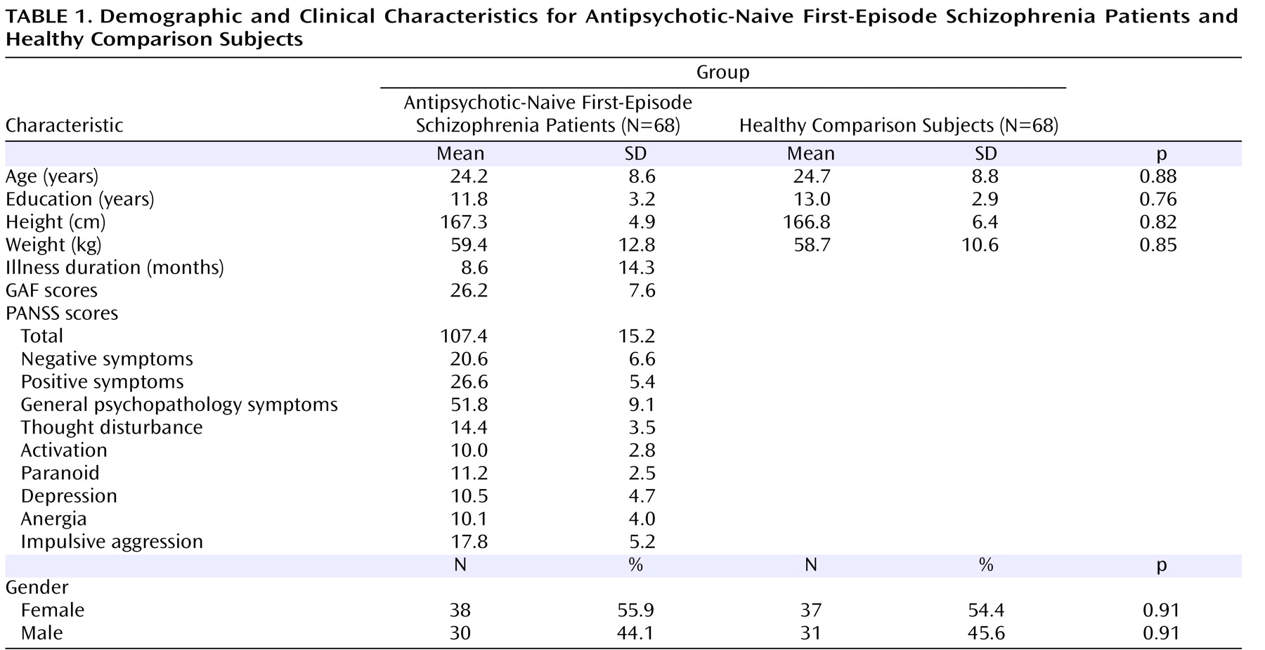
Sixty-eight antipsychotic-naive first-episode schizophrenia patients and 68 healthy comparison subjects were recruited via the Mental Health Centre of the West China Hospital, Chengdu, China ( Table 1 ). The study was approved by the local ethics committee, and all patients and healthy comparison subjects gave written informed consent. Diagnosis of schizophrenia and duration of illness were determined by consensus between the attending psychiatrist and a trained interviewer using the Structured Clinical Interview for DSM-IV (SCID)–Patient Version. Diagnoses for all patients were confirmed after ≥1-year follow-up. Healthy comparison subjects were recruited from the local area via poster advertisement and screened using the SCID–Non-Patient Version to confirm the lifetime absence of psychiatric and neurological illness. In addition, healthy subjects were interviewed to confirm that there was no history of psychiatric illness among their first-degree relatives. Data pertaining to age, sex, height, weight, handedness (based on the Annett Handedness Scale [28] ), years of education, duration of illness, and clinical symptom ratings were obtained by two experienced clinical psychiatrists prior to initiating any treatment and magnetic resonance (MR) examinations. All patients were evaluated using the Global Assessment of Functioning Scale (GAF) of DSM-IV (29) . Psychopathology associated with first-episode schizophrenia was evaluated using the Positive and Negative Syndrome Scale (PANSS) (30, 31), which determines positive and negative symptom scores and a total score as well as indices of thought disturbance, activation, paranoia, depression, and anergia by combining item ratings via a previously published six-factor structure of PANSS items (32) . Healthy comparison subjects were matched with schizophrenia patients on age, sex, height, weight, and years of education ( Table 1 ). Exclusion criteria for both groups were 1) existence of a neurological disorder; 2) alcohol or drug abuse; 3) pregnancy; and 4) any physical illness such as a brain tumor, hepatitis, or epilepsy as assessed according to clinical evaluations and medical records. Magnetic resonance imaging (MRI) was inspected by an experienced neuroradiologist, and no gross abnormalities were observed in either group.

Data Acquisition

High resolution T1-weighted images were acquired via a 3-Telsa MRI system (EXCITE, General Electric, Milwaukee, Wisc.), with a volumetric three-dimensional spoiled gradient recall sequence (TR=8.5 msec, echo time=3.4 msec, flip angle=12°, slice thickness=1 mm) using an eight-channel phase array head coil. Field of view (240×240 mm 2 ) was used with an acquisition matrix comprising 256 readings of 128 phase encoding steps that produced 156 contiguous coronal slices, with a slice thickness of 1.0 mm. The final matrix size of T1-weighted images was automatically interpolated in-plane to 512×512, which yielded an in-plane resolution of 0.47×0.47 mm 2 . MR images sensitized to changes in BOLD signal levels (TR=2000 msec, echo time=30 msec, flip angle=90°) were obtained via a gradient-echo echo-planar imaging sequence. During MR examination, subjects were instructed to relax with their eyes closed without falling asleep (confirmed by subjects immediately after the experiment). Five dummy scans were collected before fMRI scans were performed, and the first two volumes of fMRI time series were discarded for magnetization stabilization (slice thickness=5 mm [no slice gap]; matrix=64×64; field of view=240×240 mm 2 ; voxel size=3.75×3.75×5 mm 3 ). Each brain volume comprised 30 axial slices, and each functional run contained 200 image volumes.

Voxel-Based Morphometry Analysis

Optimized voxel-based morphometry (33) was conducted using Statistical Parametric Mapping–2 (SPM2) (Welcome Department of Imaging Neuroscience, London [http://www.fil.ion.ucl.ac.uk/spm]). The voxel-based morphometry–2 toolbox, which implements the optimized voxel-based morphometry approach (33), was used for data preprocessing. Optimized voxel-based morphometry consists of a two-step procedure that starts with the construction of a study-specific whole brain template and tissue priors that account for the magnetic field properties of the scanner as well as the anatomical properties of the study groups. In this first step, images from all 136 participants were normalized to the T1-Montreal Neurological Institute (MNI) brain template in SPM2 using a 12-parameter affine-only procedure, smoothed (according to SPM2 specifications) with a 4-mm isotropic Gaussian kernel and averaged. In the second step, the customized template and tissue priors were used for data segmentation and data registration to and re-segmentation in standard space. Thus, gray matter was automatically segmented from raw MRI using tissue-signal intensity values and tissue priors for the distribution of brain tissue type. An automated brain extraction step was incorporated in order to eliminate voxels from non-gray matter structures with signal intensities similar to gray matter, such as the dural venous sinuses, scalp, cranial marrow, and diplopic space. Gray matter partitions were spatially normalized (using a 12-parameter affine transformation and 7×8×7 nonlinear basis functions) to the created customized gray matter template. The deformation parameters obtained from the normalization process were applied to each participant’s original raw image (native space) in order to create optimally normalized whole brain images, which were recursively segmented for brain tissue extraction. Finally, the segmented images were modulated with the Jacobian determinants derived from spatial normalization (33) . The optimally processed images were smoothed with an isotropic Gaussian kernel (full-width half maximum=8 mm).
Voxel-by-voxel-based comparisons of gray matter volume were performed between groups using two-sample t tests. The significance of group differences in each region was estimated by distributional approximations from the theory of random Gaussian fields, and significance levels were set at p<0.05 (corrected for multiple comparisons). To identify the association between structural abnormalities and clinical symptom severity, the average gray matter volume values for all voxels in abnormal areas, revealed by voxel-based morphometry, were extracted and correlated with PANSS scores, age, and duration of illness using correlation analysis.

Functional Connectivity Analysis

Preprocessing and statistical analysis of functional images were conducted using SPM2. For each subject, echo-planar images were slice-time corrected and realigned to the first image in the first series and were subsequently unwarped to correct for susceptibility-by-movement interaction. All realigned images were spatially normalized to the MNI echo-planar imaging template in SPM2, and each voxel was resampled to 3×3×3 mm 3 . Functional connectivity was examined using a method based on a seed voxel correlation approach (24, 25) . Since voxel-based morphometry analysis showed anatomic deficits in the right superior temporal gyrus (Brodmann’s area 41), right middle temporal gyrus (Brodmann’s area 21), and right anterior cingulate gyrus (Brodmann’s area 32), areas with gray matter volume reduction in each of these three regions were defined as seeds for functional connectivity analysis. A reference time series for each seed was obtained by averaging the fMRI time series for all voxels within each of the three regions with anatomic deficits. Next, each time series was temporally bandpass filtered (0.01–0.08 Hz). Correlation analysis was conducted between the seed reference and the rest of the whole brain in a voxel-wise manner using the realigned images. The resultant r value map was subsequently normalized to MNI space for group analysis using the normalization parameters extracted previously.
For patient data, individual r value maps were analyzed with a random effect one-sample t test to identify voxels showing a significant positive or negative correlation to the seed time series, with correlations thresholded using a family-wise error correction at p<0.05. For between-group comparison, two-sample t tests were used to compare r value maps between first-episode schizophrenia patients and healthy comparison subjects, with the significance threshold set at p<0.05 after correction for multiple comparisons from the theory of random Gaussian fields. MNI coordinates for our results were transformed to Talairach coordinates via mni2tal (http://imaging.mrc-cbu.cam.ac.uk/imaging/MniTalairach). To identify the association between functional connectivity and symptoms in schizophrenia patients, a series of correlation analyses between individual r value maps and PANSS scores of interest were computed and then thresholded at p<0.05 after correction for multiple comparisons from the theory of random Gaussian fields.

Results

Morphometry Analysis

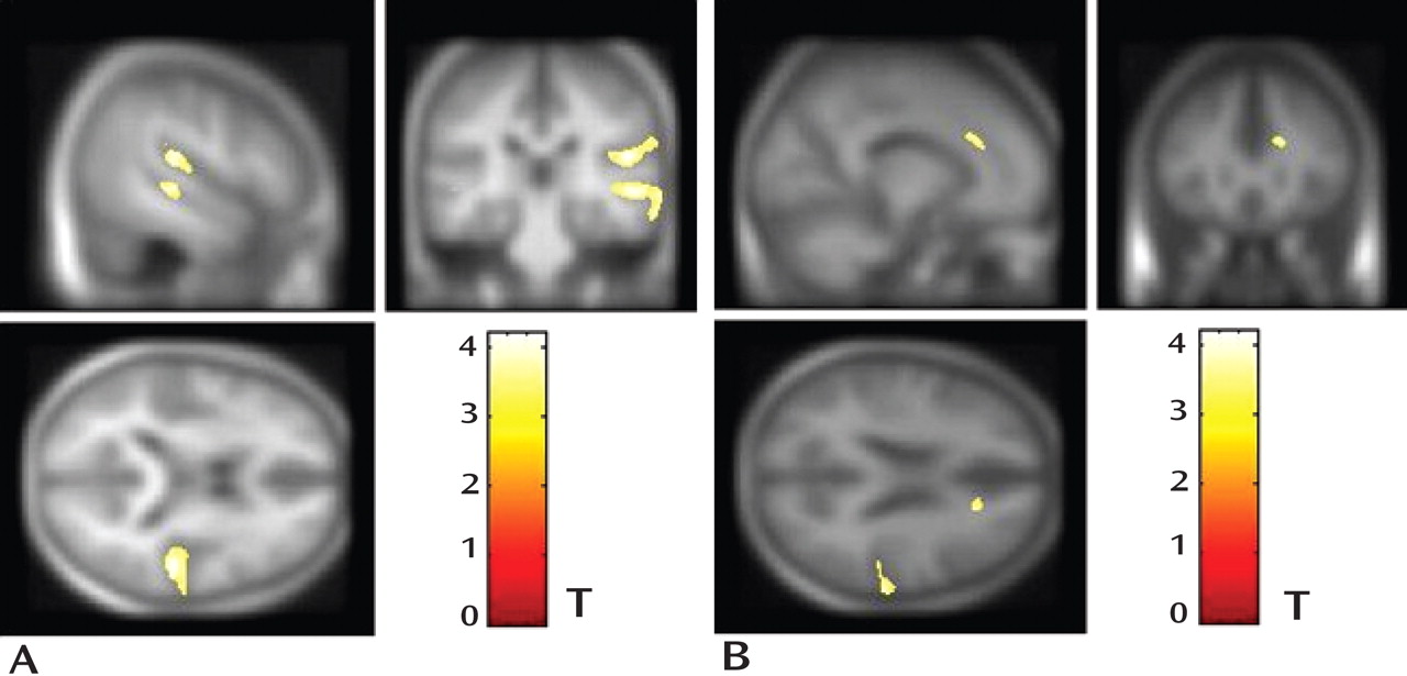
Relative to healthy comparison subjects, first-episode schizophrenia patients showed significantly decreased gray matter volume in the following three brain regions: right anterior cingulate gyrus ([Brodmann’s area 32] Talairach coordinates: 20, 22, 27; voxel size=252 mm 3 ), right middle temporal gyrus ([Brodmann’s area 21] Talairach coordinates: 54, –26, –1; voxel size=2,715 mm 3 ), and right superior temporal gyrus ([Brodmann’s area 41] Talairach coordinates: 50, –26, 16; voxel size=5,353 mm 3 ) ( Figure 1 ). No significant increases in gray matter volume were found in schizophrenia patients relative to healthy comparison subjects.
Figure 1. Statistical Parametric Images of Voxel-Based Morphometry Analysis for Antipsychotic-Naive First-Episode Schizophrenia Patients and Healthy Comparison Subjects a
a Relative to healthy comparison subjects, schizophrenia patients had significantly reduced gray matter volume in the right superior and middle temporal gyri (panel A) and right anterior cingulate gyrus (panel B).
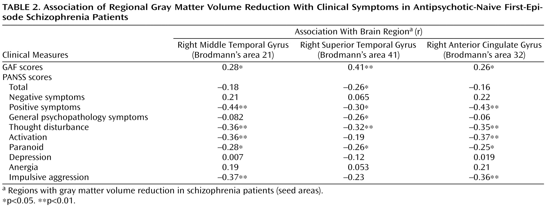
Significant positive correlations were observed between the degree of gray matter volume reduction in each of the three anatomic regions and the general functioning of schizophrenia patients as measured using GAF ( Table 2 ). Correlations were also found between PANSS scores and the gray matter volume of the three regions. Significant negative correlations were found between the gray matter volume of the right anterior cingulate gyrus and PANSS scores for positive symptoms, thought disturbance, activation, paranoia, and impulsive aggression ( Table 2 ). In addition, significant negative correlations were observed between the gray matter volume of the right middle temporal gyrus and PANSS scores for positive symptoms, thought disturbance, activation, paranoia, and impulsive aggression ( Table 2 ). Last, significant negative correlations were observed between the gray matter volume of the right superior temporal gyrus and total PANSS scores as well as scores for positive symptoms, general psychopathology symptoms, thought disturbance, and paranoia ( Table 2 ). No correlation was found between the gray matter volume of the three regions with anatomic deficits and the age and duration of illness among schizophrenia patients.

Functional Connectivity Analysis

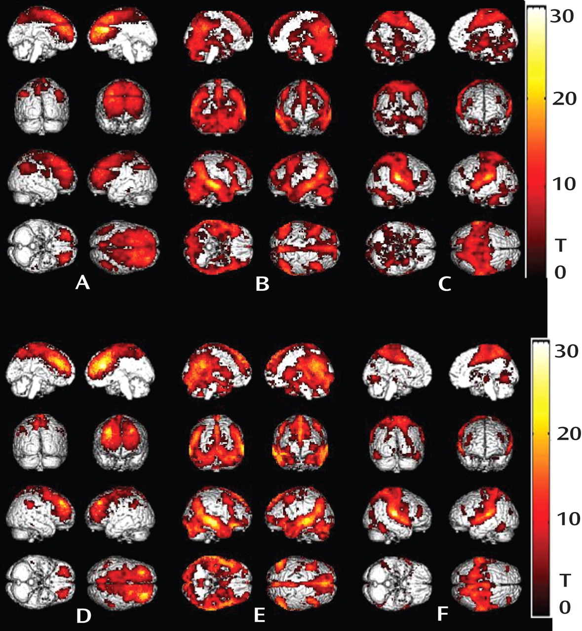
The three seed areas, where reduced gray matter volume was detected among first-episode schizophrenia patients, were selected for functional connectivity analysis. When the seed was located in the right anterior cingulate gyrus where reduced gray matter volume was detected, both schizophrenia patients and healthy comparison subjects showed significant positive correlations between the seed and activity in the contralateral cingulate gyrus, bilateral prefrontal cortex, and bilateral inferior parietal lobes ( Figure 2 ). When the seed was located in the right middle temporal gyrus where reduced gray matter volume was detected, both schizophrenia patients and healthy comparison subjects showed significant positive correlations between the seed and activity in the contralateral middle temporal gyrus, bilateral thalamus, putamen, cerebellum, dorsolateral frontal cortex, posterior cingulate gyrus, and precuneus ( Figure 2 ). When the seed was located in the right superior temporal gyrus where reduced gray matter volume was detected, both schizophrenia patients and healthy comparison subjects showed significant positive correlations between the seed and activity in the contralateral superior temporal gyrus, bilateral thalamus, occipital lobes, dorsolateral frontal cortex, bilateral parietal lobes, cingulate gyrus, and precuneus ( Figure 2 ). There were no significant differences in r value maps between schizophrenia patients and healthy comparison subjects reflecting the resting state functional connectivity of any of the three seed areas. Given the findings of previous studies (22, 34), we also examined the connectivity of the dorsolateral prefrontal cortex in our antipsychotic-naive schizophrenia sample and did not find evidence of altered functional connectivity with this region.
Figure 2. Statistical Parametric Images of Functional Connectivity Analysis for Antipsychotic-Naive First-Episode Schizophrenia Patients and Healthy Comparison Subjects a
a The seed areas (areas with gray matter volume reduction) were located in the right anterior cingulate gyrus ([Brodmann’s area 32] panels A and D), right middle temporal gyrus ([Brodmann’s area 21] panels B and E), and right superior temporal gyrus ([Brodmann’s area 41] panels C and F). Schizophrenia patients (upper row) and healthy comparison subjects (lower row) showed no significant differences in patterns of functional connectivity during the resting state.
A correlation analysis was performed between r value maps from the functional connectivity analysis and PANSS scores in order to assess the association between schizophrenia symptoms and functional connectivity values. When the seed was located in the right middle temporal gyrus, significant positive correlations were observed between PANSS thought disturbance scores and r values in the bilateral putamen ( Table 3 ). When the seed was located in the right superior temporal gyrus, significant negative correlations were observed between PANSS total scores and r values in the bilateral posterior cingulate gyrus and bilateral precuneus and between PANSS scores for negative symptoms and anergia and r values in the bilateral precuneus ( Table 3 ). When the seed was located in the right anterior cingulate gyrus, there were no areas showing a positive or negative correlation between PANSS total scores and r values.

Discussion

The present study examined gray matter abnormalities in a relatively large sample of antipsychotic-naive first-episode schizophrenia patients. Decreases in gray matter volume were observed in the right anterior cingulate gyrus, right middle temporal gyrus, and right superior temporal gyrus ( Figure 1 ). Gray matter volume reduction in these three regions was related to the severity of psychopathology ( Table 2 ).
Gray matter deficits in schizophrenia patients were reported in approximately 50 brain regions in one meta-analysis (1) . Among those regions, volume reduction in the superior temporal gyrus had 100% replicability with region-of-interest measurements (35) and approximately 50% replicability with voxel-based morphometry approaches in previous studies (1) . However, decreased gray matter volume in the right anterior cingulate gyrus and right middle temporal gyrus had replicability with voxel-based morphometry approaches of approximately 24% and 7%, respectively, in previous studies (1) . The inhomogeneous findings of different voxel-based morphometry studies may be related to numerous confounds, including illness chronicity and antipsychotic medication effects (4, 5, 36, 37) as well as methodological differences (e.g., smoothing kernel) (1) .
The study of untreated first-episode schizophrenia is especially useful for understanding the disorder-related clinical and neurophysiological correlates of morphometric changes. To our knowledge, only three voxel-based morphometry studies (3, 6, 38) have, to date, recruited antipsychotic-naive schizophrenia patients, all with a smaller sample size than that of the present study. Each of these previous studies reported decreased gray matter in the cingulate gyrus, and none found alterations in the right middle temporal gyrus. However, findings for the superior temporal gyrus differed among the three studies. To our knowledge, the present study is the first to demonstrate that gray matter deficits in the right middle temporal gyrus and right superior temporal gyrus may be present during a very early stage of schizophrenia.
In the present study, we also found positive correlations between gray matter volume values in the three brain regions with anatomic deficits (right anterior cingulate gyrus, right middle temporal gyrus, and right superior temporal gyrus) and GAF scores. This finding confirms that the gray matter abnormalities were associated with increased levels of psychopathology and functional impairment among antipsychotic-naive first-episode schizophrenia patients (39) . In a previous study using single photon emission computed tomography, activity in the anterior cingulate gyrus was reported to be correlated with some positive symptoms, such as delusion, in antipsychotic-naive schizophrenia patients (40) . In the present study, we utilized MRI to show that gray matter volume in the anterior cingulate gyrus was correlated with the psychopathology of schizophrenia, since there were negative correlations of gray matter volume in the right anterior cingulate gyrus with PANSS scores for positive symptoms, thought disturbance, activation, paranoia, and impulsive aggression.
The role of volume reduction in the superior temporal gyrus as it relates to negative symptoms (8) and thought disorder (2, 41) in schizophrenia has been discussed in previous studies, although some nonreplication has also been reported (9, 10) . Studies of patients with familial and nonfamilial schizophrenia have suggested that superior temporal gray matter volume reduction may be a familial phenotype (42) and may have a neurodevelopmental origin, since decreases in superior temporal gray matter volume in these studies occurred among unaffected individuals for whom illness progression effects would not have been a potential cause of morphometric abnormality (42, 43) . Although decreased gray matter volume in the middle temporal gyrus has been rarely reported in the literature (1), recent structural (44) and functional (41) studies have shown correlations between the severity of symptoms and severity of abnormalities in the middle temporal gyrus among first-episode schizophrenia patients. In the present study, our results confirm the presence of neuroanatomic abnormalities in the temporal and cingulate cortex and further confirm the role of these areas in the pathology of schizophrenia by demonstrating correlations of gray matter volume reduction with symptom profiles ( Table 3 ).
Alterations of functional connectivity in schizophrenia has been treated as a potential systems-level substrate of the disorder. Through the use of resting-state fMRI, previous studies (22, 23, 45) have reported alterations of functional connectivity in schizophrenia patients. Decreased fronto-parietal, fronto-cingulate, and fronto-thalamic connectivity have also been reported (22, 23, 45) . However, the findings have not been consistent. These previous studies utilized different a priori anatomic regions as seeds based on the specific hypotheses of the investigations. Furthermore, since regional changes in resting state brain physiology are known to occur after antipsychotic treatment (46, 47), previous functional connectivity studies, including those of first-episode schizophrenia patients who received treatment (22, 23, 45), may have detected effects of antipsychotic medication rather than direct illness effects on brain function. Longitudinal studies of patients evaluated before and after treatment are needed in order to assess whether effects of antipsychotic medication or direct illness effects on brain function are present.
In the present study, regions with abnormal gray matter volume were used as seeds for functional connectivity analysis. Surprisingly, patterns of resting state functional connectivity for all three structurally abnormal regions (right anterior cingulate gyrus, right middle temporal gyrus, and right superior temporal gyrus) were similar among antipsychotic-naive first-episode schizophrenia patients and healthy comparison subjects ( Figure 2 ). In addition, we did not observe altered functional connectivity with the dorsal lateral prefrontal cortex in our antipsychotic-naive schizophrenia sample, which has been found in patients who received treatment in previous studies (22, 34) . Thus, gray matter volume reduction in the three regions—although related to symptoms—did not appear to significantly alter the integration of affected brain regions within their corresponding functional networks, at least with regard to resting state indices of functional connectivity. However, correlations were found between the clinical symptoms and functional connectivity of the middle temporal gyrus with the putamen and between the clinical symptoms and functional connectivity of the superior temporal gyrus with the precuneus. Middle temporal connectivity with the putamen among first-episode schizophrenia patients was associated with thought disturbance, while superior temporal gyrus connectivity with the precuneus was associated with PANSS total scores as well as scores for negative symptoms and anergia ( Table 3 ). This suggests that heightened patterns of common activity between the basal ganglia and middle temporal gyrus may be associated with increased levels of thought disorder, an association that is perhaps mediated by increased striatal metabolic or dopaminergic activation that can occur during acute episodes of illness (48) . In contrast, functional integration of the superior temporal gyrus with the precuneus was associated with negative symptoms, which is an association that might be related to reduced attention or impaired processing of real-life events (49) .
To our knowledge, the present study is the first to identify two different patterns of associations between brain abnormalities and symptoms of first-episode schizophrenia. First, volume reduction in the right anterior cingulate gyrus, right middle temporal gyrus, and right superior temporal gyrus was directly related to the severity of several dimensions of psychopathology. Second, functional connectivity levels of the right middle temporal gyrus and right superior temporal gyrus were associated with symptom severity and thus may have clinical significance in some more severely ill patients. However, since schizophrenia patients were not followed over time, we were unable to determine whether the reported relations between symptom severity and neurophysiological and morphometric measures were restricted to periods of acute psychosis or were patterns that persisted during recovery.
The two patterns of associations between anatomic deficits and symptom severity among first-episode schizophrenia patients suggest that there may be important clinical and, perhaps, neuropsychological effects associated with disorder-related morphometric changes in the right middle temporal gyrus and superior temporal gyrus that could be examined in future studies of schizophrenia. For example, a recent study by Weinstein et al. (41) revealed that the association between decreased gray matter volume in the left superior temporal gyrus and the severity of thought disorder was mediated by the level of activation in the superior and middle temporal gyri during language processing.
Several methodological limitations regarding the use of voxel-based morphometry should be considered when interpreting the results of the present study. First, although optimized voxel-based morphometry was employed, which minimizes the contamination of brain tissue with nonbrain voxels relative to standard voxel-based morphometry (33), findings from our sample of Chinese patients may have affected the accuracy of normalization because the generic MNI template differs structurally in the brains of non-Caucasian populations (50) . However, since both schizophrenia patients and healthy comparison subjects were of a similar ethnic background, it is unlikely that the group differences observed were affected by ethnic factors. Second, some of the differences in gray matter volume may have resulted from systematic morphological differences between the two groups, since specific patterns of abnormal morphology may result in group-specific misregistration. In such a case, there can be increased sensitivity to systematic shape differences attributable to misregistration from the spatial normalization step (51) . Systematic differences in morphology are one of several potential differences between schizophrenia patients and healthy comparison subjects that may be detected by voxel-based morphometry, and they reflect a real difference between data obtained from different populations but may not necessarily be the result of changes in gray matter volume (52) . In addition, compared with the classic region of interest method, previous studies (53, 54) of individuals with schizophrenia have indicated that the results of voxel-based morphometry and region of interest are generally consistent, and voxel-based morphometry has proven to be somewhat more sensitive than region of interest methods. Last, we also recognize that voxel-based morphometry may not be sensitive to a disease that likely affects brain structure in nonlinear ways, as first demonstrated by Davatzikos (55) . Although voxel-based morphometry methods used to accommodate nonlinearities have been proposed (56), it is important to acknowledge that voxel-based morphometry is not a surrogate for multivariate volumetric analyses that have been specifically developed to characterize highly distributed nonlinear changes.
In conclusion, the present study applied morphometry analysis and resting-state functional connectivity to examine their relationship with the clinical features of first-episode schizophrenia. Our findings document that symptoms in antipsychotic-naive patients with first-episode schizophrenia are associated with both structural deficits and alterations in the functional brain networks of regions with gray matter volume reduction. However, regions with gray matter volume reduction did not have significantly altered resting state functional connectivity with other brain regions. Our results provide support for future efforts to combine anatomical and functional data to explore the complex disorder of schizophrenia.
Footnotes

Received Feb. 5, 2008; revision received May 20, 2008; accepted July 17, 2008 (doi: 10.1176/appi.ajp.2008.08020183). From the Huaxi MR Research Center, Department of Radiology, West China Hospital of Sichuan University, Chengdu, China; Department of Psychiatry, Psychiatric Laboratory, State Key Laboratory of Biotherapy, West China Hospital of Sichuan University, Chengdu, China; School of Science and Technology, University of Electronic Science and Technology of China, Chengdu, China; Magnetic Resonance Research Center, University of Illinois at Chicago, Chicago; Division of Psychological Medicine, Section of Neuroimaging, Institute of Psychiatry, King’s College London, London; Social, Genetic and Developmental Psychiatry Centre, Institute of Psychiatry, King’s College London, London; Center for Cognitive Medicine, University of Illinois at Chicago, Chicago; and the Division of Medical Imaging, Faculty of Medicine, University of Liverpool, Liverpool, United Kingdom. Address correspondence and reprint requests to Dr. Gong, Huaxi MR Research Center (HMRRC), Department of Radiology, West China Hospital of Sichuan University, Chengdu 610041, PR China; [email protected] (e-mail) or Dr. Tao Li, Department of Psychiatry and Psychiatric Laboratory, State Key Laboratory of Biotherapy, West China Hospital of Sichuan University, Chengdu 610041, PR China; [email protected] (e-mail).
The authors report no competing interests.
Drs. Su Lui and Wei Deng contributed equally to the present study.
Dr. Gong is an Honorary Fellow of the Faculty of Medicine, University of LIverpool, Liverpool, United Kingdom.
Supported by the National Natural Science Foundation of China (grants: 30530300-2006/R3, 30625024, 30125014, 30728017, and 30700256); Programs from the State Education Ministry (grants: NCET-04-0866 and SRFDP-20060610073); the National Basic Research Program of China ([973] Program No: 2007CB512301/5); the National High Technology Program of China ([863] Program No: 2008AA02Z408); the Wellcome Trust Collaborative Research Initiative grant (2003–2006); and NARSAD (Independent Investigator 2006).

References

1.
Honea R, Crow TJ, Passingham D, Mackay CE: Regional deficits in brain volume in schizophrenia: a meta-analysis of voxel-based morphometry studies. Am J Psychiatry 2005; 162:2233–2245
2.
Rajarethinam RP, DeQuardo JR, Nalepa R, Tandon R: Superior temporal gyrus in schizophrenia: a volumetric magnetic resonance imaging study. Schizophr Res 2000; 41:303–312
3.
Jayakumar PN, Venkatasubramanian G, Gangadhar BN, Janakiramaiah N, Keshavan MS: Optimized voxel-based morphometry of gray matter volume in first-episode, antipsychotic-naive schizophrenia. Prog Neuropsychopharmacol Biol Psychiatry 2005; 29:587–591
4.
Lieberman JA, Tollefson GD, Charles C, Zipursky R, Sharma T, Kahn RS, Keefe RS, Green AI, Gur RE, McEvoy J, Perkins D, Hamer RM, Gu H, Tohen M: Antipsychotic drug effects on brain morphology in first-episode psychosis. Arch Gen Psychiatry 2005; 62:361–370
5.
Velakoulis D, Wood SJ, Smith DJ, Soulsby B, Brewer W, Leeton L, Desmond P, Suckling J, Bullmore ET, McGuire PK, Pantelis C: Increased duration of illness is associated with reduced volume in right medial temporal/anterior cingulate grey matter in patients with chronic schizophrenia. Schizophr Res 2002; 57:43–49
6.
Chua SE, Cheung C, Cheung V, Tsang JT, Chen EY, Wong JC, Cheung JP, Yip L, Tai KS, Suckling J, McAlonan GM: Cerebral grey, white matter and CSF in never-medicated, first-episode schizophrenia. Schizophr Res 2007; 89:12–21
7.
Whitford TJ, Farrow TF, Gomes L, Brennan J, Harris AW, Williams LM: Grey matter deficits and symptom profile in first episode schizophrenia. Psychiatry Res 2005; 139:229–238
8.
Kim JJ, Crespo-Facorro B, Andreasen NC, O’Leary DS, Magnotta V, Nopoulos P: Morphology of the lateral superior temporal gyrus in neuroleptic naïve patients with schizophrenia: relationship to symptoms. Schizophr Res 2003; 60:173–181
9.
Yamada M, Hirao K, Namiki C, Hanakawa T, Fukuyama H, Hayashi T, Murai T: Social cognition and frontal lobe pathology in schizophrenia: a voxel-based morphometric study. Neuroimage 2007; 35:292–298
10.
Fannon D, Chitnis X, Doku V, Tennakoon L, O’Ceallaigh S, Soni W, Sumich A, Lowe J, Santamaria M, Sharma T: Features of structural brain abnormality detected in first-episode psychosis. Am J Psychiatry 2000; 157:1829–1834
11.
Andreasen NC: The Scale for the Assessment of Negative Symptoms (SANS). Iowa City, University of Iowa, 1983
12.
Andreasen NC: The Scale for the Assessment of Positive Symptoms (SAPS). Iowa City, University of Iowa, 1984
13.
Friston KJ, Frith CD: Schizophrenia: A disconnection syndrome? Clin Neurosci 1995; 3:89–97
14.
Lim KO: Connections in schizophrenia. Am J Psychiatry 2007; 164:995–998
15.
Lawrie SM, Buechel C, Whalley HC, Frith CD, Friston KJ, Johnstone EC: Reduced frontotemporal functional connectivity in schizophrenia associated with auditory hallucinations. Biol Psychiatry 2002; 51:1008–1011
16.
Shergill SS, Brammer MJ, Fukuda R, Williams SC, Murray RM, McGuire PK: Engagement of brain areas implicated in processing inner speech in people with auditory hallucinations. Br J Psychiatry 2003; 182:525–531
17.
Honey GD, Pomarol-Clotet E, Corlett PR, Honey RA, McKenna PJ, Bullmore ET, Fletcher PC: Functional dysconnectivity in schizophrenia associated with attentional modulation of motor function. Brain 2005; 128(pt 11):2597–2611
18.
Stephan KE, Magnotta VA, White T, Arndt S, Flaum M, O’Leary DS, Andreasen NC: Effects of olanzapine on cerebellar functional connectivity in schizophrenia measured by fMRI during a simple motor task. Psychol Med 2001; 31:1065–1078
19.
Schlosser R, Gesierich T, Kaufmann B, Vucurevic G, Hunsche S, Gawehn J, Stoeter P: Altered effective connectivity during working memory performance in schizophrenia: a study with fMRI and structural equation modeling. Neuroimage 2003; 19:751–763
20.
Raichle ME, Mintun MA: Brain work and brain imaging. Annu Rev Neurosci 2006; 29:449–476
21.
Cordes D, Haughton VM, Arfanakis K, Carew JD, Turski PA, Moritz CH, Quigley MA, Meyerand ME: Frequencies contributing to functional connectivity in the cerebral cortex in “resting-state” data. AJNR Am J Neuroradiol 2001; 22:1326–1333
22.
Zhou Y, Liang M, Jiang T, Tian L, Liu Y, Liu Z, Liu H, Kuang F: Functional dysconnectivity of the dorsolateral prefrontal cortex in first-episode schizophrenia using resting-state fMRI. Neurosci Lett 2007; 417:297–302
23.
Liang M, Zhou Y, Jiang T, Liu Z, Tian L, Liu H, Hao Y: Widespread functional disconnectivity in schizophrenia with resting-state functional magnetic resonance imaging. Neuroreport 2006; 17:209–213
24.
Horwitz B, Rumsey JM, Donohue BC: Functional connectivity of the angular gyrus in normal reading and dyslexia. Proc Natl Acad Sci U S A 1998; 95:8939–8944
25.
Friston KJ: Functional and effective connectivity in neuroimaging: a synthesis. Hum Brain Mapp 1994; 2:56–78
26.
Garrity AG, Pearlson GD, McKiernan K, Lloyd D, Kiehl KA, Calhoun VD: Aberrant “default mode” functional connectivity in schizophrenia. Am J Psychiatry 2007; 164:450–457
27.
Liu Y, Liang M, Zhou Y, He Y, Hao Y, Song M, Yu C, Liu H, Liu Z, Jiang T: Disrupted small-world networks in schizophrenia. Brain 2008; 131(pt 4):945–961
28.
Annett M: A classification of hand preference by association analysis. Br J Psychol 1970; 61:303–321
29.
Goldman HH, Skodol AE, Lave TR: Revising axis V for DSM-IV: a review of measures of social functioning. Am J Psychiatry 1992; 149:1148–1156
30.
Kay SR, Fiszbein A, Opler LA: The Positive and Negative Syndrome Scale (PANSS) for schizophrenia. Schizophr Bull 1987; 13:261–276
31.
Kay SR, Opler LA, Lindenmayer JP: Reliability and validity of the Positive and Negative Syndrome Scale for schizophrenics. Psychiatry Res 1988; 23:99–110
32.
Gladsjo JA, McAdams LA, Palmer BW, Moore DJ, Jeste DV, Heaton RK: A six-factor model of cognition in schizophrenia and related psychotic disorders: relationships with clinical symptoms and functional capacity. Schizophr Bull 2004; 30:739–754
33.
Good CD, Johnsrude IS, Ashburner J, Henson RN, Friston KJ, Frackowiak RS: A voxel-based morphometric study of ageing in 465 normal adult human brains. Neuroimage 2001; 14(1 pt 1):21–36
34.
Salgado-Pineda P, Caclin A, Baeza I, Junque C, Bernardo M, Blin O, Fonlupt P: Schizophrenia and frontal cortex: Where does it fail? Schizophr Res 2007; 91:73–81
35.
Shenton ME, Dickey CC, Frumin M, McCarley RW: A review of MRI findings in schizophrenia. Schizophr Res 2001; 49:1–52
36.
Konopaske GT, Dorph-Petersen KA, Pierri JN, Wu Q, Sampson AR, Lewis DA: Effect of chronic exposure to antipsychotic medication on cell numbers in the parietal cortex of macaque monkeys. Neuropsychopharmacology 2007; 32:1216–1223
37.
Dorph-Petersen KA, Pierri JN, Perel JM, Sun Z, Sampson AR, Lewis DA: The influence of chronic exposure to antipsychotic medications on brain size before and after tissue fixation: a comparison of haloperidol and olanzapine in macaque monkeys. Neuropsychopharmacology 2005; 30:1649–1661
38.
Salgado-Pineda P, Baeza I, Perez-Gomez M, Vendrell P, Junque C, Bargallo N, Bernardo M: Sustained attention impairment correlates to gray matter decreases in first episode neuroleptic-naive schizophrenic patients. Neuroimage 2003; 19(2 pt 1):365–375
39.
Vaillant GE: Mental health. Am J Psychiatry 2003; 160:1373–1384
40.
Erkwoh R, Sabri O, Steinmeyer EM, Bull U, Sass H: Psychopathological and SPECT findings in never-treated schizophrenia. Acta Psychiatr Scand 1997; 96:51–57
41.
Weinstein S, Woodward TS, Ngan ET: Brain activation mediates the association between structural abnormality and symptom severity in schizophrenia. Neuroimage 2007; 36:188–193
42.
Lui S, Deng W, Huang X, Jiang L, Ouyang L, Borgwardt SJ, Ma X, Li D, Zou L, Tang H, Chen H, Li T, McGuire P, Gong Q: Neuroanatomical differences between familial and sporadic schizophrenia and their parents: an optimized voxel-based morphometry study. Psychiatr Res Neuroimaging 2008 (in press)
43.
Antonova E, Kumari V, Morris R, Halari R, Anilkumar A, Mehrotra R, Sharma T: The relationship of structural alterations to cognitive deficits in schizophrenia: a voxel-based morphometry study. Biol Psychiatry 2005; 58:457–467
44.
Spencer MD, Moorhead TW, McIntosh AM, Stanfield AC, Muir WJ, Hoare P, Owens DG, Lawrie SM, Johnstone EC: Grey matter correlates of early psychotic symptoms in adolescents at enhanced risk of psychosis: a voxel-based study. Neuroimage 2007; 35:1181–1191
45.
Bluhm RL, Miller J, Lanius RA, Osuch EA, Boksman K, Neufeld RW, Theberge J, Schaefer B, Williamson P: Spontaneous low-frequency fluctuations in the BOLD signal in schizophrenic patients: anomalies in the default network. Schizophr Bull 2007; 33:1004–1012
46.
Lahti AC, Holcomb HH, Weiler MA, Medoff DR, Tamminga CA: Functional effects of antipsychotic drugs: comparing clozapine with haloperidol. Biol Psychiatry 2003; 53:601–608
47.
Holcomb HH, Cascella NG, Thaker GK, Medoff DR, Dannals RF, Tamminga CA: Functional sites of neuroleptic drug action in the human brain: PET/FDG studies with and without haloperidol. Am J Psychiatry 1996; 153:41–49
48.
Laruelle M, Abi-Dargham A, Gil R, Kegeles L, Innis R: Increased dopamine transmission in schizophrenia: relationship to illness phases. Biol Psychiatry 1999; 46:56–72
49.
Koren D, Seidman LJ, Goldsmith M, Harvey PD: Real-world cognitive—and metacognitive—dysfunction in schizophrenia: a new approach for measuring (and remediating) more “right stuff.” Schizophr Bull 2006; 32:310–326
50.
Sato K, Taki Y, Fukuda H, Kawashima R: Neuroanatomical database of normal Japanese brains. Neural Netw 2003; 16:1301–1310
51.
Bookstein FL: “Voxel-based morphometry” should not be used with imperfectly registered images. Neuroimage 2001; 14:1454–1462
52.
Ashburner J, Friston KJ: Why voxel-based morphometry should be used. Neuroimage 2001; 14:1238–1243
53.
Job DE, Whalley HC, McConnell S, Glabus M, Johnstone EC, Lawrie SM: Structural gray matter differences between first-episode schizophrenics and normal controls using voxel-based morphometry. Neuroimage 2002; 17:880–889
54.
Giuliani NR, Calhoun VD, Pearlson GD, Francis A, Buchanan RW: Voxel-based morphometry versus region of interest: a comparison of two methods for analyzing gray matter differences in schizophrenia. Schizophr Res 2005; 74:135–147
55.
Davatzikos C: Why voxel-based morphometric analysis should be used with great caution when characterizing group differences. Neuroimage 2004; 23:17–20
56.
Friston KJ, Ashburner J: Generative and recognition models for neuroanatomy. Neuroimage 2004; 23:21–24

Information & Authors

Information

Published In

Go to American Journal of Psychiatry
American Journal of Psychiatry
Pages: 196 - 205
PubMed: 18981063

History

Published online: 1 February 2009
Published in print: February, 2009

Authors

Details

Xiaohong Joe Zhou, Ph.D.
Andrea Mechelli, Ph.D.
David A. Collier, Ph.D.
John A. Sweeney, Ph.D.
Qiyong Gong, M.D., Ph.D.

Metrics & Citations

Metrics

Citations

Export Citations

If you have the appropriate software installed, you can download article citation data to the citation manager of your choice. Simply select your manager software from the list below and click Download.

For more information or tips please see 'Downloading to a citation manager' in the Help menu.

Format
Citation style
Style
Copy to clipboard

View Options

View options

PDF/EPUB

View PDF/EPUB

Get Access

Login options

Already a subscriber? Access your subscription through your login credentials or your institution for full access to this article.

Personal login Institutional Login Open Athens login
Purchase Options

Purchase this article to access the full text.

PPV Articles - American Journal of Psychiatry

PPV Articles - American Journal of Psychiatry

Not a subscriber?

Subscribe Now / Learn More

PsychiatryOnline subscription options offer access to the DSM-5-TR® library, books, journals, CME, and patient resources. This all-in-one virtual library provides psychiatrists and mental health professionals with key resources for diagnosis, treatment, research, and professional development.

Need more help? PsychiatryOnline Customer Service may be reached by emailing [email protected] or by calling 800-368-5777 (in the U.S.) or 703-907-7322 (outside the U.S.).

Media

Figures

Other

Tables

Share

Share

Share article link

Share