Full access
New Research
Published Online: 1 May 2011

Association of Schizophrenia in 22q11.2 Deletion Syndrome and Gray Matter Volumetric Deficits in the Superior Temporal Gyrus

This article has been corrected.
VIEW CORRECTION

Abstract

Objective:

Individuals with 22q11.2 deletion syndrome are known to be at high risk of developing schizophrenia. Previous imaging studies have provided limited data on the relation of schizophrenia expression in 22q11.2 deletion syndrome to specific regional brain volumetric changes. The authors hypothesized that the main structural brain finding associated with schizophrenia expression in 22q11.2 deletion syndrome, as for schizophrenia in the general population, would be gray matter volumetric deficits, especially in the temporal lobes.

Method:

MR brain images from 29 patients with 22q11.2 deletion syndrome and schizophrenia and 34 comparison subjects with 22q11.2 deletion syndrome and no history of psychosis were analyzed using a voxel-based morphometry method that also yielded volumes for related region-of-interest analyses. The authors compared data from the two groups using an analysis of covariance model correcting for total intracranial volume, age, sex, IQ, and history of congenital cardiac defects. The false discovery rate threshold was set at 0.05 to account for multiple comparisons.

Results:

Voxel-based morphometry analyses identified significant gray matter reductions in the left superior temporal gyrus (Brodmann's area 22) in the schizophrenia group. There were no significant between-group differences in white matter or CSF volumes. Region-of-interest analyses showed significant bilateral gray matter volume reductions in the temporal lobes and superior temporal gyri in the schizophrenia group.

Conclusions:

The structural brain expression of schizophrenia associated with the highly penetrant 22q11.2 deletion involves lower gray matter volumes in temporal lobe regions. These structural MRI findings in a 22q11.2 deletion syndrome form of schizophrenia are consistent with those from studies involving schizophrenia samples from the general population. The results provide further support for 22q11.2 deletion syndrome as a genetic subtype and as a useful neurodevelopmental model of schizophrenia.
Of the emerging copy number variations being identified as rare causes of schizophrenia, hemizygous 22q11.2 deletions are the genetic variants with the highest penetrance that convey the greatest elevation of risk for schizophrenia, estimated to be more than 20 times that of the general population (1, 2). Approximately one of every 100 patients with schizophrenia in the general population has a 22q11.2 deletion (3, 4). The associated 22q11.2 deletion syndrome, previously known as velocardiofacial or DiGeorge syndrome (Mendelian Inheritance of Man #188400, #192430), displays a variable phenotype with common features, including velopharyngeal insufficiency, congenital heart disease, and learning difficulties (5, 6). Mental retardation (IQ <70) is present in a minority of those with the syndrome (5, 7, 8). The 22q11.2 deletion syndrome has been proposed as a neurodevelopmental model of schizophrenia with enhanced genetic homogeneity that may help us understand the pathogenesis of schizophrenia, including changes in brain structure (912).
Despite many brain imaging studies of 22q11.2 deletion syndrome, there are limited data on structural findings specifically associated with the expression of schizophrenia in the presence of the 22q11.2 deletion (see reference 12 for a review). The few previous studies of adults with 22q11.2 deletion syndrome and schizophrenia involved small sample sizes (a maximum of 14 patients) (10, 13, 14). Findings from comparisons with heterogeneous comparison groups without the 22q11.2 deletion suggest that there may be gray matter volumetric deficits in 22q11.2 deletion syndrome (10), but these exploratory studies showed relatively widespread changes throughout the brain (10, 13, 14).
For this study, we predicted that the main structural brain finding associated with expression of schizophrenia in 22q11.2 deletion syndrome, as for schizophrenia in the general population (1517), would be gray matter volumetric deficits, especially in the temporal lobe. To test this hypothesis, we used whole-brain voxel-based morphometry to examine MRI data from a large, well-characterized sample of adults with 22q11.2 deletion syndrome. We compared MRI scans from adults with 22q11.2 deletion syndrome and schizophrenia with those of the most closely matched comparison group, adults with 22q11.2 deletions but with no psychosis. Region-of-interest analyses were used to follow up on significant results.

Method

Sample and Clinical Assessments

We recruited adults at least 18 years of age with 22q11.2 deletion syndrome through genetic, adult congenital cardiac, and psychiatric services across Canada and confirmed 22q11.2 deletions using standard methods (18). Of those included in this study, only two (one with schizophrenia) did not have the typical 3-Mb hemizygous 22q11.2 deletions. In a previous study (18), we showed that there was no significant effect of length of the 22q11.2 deletion on expression of schizophrenia (18).
The study was approved by the research ethics boards of the authors' institutions, and written informed consent was obtained from all participants. Comprehensive direct assessments were conducted for all participants, as described elsewhere (5, 19). Intellectual level was assessed using standard methods to obtain full-scale IQ (7). Major congenital cardiac disease status was classified according to structural complexity (20). Participants were assessed for lifetime psychiatric diagnoses by research psychiatrists (A.S.B., E.W.C.C.) using a modified version of the Structured Clinical Interview for DSM-III-R or DSM-IV, direct interview and collateral information from family members, and medical records (21). Comparable clinical data on longitudinal follow-up, obtained on average every 1–2 years, were available for all but one participant who was lost to follow-up (18). The comprehensive data obtained allowed both DSM-III-R and DSM-IV criteria to be used; DSM-IV diagnoses are reported. Cross-sectional symptom assessments using the 30-item Positive and Negative Syndrome Scale (PANSS) (19, 22) were performed when participants were in a stable clinical state (19).
Of the 63 participants included in this study, 34 did not meet criteria for a psychotic disorder, had no history of significant psychotic symptoms, and had never been treated with antipsychotic medication. Patients with schizophrenia (N=23) and schizoaffective disorder (N=6) were collectively placed in the “schizophrenia” group for this study. We recorded the type and dosage of antipsychotic medications patients were taking at the time of the scan and converted daily doses to chlorpromazine equivalents (23). Four patients were taking conventional antipsychotics (haloperidol, N=2; perphenazine, N=1; fluphenazine, N=1), and 23 were taking atypical antipsychotics (risperidone, N=9; olanzapine, N=9; clozapine, N=3; zuclopenthixol, N=1; quetiapine, N=1); two patients were not taking any antipsychotic medication. No patient was taking lithium. None of the patients had a history of significant head injury or current substance abuse. Fourteen patients and none of the nonpsychotic comparison subjects have been reported on in a previous MRI study (10).

MRI Imaging

Image acquisition

Structural MR images of brain were acquired using a single GE Signa 1.5-T scanner. All images were visually inspected for movement artifacts. Two similar coronal three-dimensional scan sequences were used that yielded 124 contiguous 1.5-mm thick T1-weighted sections: coronal three-dimensional radio frequency-spoiled fast gradient recalled echo (SPGR) (inversion time=300 msec, repetition time=25 msec, echo time=5 msec; flip angle=20°; field of view=20 cm, matrix=256×256 mm2) for 24 participants (15 with schizophrenia, nine without) and inversion-prepped radio frequency-spoiled fast gradient recalled echo (IR-SPGR) (6 minutes 20 seconds, using the same parameters as SPGR except repetition time=12 msec) for 39 participants (14 with schizophrenia, 25 without). To ensure that data from the two scan sequences could be analyzed together, we first compared the scans of 12 participants (four with schizophrenia, eight without) with both sequences obtained at the same session. Voxel-based morphometry results showed no significant intraindividual differences between the scan sequences for gray matter, white matter, or CSF. To further assess this issue, we performed separate analyses using data restricted to each individual scan sequence—that is, for the group of 36 participants with SPGR data and the group of 39 participants with IR-SPGR data—and obtained similar results. We therefore analyzed the scans of all 63 participants together for our analyses. We also conducted a post hoc analysis of covariance (ANCOVA) using the data for the entire sample and including the type of scan sequence as an additional covariate; we found no difference in the results.

Image analysis

The image processing steps have been described in detail elsewhere (24, 25). In brief, whole-brain images were analyzed by the voxel-based morphometry method using SPM5 (Wellcome Trust Centre for Neuroimaging, London) under MATLAB (MathWorks, Natick, Mass.) with the VBM5 toolbox (http://dbm.neuro.uni-jena.de/vbm/). Using this method, voxels were designated as gray matter, white matter, or CSF in a unified segmentation and normalization step using a hidden Markov random field model. This method employs standard tissue prior probability maps and final normalization into Montreal Neurological Institute standard space. A modulation step scales the gray matter and white matter concentration maps by the volume change at each voxel, preventing brain volumes on a larger spatial scale from being lost in this spatial normalization procedure. The final maps display estimates of tissue volume in cubic centimeters from the subject's original space. These images were then subjected to smoothing with a full width at half maximum isotropic Gaussian kernel of 12 mm to conform to normality. The Talairach Daemon software was used to relate Talairach voxel coordinates of the clusters showing group differences to anatomical areas.

Region-of-interest volumes.

To further examine results of the whole-brain analysis, we extracted volumes from the modulated whole-brain images using the Masks for Region-of-Interest Analysis (MARINA) program (26) to generate masks of the specific region that was identified as having significant between-group differences as well as its containing lobe. MARINA creates masks of regions based on the anatomical parcellation of the brain published by Tzourio-Mazoyer et al. (27), which then can be smoothed, edited, and saved in the SPM-Analyze format. Resulting masks may then be used for region-of-interest analysis (2830). Region-of-interest analyses therefore involved masks of the left and right superior temporal gyri and temporal lobes. Quality control was performed for each participant by inspecting whether the masks projected properly over the corresponding gyri and lobes in the image. Subsequently, these binary lobe-shaped masks were multiplied with segmented unsmoothed gray matter images to yield gyrus-specific and lobe-specific images for each participant and with conversion to gray matter volumes in cubic centimeters.

Statistical Analysis

All comparisons were between the schizophrenia group and the nonpsychotic comparison group. For our main analysis, we performed an ANCOVA of the whole-brain voxel-wise data using SPM5, correcting for age, sex, total intracranial volume, IQ, and presence of major congenital cardiac disease. We set a false discovery rate threshold at 0.05 to account for multiple comparisons and a minimum cluster size of 500 voxels. Group comparisons using the region-of-interest data were tested using an ANCOVA and the same covariates as for the voxel-wise data. We used the same covariates in within-group post hoc regression analyses of the region-of-interest data to examine duration of illness (mean=8.3 years, SD=7.8), daily chlorpromazine-equivalent dose (mean=283.5 mg, SD=290.0), and PANSS positive symptom subscale score (mean=16.8, SD=5.2) in the schizophrenia group. Mean PANSS negative and general psychopathology scores were 18.5 (SD=4.9) and 34.8 (SD=9.1), respectively. For other sample characteristics, chi-square and two-tailed Student's t tests were used to compare categorical and continuous variables, respectively. The latter analyses were conducted using SAS, version 9.1.3 (SAS Institute, Cary, N.C.). The significance threshold was set at 0.05.

Results

Sample Characteristics and General MRI Volumetric Results

There were no significant differences in sex or in age at time of scan between the two 22q11.2 deletion syndrome groups (Table 1). IQ and the proportion of participants with major congenital cardiac disease were significantly lower in the schizophrenia group. The mean age at onset of psychosis in the schizophrenia group was 20.8 years (SD=4.7). The mean age at time of scan in the nonpsychotic group was 27.8 years (SD=10.1).
TABLE 1. Characteristics of 63 Adults With 22q11.2 Deletion Syndrome With or Without Schizophrenia
CharacteristicNonpsychotic Group (N=34)Schizophrenia Group (N=29)Analysis
 N%N%χ2dfp
Males175011380.9210.340
Major congenital cardiac diseasea16476204.7810.028
 MeanSDMeanSDtdfp
Age at MRI scan (years)27.810.130.78.51.22610.200
IQ74.69.567.39.73.03610.004
a
Includes tetralogy of Fallot, pulmonary atresia, and absent pulmonary valve (20).
As shown in Table 2, total gray matter volume was lower in the schizophrenia group, but contrary to our prediction, this difference did not reach statistical significance. Total white matter and CSF volumes as well as total intracranial volume also showed no significant between-group differences.
TABLE 2. MRI Brain Volume Results for 63 Adults With 22q11.2 Deletion Syndrome With or Without Schizophrenia
 Volume (cm3)
 Nonpsychotic Group (N=34)Schizophrenia Group (N=29)Analysis
RegionMeanSDMeanSDtadfp
Total brain volumes 
Gray matter844.551.5822.553.61.66610.10
White matter510.152.7520.054.00.74610.47
Cerebrospinal fluid175.332.7177.120.90.25610.81
Total intracranial volume1,529.937.41,519.643.61.01610.32
Gray matter regions of interest 
Temporal lobe 
    Total119.324.57109.475.597.5762<0.001
    Left lobe58.822.5653.722.727.5462<0.001
    Right lobe60.492.6855.762.956.6262<0.001
Superior temporal gyrus 
    Total39.732.0236.031.737.2462<0.001
    Left gyrus19.671.0217.900.946.8062<0.001
    Right gyrus20.051.0118.120.947.1062<0.001
a
Using Tukey-Kramer's method and corrected for total intracranial volume, IQ, sex, age, and presence of major congenital cardiac disease.

Whole-Brain Voxel-Based Morphometry Results

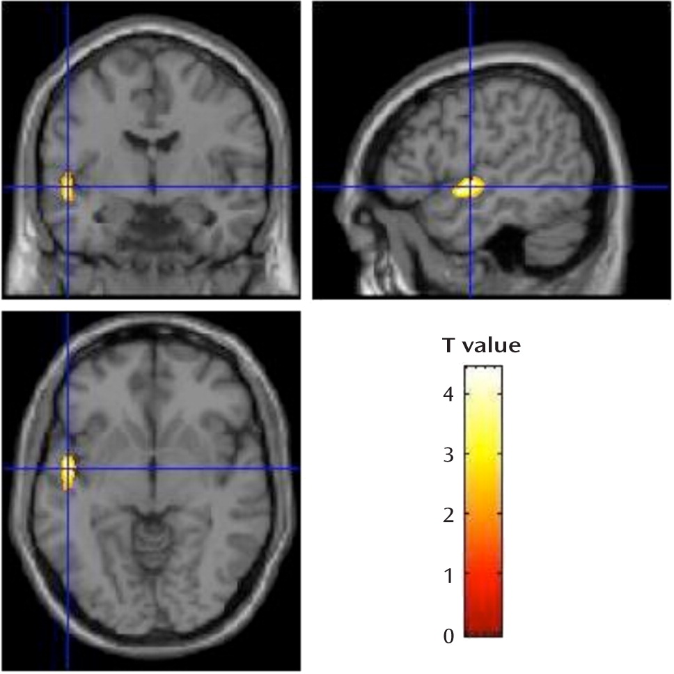
Consistent with our hypothesis, compared with the nonpsychotic group, the schizophrenia group showed significant gray matter volumetric deficits in the left superior temporal gyrus (t=4.87, df=57, false discovery rate corrected p=0.029; Brodmann's area 22; Talairach coordinates, x=–58, y=–23, z=2; cluster size=1,810 voxels of 1 mm3) (Figure 1). Results of the ANCOVA that included scan sequence as an additional covariate were similar (t=4.72, df=56, false discovery rate corrected p=0.031; Brodmann's area 22; Talairach coordinates, x=–58, y=–23, z=2; cluster size=1,158 voxels of 1 mm3). The voxel-based morphometry analysis revealed no other regions of significant between-group volumetric differences in gray matter. The analysis also found no significant differences between the schizophrenia and nonpsychotic groups in white matter volumes.
FIGURE 1. Results of Voxel-Based Morphometry in Adults With 22q11.2 Deletion Syndrome With or Without Schizophreniaa
a Rendered images of thresholded T-value maps, showing the region of superior temporal gyrus (Brodmann's area 22) with significant gray matter volumetric deficits in the schizophrenia group compared with the nonpsychotic group.

Region-of-Interest Gray Matter Volumes

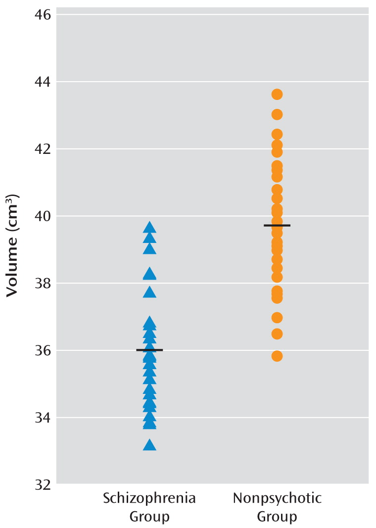
Region-of-interest analyses based on the voxel-based morphometry voxel-wise results showed significantly lower volumes of the superior temporal gyri and temporal lobes bilaterally in the schizophrenia group compared with the nonpsychotic group (Table 2, Figure 2). Results were similar when the sample was restricted to participants with an IQ >65 (N=16 schizophrenia; N=32 nonpsychotic) or when the two individuals with smaller 22q11.2 deletions were excluded (data not shown). Post hoc within-group analyses showed no significant effect of duration of illness, chlorpromazine-equivalent antipsychotic dose, or positive symptom severity on any of these gray matter volumetric results in the schizophrenia group (data not shown).
FIGURE 2. Scatterplot of Superior Temporal Gyrus Gray Matter Volumes for Adults With 22q11.2 Deletion Syndrome With (N=29) or Without (N=34) Schizophrenia

Discussion

This is the largest brain imaging study of adults with 22q11.2 deletion syndrome to date. Our goal was to determine brain structural changes associated with expression of schizophrenia in this relatively homogeneous group in which all participants carried a hemizygous 22q11.2 deletion. Taken together, voxel-based morphometry and region-of-interest analyses revealed bilateral gray matter volumetric deficits in the temporal lobes, and specifically in the superior temporal gyri, to be significantly associated with the expression of schizophrenia in adults with a 22q11.2 deletion. These findings are consistent with the most highly replicated structural imaging findings in general population samples of schizophrenia patients compared with healthy individuals (15). Gray matter volumetric deficits in the superior temporal gyrus and related temporal lobe regions have been found before, at, and after onset of psychosis, pointing to this brain region as one of those most prominently and specifically involved in the pathogenesis of schizophrenia (1517, 3134). In this study, we found no evidence that other gray matter regions, white matter, or CSF volumetric changes were significantly associated with expression of the schizophrenia phenotype in 22q11.2 deletion syndrome.
Three previous brain imaging studies of adults with 22q11.2 deletion syndrome and schizophrenia have been reported (10, 13, 14), with sample sizes of 14, 11, and 6, respectively. Only the latter two studies compared schizophrenia patients and individuals with 22q11.2 deletions with no psychotic illness (13, 14). The small sample sizes and other methodological issues likely limited the ability of those studies to identify consistent or specific MRI brain differences in 22q11.2 deletion syndrome between those with and without schizophrenia. However, consistent with the study by van Amelsvoort et al. (13), in which the participants were comparable in age to those in our study, we found no significant differences in total gray matter, white matter, and CSF volumes between the groups with and without schizophrenia. Consistent with Schaer et al. (14), we also found gray matter anomalies in the left superior temporal gyrus. As expected, our findings also differ from those of studies using different study designs. These include the more widespread MRI findings of studies that compare patients with 22q11.2 deletion syndrome and schizophrenia with individuals who have neither 22q11.2 deletion syndrome nor schizophrenia (10, 13). The differences in findings may be related to the inherent limitations in comparing a relatively homogeneous group with a specific genetic predisposition (hemizygous 22q11.2 deletion) with heterogeneous comparison samples that may have many genetic and other differences in addition to the expression of schizophrenia. This is the case even if IQ is comparably low in the comparison group (13, 14), where learning difficulties could be attributable to various causes, including other undiagnosed genetic conditions having variable predisposition to schizophrenia.
Our results may also be consistent with those of Bearden et al. (35), who found evidence of temporal lobe cortical thinning in children with 22q11.2 deletion syndrome relative to comparison children, suggesting that there may be temporal gray matter volumetric or other structural changes in some or perhaps many individuals in the sample at risk for developing schizophrenia. This raises the possibility that, as in the general population and in high-risk familial schizophrenia samples (34, 3638), individuals with 22q11.2 deletions may exhibit brain structural changes at a young age that are similar to those associated with expression of the full disease of schizophrenia later on in life. While a study of 19 adolescents with 22q11.2 deletion syndrome (39) revealed no significant differences in brain volume over 5 years when comparing the seven participants who developed psychosis with the 12 who did not, the small sample size and the young age might have limited the study's power to detect structural and developmental brain differences associated with schizophrenia. Brain imaging results for adults with 22q11.2 deletion syndrome would be expected to be different from those involving children and adolescents with 22q11.2 deletion syndrome. Studies of children must take into account the brain changes associated with development, which are known to be delayed in this condition, and with the diagnostic uncertainties given an evolving phenotype (12, 14). Prospective studies of large samples of individuals with 22q11.2 deletion syndrome followed well into adulthood will be needed to further investigate this important issue.
In the present study, comparing adults with 22q11.2 deletion syndrome with and without schizophrenia likely minimized sample-related variability and maximized the likelihood that the differences identified are attributable to expression of schizophrenia and not to other associated features of the syndrome. Our results suggest that studying the gray matter volumetric changes in the superior temporal gyrus and related temporal lobe regions will be important in understanding the pathogenesis of schizophrenia in 22q11.2 deletion syndrome, as for schizophrenia in the general population (15, 16, 34, 40, 41). Also consistent with several studies of general population samples of schizophrenia patients, we found no significant effect of duration of illness (16, 42, 43), antipsychotic dosage, or positive symptom severity (16) on the extent of gray matter volume regional losses. Longitudinal studies, particularly in the years around onset of psychosis, would be needed to investigate the issues of timing, extent of active pathogenic changes (34), and potential amelioration with antipsychotic medication (44). However, our results suggest that the main findings are more relevant to expression of schizophrenia per se than to chronicity or severity of illness.
Our sample size was adequate to withstand the well-recognized variability of expression within 22q11.2 deletion syndrome (7, 10). The mean age of the sample enhances the likelihood that diagnostic classification was stable. If some nonpsychotic individuals with 22q11.2 deletion syndrome subsequently develop schizophrenia, such diagnostic misclassification would serve to make it more difficult to detect true differences between the groups. We follow these patients longitudinally, however, minimizing this possibility. Notably, our methods accounted for IQ, presence of major congenital cardiac disease, and total intracranial volume, which are all issues of concern in studying the expression of schizophrenia in 22q11.2 deletion syndrome (12). To further evaluate the finding of reduced superior temporal gyrus volume in schizophrenia in our whole-brain voxel-based morphometry analyses, we analyzed region-of-interest volumes. These analyses might have allowed for more sensitive identification of bilateral regional findings than using the whole-brain approach, where the greater number of multiple comparisons were more strictly corrected for by the false recovery rate.
Our analysis also had limitations typical of cross-sectional and voxel-based morphometry studies. These include the inability to detect ventricular enlargement or increased CSF volume that may be associated with schizophrenia. However, such changes may be less specific; for example, they may be related more to cognitive variables in 22q11.2 deletion syndrome (12, 35). Developmental anomalies such as neuronal migration abnormalities and midline defects also may be associated with expression of schizophrenia in 22q11.2 deletion syndrome (45, 46). These would not be observable with voxel-based morphometry, although they may be compatible with the gray matter volumetric deficits observed using this imaging method. Higher-resolution scanning and other analytic methods may be needed to reveal other relevant structural anomalies associated with schizophrenia in this syndrome.
Difficulties in 22q11.2 deletion syndrome sample collection, such as pacemakers or claustrophobia precluding MRI scanning, are well known. While this is the largest MRI study of adults with 22q11.2 deletion syndrome to date, larger samples could increase the statistical power to show additional significant findings of smaller effect size than those observed. These may or may not include the frontal lobe gray matter volumetric findings seen in comparisons between adults with 22q11.2 deletion syndrome and comparison samples (10, 13) or other temporal lobe or subcortical gray matter volumetric changes seen in other genetic subtypes of schizophrenia (36).
Remaining questions of interest include why a minority of individuals with 22q11.2 deletion syndrome develop schizophrenia while the majority do not and what light these associated structural brain findings may shed on pathogenetic mechanisms. The mechanism underlying the variable expression associated with deletion 22q11.2 syndrome, even in mouse models, remains a mystery. We previously reported (18) that there were no significant effects of length of the 22q11.2 deletion, parental origin of the 22q11.2 deletion, parental age, or family history on expression of schizophrenia in 100 adults with 22q11.2 deletion syndrome. While hemizygosity of the approximately 45 genes in the commonly deleted 22q11.2 region seems to confer the major copy number-related risk factor for expression of schizophrenia, other factors may modulate this risk (3, 18). Murine models suggest that multiple gene dosage effects may play a role (47, 48). Other possibilities include altered expression related to genetic variants within the intact 22q11.2 region or in the rest of the genome (18). However, there is no evidence that the COMT functional allele influences expression of schizophrenia in 22q11.2 deletion syndrome (11, 21), and results for other genes in the 22q11.2 deletion region are inconclusive. Interacting environmental effects also deserve consideration (49). Cumulative effects of the hemizygous 22q11.2 deletion, other genetic variants, or other factors could affect the changes in neuronal cell migration, synaptogenesis, synaptic plasticity, or neurogenesis that represent plausible mechanisms for schizophrenia. Aberrant brain maturation and neuronal migration anomalies are well documented in 22q11 deletion syndrome (46, 50). Gray matter deficits in the superior temporal gyrus suggest that, as in other forms of schizophrenia, primary sensory processing cortex and cortical regions that are specialized for language and speech processes may be involved (16, 37). There is initial evidence that glutamate synapses in this region may be implicated (51).

Conclusions

This study for the first time links gray matter volumetric deficits in the superior temporal gyrus to a specific genetic etiology of schizophrenia. The findings support 22q11.2 deletion syndrome as a model for studying the development of schizophrenia from the risk conveyed by a major genetic variant to expression as a disorder. A focus on this region may be fruitful for animal models of the deletion and prospective studies of the growing population of adolescents with 22q11.2 deletions.

Footnote

Received Aug. 20, 2010; revisions received Oct. 14 and Nov. 17, 2010; accepted Nov. 22, 2010

Supplementary Material

File (ajp_168_05_522_01.pdf)

References

1.
Bassett AS, Costain G, Fung WLA, Russell KJ, Pierce L, Kapadia R, Carter RF, Chow EW, Forsythe PJ: Clinically detectable copy number variations in a Canadian catchment population of schizophrenia. J Psychiatr Res 2010; 44:1005–1009
2.
Fung WLA, McEvilly R, Fong J, Silversides C, Chow E, Bassett A: Elevated prevalence of generalized anxiety disorder in adults with 22q11.2 deletion syndrome (letter). Am J Psychiatry 2010; 167:998
3.
Bassett AS, Scherer SW, Brzustowicz LM: Copy number variations in schizophrenia: critical review and new perspectives on concepts of genetics and disease. Am J Psychiatry 2010; 167:899–914
4.
Horowitz A, Shifman S, Rivlin N, Pisante A, Darvasi A: A survey of the 22q11 microdeletion in a large cohort of schizophrenia patients. Schizophr Res 2005; 73:263–267
5.
Bassett AS, Chow EW, Husted J, Weksberg R, Caluseriu O, Webb GD, Gatzoulis MA: Clinical features of 78 adults with 22q11 deletion syndrome. Am J Med Genet A 2005; 138:307–313
6.
McDonald-McGinn DM, Kirschner R, Goldmuntz E, Sullivan K, Eicher P, Gerdes M, Moss E, Solot C, Wang P, Jacobs I, Handler S, Knightly C, Heher K, Wilson M, Ming JE, Grace K, Driscoll D, Pasquariello P, Randall P, Larossa D, Emanuel BS, Zackai EH: The Philadelphia story: the 22q11.2 deletion: report on 250 patients. Genet Couns 1999; 10:11–24
7.
Chow EW, Watson M, Young DA, Bassett AS: Neurocognitive profile in 22q11 deletion syndrome and schizophrenia. Schizophr Res 2006; 87:270–278
8.
De Smedt B, Swillen A, Devriendt K, Fryns JP, Verschaffel L, Ghesquiere P: Mathematical disabilities in children with velo-cardio-facial syndrome. Neuropsychologia 2007; 45:885–895
9.
Bassett AS, Chow EW: Schizophrenia and 22q11.2 deletion syndrome. Curr Psychiatry Rep 2008; 10:148–157
10.
Chow EW, Zipursky RB, Mikulis DJ, Bassett AS: Structural brain abnormalities in patients with schizophrenia and 22q11 deletion syndrome. Biol Psychiatry 2002; 51:208–215
11.
Murphy KC, Jones LA, Owen MJ: High rates of schizophrenia in adults with velo-cardio-facial syndrome. Arch Gen Psychiatry 1999; 56:940–945
12.
Eisenberg DP, Jabbi M, Berman KF: Bridging the gene-behavior divide through neuroimaging deletion syndromes: velocardiofacial (22q11.2 deletion) and Williams (7q11.23 deletion) syndromes. Neuroimage 2010; 53:857–869
13.
van Amelsvoort T, Daly E, Henry J, Robertson D, Ng V, Owen M, Murphy KC, Murphy DG: Brain anatomy in adults with velocardiofacial syndrome with and without schizophrenia: preliminary results of a structural magnetic resonance imaging study. Arch Gen Psychiatry 2004; 61:1085–1096
14.
Schaer M, Debbane M, Bach Cuadra M, Ottet MC, Glaser B, Thiran JP, Eliez S: Deviant trajectories of cortical maturation in 22q11.2 deletion syndrome (22q11DS): a cross-sectional and longitudinal study. Schizophr Res 2009; 115:182–190
15.
Honea R, Crow TJ, Passingham D, Mackay CE: Regional deficits in brain volume in schizophrenia: a meta-analysis of voxel-based morphometry studies. Am J Psychiatry 2005; 162:2233–2245
16.
Takahashi T, Wood SJ, Soulsby B, Kawasaki Y, McGorry PD, Suzuki M, Velakoulis D, Pantelis C: An MRI study of the superior temporal subregions in first-episode patients with various psychotic disorders. Schizophr Res 2009; 113:158–166
17.
Job DE, Whalley HC, Johnstone EC, Lawrie SM: Grey matter changes over time in high risk subjects developing schizophrenia. Neuroimage 2005; 25:1023–1030
18.
Bassett AS, Marshall CR, Lionel AC, Chow EW, Scherer SW: Copy number variations and risk for schizophrenia in 22q11.2 deletion syndrome. Hum Mol Genet 2008; 17:4045–4053
19.
Bassett AS, Chow EWC, AbdelMalik P, Gheorghiu M, Husted J, Weksberg R: The schizophrenia phenotype in 22q11 deletion syndrome. Am J Psychiatry 2003; 160:1580–1586
20.
Bassett AS, Chow EW, Husted J, Hodgkinson KA, Oechslin E, Harris L, Silversides C: Premature death in adults with 22q11.2 deletion syndrome. J Med Genet 2009; 46:324–330
21.
Bassett AS, Caluseriu O, Weksberg R, Young DA, Chow EW: Catechol-O-methyl transferase and expression of schizophrenia in 73 adults with 22q11 deletion syndrome. Biol Psychiatry 2007; 61:1135–1140
22.
Kay SR, Fiszbein A, Opler LA: The Positive and Negative Syndrome Scale (PANSS) for schizophrenia. Schizophr Bull 1987; 13:261–276
23.
Kroken RA, Johnsen E, Ruud T, Wentzel-Larsen T, Jorgensen HA: Treatment of schizophrenia with antipsychotics in Norwegian emergency wards: a cross-sectional national study. BMC Psychiatry 2009; 16:9–24
24.
Voormolen EH, Wei C, Chow EW, Bassett AS, Mikulis DJ, Crawley AP: Voxel-based morphometry and automated lobar volumetry: the trade-off between spatial scale and statistical correction. Neuroimage 2010; 49:587–596
25.
Ashburner J, Friston KJ: Unified segmentation. Neuroimage 2005; 26:839–851
26.
Walter B, Blecker C, Kirsch P, Sammer G, Schienle A, Stark R, Vaitl D: MARINA: an easy to use tool for the creation of MAsks for Region of INterest Analyses, in Proceedings of the Ninth International Conference on Functional Mapping of the Human Brain. New York, Neuroimage, 2003
27.
Tzourio-Mazoyer N, Landeau B, Papathanassiou D, Crivello F, Etard O, Delcroix N, Mazoyer B, Joliot M: Automated anatomical labeling of activations in SPM using a macroscopic anatomical parcellation of the MNI MRI single-subject brain. Neuroimage 2002; 15:273–289
28.
Gartus A, Foki T, Geissler A, Beisteiner R: Improvement of clinical language localization with an overt semantic and syntactic language functional MR imaging paradigm. AJNR Am J Neuroradiol 2009; 30:1977–1985
29.
Bergouignan L, Chupin M, Czechowska Y, Kinkingnehun S, Lemogne C, Le Bastard G, Lepage M, Garnero L, Colliot O, Fossati P: Can voxel based morphometry, manual segmentation, and automated segmentation equally detect hippocampal volume differences in acute depression? Neuroimage 2009; 45:29–37
30.
Brunner R, Henze R, Parzer P, Kramer J, Feigl N, Lutz K, Essig M, Resch F, Stieltjes B: Reduced prefrontal and orbitofrontal gray matter in female adolescents with borderline personality disorder: is it disorder specific? Neuroimage 2010; 49:114–120
31.
Keshavan MS, Haas GL, Kahn CE, Aguilar E, Dick EL, Schooler NR, Sweeney JA, Pettegrew JW: Superior temporal gyrus and the course of early schizophrenia: progressive, static, or reversible? J Psychiatr Res 1998; 32:161–167
32.
Shenton ME, Dickey CC, Frumin M, McCarley RW: A review of MRI findings in schizophrenia. Schizophr Res 2001; 49:1–52
33.
Pearlson GD: Superior temporal gyrus and planum temporale in schizophrenia: a selective review. Prog Neuropsychopharmacol Biol Psychiatry 1997; 21:1203–1229
34.
Takahashi T, Wood SJ, Yung AR, Walterfang M, Phillips LJ, Soulsby B, Kawasaki Y, McGorry PD, Suzuki M, Velakoulis D, Pantelis C: Superior temporal gyrus volume in antipsychotic-naive people at risk of psychosis. Br J Psychiatry 2010; 196:206–211
35.
Bearden CE, van Erp TG, Dutton RA, Lee AD, Simon TJ, Cannon TD, Emanuel BS, McDonald-McGinn D, Zackai EH, Thompson PM: Alterations in midline cortical thickness and gyrification patterns mapped in children with 22q11.2 deletions. Cereb Cortex 2009; 19:115–126
36.
Costain G, Ho A, Crawley AP, Mikulis DJ, Brzustowicz LM, Chow EW, Bassett AS: Reduced gray matter in the anterior cingulate gyrus in familial schizophrenia: a preliminary report. Schizophr Res 2010; 122:81–84
37.
Kasai K, Shenton ME, Salisbury DF, Hirayasu Y, Onitsuka T, Spencer MH, Yurgelun-Todd DA, Kikinis R, Jolesz FA, McCarley RW: Progressive decrease of left Heschl gyrus and planum temporale gray matter volume in first-episode schizophrenia: a longitudinal magnetic resonance imaging study. Arch Gen Psychiatry 2003; 60:766–775
38.
Kim JJ, Crespo-Facorro B, Andreasen NC, O'Leary DS, Magnotta V, Nopoulos P: Morphology of the lateral superior temporal gyrus in neuroleptic naive patients with schizophrenia: relationship to symptoms. Schizophr Res 2003; 60:173–181
39.
Gothelf D, Penniman L, Gu E, Eliez S, Reiss AL: Developmental trajectories of brain structure in adolescents with 22q11.2 deletion syndrome: a longitudinal study. Schizophr Res 2007; 96:72–81
40.
Kwon JS, McCarley RW, Hirayasu Y, Anderson JE, Fischer IA, Kikinis R, Jolesz FA, Shenton ME: Left planum temporale volume reduction in schizophrenia. Arch Gen Psychiatry 1999; 56:142–148
41.
Sumich A, Chitnis XA, Fannon DG, O'Ceallaigh S, Doku VC, Falrowicz A, Marshall N, Matthew VM, Potter M, Sharma T: Temporal lobe abnormalities in first-episode psychosis. Am J Psychiatry 2002; 159:1232–1235
42.
Marsh L, Suddath RL, Higgins N, Weinberger DR: Medial temporal lobe structures in schizophrenia: relationship of size to duration of illness. Schizophr Res 1994; 11:225–238
43.
Crespo-Facorro B, Nopoulos PC, Chemerinski E, Kim JJ, Andreasen NC, Magnotta V: Temporal pole morphology and psychopathology in males with schizophrenia. Psychiatry Res 2004; 132:107–115
44.
Lieberman JA, Tollefson GD, Charles C, Zipursky R, Sharma T, Kahn RS, Keefe RS, Green AI, Gur RE, McEvoy J, Perkins D, Hamer RM, Gu H, Tohen M: Antipsychotic drug effects on brain morphology in first-episode psychosis. Arch Gen Psychiatry 2005; 62:361–370
45.
Chow EW, Mikulis DJ, Zipursky RB, Scutt LE, Weksberg R, Bassett AS: Qualitative MRI findings in adults with 22q11 deletion syndrome and schizophrenia. Biol Psychiatry 1999; 46:1436–1442
46.
Kiehl TR, Chow EWC, Mikulis DJ, George SR, Bassett AS: Neuropathologic features in adults with 22q11.2 deletion syndrome. Cereb Cortex 2009; 19:153–164
47.
Meechan DW, Maynard TM, Gopalakrishna D, Wu Y, LaMantia AS: When half is not enough: gene expression and dosage in the 22q11 deletion syndrome. Gene Expr 2007; 13:299–310
48.
Sivagnanasundaram S, Fletcher D, Hubank M, Illingworth E, Skuse D, Scambler P: Differential gene expression in the hippocampus of the Df1/+ mice: a model for 22q11.2 deletion syndrome and schizophrenia. Brain Res 2007; 1139:48–59
49.
Chow EW, Husted J, Weksberg R, Bassett AS: Postmaturity in a genetic subtype of schizophrenia. Acta Psychiatr Scand 2003; 108:260–268
50.
Bearden CE, van Erp TG, Monterosso JR, Simon TJ, Glahn DC, Saleh PA, Hill NM, McDonald-McGinn DM, Zackai E, Emanuel BS, Cannon TD: Regional brain abnormalities in 22q11.2 deletion syndrome: association with cognitive abilities and behavioral symptoms. Neurocase 2004; 10:198–206
51.
Bowden NA, Scott RJ, Tooney PA: Altered gene expression in the superior temporal gyrus in schizophrenia. BMC Genomics 2008; 9:199

Information & Authors

Information

Published In

Go to American Journal of Psychiatry
American Journal of Psychiatry
Pages: 522 - 529
PubMed: 21362743

History

Received: 20 August 2010
Revision received: 14 October 2010
Revision received: 17 November 2010
Accepted: 22 November 2010
Published online: 1 May 2011
Published in print: May 2011

Authors

Details

Eva W.C. Chow, M.D., F.R.C.P.C.
From the Clinical Genetics Research Program, Centre for Addiction and Mental Health, Toronto; the Department of Psychiatry and the Department of Medical Imaging, University of Toronto; and the Division of Cardiology, Department of Medicine, University Health Network, Toronto.
Andrew Ho, B.Sc.
From the Clinical Genetics Research Program, Centre for Addiction and Mental Health, Toronto; the Department of Psychiatry and the Department of Medical Imaging, University of Toronto; and the Division of Cardiology, Department of Medicine, University Health Network, Toronto.
Corie Wei, M.D.
From the Clinical Genetics Research Program, Centre for Addiction and Mental Health, Toronto; the Department of Psychiatry and the Department of Medical Imaging, University of Toronto; and the Division of Cardiology, Department of Medicine, University Health Network, Toronto.
Eduard H.J. Voormolen, M.D., M.Sc.
From the Clinical Genetics Research Program, Centre for Addiction and Mental Health, Toronto; the Department of Psychiatry and the Department of Medical Imaging, University of Toronto; and the Division of Cardiology, Department of Medicine, University Health Network, Toronto.
Adrian P. Crawley, Ph.D.
From the Clinical Genetics Research Program, Centre for Addiction and Mental Health, Toronto; the Department of Psychiatry and the Department of Medical Imaging, University of Toronto; and the Division of Cardiology, Department of Medicine, University Health Network, Toronto.
Anne S. Bassett, M.D., F.R.C.P.C.
From the Clinical Genetics Research Program, Centre for Addiction and Mental Health, Toronto; the Department of Psychiatry and the Department of Medical Imaging, University of Toronto; and the Division of Cardiology, Department of Medicine, University Health Network, Toronto.

Notes

Address correspondence and reprint requests to Dr. Chow, Clinical Genetics Research Program, Centre for Addiction and Mental Health, 33 Russell St., Rm. 1085, Toronto, Ontario, Canada M5S 2S1; [email protected] (e-mail).

Funding Information

The authors report no financial relationships with commercial interests.Supported by Canadian Institutes of Health Research grants MOP-74631 (to Dr. Chow), MOP-79518, MOP-89066, and MOP-97800 (to Dr. Bassett), and by a W. Garfield Weston Foundation grant to Dr. Bassett. Dr. Bassett holds the Canada Research Chair in Schizophrenia Genetics and Genomic Disorders.

Metrics & Citations

Metrics

Citations

Export Citations

If you have the appropriate software installed, you can download article citation data to the citation manager of your choice. Simply select your manager software from the list below and click Download.

For more information or tips please see 'Downloading to a citation manager' in the Help menu.

Format
Citation style
Style
Copy to clipboard

View Options

View options

PDF/EPUB

View PDF/EPUB

Get Access

Login options

Already a subscriber? Access your subscription through your login credentials or your institution for full access to this article.

Personal login Institutional Login Open Athens login
Purchase Options

Purchase this article to access the full text.

PPV Articles - American Journal of Psychiatry

PPV Articles - American Journal of Psychiatry

Not a subscriber?

Subscribe Now / Learn More

PsychiatryOnline subscription options offer access to the DSM-5-TR® library, books, journals, CME, and patient resources. This all-in-one virtual library provides psychiatrists and mental health professionals with key resources for diagnosis, treatment, research, and professional development.

Need more help? PsychiatryOnline Customer Service may be reached by emailing [email protected] or by calling 800-368-5777 (in the U.S.) or 703-907-7322 (outside the U.S.).

Media

Figures

Other

Tables

Share

Share

Share article link

Share