Full access
Articles
Published Online: 1 July 2019

Baseline Frontoparietal Task-Related BOLD Activity as a Predictor of Improvement in Clinical Symptoms at 1-Year Follow-Up in Recent-Onset Psychosis

Abstract

Objective:

The early course of illness in psychotic disorders is highly variable, and predictive biomarkers of treatment response have been lacking. Trial and error remains the basis for care in early psychosis, and poor outcomes are common. Early prediction of nonimprovement in response to treatment could help identify those who would benefit from alternative and/or supplemental interventions. The goal of this study was to evaluate the ability of functional MRI (fMRI) measures of cognitive control–related brain circuitry collected at baseline to predict symptomatic response in patients after 1 year.

Methods:

Patients with recent-onset (<2 years) psychotic disorders (N=82) in early psychosis specialty care were classified as improvers (>20% improvement in total score on the Brief Psychiatric Rating Scale [BPRS] at 1-year follow-up compared with baseline) or as nonimprovers. Behavioral (d′ context) and fMRI (proactive control–associated activation in a priori frontoparietal regions of interest) measures of cognitive control were then evaluated on their ability to predict BPRS improvement using linear and logistic regression.

Results:

Cognitive control–associated measures significantly predicted BPRS improvement and improver status, with 70% positive predictive value, 60% negative predictive value, and 66% accuracy. Only the fMRI-based measure (and not the behavioral measure) significantly predicted status.

Conclusions:

These results suggest that frontoparietal activation during cognitive control performance at baseline significantly predicts subsequent symptomatic improvement during early psychosis specialty care. Potential implications for fMRI-based personalized patient treatment are discussed.
Although Kraepelin postulated that schizophrenia was a degenerative disorder characterized by deterioration and inevitably poor outcomes (1), longitudinal studies have found great heterogeneity in symptomatic progression over the lifespan. An early study by Ciompi (2), for example, identified eight course types based on their suddenness of onset, symptom stability (simple or undulating), and end state (recovered or otherwise). Later work by Fenton and McGlashan (3) attempted to reclassify schizophrenia based on illness progression. They found that stability varied as a function of “classic” illness subtype (paranoid, hebephrenic, undifferentiated) and that paranoid patients, who (by definition) have fewer disorganization and negative symptoms, showed the most improvement. Nonetheless, no consensus guidelines or biomarkers have been developed that can effectively predict disease progression. Development of such biomarkers would be clinically invaluable as they would not only provide mechanistic insights into what influences symptomatic response to treatment but also help identify patients who may require nonstandard treatment approaches to optimize outcome.
Ideally, such predictive biomarkers would be inexpensive, noninvasive, readily administered, and suitable for use in the majority of patients (including adolescents). Functional MRI (fMRI) has been used extensively in efforts to develop neurophysiological biomarkers for schizophrenia and other psychotic illnesses. Surprisingly, however, few fMRI studies have examined the potential for brain activation to predict treatment outcomes. In a small sample (N=23), van Veelen et al. (4) found that patients with a first episode of schizophrenia who showed >30% symptom reduction after 10 weeks of treatment had significantly greater function in the dorsolateral prefrontal cortex (DLPFC) during working memory (specifically using the contrast practice > novel stimulus set) at baseline compared with those who did not (N=12). A 2015 report by Anticevic et al. (5) in unmedicated patients observed a significant relationship between resting-state prefrontal hyperconnectivity and 12-month symptom improvement. In a 2016 study, Sarpal et al. (6) found that resting-state striatal functional connectivity distinguished treatment responders (N=24) and nonresponders (N=17) in schizophrenia with 76% and 79% positive and negative predictive values, respectively. Finally, Cao et al. (7) recently reported that resting-state connectivity between the superior temporal cortex and other cortical regions predicted treatment responders (N=25; nonresponders, N=13) after 10 weeks of risperidone treatment with 83% accuracy.
Although promising, these studies are limited by small sample sizes and brief follow-up periods. Furthermore, they were designed to predict acute treatment response in antipsychotic-naive individuals; to our knowledge, no studies have used fMRI to predict change in symptoms over a year or more in a naturalistic sample undergoing early psychosis specialty care. To that end, in this study we examined the ability of baseline brain activity during an established, validated cognitive control task, the AX continuous performance task (AX-CPT), to predict symptomatic improvement after 1-year follow-up in a sample of patients with recent-onset schizophrenia. Based on the two preliminary fMRI studies cited above and the association with better outcomes in patients with the paranoid subtype, we hypothesized that frontoparietal activation (which has been shown to be impaired in early psychosis and associated with behavioral disorganization and cognitive dysfunction [810]) would be a potential predictive biomarker of treatment response. As an additional exploratory analysis, we also examined the ability of baseline symptom dimensions (reality distortion, psychomotor poverty, and disorganization) to contribute to logistic model prediction, as previous work suggests that long-term outcome may be influenced by symptom severity at presentation (3, 11).

Methods

Sample

Baseline neuroimaging AX-CPT data were available for 171 patients (patients with schizophrenia, N=139; patients with type I bipolar disorder with psychotic features, N=32). Of this sample, follow-up clinical data were available for 82 patients (patients with schizophrenia, N=65; patients with bipolar disorder, N=17). Healthy control subjects (N=138) were included to verify that the task was activating expected frontoparietal regions (see the section “fMRI Analysis and Prespecified Region of Interest Selection” below). Neuroimaging AX-CPT data from the 82 patients with complete (baseline and follow-up) data sets have been used in previous studies as follows: 53 control subjects and 18 patients (9); 34 control subjects and 20 patients (12); 23 control subjects and 11 patients (13); 52 control subjects and 43 patients (14); and 21 control subjects and six patients (10). Imaging data from 70 (of 138) control subjects and 36 (of 82) patients in the final sample have not been published. Individuals were recruited as outpatients from the Early Diagnosis and Preventive Treatment (of Psychosis) research clinic at the University of California, Davis (https://earlypsychosis.ucdavis.edu). Treatment in the clinic follows a coordinated specialty care model for early psychosis delivered by an interdisciplinary treatment team. Treatment includes detailed clinical assessments using gold-standard structured clinical interviews and medical evaluations; targeted pharmacological treatments, including low-dose atypical antipsychotic treatment; individual and family-based psychosocial education and support; cognitive-behavioral therapy for psychosis; and support for education and employment. The Structured Clinical Interview for DSM-IV-TR Axis I Disorders (15) was used for diagnosis of psychopathology. Diagnoses were confirmed by a group of trained clinicians during case conferences. All patients reported onset of psychosis within 2 years before the date of informed consent. Patients were excluded if they had a diagnosis of major medical or neurological illness, head trauma, substance abuse in the previous 3 months (as well as a positive result on urinalysis on the day of scanning), a score <70 on the Wechsler Abbreviated Scale of Intelligence–2 (16), or contraindications for MRI (e.g., claustrophobia, metal in the body). Control participants were excluded for all of the above as well as for a history of axis I mental illness or first-degree family history of psychosis. All participants provided written informed consent and were compensated for participation. The University of California, Davis, institutional review board approved the study. Medication regimen (type and dosage) was assessed by clinical records at baseline and follow-up. Medication adherence was based on self-report. At follow-up, all patients who received medication self-reported at least medium adherence with antipsychotic medication during the treatment period (except for two individuals with schizophrenia for whom adherence data were missing at follow-up). Symptoms were assessed using the 24-point Brief Psychiatric Rating Scale (BPRS) (17), rescaled to a lowest score of 0 (i.e., a score of 24 was equal to a score of 0). At baseline, all patients had BPRS scores ≥5 to ensure sufficient resolution to detect a 20% improvement in score at follow-up. Consistent with previous work (18), syndrome scores from three core symptom dimensions were also calculated. “Psychomotor poverty” combined emotional withdrawal, motor retardation, and blunted affect from the BPRS with anhedonia/asociality, avolition/apathy, alogia, and affective flattening from the Scale for the Assessment of Negative Symptoms (SANS) (19). “Disorganization” combined conceptual disorganization, mannerisms and posturing, and disorientation scores from the BPRS with attention scores from the SANS as well as positive formal thought disorder and bizarre behavior scores from the Scale for the Assessment of Positive Symptoms (SAPS) (20). “Reality distortion” combined grandiosity, suspiciousness, hallucinations, and unusual thought content from the BPRS with hallucinations and delusions from the SAPS (18).

Task Description

The AX-CPT and associated task parameters have been described elsewhere (9, 2124). Briefly, participants are presented with a series of cues and probes and are instructed to make a target response (pressing a button with the index finger) to the probe letter “X” only if it was preceded by the cue letter “A.” All cues and nontarget probes require nontarget responses (pressing a button with the middle finger). Target sequence trials (i.e., “AX” trials) are frequent (60%−70% occurrence) and set up a prepotent tendency to make a target response when the probe letter X occurs. As a result, a nontarget sequence trial in which any non-A cue (collectively called “B” cues) is presented and followed by a probe letter X (i.e., “BX” trials) requires proactive cognitive control (e.g., maintenance of the inhibitory rule over the delay time) (22). Consistent with past work (23), individual subject data were included in analyses only if results suggested that the subject understood the AX-CPT (specifically, accuracy greater than 44% on AX trials and 50% on BY trials at both baseline and follow-up). Participants were combined across two task protocols collected from two MRI scanners over a 14-year period. Parameters for each protocol (AX-1 and AX-2) are provided in Table S1A in the online supplement. The task was presented using EPrime2 (Psychology Software Tools). The behavioral index of proactive cognitive control was d′ context, a function of AX hits minus BX false alarms (21).

fMRI Scanning Parameters and Preprocessing

Functional images were acquired with a gradient-echo T2* blood-oxygen-level-dependent (BOLD) contrast technique as outlined in Table S1B in the online supplement. The AX-1 was performed in a 1.5-T scanner (GE Healthcare), and the AX-2 in a 3.0-T scanner (Siemens).
The fMRI data were preprocessed using SPM8 (Wellcome Department of Imaging Neuroscience, London). Briefly, images were slice-timing corrected, realigned, normalized to the Montreal Neurological Institute template using a rigid-body transformation followed by nonlinear warping, and smoothed with an 8-mm full width at half maximum Gaussian kernel. All individual fMRI runs had less than 4 mm of translational within-run movement, 3° of rotational within-run movement, and 0.45 mm of average framewise displacement (calculated using the FMRIB Software Library Motion Outliers tool; https://fsl.fmrib.ox.ac.uk/fsl/fslwiki/FSLMotionOutliers). Mean displacement did not differ between improvers and nonimprovers (t=1.42, p=0.16). All participants had at least two fMRI runs surviving these criteria. Preprocessing pipelines were identical for the AX-1 and the AX-2.

fMRI Analysis and Prespecified Region of Interest Selection

First-level effects were modeled with a double-gamma function, with temporal derivatives, using the general linear model in SPM8. Rigid-body motion parameters were included as single-subject regressors to partially account for movement effects. B>A cue (correct trials only) contrast images (parameter estimates) were generated for each subject. The B>A cue contrast measures response under conditions of high and low proactive cognitive control (9, 12). All trial types were modeled (AX, AY, BX, and BY), and only correct responses were used to create first-level images, consistent with other studies (9, 12). Whole-brain analyses across the final sample (healthy control subjects and patients with follow-up data) using the B>A contrast were used to confirm significant (height threshold p<0.001; cluster threshold p<0.05 [whole brain false-discovery-rate-corrected]) activation in expected brain regions (the left and right DLPFC and superior parietal cortex [SPC]) for both protocol versions (AX-1 and AX-2).
For subsequent regression analyses using first-level images, BOLD response was extracted from prespecified 5-mm radius spherical regions of interest in the left and right DLPFC and SPC (i.e., left and right regions combined to make a single region of interest). Although this size was chosen arbitrarily, previous work from our group suggests that varying region-of-interest radii between 4 mm and 8 mm does not substantially affect AX-CPT task-associated response patterns in psychosis (14). The DLPFC region of interest was taken from a study from an independent data set (25). The SPC regions of interest were taken from a meta-analysis of executive function in schizophrenia (26). Mean task-associated response from these regions of interest was extracted using the Marsbar toolbox (27). Task-associated behavioral and functional measures were adjusted for differences in protocol version prior to further analysis by calculating standardized residuals from the linear regression of protocol version by each measure. Adjustments were calculated separately for all participants (171 patients and 138 control subjects) and for control subjects and patients with follow-up data (82 patients and 138 control subjects), as these data sets were used for different analyses (missing data comparisons [t tests] and logistic regression, respectively).

Linear Regression

Linear regression was performed in SPSS, version 25 (IBM, Armonk, N.Y.). For this analysis, the linear dependent variable was clinical improvement (increase in the total BPRS score) at follow-up, and the independent (predictor) variables were the behavioral and functional measures from the proactive cognitive control–based feature set described above. The threshold for statistical significance of the overall model and individual predictors was set to p<0.05.

Logistic Regression

Logistic regression was also performed in SPSS, version 25. For primary analyses, the binary dependent variable was clinical improvement at follow-up. Clinical improvement was defined as a >20% decrease in the total BPRS score from baseline, rescaled to a lowest score of 0 (28). An initial model was constructed using a proactive cognitive control–based feature set. An exploratory secondary model was also evaluated that added baseline core symptom dimension scores (reality distortion, psychomotor poverty, disorganization) as predictors. The SPSS classification cutoff was set to the ratio of improvers to nonimprovers. Models were evaluated for fit, specificity, sensitivity, predictive value, and accuracy.

Results

Demographic and Clinical Information

Demographic information for individuals in the study sample is presented in Table 1. Clinical information at baseline and follow-up is presented in Table 2. The mean BPRS score at baseline for all patients was 42.7 (SD=9.7). The mean BPRS score at follow-up for all patients was 37.3 (SD=9.0). Forty-seven percent of patients with bipolar disorder and 60% of patients with schizophrenia showed a >20% decrease in the total BPRS score (scaled to a lowest value of 0) at follow-up and were classified as improvers. The mean improvement in the BPRS score for improvers was 12.7 (SD=7.3), corresponding to a 59% decrease.
TABLE 1. Demographic information for participants in a study predicting improvement in clinical symptoms in early psychosis specialty care
CharacteristicHealthy Control Subjects (N=138)Patients With Bipolar Disorder (N=17)Patients With Schizophrenia (N=65)
 N%N%N%
Participants in the AX-1 protocol735314823858
Participants in the AX-2 protocol65473182742
Female53387411625
 MeanSDMeanSDMeanSD
Age (years)20.42.721.62.820.83.3
Days to follow-up429.7113.0384.7143.7
TABLE 2. Clinical information for participants at baseline and follow-up in a study predicting improvement in clinical symptoms in early psychosis specialty care
CharacteristicPatients With Bipolar Disorder (N=17)Patients With Schizophrenia (N=65)All Patients (N=82)
 N%N%N%
BPRS scorea      
 Score improved84739604757
 Score did not improve95326403543
 BaselineFollow-upBaselineFollow-upBaselineFollow-up
 N%N%N%N%N%N%
Antipsychotic medication            
 Received medication148210595585507769846073
 Did not receive medication3187411015152313162227
 MeanSDMeanSDMeanSDMeanSDMeanSDMeanSD
 Chlorpromazine equivalent dosage (mg/day)302.3156.4342.5358.2207.2148.0300.7298.9227.4154.4307.3305.9
a
Clinical improvement was defined as showing a >20% decrease (with lowest possible score [24] set to 0) on the total Brief Psychiatric Rating Scale (BPRS) score at follow-up (compared with baseline). Only patients with a total BPRS score ≥29 at baseline were included in the sample.

Behavioral and Functional AX-CPT Results

Results of comparisons between patients with follow-up data relative to those without follow-up data are presented in Table S2 in the online supplement. No differences were observed on d′ context, task-associated DLPFC or SPC response, or total BPRS score.
Across healthy control subjects and patients with follow-up clinical data (i.e., patients included in logistic regression analyses), significant activation (see the Methods section for the threshold) was observed in the DLPFC and SPC for both protocol versions (see Table S3 and Figure S1 in the online supplement). Raw behavioral and fMRI region-of-interest data segregated by protocol version are presented in Table S4 in the online supplement.

Linear Regression

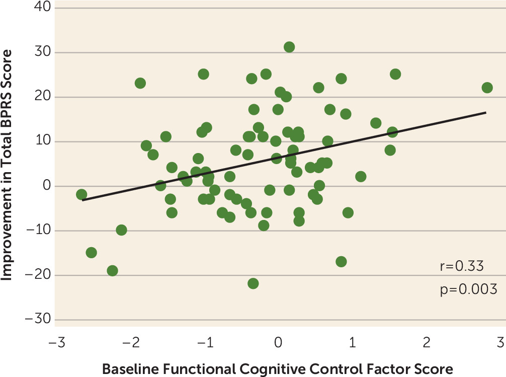
We then examined the linear relationship between BPRS improvement and baseline cognitive control measures using linear regression. Because of high covariance (0.62) between the DLPFC and SPC region-of-interest activity for the B>A cue (proactive control) fMRI contrast, BOLD response in these regions was combined into a single frontoparietal factor score. The overall model (with two predictors, behavioral and functional) was significant (F=6.50, df=2, 81, R=0.38, p=0.002), although only the fMRI-based predictor (frontoparietal factor score) significantly contributed (B=3.88, standardized coefficient [beta]=0.35, t=3.36, p=0.001). The linear relationship (Pearson’s correlation coefficient) between BPRS improvement and functional factor score is illustrated in Figure 1.
FIGURE 1. Linear relationship between improvement (from baseline) on the Brief Psychiatric Rating Scale (BPRS) and baseline frontoparietal factor score among patients in early psychosis specialty care

Logistic Regression

We next evaluated the ability of baseline proactive control measures (adjusted for protocol version) to predict BPRS improvement on a previously identified (28), clinically relevant binary scale (with an improver defined as a patient with >20% decrease in total BPRS score [rescaled to a lowest score of 0] from baseline) using logistic regression. Activation of the DLPFC and SPC was again combined into a single factor score as described for linear regression. Significance values, fit indices, and odds ratios for logistic regression models are presented in Table 3. Predictive capacity (specificity, sensitivity, positive predictive value, negative predictive value, and accuracy) for these models is presented in Table 4.
TABLE 3. Logistic regression results in a study predicting improvement in clinical symptoms in early psychosis specialty carea
Predictive FactorModel χ2Step χ2Model –2LLModel C-S R2Model Nagelkerke’s R2Predictive FactorIf Predictive Factor Removed
BetaSEpOdds Ratio95% CIChange in –2LLp
Functional cognitive control factor score (DLPFC+SPC B>A cue activation)9.5 (p<0.01)102.40.110.150.80.30.012.21.3–3.79.5<0.01
d′ context (adjusted z score)     –0.10.20.690.90.6–1.40.20.68
Constant     0.50.30.051.7  
Functional cognitive control factor score (DLPFC+SPC B>A cue activation)14.0 (p=0.02)4.49 (p=0.21)97.90.160.210.80.3<0.012.11.2–3.88.0<0.01
d′ context (adjusted z score)     0.30.20.901.00.7–1.60.020.90
Reality distortion     0.10.00.211.11.0–1.11.60.20
Disorganization     0.10.10.401.10.9–1.30.70.40
Psychomotor poverty     0.10.10.311.11.0–1.21.00.31
Constant     –1.31.00.180.3  
a
Step chi-square is for adding in the baseline syndrome scores (reality distortion, disorganization, psychomotor poverty) to the initial model (functional cognitive control factor score plus constant). C-S=Cox and Snell test for binary data; DLPFC=dorsolateral prefrontal cortex; LL=log likelihood; SPC=superior parietal cortex.
TABLE 4. Predictive metrics for each model using SPSS logistic regression in a study predicting improvement in clinical symptoms in early psychosis specialty carea
Predictive FactorSpecificity (%)Sensitivity (%)PPV (%)NPV (%)Accuracy (%)
Functional cognitive control factor score (DLPFC+SPC B>A cue activation); d′ context (adjusted z score); constant60.070.270.260.065.9
Functional cognitive control factor score (DLPFC+SPC B>A cue activation); d′ context (adjusted z score); reality distortion; psychomotor poverty; disorganization; constant68.670.275.063.269.5
a
“Positive” status for positive predictive value (PPV) was defined as showing >20% improvement in score on the Brief Psychiatric Rating Scale at follow-up. DLPFC=dorsolateral prefrontal cortex; NPV=negative predictive value; SPC=superior parietal cortex.
An initial model was constructed that included only proactive control–associated variables (behavioral and functional) as predictors. The overall model was significant (Table 3, top row), explained 15% of the variance in BPRS outcome (Nagelkerke’s R2=0.15), and was 65.9% accurate using the SPSS log-likelihood-based regression algorithm (Table 4, top row). Only the fMRI predictor significantly contributed to the model (beta=0.8, p=0.01, change in −2 log likelihood if removed=9.5 [p<0.01]; Table 3, top row).
As an exploratory measure on top of the initial model, we then evaluated a secondary model that included baseline symptom core dimension scores (reality distortion, psychomotor poverty, disorganization) as additional predictors. These additional predictors did not significantly improve fit (step χ2=4.49, p=0.21), although accuracy was slightly improved (69.5%; Table 4, bottom row).

Discussion

The results of this study suggest that patients with greater frontoparietal activation during proactive cognitive control are more likely to show symptomatic improvement at 1-year follow-up and that, conversely, poor treatment response is associated with poor activation in this circuitry. To our knowledge, this is the first study to use functional neuroimaging of cognitive control to predict 1-year treatment improver status in recent-onset psychotic illness, and consequently it may have important implications for understanding disease mechanisms and treatment. Our results also demonstrate the potential clinical utility of fMRI-based measures of cognition-related brain activity. Indeed, only functional (and not behavioral) measures associated with the task distinguished between improvers and nonimprovers.
If frontoparietal executive dysfunction is a significant predictor of outcome, how may it be targeted? Currently, clozapine is typically prescribed in patients who do not respond to more conventional forms of treatment (29) (no patients were taking clozapine at any point in this study). Interestingly, clozapine has demonstrated effects on prefrontal function that may help explain its effectiveness, including increasing P3b amplitude (an electrophysiological measure of top-down attention) (30) and decreasing resting metabolism (31). Nonetheless, clozapine has a number of highly deleterious side effects, including weight gain, agranulocytosis, seizures, and cardiomyopathy (32). Although research is still in the early stages, potential alternative methods of targeting prefrontal dysfunction in psychosis include brain stimulation (3335) and cognitive remediation (36). Future prospective studies or retrospective analyses may examine whether effects of these developmental interventions can improve outcomes in patients who show significant functional pathology at intake.
The best model in this study (fMRI plus baseline syndrome scores) correctly classified 70% of patients as being improvers. While this accuracy reflects a promising start, to be an effective diagnostic tool, fMRI should demonstrate at least 80% accuracy. Furthermore, although the correlation was significant, baseline frontoparietal activation explained only 11% of the variance in BPRS score improvement, suggesting that additional measures are necessary to fully understand why symptoms change in some patients and not others. A related point is that a number of fMRI studies in early psychosis have used classification-based analyses to differentiate patients and control subjects (or to segregate patients by diagnosis) and have performed similarly (in regard to classification accuracy) to the present study. This work has often been criticized as having statistical but not clinical significance because clinical or even lay interviewers can perform at equivalent levels of diagnostic accuracy. Unlike these studies, however, our study sought to forecast long-term symptomatic improvement, a measure impossible to predict using any established method in early-psychosis patients. Therefore, we would argue that despite not reaching an optimum level of accuracy, the present work may represent an important preliminary step toward clinical utility. Further studies using larger samples and additional predictive markers (e.g., frontal-parietal pathophysiology, structural imaging, and molecular imaging) may take us toward higher levels of prediction and closer to a precision psychiatry of early-psychosis care.
A potential limitation in interpreting our findings is that, because this was a prospective naturalistic study, we did not impose strict guidelines on medication status at either baseline or follow-up (the majority of recent-onset outpatients who enter treatment in our clinic have had some brief prior medication treatment). Furthermore, medication adherence was ascertained by self-report. Therefore, we cannot state with certainty whether differences in BPRS symptom change from baseline to follow-up are due to antipsychotics or another aspect of treatment (e.g., psychoeducation, psychotherapy). For this reason, we labeled our groups as improvers and nonimprovers rather than as responders and nonresponders. An important follow-up study would perform the same analyses in a sample of first-episode patients whose medication intake and level of psychotherapy engagement were more objectively monitored and accounted for. A second limitation was that functional outcomes (social, academic, occupational) were not examined. Given the established link between cognition and functional outcomes in schizophrenia (8, 37), additional research that evaluates the ability of fMRI neurocognitive data to predict these outcomes is strongly warranted. Despite these limitations, we believe that the present results provide important new evidence that cognitive control–related frontal-parietal brain activity may serve as a meaningful predictor of clinical improvement in early-psychosis patients and that our results may represent an important first step in developing much-needed imaging biomarkers of treatment outcomes in this important patient population.

Acknowledgments

The authors thank Amber Howell, Andrew Westphal, Julian Williams, Shaun Rafael, Natalie Hutchison, Benjamin Geib, Madison Titone, Taylor Salo, Estera (Neli) Mihov, Erika Steinbauer, Markie Benavidez, and Davis Ewbank for their assistance with data collection and processing, as well as the study participants and their families.

Supplementary Material

File (appi.ajp.2019.18101126.ds001.pdf)

References

1.
Kraepelin E: Psychiatrie: Ein Lehrbuch für Studierende und Aertze. Leipzig, Germany, Barth, 1899
2.
Ciompi L: The natural history of schizophrenia in the long term. Br J Psychiatry 1980; 136:413–420
3.
Fenton WS, McGlashan TH: Natural history of schizophrenia subtypes: I: longitudinal study of paranoid, hebephrenic, and undifferentiated schizophrenia. Arch Gen Psychiatry 1991; 48:969–977
4.
van Veelen NM, Vink M, Ramsey NF, et al: Prefrontal lobe dysfunction predicts treatment response in medication-naive first-episode schizophrenia. Schizophr Res 2011; 129:156–162
5.
Anticevic A, Hu X, Xiao Y, et al: Early-course unmedicated schizophrenia patients exhibit elevated prefrontal connectivity associated with longitudinal change. J Neurosci 2015; 35:267–286
6.
Sarpal DK, Argyelan M, Robinson DG, et al: Baseline striatal functional connectivity as a predictor of response to antipsychotic drug treatment. Am J Psychiatry 2016; 173:69–77
7.
Cao B, Cho RY, Chen D, et al: Treatment response prediction and individualized identification of first-episode drug-naive schizophrenia using brain functional connectivity. Mol Psychiatry (Epub ahead of print, June 19, 2018)
8.
Lesh TA, Niendam TA, Minzenberg MJ, et al: Cognitive control deficits in schizophrenia: mechanisms and meaning. Neuropsychopharmacology 2011; 36:316–338
9.
Lesh TA, Westphal AJ, Niendam TA, et al: Proactive and reactive cognitive control and dorsolateral prefrontal cortex dysfunction in first episode schizophrenia. Neuroimage Clin 2013; 2:590–599
10.
Yoon JH, Minzenberg MJ, Ursu S, et al: Association of dorsolateral prefrontal cortex dysfunction with disrupted coordinated brain activity in schizophrenia: relationship with impaired cognition, behavioral disorganization, and global function. Am J Psychiatry 2008; 165:1006–1014; correction, 165:1359
11.
Fenton WS, McGlashan TH: Natural history of schizophrenia subtypes: II: positive and negative symptoms and long-term course. Arch Gen Psychiatry 1991; 48:978–986
12.
Lesh TA, Tanase C, Geib BR, et al: A multimodal analysis of antipsychotic effects on brain structure and function in first-episode schizophrenia. JAMA Psychiatry 2015; 72:226–234
13.
Niendam TA, Lesh TA, Yoon J, et al: Impaired context processing as a potential marker of psychosis risk state. Psychiatry Res 2014; 221:13–20
14.
Smucny J, Lesh TA, Newton K, et al: Levels of cognitive control: a functional magnetic resonance imaging–based test of an RDoC domain across bipolar disorder and schizophrenia. Neuropsychopharmacology 2018; 43:598–606
15.
First MB, Spitzer RL, Gibbon M, et al: Structured Clinical Interview for DSM-IV-TR Axis I Disorders, Research Version, Patient Edition. New York, Biometrics Research, New York State Psychiatric Institute, 2002
16.
Wechsler D: Wechsler Abbreviated Scale of Intelligence (WASI). San Antonio, Tex, Harcourt Assessment, 1999
17.
Ventura J, Lukoff D, Nuechterlein KH, et al: Manual for the expanded brief psychiatric rating scale. Int J Methods Psychiatr Res 1993; 3:227–244
18.
Barch DM, Carter CS, MacDonald AW III, et al: Context-processing deficits in schizophrenia: diagnostic specificity, 4-week course, and relationships to clinical symptoms. J Abnorm Psychol 2003; 112:132–143
19.
Andreasen NC: Scale for the Assessment of Negative Symptoms (SANS). Iowa City, Iowa, Department of Psychiatry, Carver College of Medicine, University of Iowa, 1984
20.
Andreasen NC: Scale for the Assessment of Positive Symptoms (SAPS). Iowa City, Iowa, Department of Psychiatry, Carver College of Medicine, University of Iowa, 1984
21.
Cohen JD, Barch DM, Carter C, et al: Context-processing deficits in schizophrenia: converging evidence from three theoretically motivated cognitive tasks. J Abnorm Psychol 1999; 108:120–133
22.
Braver TS, Paxton JL, Locke HS, et al: Flexible neural mechanisms of cognitive control within human prefrontal cortex. Proc Natl Acad Sci USA 2009; 106:7351–7356
23.
Henderson D, Poppe AB, Barch DM, et al: Optimization of a goal maintenance task for use in clinical applications. Schizophr Bull 2012; 38:104–113
24.
Phillips RC, Salo T, Carter CS: Distinct neural correlates for attention lapses in patients with schizophrenia and healthy participants. Front Hum Neurosci 2015; 9:502
25.
MacDonald AW III, Cohen JD, Stenger VA, et al: Dissociating the role of the dorsolateral prefrontal and anterior cingulate cortex in cognitive control. Science 2000; 288:1835–1838
26.
Minzenberg MJ, Laird AR, Thelen S, et al: Meta-analysis of 41 functional neuroimaging studies of executive function in schizophrenia. Arch Gen Psychiatry 2009; 66:811–822
27.
Brett M, Anton J-L, Valabregue R, et al: Region-of-interest analysis using an SPM toolbox. 8th International Conference on Functional Mapping of the Human Brain, 2002
28.
Howes OD, McCutcheon R, Agid O, et al: Treatment-resistant schizophrenia: Treatment Response and Resistance in Psychosis (TRRIP) working group consensus guidelines on diagnosis and terminology. Am J Psychiatry 2017; 174:216–229
29.
Stroup TS, Gerhard T, Crystal S, et al: Comparative effectiveness of clozapine and standard antipsychotic treatment in adults with schizophrenia. Am J Psychiatry 2016; 173:166–173
30.
Schall U, Catts SV, Chaturvedi S, et al: The effect of clozapine therapy on frontal lobe dysfunction in schizophrenia: neuropsychology and event-related potential measures. Int J Neuropsychopharmacol 1998; 1:19–29
31.
Molina V, Gispert JD, Reig S, et al: Cerebral metabolic changes induced by clozapine in schizophrenia and related to clinical improvement. Psychopharmacology (Berl) 2005; 178:17–26
32.
Young CR, Bowers MB Jr, Mazure CM: Management of the adverse effects of clozapine. Schizophr Bull 1998; 24:381–390
33.
Mervis JE, Capizzi RJ, Boroda E, et al: Transcranial direct current stimulation over the dorsolateral prefrontal cortex in schizophrenia: a quantitative review of cognitive outcomes. Front Hum Neurosci 2017; 11:44
34.
Barr MS, Farzan F, Rajji TK, et al: Can repetitive magnetic stimulation improve cognition in schizophrenia? Pilot data from a randomized controlled trial. Biol Psychiatry 2013; 73:510–517
35.
Lett TA, Voineskos AN, Kennedy JL, et al: Treating working memory deficits in schizophrenia: a review of the neurobiology. Biol Psychiatry 2014; 75:361–370
36.
Wykes T, Huddy V, Cellard C, et al: A meta-analysis of cognitive remediation for schizophrenia: methodology and effect sizes. Am J Psychiatry 2011; 168:472–485
37.
Green MF: What are the functional consequences of neurocognitive deficits in schizophrenia? Am J Psychiatry 1996; 153:321–330

Information & Authors

Information

Published In

Go to American Journal of Psychiatry
American Journal of Psychiatry
Pages: 839 - 845
PubMed: 31256610

History

Received: 1 October 2018
Revision received: 18 January 2019
Revision received: 25 February 2019
Revision received: 26 March 2019
Accepted: 22 April 2019
Published online: 1 July 2019
Published in print: October 01, 2019

Keywords

  1. Psychosis
  2. Brain Imaging Techniques

Authors

Details

Jason Smucny, Ph.D. [email protected]
Department of Psychiatry and Behavioral Sciences, University of California, Davis, Sacramento.
Tyler A. Lesh, Ph.D.
Department of Psychiatry and Behavioral Sciences, University of California, Davis, Sacramento.
Cameron S. Carter, M.D.
Department of Psychiatry and Behavioral Sciences, University of California, Davis, Sacramento.

Notes

Send correspondence to Dr. Smucny ([email protected]).

Competing Interests

The authors report no financial relationships with commercial interests.

Funding Information

National Institutes of Healthhttp://dx.doi.org/10.13039/100000002: 5F32-MH11432, 5R01-MH059883
Supported by NIMH grant MH-059883 to Dr. Carter and grant MH-114325 to Dr. Smucny.

Metrics & Citations

Metrics

Citations

Export Citations

If you have the appropriate software installed, you can download article citation data to the citation manager of your choice. Simply select your manager software from the list below and click Download.

For more information or tips please see 'Downloading to a citation manager' in the Help menu.

Format
Citation style
Style
Copy to clipboard

View Options

View options

PDF/EPUB

View PDF/EPUB

Login options

Already a subscriber? Access your subscription through your login credentials or your institution for full access to this article.

Personal login Institutional Login Open Athens login
Purchase Options

Purchase this article to access the full text.

PPV Articles - American Journal of Psychiatry

PPV Articles - American Journal of Psychiatry

Not a subscriber?

Subscribe Now / Learn More

PsychiatryOnline subscription options offer access to the DSM-5-TR® library, books, journals, CME, and patient resources. This all-in-one virtual library provides psychiatrists and mental health professionals with key resources for diagnosis, treatment, research, and professional development.

Need more help? PsychiatryOnline Customer Service may be reached by emailing [email protected] or by calling 800-368-5777 (in the U.S.) or 703-907-7322 (outside the U.S.).

Media

Figures

Other

Tables

Share

Share

Share article link

Share