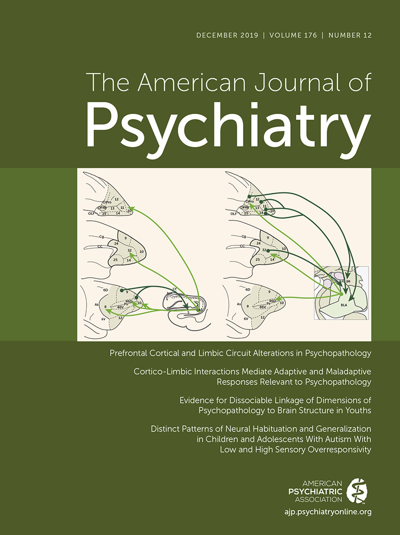
In this issue, McHugo and colleagues (
1) build upon a growing body of empirical and theoretical work nominating baseline hyperactivity in the anterior hippocampus as a promising biomarker for identifying individuals who are at risk for developing a first episode of psychosis. The appeal of this biomarker is that it provides a link between neuroanatomy, neurochemistry, and clinical features, with several studies providing evidence of predictive validity. However, as the authors acknowledge, the link between this putative biomarker and group differences in the phasic response of the hippocampus to cognitive demands is less clearly established. The study reported here provides a link between baseline hyperactivity and reduced activation in patients during a scene processing task. However, a clear link to patient deficits in anterior hippocampal engagement during the relational and associative episodic memory conditions closely associated with hippocampal function and disproportionate memory deficits in people with schizophrenia remains an elusive goal. Nevertheless, the multimodal imaging procedures described here illustrate a promising path forward.
The hippocampus is a well-documented locus of pathophysiology in people with psychotic disorders. Beginning with CT studies documenting enlarged lateral and third ventricles in patients relative to healthy control subjects (
2), speculation arose as to whether ventricular enlargement reflected volume loss in adjacent structures, including the hippocampus. Subsequent MRI research consistently documented smaller hippocampi bilaterally in people with schizophrenia, occurring early in the illness and with little progression, supporting a neurodevelopmental model (see reference
3 for a review). Most recently, a large multisite study of more than 2,000 people with schizophrenia (
4) confirmed a medium-sized decrease in overall hippocampal volume (Cohen’s d=−0.46) in patients relative to an equally large sample of healthy control subjects.
Attempts to identify an anterior-posterior gradient or specific subregions of hippocampal volume loss yielded less consistent findings, with a similar number of studies finding selective anterior or posterior deficits, or no evidence of an anterior-posterior gradient (
5,
6). However, when investigators began to examine individuals who were at clinical high risk for psychosis (
7) or were early in their first episode of psychosis (
8), a selective volume loss in the anterior hippocampus was more consistently observed, suggesting that the anterior hippocampus may be a promising target for early intervention efforts.
Evidence that the anterior hippocampus is more physiologically active in a baseline or resting state in people with schizophrenia provided a theoretical link to earlier postmortem studies that had documented molecular changes in both inhibitory (GABA) and excitatory (glutamate) neurotransmitters in the same region of the brain. For example, in a positron emission tomography study utilizing low-level baseline and word-stem completion memory tasks, Heckers and colleagues (
9) revealed that although people with schizophrenia had reduced hippocampal activation relative to control subjects during memory retrieval, their hippocampus was hyperactive during baseline conditions. Benes (
10) seized upon this finding and proposed that this hyperactivity may be secondary to the loss of GABAergic inhibition that she had observed in the hippocampus of patients during postmortem studies. This led to a theoretical model proposing that reduced GABAergic inhibition leads to hippocampal hyperactivity, resulting in excitotoxic damage to interneurons in CA2 and other portions of the hippocampus, culminating in the previously noted volume loss (
10,
11).
Evidence of baseline hyperactivity was subsequently replicated and localized to the anterior hippocampus using measures of cerebral blood flow and cerebral blood volume (CBV) (
12,
13). Moreover, evidence that resting-state anterior hippocampal hyperactivity predicted conversion from a prodromal state to a first episode of psychosis (
12) and was associated with more severe overall psychopathology (
14) and greater auditory hallucinations (
15) further strengthened the bridge from neurophysiology to clinical features and encouraged several other theoretical models supported by human and animal research. For example, Lodge and Grace (
16) proposed that anterior hippocampal hyperactivity produces aberrant dopamine signaling in the striatum and the resulting positive symptoms. In this model, a hyperactive hippocampus increases excitatory glutamatergic input to the nucleus accumbens, causing it to increase GABAergic inhibitory input to the ventral pallidum, which disinhibits dopamine neuron activity in the ventral tegmental area of the striatum. A third model (
17) proposed that decreased glutamatergic signaling in the dentate gyrus disrupts signaling in the CA3 subfield of the hippocampus, which contributes to formation of aberrant associations (i.e., increased pattern completion) and a resulting increase in positive symptoms. Thus, there are a number of alternative models that link these multiple levels of analysis and provide a potential mechanism for the development of psychosis. A remaining question is whether or not these models can also explain group differences in phasic hippocampal activation when individuals are presented with memory and other task paradigms.
McHugo et al. attempt to address this question by performing the first study to date that combines resting measures of CBV with task-related measures of blood-oxygen-level-dependent functional MRI (fMRI) cognitive activation. The experiment examined a subset of previously studied early-psychosis patients and healthy control subjects who had performed a scene memory task during fMRI (
18) and also had baseline CBV data. As previously observed, anterior hippocampal CBV was increased in patients compared with control subjects. This hyperactivity was correlated with reduced fMRI activation in the same region when patients were processing visual scenes (composed of faces overlaid on outdoor scenes) relative to a baseline condition (scrambled faces and scenes). Although the previous report (
18) revealed a patient deficit in relational memory for face and scene pairings, the present study did not report associations with memory conditions. In discussing previous fMRI evidence of both anterior hippocampal hypoactivation and hyperactivation in patients during memory conditions, the authors proposed that this variability may relate to whether the task paradigms engaged the anterior hippocampus in healthy control subjects. As discussed below, it may also be that memory tasks that most strongly engage the anterior hippocampus measure aspects of episodic memory that are least impaired in people with schizophrenia.
There is consistent research in rodents demonstrating that the ventral hippocampus (corresponding to anterior in humans) and dorsal hippocampus (corresponding to posterior in humans) are functionally distinct (
19,
20). In humans, functional dissociations along the longitudinal axis of the hippocampus remain an area of active debate, but fMRI studies have generally shown that the anterior hippocampus is preferentially involved in novelty detection and memory for faces (
21) and affective stimuli (
22), whereas the posterior hippocampus may have preferential involvement in processing spatial or contextual information (
23,
24). Moreover, the anterior hippocampus has stronger connections with an anterior temporal memory network involved in processing item information, and the posterior hippocampus has stronger connections with a posterior medial memory network involved in processing context information (
25). Given evidence that memory appears to be most impaired in individuals with psychosis when they are required to use contextual information to support relational encoding, associative retrieval, and recollection (
26,
27), it is important to determine whether or not baseline anterior hippocampal hyperactivity is associated with patient deficits in fMRI tasks that differentially engage the posterior and the anterior hippocampus. This could be accomplished by combining resting CBV measures with fMRI item recognition and source retrieval tasks, or similar paradigms that differentiate item encoding conditions that support familiarity-based recognition from relational encoding conditions that promote retrieval of associated contextual information to support a sense of recollection. Without such research, the anterior hippocampus remains a compelling pathophysiological model that links brain chemistry, anatomy, and clinical outcome, but a bridge to the types of episodic memory functions that are disproportionately impaired in individuals with schizophrenia and other psychotic disorders appears to remain under construction.
Acknowledgments
Supported by NIMH grant R01MH105411.