Full access
Regular Article
Published Online: 1 February 2002

Frontal Cortex Atrophy Predicts Cognitive Impairment in Multiple Sclerosis

Publication: The Journal of Neuropsychiatry and Clinical Neurosciences

Abstract

The association between regional measures of cortical atrophy and neuropsychological (NP) dysfunction was studied in 35 multiple sclerosis (MS) patients. Patients underwent neurological examination, MRI, and NP testing. Blind quantitative MRI analysis yielded total T2 lesion area (TLA) and third ventricle width (3VW). Cortical atrophy, rated by blind visual inspection, was more extensive in superior frontal and parietal cortices than in other regions. No MRI measures were correlated with depression scores. TLA and 3VW were significantly correlated with each NP test. Cortical atrophy measures for bilateral superior frontal cortex were retained in regression models predicting impairments in verbal learning, spatial learning, attention, and conceptual reasoning. The authors conclude that cerebral atrophy predicts NP impairment while accounting for the influence of TLA or 3VW. Regions of cortex most susceptible to atrophic and cognitive changes in MS are the right and left superior frontal lobes.
It is well established that MS-associated cognitive impairment is associated with general MRI measures of disease burden such as the total area or volume of T2 lesions (TLA),18 cerebral volume,2,7,9 corpus callosum size,5,7,10 and third ventricle volume or width (3VW).5,11,12 In contrast, correlations between region-specific pathology on MRI and specific cognitive deficits are rarely reported. Arnett and colleagues1 showed that patients with a high ratio of frontal to nonfrontal T2 lesion area were more likely to exhibit defective performance on the Wisconsin Card Sorting Test, a measure of executive ability. However, this finding was not replicated in a more recent study using different methodology.13 Most of these studies have employed T2 lesion area as the primary MRI measure. In MS, T2 hyperintensities most often occur in the cerebral white matter, and as such they are likely to have wide-ranging neurobehavioral effects that involve both anterior and posterior neural networks. Therefore, statistical association between T2 lesion area and specific neuropsychological (NP) tests should be difficult to identify in the MS population.
Recent research has focused on the destructive potential of the MS disease process in causing tissue loss and atrophy in the brain.14,15 Such atrophy occurs early in the disease process and shows a closer correlation with physical disability than T2 lesions.15 Although atrophy is an anatomically general measure of disease burden, it may also be fruitful to examine atrophic change in specified regions of the cortex. Global atrophy measures have been examined in MS patients, but little is known about how regional measures of cortical atrophy interact with neuropsychological function.
The goal of this study was to determine if there is an association between regional ratings of cortical atrophy and NP impairment in MS patients. There were two specific objectives. First, we investigated whether quantitative computer-assisted measures of TLA and/or 3VW predict NP dysfunction. Second, after accounting for variance in NP testing associated with these general measures, we asked if additional predictive variance would be accounted for by region-specific ratings of cortical atrophy. When both global and regional MRI atrophy measures were regressed on objective tests of language, spatial processing, memory, attention, and conceptual reasoning, the regional measures contributed significant predictive variance.

METHODS

Subjects

We studied 35 patients with clinically definite or laboratory-supported MS.16 Recruitment procedures were described in a previous publication.17 In brief, the patients were either consecutive clinical referrals for NP evaluation or respondents to advertisement for MS patients with cognitive/emotional problems. The study was approved by the State University of New York at Buffalo Institutional Review Board (IRB), and informed consent was obtained prior to each patient's participation. In addition to NP testing, patients underwent MRI and neurological examination to derive an Expanded Disability Status Scale (EDSS) rating of physical disability.18 Some patients also participated in additional studies of personality and a psychological intervention designed to diminish the frequency of socially aggressive behaviors. The results of the psychological studies will be presented in separate reports.
Twenty-one patients had a progressive form of MS and 14 had relapsing disease. EDSS scores ranged from 1.0 to 8.5 (mode=3.0). There were 32 Caucasian patients and 3 African-Americans. Sixty-six percent (n=23) of the patients were female. Premorbid IQs were estimated by the method of Barona et al.,19 and the sample mean was 109.3 (SD=7.8). In contrast, the mean current IQ derived from a short form of the Wechsler Adult Intelligence Scale–Revised20 was 92.4 (SD=14.4).
Patients were excluded if structured interview revealed history of other neurological disease, drug or alcohol dependence, psychiatric disorder other than psychological problems developing in the context of MS, or clinical MS relapse (or corticosteroid treatment) within 3 weeks of participation. None of the patients reported psychiatric treatment predating MS diagnosis. Use of anxiolytic and antidepressant medications was permitted, although no patient met diagnostic criteria for major depressive episode at the time of the study.

Neuropsychological Testing

Neuropsychological testing was performed by a trained technician, supervised by a board-certified clinical neuropsychologist. Depression was quantified by using the Beck Depression Inventory (BDI).21 The original battery of cognitive tests was based on review of previous MS studies,22,23 and was then reduced to one variable per domain to limit the number of statistical tests as much as possible. Language was assessed by the Boston Naming Test, a measure of visual confrontation naming.24 Spatial processing was assessed with the Judgment of Line Orientation Test, which emphasizes the perception of line angles.25 The learning recall trials from the California Verbal Learning Test26 and the revised Brief Visuospatial Memory Test27 were used to assess new learning capacity. Attention and/or rate of processing was examined by the Paced Auditory Serial Addition Test.28 Set shifting and conceptual reasoning were examined by using the perseverative response index from the Wisconsin Card Sorting Test29 and the error score derived from the Booklet Category Test,30 respectively.

MRI Acquisition and Analysis

Brain MRI was performed on a 1.5-T scanner (General Electric, Milwaukee, WI). The protocol included a 5-mm-thick (2.5-mm interslice gap) axial T1-weighted series (450/19) with a 256×256 matrix, a 22-cm field of view, and two excitations and an axial 5-mm (2.5-mm interslice gap) dual-echo, conventional spin-echo series acquired with intermediate and T2 weighting (2,100, 30/85), with a 256×256 matrix, a 22-cm field of view, and one excitation. Individuals who were unaware of clinical details made the MRI measurements (R. Bakshi, J.H.S.).
Two quantitative, general MRI measures of disease burden were obtained at the University of Colorado Health Sciences Center (Denver, CO), based on manual tracings on a computer workstation using previously described methods.14,31 Total T2 hyperintense lesion area (TLA) was determined by using tracings from paired proton-density and standard T2-weighted images by an experienced image analysis technician. The values were calculated by multiplying lesion area by the sum of slice thickness and slice gap. Third ventricular width (3VW) was based on axial T1-weighted series determined along a plane corresponding to the (anteroposterior) midpoint of the ventricle. The intra-observer mean coefficient of variation was previously shown to be 3% for this method.14
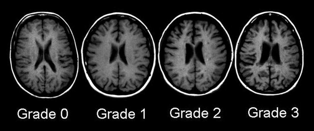
Cortical atrophy was measured in relation to age-matched control subjects by an experienced observer using an ordinal rating scale according to the method of Bakshi and co-workers.15,3236 Cortical atrophy was rated in six lobar regions in each hemisphere: superior frontal, inferior frontal, superior parietal, inferior parietal, occipital, and temporal. Standard fissures were used to separate the various lobes, including the central (rolandic), lateral (sylvian), and parieto-occipital sulci. The third ventricle demarcated superior versus inferior frontal and parietal lobes. All axial slices showing the region were analyzed. We used an ordinal rating system that assessed severity of enlargement of subarachnoid spaces (sulci and fissures) in the above-mentioned regions on axial noncontrast T1-weighted images. Each patient was rated against an age- and gender-matched normal control scan, selected from a standard, normative MRI database of 100 control subjects. The degree of atrophy was rated as normal (grade 0), mild (<10%, grade 1), moderate (10%–25%, grade 2), or severe (>25%, grade 3), according to the percentage of volume loss of parenchyma in the entire region. Examples of these ratings from subjects in our database are presented in Figure 1. The intra-observer agreement of data obtained by this MRI atrophy technique is high (mean kappa 0.9, range 0.8–1.0) and the inter-observer agreement is moderate to very good (mean kappa 0.8, range 0.6–1.0).15,34

Procedure

All participants underwent an initial structured telephone screening interview, emphasizing previous medical history and DSM-IV37 criteria for psychiatric disorder. The interview was standardized across participants and included questions pertaining to previous psychiatric history and current symptoms of depressive disorder. After screening, participants were scheduled for neuropsychological and neurological examinations. MRI scans were performed for patients without a clinical study within 6 months of testing. The MRI data were digitized and shipped to the Colorado Health Sciences Center, where they were analyzed with quantitative techniques. Hard copy images were read to obtain the ordinal measures of cortical atrophy. All MRI analyses were blind to NP and clinical data. All participants provided signed, IRB-approved, informed consent prior to participation.

Statistical Analyses

All group comparisons and correlations were evaluated by using a conservative threshold of P<0.01. Linear regression analysis with forward stepwise selection was the primary method of analysis. First, TLA and 3VW were regressed on the NP measures in separate models. Conservative selection criteria (entrance 0.01, exit 0.05) were employed to reduce the likelihood of a chance finding. In the second stage of analysis, cortical atrophy measures were regressed on the same criterion measures using the same selection criteria. However, in this stage, the forward selection procedure was preceded by the entrance of either TLA or 3VW, depending on which general measure had accounted for most variance in the previous stage. The general measure was retained in each model in fixed fashion. In this way, we answered the question of whether cortical atrophy accounts for predictive variance in cognitive function after accounting for the influence of general cerebral pathology.
Unlike the NP and volumetric MRI variables, the cortical MRI ratings were not, in most cases, normally distributed by Kolmogorov-Smirnov test. Therefore, where possible, nonparametric statistical tests were employed for these measures. Such an accommodation was not possible for testing the primary hypotheses with linear regression.

RESULTS

Descriptive and Correlational Analyses of MRI Data

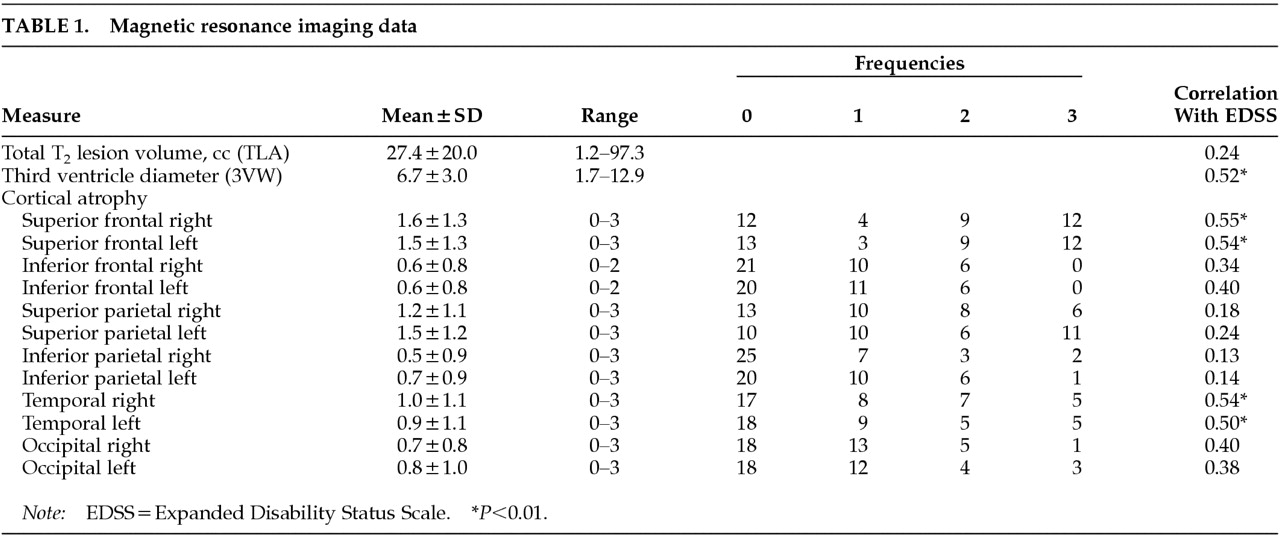
Descriptive MRI data are presented in Table 1. For the ordinal, cortical atrophy variables, kurtosis values were all negative or near 0 except in the left inferior parietal cortex. In this single case, which had 25 ratings of grade 0, markedly positive kurtosis (2.1, SD=0.76) was found. There were no differences between left and right ordinal ratings of atrophy across region by Marginal Homogeneity Test, with the exception of the superior parietal comparison (P<0.01), where there was a higher frequency of abnormal rankings in the left versus the right hemisphere. Superior/inferior comparisons were also performed for each hemisphere (e.g., right superior vs. inferior frontal). All comparisons were significant (P<0.001), with the superior ratings showing more atrophic change. In the right frontal lobe, for example, 12 cases were graded 3 (severe atrophy) in the superior region, whereas no cases were graded 3 in the inferior region.
The Pearson correlation coefficient for TLA and 3VW was 0.60 (P<0.0001). As would be expected, the regional atrophy measures were strongly intercorrelated. The Spearman rho coefficients ranged from 0.41 (superior frontal left vs., inferior parietal right) to 0.97 (inferior frontal left vs. right).
Spearman rho correlations between the general and regional MRI measures were significant for the superior frontal and parietal regions and for the temporal lobes. Correlations in the inferior frontal and parietal regions and the occipital lobe were consistently smaller in magnitude and were inconsistently significant. Overall, these correlations ranged from 0.23 (3VW vs. inferior parietal right) to 0.77 (TLA vs. superior frontal left and right).
There were no significant correlations of age or premorbid IQ with any of the MRI measures. As shown in Table 1, EDSS was correlated significantly with 3VW (0.52, P<0.01), but not with TLA. Superior frontal and temporal atrophy ratings were significantly correlated with EDSS, bilaterally.

Prediction of NP Impairment, Stage One, General Measures

The general MRI measures were regressed on to each NP measure in separate models. In each case, a single predictor variable was retained, as summarized in Table 2. Neither TLA or 3VW was significantly correlated with BDI. For all but one cognitive criterion measure, TLA accounted for more variance.

Prediction of NP Impairment, Stage Two, Cortical Atrophy

To test the hypothesis that regional cortical atrophy accounts for additional variance in NP impairment, the retained general measures listed in Table 2 were entered along with the ordinal atrophy ratings to predict NP performance. In each analysis, the general measure was entered first and retained. Regional atrophy measures followed, en bloc, in a forward stepwise procedure. There were no statistically reliable predictors of BDI among the regional cortical MR measures, as no correlation coefficient approached significance. Cortical atrophy failed to add predictive variance on tests of confrontation naming, spatial processing, and set shifting. For Boston Naming Test, partial correlations (controlling for TLA) approached significance for superior frontal left (–0.30) and superior parietal left (–0.31), but these variables were not retained in the final model. For Judgment of Line Orientation, only the partial correlation for occipital lobe left approached significance (–0.38). There were no partial correlations above 0.3 between cortical atrophy and Wisconsin Card Sorting perseverations.
Cortical atrophy measures were retained in models predicting impairment in verbal learning, spatial learning, attention, and conceptual reasoning (Table 2). In all cases, the added variance arose from superior frontal cortex. Right superior frontal atrophy accounted for variance in models predicting total words recalled on learning trials 1–5 from the California Verbal Learning Test (P<0.01), design recall trials 1–3 from the Brief Visuospatial Memory Test (P<0.01), and Paced Auditory Serial Addition Test number correct (P<0.01). Left frontal atrophy was retained in the model predicting errors on the Booklet Category Test (P<0.01).
Finally, these significant regression analyses were repeated twice, with either EDSS or disease course (relapsing vs. progressive) added to the second block of predictor variables. In these post hoc models incorporating either EDSS or disease course, the results were unchanged for Brief Visuospatial Memory Test, Paced Auditory Serial Addition Test, and Booklet Category Test. In contrast, both California Verbal Learning Test models retained EDSS or disease course rather than cortical atrophy. However, it was noted that Step 1 partial correlations controlling for TLA were similar for EDSS, disease course, and superior cortical atrophy; all partial r's were between –0.44 and –0.50.

DISCUSSION

Previous attempts to identify relationships between specific lesion locations and cognitive defects have relied primarily on measures of regional white matter (WM) lesion load. Swirsky-Sacchetti et al.8 found that left frontal lesion area predicted perseverative responses on the Wisconsin Card Sorting Test (WCST). Although the authors did not control for total lesion load, Arnett et al.1 controlled for this factor by dividing patients into those with and without predominantly frontal lesions. These subgroups were matched on TLA. Frontal pathology was again associated with WCST errors. Yet this finding was not replicated by Foong et al.4 in a more recent study of executive ability and quantified lesion volume. In this study, lesion volumes were quantified using an automated method with a line drawn through the central sulcus to demarcate the frontal regions. Frontal lesion area correlated highly with TLA, and both measures were correlated with failures on executive tasks. When the sample was divided based on a median split of the frontal/total lesion volume ratio, there were no differences on any of the executive measures. Thus, the evidence supporting a reliable relationship between localized T2 lesions and specific cognitive impairments is, at best, inconsistent.
Given that the cerebral white matter connects various regions of the cerebral cortex, such difficulty in identifying specific WM lesion–cognition correlations is not unexpected. On the other hand, atrophic change in specified areas of the cortex may be more likely to show focal cognitive effects. We found this to be the case in four NP tests emphasizing new learning, divided attention, and conceptual reasoning. Not only were failures on these tests correlated strongly with superior frontal cortex atrophy, but the predictive relationship held after controlling for the influence of TLA and 3VW. Superior frontal cortex has been shown to be active during a range of new learning and executive ability tasks.3841 Consequently, the importance of this region in our statistical models of cognitive dysfunction is compatible with previous neuropsychological research.
Brain atrophy is a topic of increasing interest to neurologists caring for MS patients. Atrophy correlates well with neurologic function and is associated with progressive neurologic disability.14,15 Unfortunately, the etiology of MS-associated brain atrophy is not well understood, but it may be due to focal white matter damage in tracts that underlie the affected region of the cortex.14,15 Wallerian degeneration may occur distal from the focal WM lesion and contribute to tissue loss. In this regard, it is remarkable that much higher ratings of atrophy were discovered in the superior aspect of the frontal lobe, as compared with the inferior aspect. As these regions are also more susceptible to WM lesions,42 our data support a role of WM disease in the development of focal atrophy. This is also consistent with previous fluorodeoxyglucose PET work from our group showing more prominent hypometabolism in the superior versus inferior aspect of the frontal cortex in MS.43
In the present study, the severity of cortical sulcal enlargement was measured in comparison to age-matched normal control subjects. We recently demonstrated that these visually determined region-by-region severity measures of atrophy show significant correlations with brain hypometabolism detected quantitatively by fluorodeoxyglucose brain PET,34 supporting the validity of this MRI rating method. Although this method is qualitative and rater-dependent, and does not account for subtle sulcal variability in normal individuals, these atrophy measures can be obtained rapidly from hard copies of images and are practical in the clinical setting. Computer-assisted, quantitative methods of assessing cortical atrophy have been developed,44 but they require extensive, time-consuming analyses and, as such, are not yet applicable to the clinic setting. Given the robust correlations between cognitive function and the ordinal cortical atrophy indices obtained here, we tentatively propose that this and similar methods might be used to identify patients at high risk for cognitive impairment, and in turn, vocational disability. Future studies should be pursued to confirm and extend our findings, using computer-assisted methods that will further validate the rating method and more precisely quantify regional cortical atrophy.
One caveat concerns the composition of our study sample, which included clinical referrals and patients responding to advertisement. Although we previously showed that this sample is impaired on NP testing,17 it is likely that these patients reflect a subpopulation of MS who experience cognitive and personality change, and thus are more likely to present with cerebral pathology. Although many of our patients presented with some symptoms of depressive or adjustment disorder, none met diagnostic criteria for major depressive episode.37 Depressive symptoms are common in MS,4548 and the mean BDI score reported here is similar to that reported in other studies.49 On the other hand, the degree of atrophy and T2 lesion volume was high in comparison to other recent research using similar methods.14,31 Although the degree of impairment in our sample may limit the generalizability of our findings, it is also important to note that the range of MRI values was probably restricted. It is likely that a more heterogeneous and representative sample would result in more robust correlative findings. Another methodological concern was the use of predictor variables that are not normally distributed (i.e., ordinal rather than ratio scale). The criterion measures were, however, normal, as were the general MRI predictors. The use of predictor variables that deviate from normal would not cause spurious results, but rather would reduce statistical power. Had our study employed computer-assisted continuous measurement of cortical atrophy, the test of the primary hypothesis would have been stronger. Such an approach is currently under development at our center.
These methodological concerns notwithstanding, we conclude that cortical atrophy as measured by blind ratings of enlargement of subarachnoid spaces (sulci, fissures, cisterns) is associated with cognitive dysfunction in MS. The association is strong when examined with simple correlation coefficients, and it remains reliable after controlling for the influence of total T2 lesion area and the diameter of the third ventricle. The regions of the brain most integral to cognitive morbidity are the superior frontal cortices.
ACKNOWLEDGMENTS

The authors gratefully acknowledge the Western New York Chapter of the National Multiple Sclerosis Society, Buffalo, NY, for assistance with subject recruitment, and Lawrence Jacobs, M.D., for his comments concerning research design and manuscript preparation. This research was supported, in part, by Pilot Research Project Award PP0574 from the National Multiple Sclerosis Society.
FIGURE 1. Representative T1-weighted images showing all 4 grades of cortical atrophy in the superior frontal region in 4 MS patientsAtrophy was defined as enlargement of subarachnoid spaces (sulci/fissures) in relation to age-matched control subjects in a given region. The degree of atrophy was rated according the percentage of loss of tissue volume, as shown on all axial slices from a given region, as normal (grade 0), mild (<10%, grade 1), moderate (10%–25%, grade 2), or severe (>25%, grade 3), according to the percentage of volume loss of parenchyma in the entire region as shown on all axial slices from that region.
TABLE 1. Magnetic resonance imaging data
TABLE 2. Regression analysis results

References

1.
Arnett PA, Rao SM, Bernardin L, et al: Relationship between frontal lobe lesions and Wisconsin Card Sorting Test performance in patients with multiple sclerosis. Neurology 1994; 44:420-425
2.
Camp SJ, Stevenson VL, Thompson AJ, et al: Cognitive function in primary progressive and transitional progressive multiple sclerosis: a controlled study with MRI correlates. Brain 1999; 122:1341-1348
3.
Comi G, Filippi M, Martinelli V, et al: Brain MRI correlates of cognitive impairment in primary and secondary progressive multiple sclerosis. J Neurol Sci 1995; 132:222-227
4.
Foong J, Rozewicz L, Quaghebeur G, et al: Executive function in multiple sclerosis: the role of frontal lobe pathology. Brain 1997; 120:15-26
5.
Huber SJ, Bornstein RA, Rammohan KW, et al: Magnetic resonance imaging correlates of neuropsychological impairment in multiple sclerosis. J Neuropsychiatry Clin Neurosci 1992; 4:152-158
6.
Patti F, Di Stefano M, De Pascalis D, et al: May there exist specific MRI findings predictive of dementia in multiple sclerosis patients? Functional Neurology 1995; 10:83-90
7.
Rao SM, Leo GJ, Haughton VM, et al: Correlation of magnetic resonance imaging with neuropsychological testing in multiple sclerosis. Neurology 1989; 39:161-166
8.
Swirsky-Sacchetti T, Mitchell DR, Seward J, et al: Neuropsychological and structural brain lesions in multiple sclerosis: a regional analysis. Neurology 1992; 42:1291-1295
9.
Hohol MJ, Guttmann CRG, Orav J, et al: Serial neuropsychological assessment and magnetic resonance imaging analysis in multiple sclerosis. Arch Neurol 1997; 54:1018-1024
10.
Ryan L, Clark CM, Klonoff H, et al: Patterns of cognitive impairment in relapsing-remitting multiple sclerosis and their relationship to neuropathology on magnetic resonance images. Neuropsychology 1996; 10:176-193
11.
Rabins PV, Brooks BR, O'Donnell P, et al: Structural brain correlates of emotional disorders in multiple sclerosis. Brain 1986; 109:585-597
12.
Rao SM, Glatt S, Hammeke TA, et al: Chronic progressive multiple sclerosis: relationship between cerebral ventricular size and neuropsychological impairment. Arch Neurol 1985; 42:678-682
13.
Foong J, Rozewicz L, Davie CA, et al: Correlates of executive function in multiple sclerosis: the use of magnetic resonance spectroscopy as an index of focal pathology. J Neuropsychiatry Clin Neurosci 1999; 11:45-50
14.
Simon JH, Jacobs LD, Campion M, et al: A longitudinal study of brain atrophy in relapsing multiple sclerosis. Neurology 1999; 53:139-148
15.
Bakshi R, Benedict RHB, Bermel RA, et al: Regional brain atrophy is associated with physical disability in multiple sclerosis: semiquantitative MRI and relationship to clinical findings. J Neuroimaging 2001; 11:129-136
16.
Poser CM, Paty DW, Scheinberg L, et al: New diagnostic criteria for multiple sclerosis: guidelines for research protocols. Ann Neurol 1983; 13:655-661
17.
Benedict RHB, Priore RL, Miller C, et al: Personality disorder in MS correlates with cognitive impairment. J Neuropsychiatry Clin Neurosci 2001; 13:70-76
18.
Kurtzke JF: Rating neurologic impairment in multiple sclerosis: an expanded disability status scale (EDSS). Ann Neurol 1983; 13:227-231
19.
Barona A, Reynolds CR, Chastain R: A demographically based index of pre-morbid intelligence for the WAIS-R. J Consult Clin Psychol 1984; 52:885-887
20.
Wechsler D: WAIS-R Manual. New York, Psychological Corporation, 1981
21.
Beck AT: Beck Depression Inventory. San Antonio, TX, Psychological Corporation, 1993
22.
Rao SM: Neuropsychology of multiple sclerosis: a critical review. J Clin Exp Neuropsychol 1986; 8:503-542
23.
Fischer JS, Foley FW, Aikens JE, et al: What do we really know about cognitive dysfunction, affective disorders, and stress in multiple sclerosis? A practitioner's guide. J Neurol Rehab 1994; 8:151-164
24.
Kaplan EF, Goodglass H, Weintraub S: The Boston Naming Test. Philadelphia, Lea and Febiger, 1983
25.
Benton AL, Sivan AB, Hamsher K, et al: Contributions to Neuropsychological Assessment, 2nd edition. New York, Oxford University Press, 1994
26.
Delis DC, Kramer JH, Kaplan E, et al: California Verbal Learning Test: Adult Version. San Antonio, TX, Psychological Corporation, 1987
27.
Benedict RHB: Brief Visuospatial Memory Test-Revised: Professional Manual. Odessa, FL, Psychological Assessment Resources, 1997
28.
Gronwall DMA: Paced Auditory Serial Addition Task: a measure of recovery from concussion. Percept Mot Skills 1977; 44:367-373
29.
Heaton RK: A Manual for the Wisconsin Card Sorting Test. Odessa, FL, Psychological Assessment Resources, 1981
30.
De Filippis, NA, McCampbell E: Manual for the Booklet Category Test. Odessa, FL, Psychological Assessment Resources, 1991
31.
Simon JH, Jacobs LD, Campion M, et al: Magnetic resonance studies of intramuscular interferon B-1a for relapsing multiple sclerosis. Ann Neurol 1998; 43:79-87
32.
Bakshi R, Miletich RS, Henschel K, et al: Fatigue in multiple sclerosis: cross-sectional correlation with brain MRI findings in 71 patients. Neurology 1999; 43:1151-1153
33.
Bakshi R, Kamran S, Kinkel Pr, et al: Fluid-attenuated inversion-recovery MR imaging in acute and subacute cerebral intraventricular hemorrhage. Am J Neuroradiology 1999; 20:629-636
34.
Bakshi R, Miletich RS, Kinkel PR, et al: MRI hemispheric atrophy is associated with FDG-PET hypometabolism and MRI white matter lesions in multiple sclerosis. Neurology 1999; 52(suppl):A361-A361
35.
Bakshi R, Czarnecki D, Shaikh ZA, et al: Brain MRI lesions and atrophy are related to depression in multiple sclerosis. Neuroreport 2000; 11:1153-1158
36.
Janardhan V, Bakshi R: Quality of life in multiple sclerosis and its relationship to brain MRI lesions and atrophy in 60 patients. Arch Neurol 2000; 57:1485-1491
37.
American Psychiatric Association: Diagnostic and Statistical Manual of Mental Disorders, 4th edition. Washington, DC, American Psychiatric Association, 1994
38.
Grasby PM, Frith CD, Friston KJ, et al: A graded task approach to the functional mapping of brain areas implicated in auditory-verbal memory. Brain 1994; 117:1271-1282
39.
Braver TS, Cohen JD, Nystrom LE, et al: A parametric study of prefrontal cortex involvement in human working memory. Neuroimage 1997; 5:49-62
40.
Sarazin M, Pillon B, Giannakopoulos P, et al: Clinicometabolic dissociation of cognitive functions and social behavior in frontal lobe lesions. Neurology 1998; 51:142-148
41.
Corbetta M, Miezin FM, Dobmeyer S, et al: Selective and divided attention during visual discriminations of shape, color, and speed: functional anatomy by positron emission tomography. J Neurosci 1991; 11:2383-2402
42.
Brownell B, Hughes TJ: The distribution of plaques in the cerebrum in multiple sclerosis. J Neurol Neurosurg Psychiatry 1962; 25:315-320
43.
Bakshi R, Miletich RS, Kinkel PR, et al: High-resolution fluorodeoxyglucose positron emission tomography shows both global and regional cerebral hypometabolism in multiple sclerosis. J Neuroimaging 1998; 8:228-234
44.
Goldstein JM, Goodman JM, Seidman LJ, et al: Cortical abnormalities in schizophrenia identified by structural magnetic resonance imaging. Arch Gen Psychiatry 1999; 56:537-547
45.
Devins GM, Seland TP: Emotional impact of multiple sclerosis: recent findings and suggestions for future research. Psychol Bull 1987; 101:363-375
46.
Murray TJ: The psychosocial aspects of multiple sclerosis. Neurol Clin 1995; 13:197-223
47.
Cottrell SS, Wilson SAK: The affective symptomatology of disseminated sclerosis. Journal of Neurology and Psychopathology 1926; 1-30
48.
Sadovnick AD, Remick RA, Allen J, et al: Depression and multiple sclerosis. Neurology 1996; 46:628-632
49.
Hiber SJ, Rammohan KW, Bornstein RA, et al: Depressive symptoms are not influenced by severity of multiple sclerosis. Neuropsychiatry Neuropsychol Behav Neurol 1993; 6:177-180

Information & Authors

Information

Published In

Go to The Journal of Neuropsychiatry and Clinical Neurosciences
The Journal of Neuropsychiatry and Clinical Neurosciences
Pages: 44 - 51
PubMed: 11884654

History

Published online: 1 February 2002
Published in print: February 2002

Authors

Details

Ralph H.B. Benedict, Ph.D.
Received September 1, 2000; revised November 21, 2000; accepted November 27, 2000. From the Department of Neurology, State University of New York at Buffalo School of Medicine, Buffalo, New York, and the University of Colorado Health Sciences Center, Denver, Colorado. Address correspondence to Dr. Benedict, Neurology, Suite D-6, Buffalo General Hospital, 100 High Street, Buffalo, NY 14203. E-mail: [email protected].
Rohit Bakshi, M.D.
Received September 1, 2000; revised November 21, 2000; accepted November 27, 2000. From the Department of Neurology, State University of New York at Buffalo School of Medicine, Buffalo, New York, and the University of Colorado Health Sciences Center, Denver, Colorado. Address correspondence to Dr. Benedict, Neurology, Suite D-6, Buffalo General Hospital, 100 High Street, Buffalo, NY 14203. E-mail: [email protected].
Jack H. Simon, M.D., Ph.D.
Received September 1, 2000; revised November 21, 2000; accepted November 27, 2000. From the Department of Neurology, State University of New York at Buffalo School of Medicine, Buffalo, New York, and the University of Colorado Health Sciences Center, Denver, Colorado. Address correspondence to Dr. Benedict, Neurology, Suite D-6, Buffalo General Hospital, 100 High Street, Buffalo, NY 14203. E-mail: [email protected].
Roger Priore, Sc.D.
Received September 1, 2000; revised November 21, 2000; accepted November 27, 2000. From the Department of Neurology, State University of New York at Buffalo School of Medicine, Buffalo, New York, and the University of Colorado Health Sciences Center, Denver, Colorado. Address correspondence to Dr. Benedict, Neurology, Suite D-6, Buffalo General Hospital, 100 High Street, Buffalo, NY 14203. E-mail: [email protected].
Colleen Miller, D.N.S.
Received September 1, 2000; revised November 21, 2000; accepted November 27, 2000. From the Department of Neurology, State University of New York at Buffalo School of Medicine, Buffalo, New York, and the University of Colorado Health Sciences Center, Denver, Colorado. Address correspondence to Dr. Benedict, Neurology, Suite D-6, Buffalo General Hospital, 100 High Street, Buffalo, NY 14203. E-mail: [email protected].
Frederick Munschauer, M.D.
Received September 1, 2000; revised November 21, 2000; accepted November 27, 2000. From the Department of Neurology, State University of New York at Buffalo School of Medicine, Buffalo, New York, and the University of Colorado Health Sciences Center, Denver, Colorado. Address correspondence to Dr. Benedict, Neurology, Suite D-6, Buffalo General Hospital, 100 High Street, Buffalo, NY 14203. E-mail: [email protected].

Metrics & Citations

Metrics

Citations

Export Citations

If you have the appropriate software installed, you can download article citation data to the citation manager of your choice. Simply select your manager software from the list below and click Download.

For more information or tips please see 'Downloading to a citation manager' in the Help menu.

Format
Citation style
Style
Copy to clipboard

View Options

View options

PDF/EPUB

View PDF/EPUB

Login options

Already a subscriber? Access your subscription through your login credentials or your institution for full access to this article.

Personal login Institutional Login Open Athens login
Purchase Options

Purchase this article to access the full text.

PPV Articles - Journal of Neuropsychiatry and Clinical Neurosciences

PPV Articles - Journal of Neuropsychiatry and Clinical Neurosciences

Not a subscriber?

Subscribe Now / Learn More

PsychiatryOnline subscription options offer access to the DSM-5-TR® library, books, journals, CME, and patient resources. This all-in-one virtual library provides psychiatrists and mental health professionals with key resources for diagnosis, treatment, research, and professional development.

Need more help? PsychiatryOnline Customer Service may be reached by emailing [email protected] or by calling 800-368-5777 (in the U.S.) or 703-907-7322 (outside the U.S.).

Media

Figures

Other

Tables

Share

Share

Share article link

Share