Full access
Article
Published Online: 1 November 2001

Impaired Sensory Processing as a Basis for Object-Recognition Deficits in Schizophrenia

Abstract

OBJECTIVE: Individuals are able to recognize common objects even when portions of them are obscured from view, reflecting the operation of neural perceptual closure processes. This study evaluates the integrity of object recognition and perceptual closure as a function of sensory and cognitive manipulations. METHOD: Object recognition was examined in 26 subjects with schizophrenia and 23 nonpsychiatric comparison subjects of similar age with a presentation of fragmented pictures by means of the ascending method of limits. The effects of prior exposure to subsets of stimuli and of word prompting were examined in separate testing phases. Demographic and clinical characteristics were evaluated as covariates. RESULTS: Although they had impairments in perceptual closure, schizophrenic patients showed improvement in performance equivalent to that of nonpatient comparison subjects with prior exposure to the pictures (i.e., repetition priming) and with presentation of valid word prompts. A significant correlation was found between impaired performance and the severity of negative symptoms. CONCLUSIONS: The results support models of widespread dysfunction in information processing in patients with schizophrenia involving both sensory and cognitive regions. Perceptual closure is significantly impaired in schizophrenic patients; however, this deficit in sensory precision is dissociated from the effects of higher-order repetition priming and word prompting. Furthermore, this work suggests that deficits in perceptual closure may contribute to the muted world experience of patients with the persistent negative symptoms of schizophrenia.
Schizophrenia is manifested across a range of symptom categories and cognitive deficits. One tradition has been to ascribe such deficits to dysfunction within a limited number of critical brain regions, such as the prefrontal or temporoparietal cortex (1, 2). More recent studies (3, 4), however, have suggested that patients show a widespread pattern of dysfunction that cannot easily be ascribed to dysfunction within such circumscribed brain regions. Furthermore, studies of cortical blood flow show impaired activation patterns not only in the prefrontal and temporoparietal cortices but also in auditory and visual regions and the cerebellum (5). Despite renewed interest in brain areas outside the prefrontal/temporoparietal cortices as mediators of cognitive dysfunction in patients with schizophrenia, relatively few studies have investigated the integrity of basic sensory processing in schizophrenia independent of top-down influences. The present study evaluates the integrity of sensory processing within the visual system in patients with schizophrenia by examining the phenomenon of perceptual closure (69).
Perceptual closure refers to the ability of the brain to form complete object representations on the basis of fragmentary visual information (6, 9). It is assumed that the brain automatically “fills in” missing object information, which is consistent with results of single-cell studies of the primate visual cortex (1012). Investigation of perceptual closure in patients with schizophrenia provides an index of their ability to form complete representations on the basis of partial information. The phenomenon of perceptual closure also represents an ideal domain within which to examine the dependence of sensory processing dysfunction on top-down regulation in patients with schizophrenia. Although the ability to “close” an image is primarily tied to the amount of visual information provided, the level of information required for object recognition can be manipulated (6, 8). Thus, for example, if an image is repeated either immediately or after some delay, participants are able to make an identification with significantly less visual information (i.e., at significantly greater levels of stimulus fragmentation). Also, if participants are given a word prompt that potentially names the object, less visual information is required for recognition when the prompt names the object pictured than when it does not. Repetition priming assesses the ability of the visual system to maintain stimulus representations over time, while word prompting assesses the use of nonfigural information to aid figural interpretation. For word prompting to occur, visual regions must receive top-down information flow from language regions and other areas of the cortex (13, 14). The present study assesses the integrity of modulatory influences on perceptual closure in patients with schizophrenia, along with the integrity of perceptual closure itself.
Perceptual closure is subserved primarily by the lateral occipital complex (9, 15, 16), which lies at the height of the ventral visual-stream object-processing hierarchy (the “what” stream) (15, 17). Presentation of identifiable objects in normal subjects leads to focal bilateral lateral occipital activation (18, 19). Moreover, object recognition in circumstances of partial information is associated with the generation of an event-related potential component that we termed “Ncl,” which is localized over the lateral occipital complex (9). Given that perceptual closure has a specific neural correlate, compromised integrity of object recognition in patients with schizophrenia implicates the structures of the lateral occipital complex that generate it. Thus, the present study permits analysis of information processing in patients with schizophrenia at the level of the visual sensory cortex.
This study employed the presentation of fragmented pictures by means of the ascending method of limits to determine the amount of visual information required by patients with schizophrenia and comparison participants to identify progressively less fragmented line drawings of common objects. Patients were drawn from both outpatient and inpatient facilities. Positive and Negative Syndrome Scale (20) ratings were obtained to examine the relationship between dysfunction in perceptual closure and positive versus negative symptoms.

Method

Participants

Written informed consent was obtained from 26 patients with chronic schizophrenia (14 female, one left-handed) and 23 nonpsychiatric comparison participants of similar age (eight female, four left-handed) (schizophrenia patients: mean=40.2 years, SD=6.7; comparison subjects: mean=37.6 years, SD=10.5). All participants reported normal or corrected-to-normal vision. The patients with schizophrenia were recruited from inpatient and outpatient facilities associated with the Nathan Kline Institute for Psychiatric Research and were receiving medication at the time of testing. Their mean medication dose was 1053.1 mg/day in chlorpromazine equivalents. Positive and Negative Syndrome Scale ratings were performed by a single rater (G.S.) and analyzed by using predetermined factors (21). IQ was assessed by the same rater with the Quick Test (22).
All subjects were screened with the Structured Clinical Interview for DSM-IV. Both patient and comparison subjects were excluded if they reported recent or current drug or alcohol use or had a past diagnosis of alcohol or drug dependence and use in the 6 months before the study. Subjects in both groups were also excluded if they had mental retardation, dementia, a developmental disability, or a neurological, visual, or hearing impairment. Patients were included in the study if they met DSM-IV criteria for a diagnosis of schizophrenia or schizoaffective disorder and were or were not taking medication. The comparison subjects were excluded if they met criteria for a DSM-IV axis I diagnosis. Additional demographic and clinical information on the patient group is provided in Table 1.

Stimuli and Tasks

The testing procedure consisted of three parts. Stimuli in each part were taken from “equated” stimulus sets in a study by Snodgrass and Corwin (23) and selected across conditions to be roughly equivalent in terms of difficulty of identification and category membership. Identical stimuli were used for patients and comparison subjects. The same stimuli were used across participants.
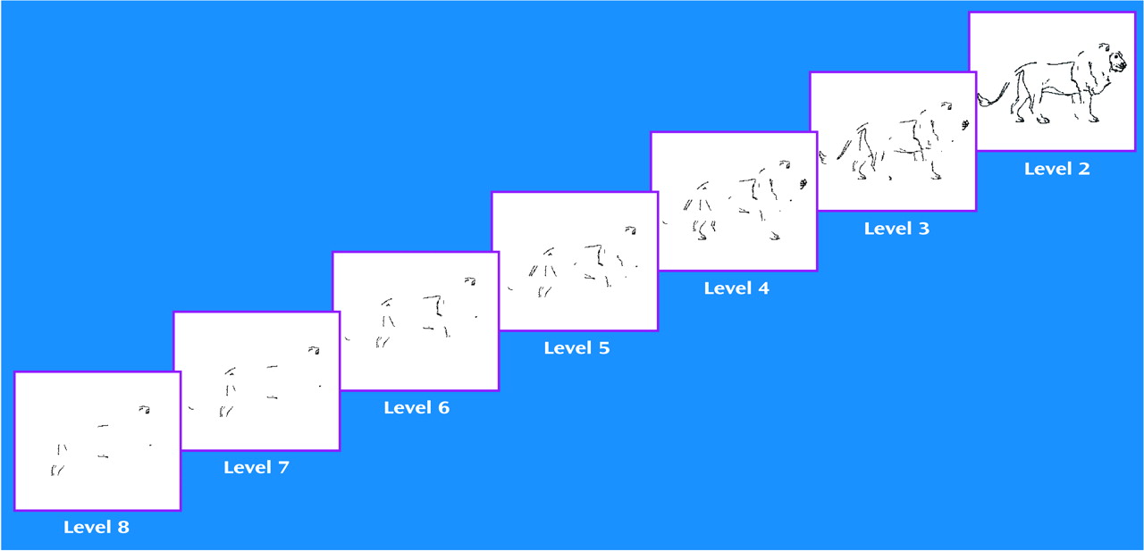
Part 1 consisted of a test of object recognition in the absence of sensory or cognitive manipulation. The participants were shown 18 line drawings (black ink on a white background) of animate and inanimate objects from a normed set of images in a study by Snodgrass and Vanderwart (24). The pictures were manipulated to produce eight incrementally fragmented images (23). “Level 1” refers to a complete picture, and “level 8” refers to the most fragmented version of an object.
The images were presented in accordance with the ascending method of limits, from least (level 8) to most (level 1) complete (Figure 1). Increasingly complete images were presented until the participant pressed a button, indicating that he or she could identify the object pictured. The experimenter then recorded the subject’s response, after which he or she again pressed the button to initiate the next picture sequence. Images subtended ∼9.3° of the vertical visual angle and ∼8.3° of the horizontal angle. Each image appeared for 750 msec, followed by a blank screen for 1,250 msec. The participants’ response window extended for 1,750 msec from the appearance of the image.
Part 2 of the testing examined the effects of repetition priming on perceptual closure and consisted of a preexposure phase followed by a testing phase. The testing phase was identical to part 1 except that it included both new pictures and those to which the participants had already been exposed. In the preexposure phase, nine nonfragmented stimuli were presented for 750 msec each. The participants were instructed to name the object the picture represented. If the participant correctly named the object, the next picture was presented. If the participant’s answer was incorrect, the experimenter asked the participant to try again. The participants were given up to three chances to name each object correctly. Both the patients and comparison subjects were able to correctly name the object pictured at its first presentation more than 99% of the time; there was no difference between the groups on performance. Immediately after the preexposure phase, the participants worked on multiplication problems for 5 minutes as a distraction.
After the preexposure phase, 18 pictures were presented by means of the ascending method of limits, including the nine pictures that had been presented in the preexposure phase, and nine new pictures. The effects of repetition priming were evaluated by comparing the level of fragmentation at identification for new and repeated pictures.
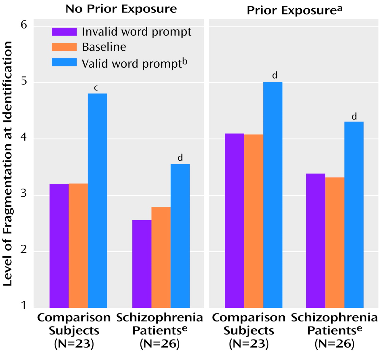
Part 3 of the testing focused on the effects of both repetition priming and word prompting on perceptual closure. As in part 2, the participants were preexposed to nine previously unseen pictures and then distracted for 5 minutes. During the test phase, 18 pictures were presented by means of the ascending method of limits, including nine from the preexposure phase and nine new ones. In addition, a visual word prompt was given before presentation of each stimulus. For six pictures (three new and three old), the prompt words correctly named the objects pictured (“valid prompt”); for another six pictures (three new and three old), the prompt word did not correctly name the object pictured (“invalid prompt”); and for the six remaining pictures (three new and three old), a meaningless string of six “X”s (baseline prompt) was presented. The order of new/old pictures and prompt types were randomly assigned to all participants. Invalid prompt words were taken from an unpresented picture set by Snodgrass and Corwin (23). Prompt words were presented for 750 msec and followed 1,250 msec later by the appearance of the most fragmented image in the picture sequence. The participants’ task was identical to the object-recognition task in parts 1 and 2 of the testing.
The order of the pictures in each test and in the preexposure phase remained fixed across participants. All participants were tested first on object recognition alone (part 1). Parts 2 and 3 of the testing were conducted in counterbalanced order. In parts 2 and 3, the pictures used for preexposure were counterbalanced across participants. In addition, in part 3 of the testing the test pictures preceded by valid, invalid, and baseline prompts were also counterbalanced across participants.

Statistical Analyses

Analyses were conducted only on data for the pictures of objects that were identified correctly by the end of the picture sequence. Between-group comparisons were performed by using multivariate analysis of variance (ANOVA) (25). Follow-up planned comparisons were performed by using paired or independent-samples t tests, as appropriate. Two-tailed statistics were used throughout the study.

Results

This study consisted of three parts. Part 1 tested the participants’ ability to identify the objects in partially fragmented pictures to which they had no previous exposure. The level of fragmentation at which correct object recognition occurred (identification threshold, referred to here as “the level of identification”) was treated as the primary dependent variable. Parts 2 and 3 of the testing evaluated the degree to which the identification threshold during the test phase was modulated by preexposure to the complete pictures. Part 3 also tested the effect of word prompting on object recognition. Higher numbers of images reflected more fragmentation; thus a higher number at object identification indicated better performance. When object recognition occurred during the test phases, both the patients with schizophrenia and the normal comparison subjects demonstrated accuracy above 93%; there was no significant difference in performance between the groups.
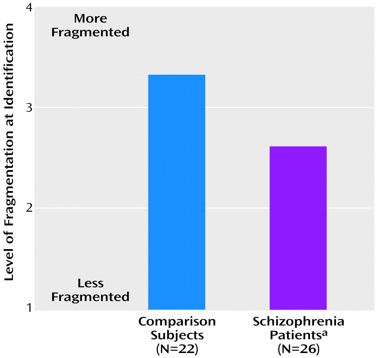
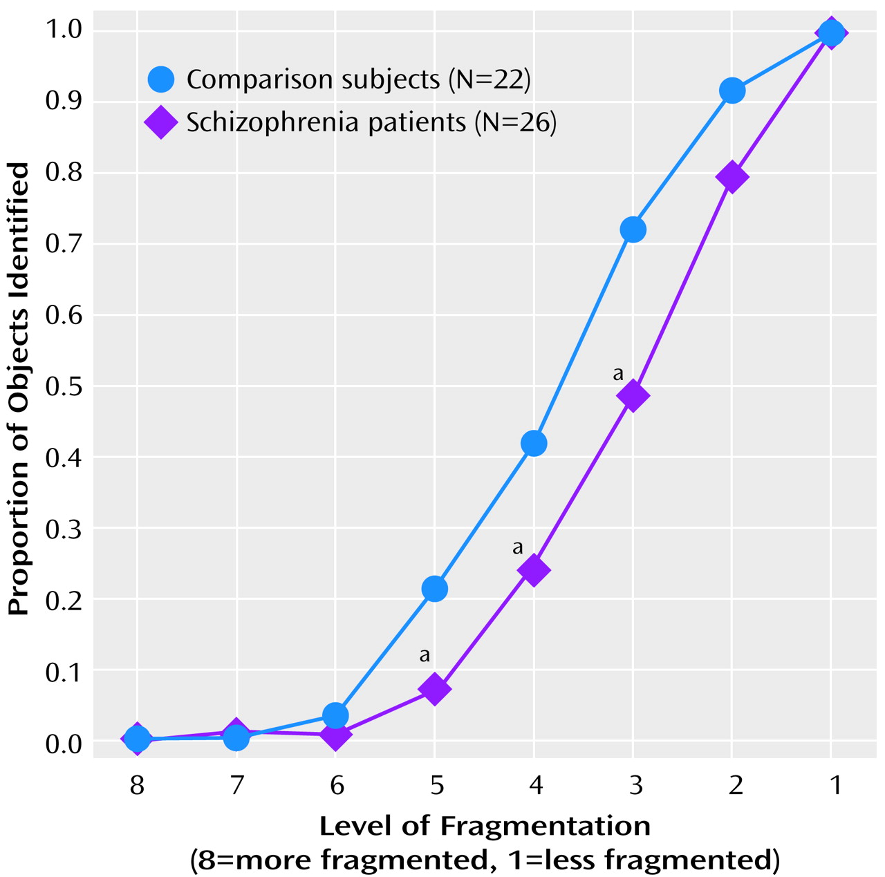
In part 1, data were analyzed by using a one-way, between-group ANOVA. The patients with schizophrenia showed significant impairment in object recognition, as evidenced by the necessity for a more complete image to appear before object recognition was possible (higher threshold of identification; Figure 2). Similarly, when the proportions of pictures recognized at each level by the two groups were analyzed, there was a rightward shift in the psychometric function for schizophrenic patients in relation to the comparison subjects (Figure 3). Slopes for the two groups, however, were similar.
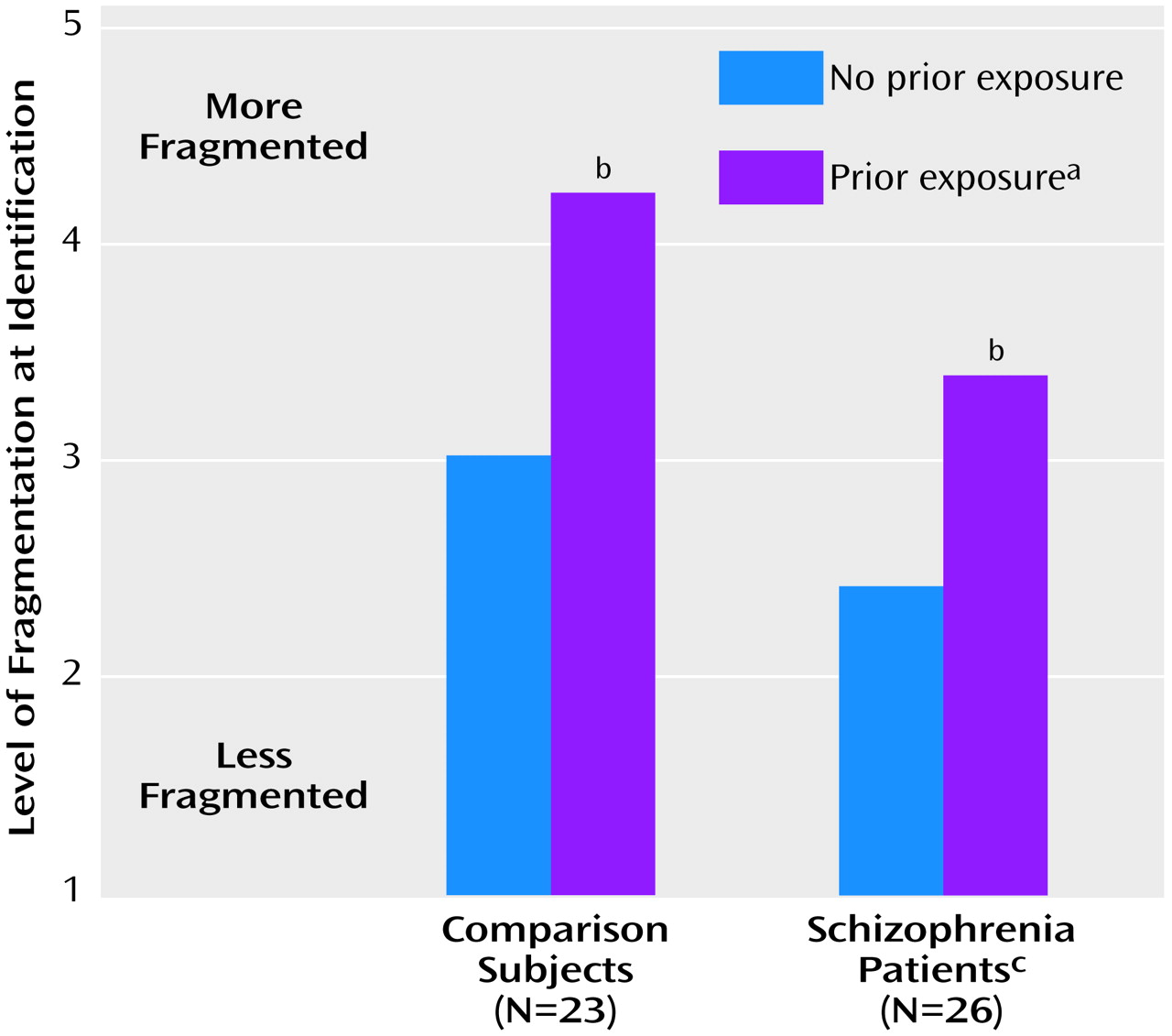
In part 2, data were analyzed by using a two-way (group-by-repetition) ANOVA, with repetition coded as a within-group factor. Both schizophrenic patients and nonpsychiatric comparison participants experienced the expected repetition priming effect, as evidenced by a significant shift in object-recognition threshold (level of identification) as a function of previous exposure (Figure 4). As in part 1, schizophrenic patients required significantly more complete pictures than the comparison subjects before they were able to identify the objects, whether or not the pictures had been presented previously. However, the schizophrenia patients benefited as much as the comparison subjects from previous exposure to the complete pictures, as reflected by a significant improvement within both groups (comparison subjects: t=10.28, df=22, p<0.001; patients: t=11.06, df=25, p<0.001) and a nonsignificant group-by-repetition interaction (F=2.4, df=1, 47, p=0.13).
In part 3, the combined effects of repetition priming and word prompting were assessed in a three-way (group-by-repetition-by-prompt type) ANOVA. Repetition and prompt type were coded as within-group factors. Prior exposure and word prompting contributed independently to performance, as reflected by significant main effects of repetition and prompt type (Figure 5) and the lack of a significant repetition-by-prompt-type interaction (F=1.9, df=2, 94, p=0.16). As in parts 1 and 2 of testing, the schizophrenic participants were highly impaired in their ability to identify fragmented pictures of objects across conditions, as reflected in a significantly higher threshold level of identification (Figure 5). Significant, similar effects in magnitude of word prompting were observed for the schizophrenia patients and the comparison subjects in both conditions of no prior exposure (comparison subjects: t=6.91, df=22, p<0.001; schizophrenia patients: t=4.71, df=25, p<0.001) and prior exposure (comparison subjects: t=3.21, df=22, p<0.005; schizophrenia patients: t=4.72, df=25, p<0.001). Furthermore, neither the group-by-repetition (F=0.1, df=1, 47, p=0.80) nor the group-by-prompt-type (F=1.6, df=2, 94, p=0.22) interaction effects were significant, indicating that the patients with schizophrenia benefited as much as the comparison subjects from both prior experience and having seen valid word prompts.
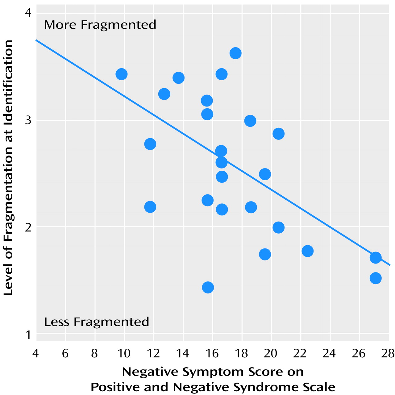
A significant positive correlation was found between level of identification, as measured in part 1 of the testing, and negative symptom score on the Positive and Negative Syndrome Scale (r=–0.54, p=0.005) (Figure 6). Similar correlations were found for results from parts 2 and 3 of the testing. The level of identification did not correlate with positive symptom or autistic preoccupation subscale scores. Mean IQs for the patient and comparison groups were 98.7 and 110.4, respectively. Although there was a between-group difference in IQ (t=–3.73, df=46, p=0.001), the between-group difference in level of identification, as measured in part 1 of the testing, remained significant after covariation for IQ (F=14.5, df=1, 44. p<0.001). Similar significant differences remained after covariation for results from parts 2 and 3 of the testing.

Discussion

The term “perceptual closure” refers to the collection of neural processes that permits object recognition to occur when only partial visual information is present. Physiologic evidence suggests that these processes may involve interpolation, or “filling in,” of the missing information (12). The phenomenon of perceptual closure has been extensively studied with the use of fragmented images, both behaviorally (68) and electrophysiologically (9, 16), in normal subjects, but, to our knowledge, it has not been evaluated previously in subjects with schizophrenia. This study provides the first demonstration that perceptual closure is impaired in patients with schizophrenia, leading to deficits in object recognition. Nonetheless, patients derive benefit comparable to that of comparison subjects from prior exposure to complete pictures and from having seen valid word prompts that name the object subsequently presented in a fragmented picture, suggesting intact top-down regulation.
Processing of object forms occurs primarily within the ventral visual stream (26), particularly in the lateral occipital complex (27). Impaired object recognition in patients with schizophrenia, as reflected in this study, would thus be consistent with dysfunction within ventral visual-stream sensory regions and especially in the lateral occipital complex. Lower cerebral blood flow has been observed in the vicinity of the lateral occipital complex in schizophrenia patients (5), supporting the notion that deficits in perceptual closure reflect impaired local processing within visual brain regions. Furthermore, because the inferior temporal area in the monkey, which is likely homologous to the lateral occipital complex in humans, plays a critical role in visual backward masking (28), it is possible that dysfunction in the lateral occipital complex could also account for the well-described visual backward-masking deficits associated with schizophrenia (2931). In our previous high-density electrical mapping studies (9, 16), we identified a novel event-related potential component—Ncl—that reflects an incremental increase in activity in object-recognition areas of the lateral occipital complex during the presentation of fragmented pictures of objects by means of the ascending method of limits. We proposed that Ncl reflects the processes of perceptual closure, in which the brain attempts to “fill in” the missing pieces of an object until recognition is achieved. It is these neural processes that we believe to be critical for performance in the present study, but they appear to be unaffected by priming or prompting manipulations. This conclusion is supported by our work showing an “early” electrophysiologic index of repetition priming in a perceptual closure task (16) and other work showing “late” indices of semantic priming (32).
In addition to demonstrating a deficit in visual sensory processing, the present study also provides evidence that the deficit is not attributable to top-down dysregulation of structures in the ventral visual stream. Two manipulations have been shown to affect the level of sensory information needed to successfully identify an object. First, if an object has been shown previously in identifiable form (either as complete or fragmentary), subsequent recognition is facilitated (6), as is generation of Ncl (16). Second, if participants are given a prompt that potentially identifies the object pictured, recognition is facilitated (33; unpublished report by Matsukawa et al.). In the present study, the patients with schizophrenia derived benefit similar to that of comparison subjects from stimulus repetition and word prompting, although across conditions, the patients required more information than the comparison subjects to successfully identify an object. Thus, the study shows a clear differential deficit whereby patients were impaired in the sensory aspects of the task but were relatively unimpaired with regard to higher-order functions.
The pattern of impairment observed in this study differs from that of other neuropsychiatric disorders. Thus, although Alzheimer’s disease has been associated with deficits in object recognition, these are accompanied by profound deficits in repetition priming (3437). In contrast, subjects with Huntington’s disease may show enhanced priming (37). Finally, subjects with Parkinson’s disease have not shown deficits in object recognition or priming (35). Thus, the present pattern may be specific to subjects with schizophrenia.
A study by Newcombe and Russell (38) revealed that patients with temporal damage show poor performance on a “closure” task that requires age and gender judgments, supporting localization of perceptual closure to the lateral occipital complex. Patients with right parietal lesions have been shown to have a syndrome termed “apperceptive agnosia” (39) characterized by impaired shape perception and suggesting that impaired parietal input to the lateral occipital cortex may contribute to the observed deficit (40, 41). In contrast, patients with predominant frontocortical lesions showed more robust deficits in priming than in object recognition itself, supporting the notion that the present pattern of impairment cannot be attributed to prefrontal dysfunction (42).
The present findings are consistent with deficits demonstrated in patients with schizophrenia in a variety of perceptual organization tasks (4345). However, an advantage of the present tasks is that “perceptual closure” is a highly specific term that refers to a distinct set of processes that underlie the identification of objects in fragmented pictures, such as those in the present study. The current study sets the stage for future electrophysiological studies of perceptual closure in patients with schizophrenia, in which the neural processes underlying these behavioral effects can be examined directly. Our findings are also consistent with studies showing intact repetition priming and implicit learning in subjects with schizophrenia (46, 47). Again, implicit learning in the context of a perceptual closure task has a well-defined electrophysiological signature (16), whereas such is not the case with other forms of implicit learning.
This is the first study of which we are aware of object recognition in patients with schizophrenia. However, at least one prior study investigated the phenomenon of perceptual closure (48). In that study, patients were asked to reproduce line drawings with gaps. Comparison subjects automatically filled in the gaps because of the operation of perceptual closure, drawing more complete pictures of objects than had been requested. In contrast, the patients did not fill in the gaps; thus, paradoxically, they drew pictures that better approximated the fragmented originals than did the comparison subjects. Since the time of this 1961 study, little work has been done on the impairment of perceptual closure in patients with schizophrenia. The present study builds on earlier work and offers an improved method for quantifying the perceptual closure deficit.
It has been appreciated for at least 30 years that schizophrenic patients perform poorly on a wide variety of cognitive tasks. To some extent, this observation has been used to suggest that individual deficits are significant only to the degree that they stand out from a background of widespread dysfunction (49). More recently, however, it has been accepted that a widespread, multifocal deficit is, of itself, a crucial aspect of schizophrenia and a primary predictor of poor outcome (3, 4). The present study suggests that such deficits arise from distributed dysfunction within multiple brain regions, involving sensory as well as cognitive areas. These findings are consistent with those of parallel studies that have demonstrated sensory processing deficits within auditory (50) and somatosensory (51) brain regions.
There are several indications that the observed deficit in perceptual closure was not due to factors such as impaired cooperation or motivation on the part of the participants. First, with sufficient information, the patients recognized the objects correctly as often as the comparison subjects; both groups correctly identified the objects over 90% of the time. Only correct responses were analyzed. Second, the slopes of the performance curves were very similar for schizophrenia patients and comparison subjects, although the patient curve was shifted significantly to the right (Figure 3). Thus, similar processes were engaged in both groups, although processing precision in the patient group was less than that of the comparison group. Finally and most important, the patients with schizophrenia were able to derive as much benefit as the comparison subjects from repetition priming and word prompting. Repetition priming and word prompting require at least as much task engagement as object recognition. Overall, therefore, the present study suggests that deficits in sensory-level processing may contribute significantly to widespread cognitive dysfunction in patients with schizophrenia and that such deficits reflect intrinsic dysfunction within sensory brain regions.
Although the present study was designed to assess the integrity of object recognition, a ventral visual-stream process, it should be noted that deficits in visual processing in patients with schizophrenia are not likely confined to this pathway. Deficits in motion detection have also been reported (52), as have deficits in regulation of smooth-pursuit eye movements (53). Both sets of deficits are postulated to reflect dysfunction of the dorsal-stream visual pathway. We have recently observed selective deficits in magnocellular processing, suggesting dysfunction even at the level of the lateral geniculate nucleus or extrastriate cortex (54). Thus, in addition to reflecting dysfunction within the lateral occipital complex, the deficit in perceptual closure may also reflect impaired information processing within earlier sensory regions.
The patients in the present study were recruited from outpatient and inpatient facilities. This permitted recruitment of schizophrenic patients with a wide range of positive, negative, and cognitive symptoms. A significant correlation was found between negative symptoms and level of fragmentation at object recognition. Studies of the information-processing correlates of positive and negative symptoms (55) have found that patients with negative symptoms have elevated stimulus-recognition thresholds and deficits in the time-dependent processing of information. The present findings are consistent with this earlier work.
Many real-world stimuli to which individuals are exposed are observed in fragmentary form. Perceptual closure permits relatively automatic identification of such objects (e.g., “a cat behind venetian blinds”). The correlation between the deficit in perceptual closure and the negative symptom score suggests that patients with negative symptoms may navigate a world that is form-impoverished and that the absence of salient forms may directly contribute to such diagnostic features as a lack of interest in the environment. The correlation may also reflect operation of a common mediating variable. For example, dysfunction of N-methyl-d-aspartate (NMDA) receptors is postulated to play a key role in the mediation of negative symptoms (56). Furthermore, these receptors contribute significantly to the development of feature-detection circuits within visual sensory regions (57). NMDA receptor dysfunction could thus contribute to both symptoms and information-processing aspects of the disorder.
A limitation of the present study is that all of the patients with schizophrenia were receiving medication at the time of testing (58). However, there was no significant correlation between neuroleptic dose and performance. Furthermore, antipsychotic effects would not explain the finding of preserved higher-order processing despite impaired perceptual closure within the patient group. Because dopamine is an important neurotransmitter in the retina (59, 60), the possibility that endogenous dopamine deficiency contributes to the observed deficit in perceptual closure must be considered (61). It has been shown, however, that visual acuity is unaffected by the dopamine-blocking antipsychotic haloperidol (62). Furthermore, the recognition of objects in fragmented pictures has been found to be unimpaired in patients with Parkinson’s disease, which is characterized by a deficiency of dopamine (35). Hence, it is unlikely that the effects of antipsychotic dopamine blockers can account for the deficit in perceptual closure observed in the present study.
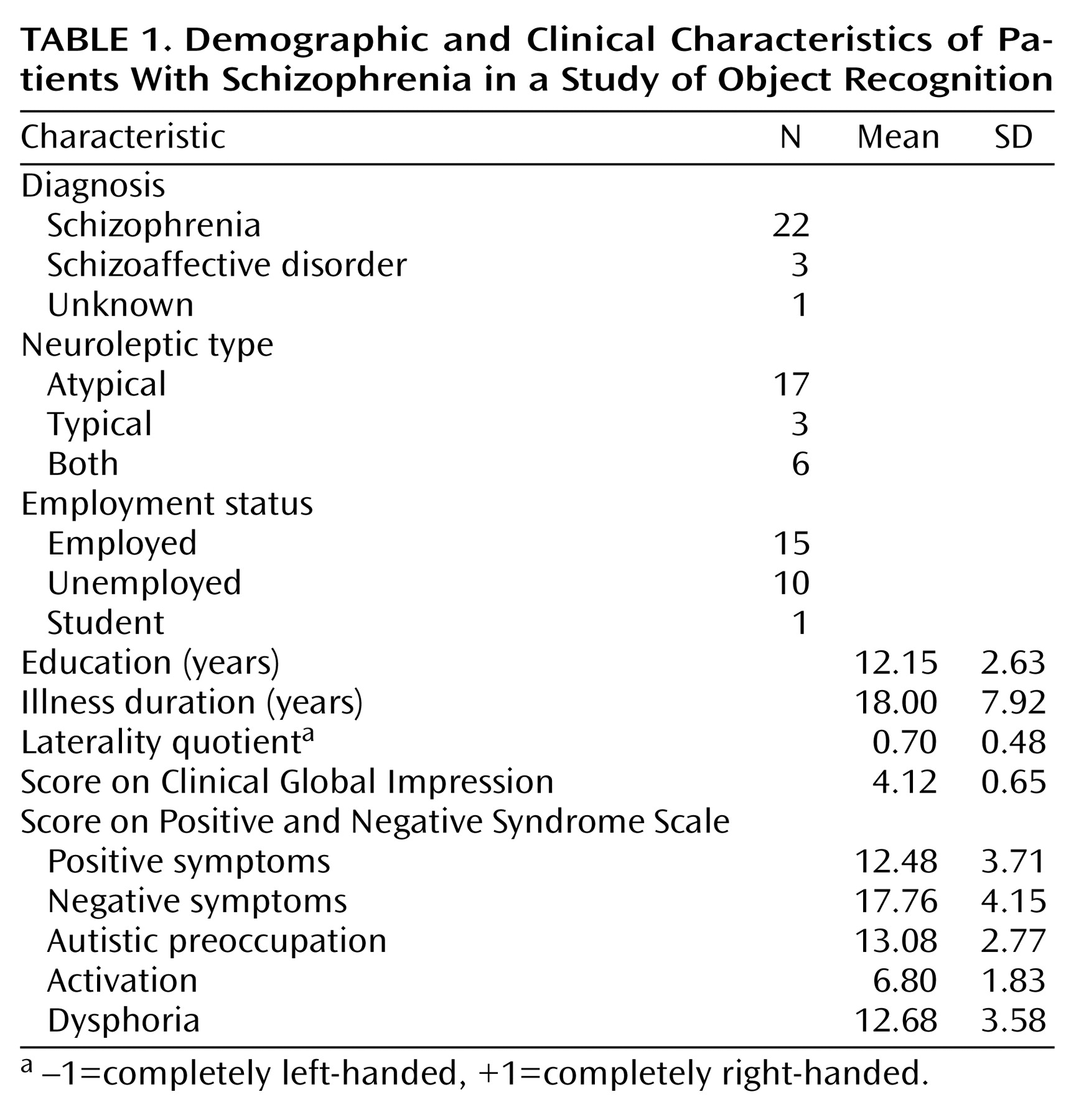
TABLE 1
Figure 1. Representative Picture Sequence From Perceptual Closure Task Administered to 26 Patients With Schizophrenia and 23 Nonpatient Comparison Subjects (level 1 not shown)
Figure 2. Level of Fragmentation at Which Object Was Recognized by Patients With Schizophrenia and Nonpatient Comparison Subjects
aSignificant group effect (ANOVA: F=16.9, df=1, 49, p<0.001).
Figure 3. Effect of Level of Fragmentation on Proportion of Objects Recognized by Patients With Schizophrenia and Nonpatient Comparison Subjects
aSignificant difference between patients and comparison subjects (p<0.005, t test, two-tailed).
Figure 4. Effect of Prior Exposure on Level of Fragmentation at Which Objects Were Recognized by Patients With Schizophrenia and Nonpatient Comparison Subjects
aMain effect of prior exposure (ANOVA: F=227.2, df=1, 47, p<0.001).
bSignificant difference from trials with subjects with no prior exposure (within group) (p<0.001, t test, two-tailed).
cMain effect of group (ANOVA: F=18.5, df=1, 47, p<0.001).
Figure 5. Effect of Prior Exposure and Word Prompting on Level of Fragmentation at Which Objects Were Recognized by Patients With Schizophrenia and Nonpatient Comparison Subjects
aMain effect of prior exposure (ANOVA: F=84.6, df=1, 47, p<0.001).
bMain effect of prompt type (ANOVA: F=58.7, df=1, 47, p<0.001).
cSignificant difference from baseline condition (p<0.001, t test, two-tailed).
dSignificant difference from baseline condition (p<0.01, t test, two-tailed).
eMain effect of group (ANOVA: F=12.1, df=1, 47, p<0.005).
Figure 6. Effect of Negative Symptom Score on Level of Fragmentation at Which Objects Were Recognized by 25 Patients With Schizophreniaa
ar=–0.54, p=0.005.

Footnote

Received Feb. 15, 2000; revisions received June 19 and Oct. 26, 2000, and May 30, 2001; accepted June 6, 2001. From the Cognitive Neuroscience and Schizophrenia Program, Nathan Kline Institute for Psychiatric Research. Address reprint requests to Dr. Javitt, Cognitive Neuroscience and Schizophrenia Program, Nathan Kline Institute for Psychiatric Research, 140 Old Orangeburg Rd., Orangeburg, NY 10962; [email protected] (e-mail). Supported by NIMH grants MH-49334 and MH-01439 and a grant from the Burroughs Wellcome Fund to Dr. Javitt. The authors thank Lisa Falbo and her staff at the Residential-Care Center for Adults, Orangeburg, N.Y., G. Gail Neffinger, Mara Cohen, and their staff at the Young Adult Center, Department of Mental Health, County of Rockland, Pomona, N.Y.; and Robert Lindsley.

References

1.
Buchsbaum MS: The frontal lobes, basal ganglia, and temporal lobes as sites for schizophrenia. Schizophr Bull 1990; 16:379-389
2.
Heckers S: Neuropathology of schizophrenia: cortex, thalamus, basal ganglia, and neurotransmitter-specific projection systems. Schizophr Bull 1997; 23:403-421
3.
Bilder RM, Goldman RS, Robinson D, Reiter G, Bell L, Bates JA, Pappadopulos E, Willson DF, Alvir JMJ, Woerner MG, Geisler S, Kane JM, Lieberman JA: Neuropsychology of first-episode schizophrenia: initial characterization and clinical correlates. Am J Psychiatry 2000; 157:549-559
4.
Mohamed S, Paulsen JS, O’Leary D, Arndt S, Andreasen N: Generalized cognitive deficits in schizophrenia: a study of first-episode patients. Arch Gen Psychiatry 1999; 56:749-754
5.
Kim J-J, Mohamed S, Andreasen NC, O’Leary DS, Watkins GL, Boles Ponto LL, Hichwa RD: Regional neural dysfunctions in chronic schizophrenia studied with positron emission tomography. Am J Psychiatry 2000; 157:542-548
6.
Snodgrass JG, Feenan K: Priming effects in picture fragment completion: support for the perceptual closure hypothesis. J Exp Psychol Gen 1990; 119:276-296
7.
Foley MA, Foley HJ, Durso FT, Smith NK: Investigations of closure processes: what source-monitoring judgments suggest about what is “closing.” Mem Cognit 1997; 25:140-155
8.
Snodgrass JG, Kinjo H: On the generality of the perceptual closure effect. J Exp Psychol Learn Mem Cogn 1998; 24:645-658
9.
Doniger GM, Foxe JJ, Murray MM, Higgins BA, Snodgrass JG, Schroeder CE, Javitt DC: Activation timecourse of ventral visual stream object-recognition areas: high density electrical mapping of perceptual closure processes. J Cogn Neurosci 2000; 12:615-621
10.
von der Heydt R, Peterhans E, Baumgartner G: Illusory contours and cortical neuron responses. Science 1984; 224:1260-1262
11.
Fiorani M, Rosa M, Gattass R, Rocha-Miranda C: Dynamic surrounds of receptive fields in primate striate cortex: a physiological basis for perceptual completion. Proc Natl Acad Sci USA 1992; 89:8547-8551
12.
De Weerd P, Gattass R, Desimone R, Ungerleider L: Responses of cells in monkey visual cortex during perceptual filling-in of an artificial scotoma. Nature 1995; 377:731-734
13.
Nobre AC, Allison T, McCarthy G: Word recognition in the human inferior temporal lobe. Nature 1994; 372:260-263
14.
Moore CJ, Price CJ: Three distinct ventral occipitotemporal regions for reading and object naming. Neuroimage 1999; 10:181-192
15.
Grill-Spector K, Kushnir T, Hendler T, Malach R: The dynamics of object-selective activation correlate with recognition performance in humans. Nat Neurosci 2000; 3:837-843
16.
Doniger GM, Foxe JJ, Schroeder CE, Murray MM, Higgins BA, Javitt DC: Visual perceptual learning in human object recognition areas: a repetition priming study using high-density electrical mapping. Neuroimage 2001; 13:305-313
17.
Ungerleider LG, Mishkin M: Two cortical visual systems, in Analysis of Visual Behavior. Edited by Ingle D, Goodale M, Mansfield R. Cambridge, Mass, MIT Press, 1982, pp 549-586
18.
Grill-Spector K, Kushnir T, Edelman S, Itzchak Y, Malach R: Cue-invariant activation in object-related areas of the human occipital lobe. Neuron 1998; 21:191-202
19.
Grill-Spector K, Kushnir T, Hendler T, Edelman S, Itzchak Y, Malach R: A sequence of object-processing stages revealed by fMRI in the human occipital lobe. Hum Brain Mapp 1998; 6:316-328
20.
Kay SR, Fiszbein A, Opler LA: The Positive and Negative Syndrome Scale (PANSS) for schizophrenia. Schizophr Bull 1987; 13:261-276
21.
White L, Harvey PD, Opler L, Lindenmayer JP (PANSS Study Group): Empirical assessment of the factorial structure of clinical symptoms in schizophrenia: a multisite, multimodel evaluation of the factorial structure of the Positive and Negative Syndrome Scale. Psychopathology 1997; 30:263-274
22.
Ammons R, Ammons C: The Quick Test (QT): provisional manual. Psychol Rep 1962; 11:111-162
23.
Snodgrass JG, Corwin J: Perceptual identification thresholds for 150 fragmented pictures from the Snodgrass and Vanderwart picture set. Percept Mot Skills 1988; 67:3-36
24.
Snodgrass JG, Vanderwart M: A standardized set of 260 pictures: norms for name agreement, image agreement, familiarity, and visual complexity. J Exp Psychol Hum Learn Mem 1980; 6:174-215
25.
Howell DC: Statistical Methods in Psychology. Belmont, Calif, Duxbury, 1997
26.
Ungerleider LG, Courtney SM, Haxby JV: A neural system for human visual working memory. Proc Natl Acad Sci USA 1998; 95:883-890
27.
Malach R, Reppas JB, Benson RR, Kwong KK, Jiang H, Kennedy WA, Ledden PJ, Brady TJ, Rosen BR, Tootell RB: Object-related activity revealed by functional magnetic resonance imaging in human occipital cortex. Proc Natl Acad Sci USA 1995; 92:8135-8139
28.
Kovacs G, Vogels R, Orban GA: Cortical correlate of pattern backward masking. Proc Natl Acad Sci USA 1995; 92:5587-5591
29.
Cadenhead KS, Serper Y, Braff DL: Transient versus sustained visual channels in the visual backward masking deficits of schizophrenia patients. Biol Psychiatry 1998; 43:132-138
30.
Butler PB, Harkavy-Friedman JM, Amador XF, Gorman JM: Backward masking in schizophrenia: relationship to medication status, neuropsychological functioning, and dopamine metabolism. Biol Psychiatry 1996; 40:295-298
31.
Green MF, Nuechterlein KH, Breitmeyer B, Mintz J: Backward masking in unmedicated schizophrenic patients in psychotic remission: possible reflection of aberrant cortical oscillation. Am J Psychiatry 1999; 156:1367-1373
32.
Weisbrod M, Kiefer M, Winkler S, Maier S, Hill H, Roesch-Ely D, Spitzer M: Electrophysiological correlates of direct versus indirect semantic priming in normal volunteers. Brain Res Cogn Brain Res 1999; 8:289-298
33.
Carr TH, McCauley C, Sperber RD, Parmelee CM: Words, pictures, and priming: on semantic activation, conscious identification, and the automaticity of information processing. J Exp Psychol Hum Percept Perform 1982; 8:757-777
34.
Mack J, Patterson M, Schell A, Whitehouse P: Performance of subjects with probable Alzheimer disease and normal elderly controls on the Gollin Incomplete Pictures Test. Percept Mot Skills 1993; 77:951-969
35.
Bondi M, Kaszniak A: Implicit and explicit memory in Alzheimer’s disease and Parkinson’s disease. J Clin Exp Neuropsychol 1991; 13:339-358
36.
Mielke R, Kessler J, Fink G, Herholz K, Heiss W: Dysfunction of visual cortex contributes to disturbed processing of visual information in Alzheimer’s disease. Int J Neurosci 1995; 82:1-9
37.
Heindel W, Salmon D, Butters N: Pictorial priming and cued recall in Alzheimer’s and Huntington’s disease. Brain Cogn 1990; 13:282-295
38.
Newcombe F, Russell W: Dissociated visual perceptual and spatial deficits in focal lesions of the right hemisphere. J Neurol Neurosurg Psychiatry 1969; 32:73-81
39.
Lissauer H: Ein fall von seelenblindheit nebst einem Beitrage zur Theori derselben. Arch Psychiatr Nervenkrankheiten 1890; 21:222-270
40.
Warrington E, James M: Disorders of visual perception in patients with localised cerebral lesions. Neuropsychologia 1967; 5:253-266
41.
Lewis R, Kamptner N: Sex differences in spatial task performance of patients with and without unilateral cerebral lesions. Brain Cogn 1987; 6:142-152
42.
Markowitsch H, Harting C: Interdependence of priming performance and brain-damage. Int J Neurosci 1996; 85:291-300
43.
Rabinowicz EF, Opler LA, Owen DR, Knight RA: Dot Enumeration Perceptual Organization Task (DEPOT): evidence for a short-term visual memory deficit in schizophrenia. J Abnorm Psychol 1996; 105:336-348
44.
Silverstein SM, Knight RA, Schwarzkopf SB, West LL, Osborn LM, Kamin D: Stimulus configuration and context effects in perceptual organization in schizophrenia. J Abnorm Psychol 1996; 105:410-420
45.
Chey J, Holzman P: Perceptual organization in schizophrenia: utilization of the Gestalt principles. J Abnorm Psychol 1997; 106:530-538
46.
Schmand B, Kop W, Kuipers T, Bosveld J: Implicit learning in psychotic patients. Schizophr Res 1992; 7:55-64
47.
Bazin N, Perruchet P: Implicit and explicit associative memory in patients with schizophrenia. Schizophr Res 1996; 22:241-248
48.
Snyder S, Rosenthal D, Taylor IA: Perceptual closure in schizophrenia. J Abnorm Soc Psychol 1961; 63:131-136
49.
Chapman LJ, Chapman JP: The measurement of differential deficit. J Psychiatr Res 1978; 14:303-311
50.
Javitt DC, Strous RD, Grochowski S, Ritter W, Cowan N: Impaired precision, but normal retention, of auditory sensory (“echoic”) memory information in schizophrenia. J Abnorm Psychol 1997; 106:315-324
51.
Javitt DC, Liederman E, Cienfuegos A, Shelley AM: Panmodal processing imprecision as a basis for dysfunction of transient memory storage systems in schizophrenia. Schizophr Bull 1999; 23:763-775
52.
Schwartz BD, Maron BA, Evans WJ, Winstead DK: Smooth pursuit tracking deficits of patients with schizophrenia at specific within-sine wave bins. Neuropsychiatry Neuropsychol Behav Neurol 2000; 12:221-229
53.
Schwartz BD, Maron BA, Evans WJ, Winstead DK: High velocity transient visual processing deficits diminish ability of patients with schizophrenia to recognize objects. Neuropsychiatry Neuropsychol Behav Neurol 1999; 12:170-177
54.
Butler PB, Schechter I, Zemon V, Schwartz SG, Greenstein VC, Gordon J, Schroeder CE, Javitt DC: Dysfunction of early-stage visual processing in schizophrenia. Am J Psychiatry 2001; 158:1126-1133
55.
Braff DL: Sensory input deficits and negative symptoms in schizophrenic patients. Am J Psychiatry 1989; 146:1006-1011
56.
Javitt DC, Zukin SR: Recent advances in the phencyclidine model of schizophrenia. Am J Psychiatry 1991; 148:1301-1308
57.
Bear MF, Kleinschmidt A, Singer W: Disruption of experience-dependent synaptic modifications in striate cortex by infusion of an NMDA receptor antagonist. J Neurosci 1990; 10:909-925
58.
Spohn HE, Lacoursiere RB, Thompson RN, Coyne L: Phenothiazine effects on psychological and psychophysiological dysfunction in chronic schizophrenics. Arch Gen Psychiatry 1977; 34:633-644
59.
Nguyen-Legros J, Versaux-Botteri C, Vernier P: Dopamine receptor localization in the mammalian retina. Mol Neurobiol 1999; 19:181-204
60.
Ehinger B: Functional role of dopamine in the retina, in Progress in Retinal Research, vol 2. Edited by Osborne N, Chader J. Oxford, UK, Pergamon Press, 1983, pp 213-232
61.
Hankins M: Functional dopamine deficits in the senile rat retina. Vis Neurosci 2000; 17:839-845
62.
Harris J, Gelbtuch M, Phillipson O: Effects of haloperidol and nomifensine on the visual aftereffects of tilt and movement. Psychopharmacology (Berl) 1986; 89:177-182

Information & Authors

Information

Published In

Go to American Journal of Psychiatry
American Journal of Psychiatry
Pages: 1818 - 1826
PubMed: 11691687

History

Published online: 1 November 2001
Published in print: November 2001

Authors

Details

Glen M. Doniger, Ph.D.
Esther F. Rabinowicz, Ph.D.
Joan G. Snodgrass, Ph.D.
Daniel C. Javitt, M.D., Ph.D.

Metrics & Citations

Metrics

Citations

Export Citations

If you have the appropriate software installed, you can download article citation data to the citation manager of your choice. Simply select your manager software from the list below and click Download.

For more information or tips please see 'Downloading to a citation manager' in the Help menu.

Format
Citation style
Style
Copy to clipboard

View Options

View options

PDF/EPUB

View PDF/EPUB

Login options

Already a subscriber? Access your subscription through your login credentials or your institution for full access to this article.

Personal login Institutional Login Open Athens login
Purchase Options

Purchase this article to access the full text.

PPV Articles - American Journal of Psychiatry

PPV Articles - American Journal of Psychiatry

Not a subscriber?

Subscribe Now / Learn More

PsychiatryOnline subscription options offer access to the DSM-5-TR® library, books, journals, CME, and patient resources. This all-in-one virtual library provides psychiatrists and mental health professionals with key resources for diagnosis, treatment, research, and professional development.

Need more help? PsychiatryOnline Customer Service may be reached by emailing [email protected] or by calling 800-368-5777 (in the U.S.) or 703-907-7322 (outside the U.S.).

Media

Figures

Other

Tables

Share

Share

Share article link

Share