Full access
New Research
Published Online: 1 January 2011

Cortical Activations During Auditory Verbal Hallucinations in Schizophrenia: A Coordinate-Based Meta-Analysis

Abstract

Objective:

Auditory verbal hallucinations (AVHs) constitute severe, incapacitating symptoms of schizophrenia. Despite increasing interest in the functional exploration of AVHs, the available findings remain difficult to integrate because of their considerable variability. The authors' aim was to perform a robust quantitative review of existing functional data in order to elucidate consistent patterns observed during the emergence of AVHs and to orient new pathophysiological models of hallucinations.

Method:

Ten positron emission tomography or functional magnetic resonance imaging studies were selected for the meta-analysis after systematic review. A total of 68 patients with schizophrenia spectrum disorders experiencing AVHs during scanning were included. According to a random-effects activation likelihood estimation algorithm, stereotaxic coordinates of 129 foci, reported as significant in the source studies, were extracted and computed to estimate the brain locations most consistently associated with AVHs across studies (cluster-extent threshold: 200 mm3).

Results:

Patients experiencing AVHs demonstrated significantly increased activation likelihoods in a bilateral neural network, including the Broca's area (activation likelihood estimation=1.84×10⁻³), anterior insula (1.78×10⁻³), precentral gyrus (1.46×10⁻³), frontal operculum (1.29×10⁻³), middle and superior temporal gyri (1.59×10⁻³), inferior parietal lobule (1.33×10⁻³), and hippocampus/parahippocampal region (1.90×10⁻³).

Conclusions:

This meta-analysis demonstrated that experiencing AVHs is associated with increased activity in fronto-temporal areas involved in speech generation and speech perception, but also within the medial temporal lobe, a structure notably involved in verbal memory. Such findings support a model for AVHs in which aberrant cortical activations emerge within a distributed network involved at different levels of complexity in the brain architecture. Critical future directions are considered.
Hallucinations can be defined as perceptions without corresponding sources in the external world. This feature represents one of the main positive symptoms of schizophrenia spectrum disorders, and 60%–70% of patients meeting the diagnostic criteria for this pathology experience hallucinations (1). Even though they may involve any of the senses, auditory verbal hallucinations (AVHs) are most prevalent in such a psychiatric context. Patients experiencing AVHs usually describe hearing words, sentences, and conversations that are often intrusive or comment on their thoughts. In about 25% of patients, this symptom can be drug-resistant and become chronic (2), causing an impaired quality of life. The pathophysiology of AVHs is still poorly understood, even though neuroimaging exploration has expanded in the recent decades to address various morphometric, functional, and connectivity issues in patients with schizophrenia. Many underlying mechanisms for AVHs have been proposed (3), most of which are not mutually exclusive. Three main mechanisms are briefly presented in the present meta-analysis. First, some authors have postulated that AVHs could result from aberrant perceptions generated in auditory regions. Primary support for this theory came from the generation of involuntary auditory or verbal material during per-operative electrical stimulations of the temporal cortex in nonschizophrenia subjects (4). Another influential hypothesis concerning the origin of AVHs is external misattribution of self-inner speech. According to this model, patients with schizophrenia are unable to identify their own thoughts as self-generated and, furthermore, interpret them as intrusive alien voices within their heads (5). Finally, possible dysfunctions in the neural substrates of episodic verbal memory have been proposed to account for the involuntary emergence of AVHs (6).
An initial reappraisal of the functional imaging procedures developed to test these pathophysiological hypotheses allowed us to conceptually distinguish between two main study categories. First are the cognitive studies comparing hallucinators and nonhallucinators. These studies, called trait-studies, investigate the neural bases of the susceptibility to hallucinate, independent of the subjects' experience during scanning. Second are state-studies conducted during the occurrence of an AVH, which are of particular significance for our purpose, since they directly measure brain activations associated with symptom emergence. However, the problem of disentangling the previously evoked hypotheses regarding the origin of AVHs is actually exacerbated by the fact that only a few studies have directly explored the AVH state, resulting in inconsistencies between findings. Some authors have identified restrictive activations in the Heschl's gyrus (7, 8) or in Broca's area (9, 10) in support of a strict sensory or motor origin for AVHs. Meanwhile, other studies have identified more distributed fronto-temporal networks coupled with subcortical structures (1113). Notable reasons for this difference include the fact that an AVH constitutes an unpredictable subjective event, for which various detection designs have been proposed. In a first subset of studies, symptom occurrence was signaled by pressing a response button when experiencing an AVH during scanning (7, 10, 13, 14). Other authors have employed a discontinuous acquisition method: the random sampling approach, in which a large number of functional magnetic resonance imaging (fMRI) volumes were acquired at random intervals during AVHs (11, 15). Patients reported their sensory experiences immediately after each acquisition. Besides these hypothesis-driven methods, some studies have used more data-driven methods, such as spatial Independent Component Analysis. This method does not rely on a predefined model of brain activity (16). In these studies, AVH occurrences were sometimes signaled by button presses (8, 17) or on the basis of a posteriori standardized interviews (12, 18) to help select the components of interest.
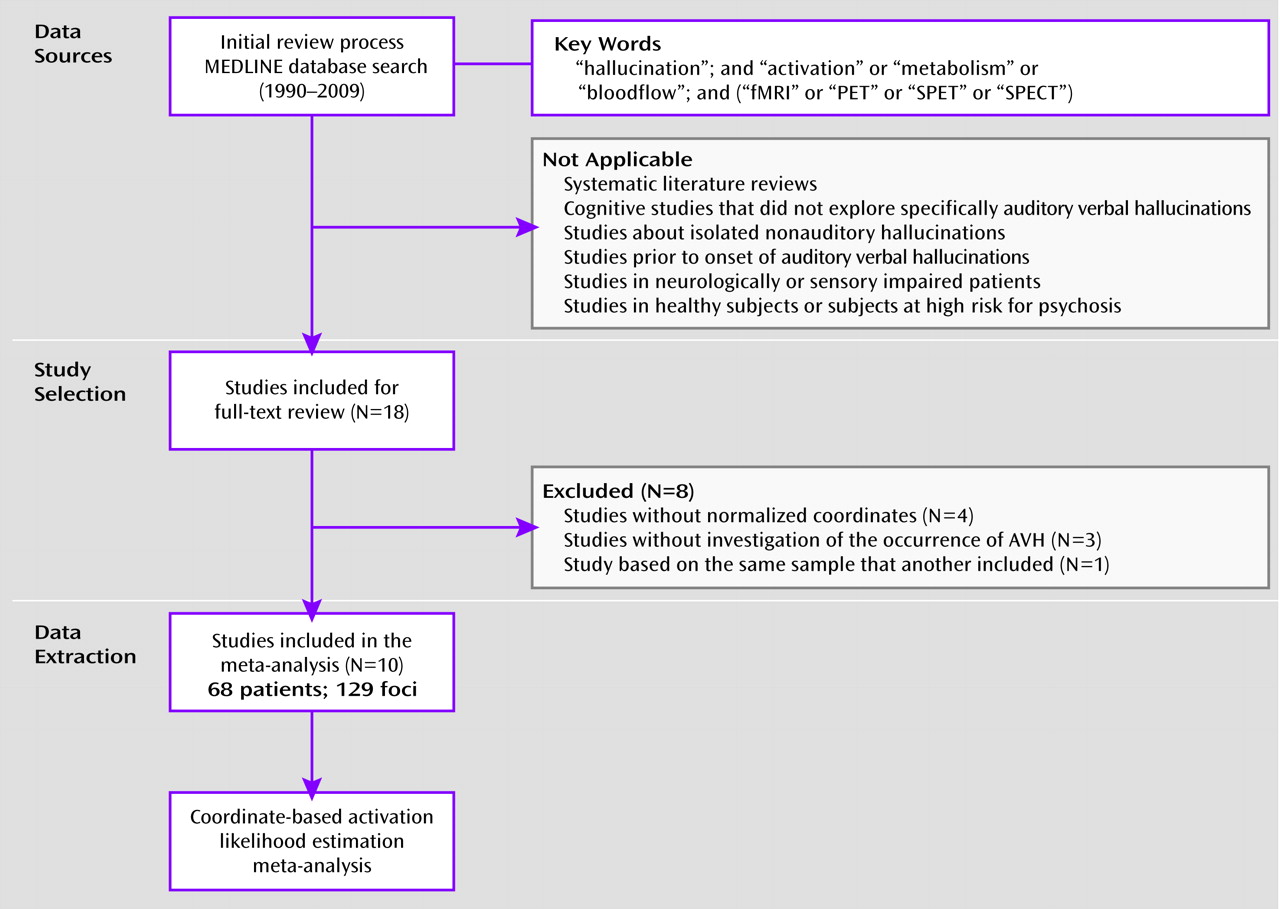
Another plausible explanation for disparities across studies could be considerable interindividual variability of the brain areas involved in AVHs, supporting several possible underlying mechanisms. However, since the available reports often included small samples or did not perform group analyses, it remains difficult to draw definitive conclusions and generalize to the whole population of patients based on the findings. Quantitative meta-analytic techniques have been precisely developed to provide objective measures of functional data and resolve such conflicting views. In the present review, we employed a revised version of the activation likelihood estimation algorithm (19) to describe the brain locations most consistently active during the activation likelihood estimation state across studies. Activation likelihood estimation was recently judged to be the best coordinate-based meta-analysis method when compared with the gold standard of image-based procedures (20). This method allowed us to perform a random-effects meta-analysis and to control for one of the major drawbacks of the previous fixed-effects procedures (i.e., their strong tendency to be dominated by one or a few individual studies) (21). In the present meta-analysis, we postulated that AVHs in patients suffering from schizophrenia spectrum disorders could rely on the interaction between several brain areas involved in a widespread cortical network rather than on restricted sensory or motor activations.

Method

Literature Selection, Data Collection, and Preparation

We first conducted systematic MEDLINE searches to identify all neuroimaging studies about the hallucinatory phenomenon that were published between 1990 and May 2009 (Figure 1). The following key words were employed: “hallucination,” “activation,” “blood flow,” “metabolism,” “fMRI,” “PET” (positron emission tomography), and “SPECT.” We also used the related articles function of the PubMed database and the reference list of retained studies to identify additional articles. A total of 59 studies were collected using this process. We then specifically selected state studies of the AVH phenomenon in people suffering from schizophrenia spectrum disorders. Each article under consideration was independently assessed on this main judgment criterion by two raters (Drs. Jardri and Pouchet) prior to reaching a consensus according to the Preferred Reporting Items for Systematic Reviews and Meta-Analyses statement (http://www.prisma-statement.org/statement.htm). After reviewing all abstracts, articles that did not meet this criterion were excluded. This was the case for systematic reviews (22, 23), activation studies exploring AVHs in people outside of the schizophrenia spectrum (2434), trait studies comparing patients with hallucinations with nonhallucinators (3558), studies of isolated extraauditory hallucinations (59), and studies measuring brain activation prior to the onset of AVHs (6062). After full-text review, secondary exclusions were made of resting state perfusion studies that did not address AVH occurrence (6365) and studies in which stereotaxic results were not reported (9, 6668). E-mail contact was made directly with the authors of the latter articles in an attempt to establish these stereotaxic coordinates, but this attempt was unsuccessful. Finally, since two studies reported partially overlapping samples (7, 8), only data resulting from brain-wide analysis methods were retained (7) to minimize the possibility of nonindependent observations. Importantly, it was not necessary to exclude single-case reports from the meta-analysis, since a weighting factor for the sample size of the retained studies was implemented in the activation likelihood estimation algorithm (19). In summary, 10 whole-brain activation studies using PET or fMRI were selected. Five studies used a button-press method to signal AVH occurrences (6, 7, 10, 13, 14), two employed the random sampling method (11, 15), and three used either data-driven analyses combined with a response box (17) or a posteriori interviews (12, 18). For each of the 10 remaining articles listed in Table 1, we extracted the coordinates (x, y, z) for the 129 foci of interest and the corresponding number of subjects. Only overactivation foci reported as significant at a p value <0.05 in the source studies were included. Nevertheless, when different sensory modalities were involved during hallucinations (15, 17), only foci related to AVHs were selected for further analysis. When necessary, a transformation from the Montreal Neurological Institute to the Talairach space (69) was performed using the icbm2tal algorithm (70), implemented in the GingerALE software (www.brainmap.org/ale/).
FIGURE 1. Flow Diagram of Article Selection Processa
a Data include the numbers of studies initially selected and reasons for exclusion; 10 studies were finally selected, with a total of 68 patients and 129 foci of interest.
TABLE 1. Characteristics of Included Studies Measuring Functional Brain Activity Associated With Auditory Verbal Hallucinations in Schizophrenia Spectrum Disorders
StudyImaging ModalityPatientsDiagnostic CriteriaDesignNumber of FociOriginal Stereotaxic Space
Copolov et al. (6)PETSchizophrenia (N=7); schizoaffective disorder (N=1)DSM-IVOnline button press during auditory verbal hallucinations6Talairach and Tournoux standardized
Dierks et al. (7)fMRISchizophrenia (N=3)DSM-III-ROnline button press during auditory verbal hallucinations23Talairach and Tournoux standardized
Jardri et al. (18)fMRIChildhood-onset schizophrenia (N=1)DSM-IV-TRIndependent component analysis + a posteriori interview4Talairach and Tournoux standardized
Jardri et al. (17)fMRISchizophrenia (N=1)DSM-IV-TRIndependent component analysis + online button press during auditory verbal hallucinations10Talairach and Tournoux standardized
Jardri (12)fMRIFirst-episode schizophrenia (N=15)DSM-IV-TRIndependent component analysis + a posteriori interview14Talairach and Tournoux standardized
Lennox et al. (14)fMRISchizophrenia (N=4)ICD-10Online button press during auditory verbal hallucinations19Talairach and Tournoux standardized
Shergill et al. (11)fMRISchizophrenia (N=6)DSM-IVRandom sampling20Talairach and Tournoux standardized
Shergill et al. (15)fMRISchizophrenia (N=1)DSM-IVRandom sampling3Talairach and Tournoux standardized
Silbersweig et al. (13)PETSchizophrenia (N=5)DSM-IVOnline button press during auditory verbal hallucinations9Talairach and Tournoux standardized
Sommer et al. (10)fMRISchizophrenia (N=18); schizoaffective disorder (N=3); psychosis not otherwise specified (N=3)DSM-IVOnline button press during auditory verbal hallucinations21Montreal Neurological Institute

Meta-Analysis Procedure

A widely used technique for coordinate-based meta-analyses of neuroimaging data is activation likelihood estimation, which treats reported foci not as points but as spatial probability distributions centered at the given coordinates (71). In the present research, we used a revised version of the activation likelihood estimation algorithm implemented in the GingerALE Version 2.0 software (Research Imaging Center, University of Texas at San Antonio, San Antonio, Tex.) (72). This revised activation likelihood estimation implementation has been shown to be more specific than previous algorithms, while retaining comparable sensitivity (19). The activation likelihood estimation meta-analysis followed three steps. The first step was to compute modeled activation maps for each included study. All of the foci reported for a given study were modeled as Gaussian distributions and then merged into a single three-dimensional volume. Rather than using a -prespecified full-width half maximum for the Gaussian distribution, an uncertainty modeling algorithm implemented in GingerALE Version 2.0 was employed to empirically estimate the between-subjects and between-templates variability of the included studies. The second step was to compute the activation likelihood estimation values on a voxel-to-voxel basis by taking the union of these individual modeled activation maps. This analysis was constrained to a gray matter mask that defined the outer limit of the Talairach space. Finally, to assess above-chance clustering between experiments, an empirical null distribution of random spatial association was established to distinguish between noise and true convergences. To do so, an iterative permutation procedure (1011) was used by sampling each activation likelihood estimation result at an independently chosen random location. This test was corrected for multiple comparison bias using the false discovery rate (73) method, with standard values recommended by the authors of the software, so that the q value (the number of expected false positives) was set at 0.05 and a cluster-extent threshold of 200 mm3 was chosen. Final activation likelihood estimation results were exported as a NIfTI file into the Mango software (http://ric.uthscsa.edu/mango/) and were overlaid onto an anatomical template generated by spatially normalizing the International Consortium for Brain Mapping template to Talairach space (74).

Results

When experiencing AVHs, patients with schizophrenia spectrum disorders demonstrated significantly increased activation likelihoods in five clusters distributed in temporal, parietal, frontal, and subcortical sites. The meta-analysis results of these 10 studies are synthesized in Table 2 and Figure 2. The largest clusters were located in the left inferior frontal gyrus at the level of the pars opercularis (Brodmann's area 44), the left precentral gyrus (Brodmann's area 6), the bilateral anterior insula (Brodmann's area 13), and the frontal operculum (Brodmann's area 47). Increased values were also measured in the left middle temporal (Brodmann's area 21) and superior temporal gyri (Brodmann's area 22). This was in addition to the left hippocampus/parahippocampal region (Brodmann's area 27). Additional elevated activation likelihood was measured in the inferior parietal lobule at the level of the left supramarginalis gyrus (Brodmann's area 40). Finally, increased likelihoods were measured in the right-sided internal globus pallidus.
TABLE 2. Brain Regions With Significantly Elevated Likelihoods of Activation During Auditory Verbal Hallucinations in Subjects With Schizophrenia Spectrum Disorders
Identified ClustersBrodmann's AreaBrain LateralityTalairach and Tournoux Coordinates (x,y,z)aCluster Size (mm3)Activation Likelihood Estimates (×10−3)b
Cluster A     
    Broca's convolution44Left−48, 10, 71,3121.84
    Anterior insula13Left−42, 0, 61,2401.78
    Precentral gyrus6Left−54, 0, 144881.46
Cluster B     
    Hippocampus/parahippocampal gyrus27Left−24, −32, −41,6641.90
Cluster C     
    Anterior insula13Right44, 6, −49641.66
    Frontal operculum47Right42, 12, −102651.29
Cluster D     
    Superior and middle temporal gyri21−22Left−54, −44, 168001.59
Cluster E     
    Supramarginalis gyrus40Left−52, −20, 153041.33
a
Data indicate coordinates in the stereotaxic space of the weighted center for each cluster showing greater probability of activation during auditory verbal hallucinations.
b
Estimates are reported for each cluster with a significance of a corrected value (p<0.05).
FIGURE 2. Results of Included Studies Measuring Functional Brain Activity Associated With Auditory Verbal Hallucinations in Subjects With Schizophrenia Spectrum Disordersa
a The first three columns depict the activation likelihood estimation (ALE) results on coronal (COR) views (upper panel) as well as on transverse (TRA) views (lower panel) of the brain anatomy. The fourth column depicts slice levels shown on sagittal views. The fifth column shows clusters (Cl.a to CI.e) of consistent activity among patients with schizophrenia spectrum disorders experiencing auditory verbal hallucinations, projected over a standardized template (see Table 2 for peak coordinates; all clusters were >200 mm3, with false discovery rate [FDR]-corrected p values <0.05). Greater likelihoods were measured within the left inferior parietal lobule, left hippocampus/parahippocampal region, left superior temporal gyrus, Globus pallidum, Broca's convolution, right anterior insula, and frontal operculum. Abbreviations: L=Left; R=Right.

Discussion

In the present review, we used coordinate-based meta-analysis to determine the brain areas predominantly recruited during AVHs in people suffering from schizophrenia spectrum disorders. Critically, the random-effects activation likelihood estimation method used allowed generalization of the results to the entire population of studies from which the analyzed experiments were drawn. Its robustness was reinforced by a weighting procedure of the localizing power in favor of the studies with larger sample sizes.
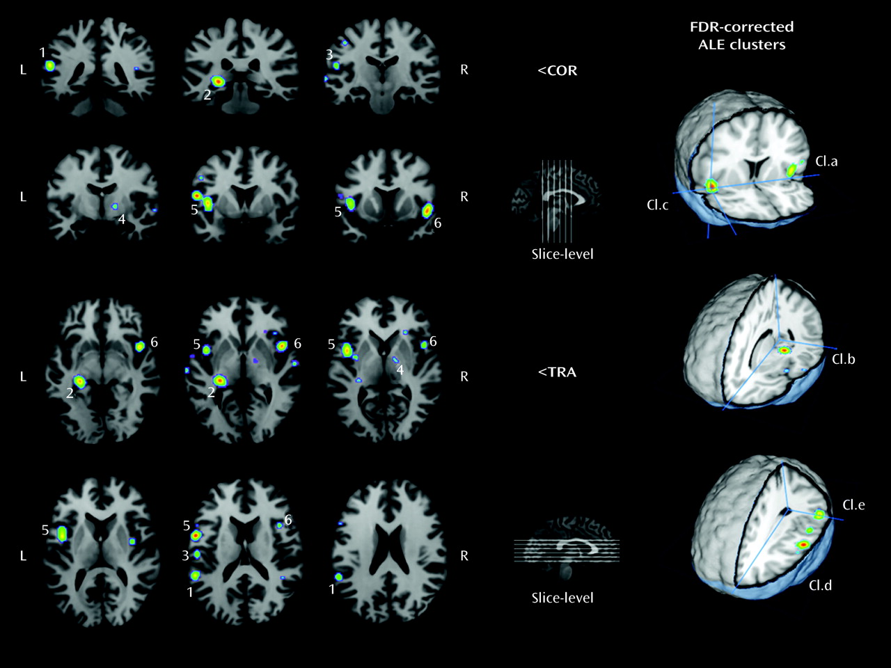
This meta-analysis first identified a widespread set of dysfunctional language-related areas that present increased activity when patients experience AVHs. The largest clusters were identified in cortical areas involved in speech generation. The left pars opercularis (Brodmann's area 44), located in the inferior frontal gyrus, is part of Broca's area. This region is bounded by the premotor precentral gyrus (Brodmann's area 6) to its posterior and in depth by the anterior insula (Brodmann's area 13). Overall, the insula constitutes one element of the homologous region of Broca's area on the right side of the brain. Lesion and functional imaging studies have revealed the critical involvement of this extended Broca's convolution in syntactic processing (7577) and also during verbal imagery (78). In up to 90% of healthy right-handed subjects, this region shows strong functional left lateralization (79). Some authors have suggested that reduced language lateralization in the frontal lobes of schizophrenia patients could account for the emergence of AVHs (8082). Our meta-analytic data, which show significant activations during AVHs within the left Brod-mann's area 44 and the right Brodmann's area 13, fully support this theory.
The present meta-analysis also showed increased likelihoods within the left middle (Brodmann's area 21) and superior temporal gyri (Brodmann's area 22), which compose the associative auditory cortices. The inferior parietal lobule (Brodmann's area 40), part of Wernicke's convolution on the left side of the brain, was also identified as a region of increased activity and is notably involved in speech processing (83). Interestingly, structural imaging studies report a correlation between the severity of hallucinations and gray matter volume reductions within the left superior (84, 85) and middle temporal gyri (86). The potential involvement of these language-related perceptual and motor areas in AVHs is reinforced by research using gyrification and diffusion measures. First, abnormalities in cortical gyrification of the bilateral superior temporal sulci, the left middle frontal sulcus, and the left sylvian fissure (Broca's area) have been seen in chronic hallucinators suffering from schizophrenia, which suggests a neurodevelopmental susceptibility to AVHs in schizophrenia populations (87). Second, the measured fractional anisotropy within the arcuate fasciculus, a white matter bundle connecting Broca's and Wernicke's regions (88), was significantly increased in a subgroup of patients experiencing frequent AVHs relative to nonhallucinators (89, 90), which speaks in favor of a fronto-temporal disconnectivity.
Aside from the language network, other regions of significance were identified by our meta-analytic procedure. Activation of the left hippocampus/parahippocampal region (Brodmann's area 27) was apparent. This region is known to be involved in the formation of new memories about autobiographical events and conscious recollection (91), and it connects widely distributed association cortices, including the language areas. Moreover, severe damage to this structure usually results in retrograde amnesia, whereas its ictal stimulation may cause experiential hallucinations, such as those experienced during the dreamy state phenomenon (92). Although abnormalities of this region have been frequently reported in schizophrenia independently of AVHs (93), some authors have proposed that hippocampal dysfunction might alter dopamine release in the basal ganglia, potentially causing positive psychotic symptoms (94). Interestingly, other studies investigating cortical activations prior to the onset of AVHs have reported deactivation of the parahippocampal region before symptom onset as opposed to activation during hallucinations (60, 95). Such parahippocampal deactivation has been shown to be associated with the memory recollection process (96) and could be involved in the inadequate trigger of activations in language-related areas responsible for the hallucinatory experience. Taken together, these data support models of abnormal remembered episodic memories of speech and suggest the plausible involvement of memory retrieval during AVHs (6).
The right basal ganglia focus deserves further attention. Strangely, this activation is rarely discussed in the source studies. First, it seems unlikely that this cluster could be related to motor control because more than 80% of the patients involved in studies using a button press paradigm signaled the occurrence of AVHs with their right hand and no complementary activation was measured within the precentral gyrus or the cerebellar cortex. Second, a dysregulation of dopamine systems within thalamo-cortico-striatal circuitry is regularly proposed to account for delusions and hallucinations. However, the medial globus pallidus, which has been shown to be active during AVHs, is devoid of dopaminergic afferents (97). Finally, the main contributors to this cluster are PET studies (6, 13). We argue that fMRI might be less sensitive than PET studies for detecting activation within this area. A loss in the blood-oxygen-level-dependent signal could be a consequence of the particular vascular system of the globus pallidus compared with the rest of the capillary bed, or it could be a consequence of an elevated tissue iron level (97). These data need to be considered in the design of future studies, since explorations of the subcortical structures involved in AVHs move beyond a strict dopamine hypothesis.
Altogether, the findings of the present meta-analysis allow us to discuss the three main pathophysiological -theories of AVHs that were previously mentioned. Our results fully support the following two hypotheses: 1) aberrant activations within sensorimotor cortices and 2) a dysfunction of the verbal memory system during the emergence of AVHs. Therefore, we postulate that abnormal memory retrieval involving the hippocampal/parahippocampal region could trigger dispersed neocortical storage sites, notably those within the language areas, which are responsible for the involuntary emergence of AVHs. Interestingly, in psychotic patients experiencing AVHs, reduced connectivity has been found in neural substrates of episodic verbal memory (hippocampus) and central auditory processing (98), in accordance with the present meta-analytic findings. A third model suggests that a potential underlying mechanism for AVHs could be a reduced ability to attribute the source of speech (5, 43, 44, 51, 99, 100). The present review did not find evidence for activations of the cortical midline structures commonly involved in source attribution during AVHs. However, in our view, this does not rule out the mechanism of misperceptions of unbidden thoughts as external speech in hallucinators. Such cognitive dysfunctions in patients with AVHs could be present independent of the hallucinatory state. We believe that further insight in the validation of the misattribution model could be provided by another quantitative review of brain imaging trait studies that compare patients with and without hallucinations during verbal monitoring tasks.
We are aware that this study has some limitations. First, a minimum of 20 to 100 coordinates are usually needed to produce a robust meta-analysis map, depending on the complexity of the underlying cognitive processing. Even though the number of foci included in the present research was substantial (129 foci of interest), we were only able to integrate a modest number of articles and few high-quality studies that did not report stereotaxic coordinates were excluded (9, 6668), limiting the power of our analysis to detect more subtle activations. Furthermore, we were not able to control for medication status or patient age across studies as covariates of interest. Incorporation of additional weighting factors for the acquisition methods (magnetic resonance field strengths, etc.) and the intensity scores of activation for all clusters will be incorporated into an upcoming version of the activation likelihood estimation algorithm (72) and should be considered in future meta-analytic studies of AVHs. Nevertheless, the goal of the present research was to clearly define the spatial localizations of the most frequently replicated activations during AVHs rather than to estimate their magnitudes. Despite the cited shortcomings inherent to the activation likelihood estimation method, our data provide strong evidence for concomitant activations in brain areas involved at different levels of complexity in patients' brain architectures. Thus, they allow us to propose an original view of the pathophysiology of AVHs, integrating previous hypotheses that focus on aberrant activations resulting from disturbed interactions within language and verbal-memory networks. Thus, in our view, the experience of voices can be understood as the combination of distinct mechanisms. First, unbidden auditory memories activate verbal areas of the auditory cortex, making the experience sensory. Then, because the self-tag is missing from these sensory experiences, the phenomenon is experienced as voices. These results also invite new theoretical perspectives because although hyperactivation of the primary sensory cortex (Heschl's gyrus) was identified in some reports (7, 8, 14), results for this area were not significant after quantitative meta-analysis, and thus the area does not seem necessary for the emergence of AVHs. Even if the present data do not permit drawing conclusions about causality, such a hypothesis is consistent with previous reports about the generation of inner speech or auditory verbal imagery, in which Heschl's gyrus is not activated (39). We postulate that activation of the primary auditory cortex, sometimes measured during AVHs, might result from the backpropagation of activity in associative cortices. It is possible that increased severity, vividness, or external spatial voice localizations may be related to propagation of such activation to the brain areas directly receiving sensory inputs. Further research will be needed to confirm this last proposal.

Acknowledgments

The authors thank Dr. Jack Foucher for advice on a preliminary version of this article.

Footnote

Received Oct. 24, 2009; revisions received May 4 and July 12, 2010; accepted July 30, 2010

References

1.
Andreasen NC, Flaum M: Schizophrenia: the characteristic symptoms. Schizophr Bull 1991; 17:27–49
2.
Shergill SS, Murray RM, McGuire PK: Auditory hallucinations: a review of psychological treatments. Schizophr Res 1998; 32:137–150
3.
Aleman A, Larøi F: Hallucinations: The Science of Idiosyncratic Perception. Washington, DC, American Psychological Association, 2008
4.
Penfield W: Some mechanisms of consciousness discovered during electrical stimulation of the brain. Proc Natl Acad Sci U S A 1958; 44:51–66
5.
McGuire PK, Silbersweig DA, Wright I, Murray RM, David AS, Frackowiak RS, Frith CD: Abnormal monitoring of inner speech: a physiological basis for auditory hallucinations. Lancet 1995; 346:596–600
6.
Copolov DL, Seal ML, Maruff P, Ulusoy R, Wong MT, Tochon-Danguy HJ, Egan GF: Cortical activation associated with the experience of auditory hallucinations and perception of human speech in schizophrenia: a PET correlation study. Psychiatry Res 2003; 122:139–152
7.
Dierks T, Linden DE, Jandl M, Formisano E, Goebel R, Lanfermann H, Singer W: Activation of Heschl's gyrus during auditory hallucinations. Neuron 1999; 22:615–621
8.
van de Ven VG, Formisano E, Roder CH, Prvulovic D, Bittner RA, Dietz MG, Hubl D, Dierks T, Federspiel A, Esposito F, Di Salle F, Jansma B, Goebel R, Linden DE: The spatiotemporal pattern of auditory cortical responses during verbal hallucinations. Neuroimage 2005; 27:644–655
9.
McGuire PK, Shah GM, Murray RM: Increased blood flow in Broca's area during auditory hallucinations in schizophrenia. Lancet 1993; 342:703–706
10.
Sommer IE, Diederen KM, Blom JD, Willems A, Kushan L, -Slotema K, Boks MP, Daalman K, Hoek HW, Neggers SF, Kahn RS: Auditory verbal hallucinations predominantly -activate the right inferior frontal area. Brain 2008; 131(pt 12):3169–3177
11.
Shergill SS, Brammer MJ, Williams SC, Murray RM, McGuire PK: Mapping auditory hallucinations in schizophrenia using functional magnetic resonance imaging. Arch Gen Psychiatry 2000; 57:1033–1038
12.
Jardri R: Functional MRI to define rTMS targets in the case of complex multisensory hallucinations. Eur Arch Psychiatry Clin Neurosci 2009; 259(suppl 1):31
13.
Silbersweig DA, Stern E, Frith C, Cahill C, Holmes A, Grootoonk S, Seaward J, McKenna P, Chua SE, Schnorr L, et al.: A functional neuroanatomy of hallucinations in schizophrenia. Nature 1995; 378:176–179
14.
Lennox BR, Park SB, Medley I, Morris PG, Jones PB: The functional anatomy of auditory hallucinations in schizophrenia. Psychiatry Res 2000; 100:13–20
15.
Shergill SS, Cameron LA, Brammer MJ, Williams SC, Murray RM, McGuire PK: Modality specific neural correlates of auditory and somatic hallucinations. J Neurol Neurosurg Psychiatry 2001; 71:688–690
16.
Formisano E, Esposito F, Di Salle F, Goebel R: Cortex-based independent component analysis of fMRI time series. Magn Reson Imaging 2004; 22:1493–1504
17.
Jardri R, Pins D, Bubrovszky M, Lucas B, Lethuc V, Delmaire C, Vantyghem V, Despretz P, Thomas P: Neural functional organization of hallucinations in schizophrenia: multisensory dissolution of pathological emergence in consciousness. Conscious Cogn 2009; 18:44.9–457
18.
Jardri R, Lucas B, Delevoye-Turrell Y, Delmaire C, Delion P, Thomas P, Goeb JL: An 11-year-old boy with drug-resistant schizophrenia treated with temporo-parietal rTMS. Mol Psychiatry 2007; 12:320
19.
Eickhoff SB, Laird AR, Grefkes C, Wang LE, Zilles K, Fox PT: Coordinate-based activation likelihood estimation meta-analysis of neuroimaging data: a random-effects approach based on empirical estimates of spatial uncertainty. Hum Brain Mapp 2009; 30:2907–2926
20.
Salimi-Khorshidi G, Smith SM, Keltner JR, Wager TD, Nichols TE: Meta-analysis of neuroimaging data: a comparison of image-based and coordinate-based pooling of studies. Neuroimage 2009; 45:810–823
21.
Wager TD, Lindquist M, Kaplan L: Meta-analysis of functional neuroimaging data: current and future directions. Soc Cogn Affect Neurosci 2007; 2:150–158
22.
Allen P, Larøi F, McGuire PK, Aleman A: The hallucinating brain: a review of structural and functional neuroimaging -studies of hallucinations. Neurosci Biobehav Rev 2008; 32:175–191
23.
Weiss AP, Heckers S: Neuroimaging of hallucinations: a review of the literature. Psychiatry Res 1999; 92:61–74
24.
Ffytche DH, Howard RJ, Brammer MJ, David A, Woodruff P, Williams S: The anatomy of conscious vision: an fMRI study of visual hallucinations. Nat Neurosci 1998; 1:738–742
25.
Szechtman H, Woody E, Bowers KS, Nahmias C: Where the imaginal appears real: a positron emission tomography study of auditory hallucinations. Proc Natl Acad Sci U S A 1998; 95:1956–1960
26.
Kasai K, Asada T, Yumoto M, Takeya J, Matsuda H: Evidence for functional abnormality in the right auditory cortex during musical hallucinations. Lancet 1999; 354:1703–1704
27.
Izumi Y, Terao T, Ishino Y, Nakamura J: Differences in regional cerebral blood flow during musical and verbal hallucinations. Psychiatry Res 2002; 116:119–123
28.
Mori T, Ikeda M, Fukuhara R, Sugawara Y, Nakata S, Matsumoto N, Nestor PJ, Tanabe H: Regional cerebral blood flow change in a case of Alzheimer's disease with musical hallucinations. Eur Arch Psychiatry Clin Neurosci 2006; 256:236–239
29.
Holroyd S, Wooten GF: Preliminary fMRI evidence of visual system dysfunction in Parkinson's disease patients with visual hallucinations. J Neuropsychiatry Clin Neurosci 2006; 18:402–404
30.
De Haan EH, Nys GM, van Zandvoort MJ, Ramsey NF: The physiological basis of visual hallucinations after damage to the primary visual cortex. Neuroreport 2007; 18:1177–1180
31.
Ramirez-Ruiz B, Marti MJ, Tolosa E, Falcon C, Bargallo N, Valldeoriola F, Junqué C: Brain response to complex visual stimuli in Parkinson's patients with hallucinations: a functional magnetic resonance imaging study. Mov Disord 2008; 23:2335–2343
32.
Perneczky R, Drzezga A, Boecker H, Forstl H, Kurz A, Haussermann P: Cerebral metabolic dysfunction in patients with dementia with Lewy bodies and visual hallucinations. Dement Geriatr Cogn Disord 2008; 25:531–538
33.
Matsui H, Nishinaka K, Miyoshi T, Hara N, Oda M, Kubori T, Udaka F: Thalamic hyperperfusion in verbal hallucination of parkinsonian patients. Intern Med 2007; 46:1765–1769
34.
Whalley HC, Gountouna VE, Hall J, McIntosh A, Whyte MC, Simonotto E, Job DE, Owens DG, Johnstone EC, Lawrie SM: Correlations between fMRI activation and individual psychotic symptoms in un-medicated subjects at high genetic risk of schizophrenia. BMC Psychiatry 2007; 7:61
35.
Wible CG, Lee K, Molina I, Hashimoto R, Preus AP, Roach BJ, Ford JM, Mathalon DH, McCarthey G, Turner JA, Potkin SG, O'Leary D, Belger A, Diaz M, Voyvodic J, Brown GG, Notestine R, Greve D, Lauriello J, FBIRN: fMRI activity correlated with auditory hallucinations during performance of a working memory task: data from the FBIRN Consortium Study. Schizophr Bull 2009; 35:47–57
36.
Bentaleb LA, Beauregard M, Liddle P, Stip E: Cerebral activity associated with auditory verbal hallucinations: a functional magnetic resonance imaging case study. J Psychiatry Neurosci 2002; 27:110–115
37.
Howard R, Williams S, Bullmore E, Brammer M, Mellers J, Woodruff P, David A: Cortical response to exogenous visual stimulation during visual hallucinations. Lancet 1995; 345:70
38.
David AS, Woodruff PW, Howard RJ, Mellers JD, Brammer M, Bullmore E: Auditory hallucinations inhibit exogenous activation of auditory association cortex. Neuroreport 1996; 7:932–936
39.
McGuire PK, Silbersweig DA, Wright I, Murray RM, Frackowiak RS, Frith CD: The neural correlates of inner speech and auditory verbal imagery in schizophrenia: relationship to auditory verbal hallucinations. Br J Psychiatry 1996; 169:148–159
40.
McGuire PK, Paulesu E, Frackowiak RS, Frith CD: Brain activity during stimulus independent thought. Neuroreport 1996; 7:2095–2099
41.
Woodruff PW, Wright IC, Bullmore ET, Brammer M, Howard RJ, Williams SC, Shapleske J, Rossell S, David AS, McGuire PK, Murray RM: Auditory hallucinations and the temporal cortical response to speech in schizophrenia: a functional magnetic resonance imaging study. Am J Psychiatry 1997; 154:1676–1682
42.
Shergill SS, Bullmore E, Simmons A, Murray R, McGuire P: Functional anatomy of auditory verbal imagery in schizophrenic patients with auditory hallucinations. Am J Psychiatry 2000; 157:1691–1693
43.
Shergill SS, Brammer MJ, Fukuda R, Williams SC, Murray RM, McGuire PK: Engagement of brain areas implicated in processing inner speech in people with auditory hallucinations. Br J Psychiatry 2003; 182:525–531
44.
Fu CH, Vythelingum GN, Brammer MJ, Williams SC, Amaro E, Andrew CM, Yágüez L, van Haren NE, Matsumoto K, McGuire PK: An fMRI study of verbal self-monitoring: neural correlates of auditory verbal feedback. Cereb Cortex 2006; 16:969–977
45.
Plaze M, Bartres-Faz D, Martinot JL, Januel D, Bellivier F, De Beaurepaire R, Chanraud S, Andoh J, Lefaucheur JP, Artiges E, Pallier C, Paillère-Martinot ML: Left superior temporal gyrus activation during sentence perception negatively correlates with auditory hallucination severity in schizophrenia patients. Schizophr Res 2006; 87:109–115
46.
Aleman A, Formisano E, Koppenhagen H, Hagoort P, de Haan EH, Kahn RS: The functional neuroanatomy of metrical stress evaluation of perceived and imagined spoken words. Cereb Cortex 2005; 15:221–228
47.
Allen P, Amaro E, Fu CH, Williams SC, Brammer MJ, Johns LC, McGuire PK: Neural correlates of the misattribution of speech in schizophrenia. Br J Psychiatry 2007; 190:162–169
48.
Stephane M, Hagen MC, Lee JT, Uecker J, Pardo PJ, Kuskowski MA, Pardo JV: About the mechanisms of auditory verbal hallucinations: a positron emission tomographic study. J Psychiatry Neurosci 2006; 31:396–405
49.
Hashimoto RI, Lee K, Preus A, McCarley RW, Wible CG: An fMRI study of functional abnormalities in the verbal working memory system and the relationship to clinical symptoms in chronic schizophrenia. Cereb Cortex 2010; 20:46–60
50.
Jardri R, Delevoye-Turrell Y, Lucas B, Pins D, Bulot V, Delmaire C, Thomas P, Delion P, Goeb JL: Clinical practice of rTMS reveals a functional dissociation between agency and hallucinations in schizophrenia. Neuropsychologia 2009; 47:132–138
51.
Kumari V, Fannon D, Ffytche DH, Raveendran V, Antonova E, Premkumar P, Cooke MA, Anilkumar AP, Williams SC, Andrew C, Johns LC, Fu CH, McGuire PK, Kuipers E: Functional MRI of verbal self-monitoring in schizophrenia: performance and illness-specific effects. Schizophr Bull 2010; 36:740–755
52.
Ford JM, Roach BJ, Jorgensen KW, Turner JA, Brown GG, Notestine R, Bischoff-Grethe A, Greve D, Wible C, Lauriello J, Belger A, Mueller BA, Calhoun V, Preda A, Keator D, O'Leary DS, Lim KO, Glover G, Potkin SG, FBIRN, Mathalon DH: Tuning in to the voices: a multisite fMRI study of auditory hallucinations. Schizophr Bull 2009; 35:58–66
53.
Kang JI, Kim JJ, Seok JH, Chun JW, Lee SK, Park HJ: Abnormal brain response during the auditory emotional processing in schizophrenic patients with chronic auditory hallucinations. Schizophr Res 2009; 107:83–91
54.
Brune M, Lissek S, Fuchs N, Witthaus H, Peters S, Nicolas V, Juckel G, Tegenthoff M: An fMRI study of theory of mind in schizophrenic patients with “passivity” symptoms. Neuropsychologia 2008; 46:1992–2001
55.
Fu CH, Brammer MJ, Yaguez L, Allen P, Matsumoto K, Johns L, Weinstein S, Borgwardt S, Broome M, van Haren N, McGuire PK: Increased superior temporal activation associated with external misattributions of self-generated speech in schizophrenia. Schizophr Res 2008; 100:361–363
56.
Zhang Z, Shi J, Yuan Y, Hao G, Yao Z, Chen N: Relationship of auditory verbal hallucinations with cerebral asymmetry in patients with schizophrenia: an event-related fMRI study. J Psychiatr Res 2008; 42:477–486
57.
Hoffman RE, Hampson M, Wu K, Anderson AW, Gore JC, Buchanan RJ, Constable RT, Hawkins KA, Sahay N, Krystal JH: Probing the pathophysiology of auditory/verbal hallucinations by combining functional magnetic resonance imaging and transcranial magnetic stimulation. Cereb Cortex 2007; 17:2733–2743
58.
Kopecek M, Spaniel F, Novak T, Tislerova B, Belohlavek O, -Horacek J: 18FDG PET in hallucinating and non-hallucinating patients. Neuro Endocrinol Lett 2007; 28:53–59
59.
Jardri R, Pins D, Thomas P: A case of fMRI-guided rTMS treatment of coenesthetic hallucinations. Am J Psychiatry 2008; 165:1490–1491
60.
Hoffman RE, Anderson AW, Varanko M, Gore JC, Hampson M: Time course of regional brain activation associated with onset of auditory/verbal hallucinations. Br J Psychiatry 2008; 193:424–425
61.
Shergill SS, Brammer MJ, Amaro E, Williams SC, Murray RM, McGuire PK: Temporal course of auditory hallucinations. Br J Psychiatry 2004; 185:516–517
62.
Lennox BR, Park SB, Jones PB, Morris PG: Spatial and temporal mapping of neural activity associated with auditory hallucinations. Lancet 1999; 353:644
63.
Liddle PF, Friston KJ, Frith CD, Hirsch SR, Jones T, Frackowiak RS: Patterns of cerebral blood flow in schizophrenia. Br J Psychiatry 1992; 160:179–186
64.
Gur RE, Mozley PD, Resnick SM, Mozley LH, Shtasel DL, Gallacher F, Arnold SE, Karp JS, Alavi A, Reivich M, et al.: Resting cerebral glucose metabolism in first-episode and previously treated patients with schizophrenia relates to clinical features. Arch Gen Psychiatry 1995; 52:657–667
65.
Lahti AC, Weiler MA, Holcomb HH, Tamminga CA, Carpenter WT, McMahon R: Correlations between rCBF and symptoms in two independent cohorts of drug-free patients with schizophrenia. Neuropsychopharmacology 2006; 31:221–230
66.
Suzuki M, Yuasa S, Minabe Y, Murata M, Kurachi M: Left superior temporal blood flow increases in schizophrenic and schizophreniform patients with auditory hallucination: a longitudinal case study using 123I-IMP SPECT. Eur Arch Psychiatry Clin Neurosci 1993; 242:257–261
67.
Woodruff P, Brammer M, Mellers J, Wright I, Bullmore E, -Williams S: Auditory hallucinations and perception of external speech. Lancet 1995; 346:1035
68.
Parellada E, Lomena F, Font M, Pareto D, Gutierrez F, Simo M, Fernández-Egea E, Pavia J, Ros D, Bernardo M: Fluordeoxyglucose-PET study in first-episode schizophrenic patients during the hallucinatory state, after remission and during linguistic-auditory activation. Nucl Med Commun 2008; 29:894–900
69.
Talairach J, Tournoux P: A Coplanar Stereotactic Atlas of the -Human Brain. New York, Thieme Medical, 1988
70.
Lancaster JL, Tordesillas-Gutierrez D, Martinez M, Salinas F, -Evans A, Zilles K, Mazziotta JC, Fox PT: Bias between MNI and Talairach coordinates analyzed using the ICBM-152 brain template. Hum Brain Mapp 2007; 28:1194–1205
71.
Turkeltaub PE, Eden GF, Jones KM, Zeffiro TA: Meta-analysis of the functional neuroanatomy of single-word reading: method and validation. Neuroimage 2002; 16:765–780
72.
Laird AR, Fox PM, Price CJ, Glahn DC, Uecker AM, Lancaster JL, Turkeltaub PE, Kochunov P, Fox PT: ALE meta-analysis: controlling the false discovery rate and performing statistical contrasts. Hum Brain Mapp 2005; 25:155–164
73.
Genovese CR, Lazar NA, Nichols T: Thresholding of statistical maps in functional neuroimaging using the false discovery rate. Neuroimage 2002; 15:870–878
74.
Kochunov P, Lancaster J, Thompson P, Toga AW, Brewer P, Hardies J, Fox P: An optimized individual target brain in the Talairach coordinate system. Neuroimage 2002; 17:922–927
75.
Stromswold K, Caplan D, Alpert N, Rauch S: Localization of syntactic comprehension by positron emission tomography. Brain Lang 1996; 52:452–473
76.
Fiebach CJ, Schlesewsky M, Lohmann G, von Cramon DY, Friederici AD: Revisiting the role of Broca's area in sentence processing: syntactic integration versus syntactic working memory. Hum Brain Mapp 2005; 24:79–91
77.
Dronkers NF: A new brain region for coordinating speech articulation. Nature 1996; 384:159–161
78.
Shergill SS, Bullmore ET, Brammer MJ, Williams SC, Murray RM, McGuire PK: A functional study of auditory verbal imagery. Psychol Med 2001; 31:241–253
79.
Knecht S, Drager B, Deppe M, Bobe L, Lohmann H, Floel A, Ringelstein EB, Henningsen H: Handedness and hemispheric language dominance in healthy humans. Brain 2000; 123:2512–2518
80.
Sommer IE, Ramsey NF, Kahn RS: Language lateralization in schizophrenia: an fMRI study. Schizophr Res 2001; 52:57–67
81.
Sommer IE, Diederen KM: Language production in the non-dominant hemisphere as a potential source of auditory verbal hallucinations. Brain 2009; 132:e124
82.
Mitchell RL, Crow TJ: Right hemisphere language functions and schizophrenia: The forgotten hemisphere? Brain 2005; 128:963–978
83.
Binder JR, Frost JA, Hammeke TA, Cox RW, Rao SM, Prieto T: Human brain language areas identified by functional magnetic resonance imaging. J Neurosci 1997; 17:353–362
84.
Barta PE, Pearlson GD, Powers RE, Richards SS, Tune LE: Auditory hallucinations and smaller superior temporal gyral volume in schizophrenia. Am J Psychiatry 1990; 147:1457–1462
85.
Flaum M, O'Leary DS, Swayze VW 2nd, Miller DD, Arndt S, -Andreasen NC: Symptom dimensions and brain morphology in schizophrenia and related psychotic disorders. J Psychiatr Res 1995; 29:261–276
86.
Onitsuka T, Shenton ME, Salisbury DF, Dickey CC, Kasai K, Toner SK, Frumin M, Kikinis R, Jolesz FA, McCarley RW: Middle and inferior temporal gyrus gray matter volume abnormalities in chronic schizophrenia: an MRI study. Am J Psychiatry 2004; 161:1603–1611
87.
Cachia A, Paillere-Martinot ML, Galinowski A, Januel D, de -Beaurepaire R, Bellivier F, Artiges E, Andoh J, Bartrés-Faz D, Duchesnay E, Rivière D, Plaze M, Mangin JF, Martinot JL: Cortical folding abnormalities in schizophrenia patients with resistant auditory hallucinations. Neuroimage 2008; 39:927–935
88.
Catani M, Jones DK, Ffytche DH: Perisylvian language networks of the human brain. Ann Neurol 2005; 57:8–16
89.
Hubl D, Koenig T, Strik W, Federspiel A, Kreis R, Boesch C, Maier SE, Schroth G, Lovblad K, Dierks T: Pathways that make voices: white matter changes in auditory hallucinations. Arch Gen Psychiatry 2004; 61:658–668
90.
Shergill SS, Kanaan RA, Chitnis XA, O'Daly O, Jones DK, Frangou S, Williams SC, Howard RJ, Barker GJ, Murray RM, McGuire P: A diffusion tensor imaging study of fasciculi in schizophrenia. Am J Psychiatry 2007; 164:467–473
91.
Squire LR, Schacter DL: The Neuropsychology of Memory. New York, Guilford Press, 2002
92.
Vignal JP, Maillard L, McGonigal A, Chauvel P: The dreamy state: hallucinations of autobiographic memory evoked by temporal lobe stimulations and seizures. Brain 2007; 130:88–99
93.
Harrison PJ: The hippocampus in schizophrenia: a review of the neuropathological evidence and its pathophysiological implications. Psychopharmacology (Berl) 2004; 174:151–162
94.
Goto Y, Grace AA: Limbic and cortical information processing in the nucleus accumbens. Trends Neurosci 2008; 31:552–558
95.
Diederen KM, Neggers SF, Daalman K, Blom JD, Goekoop R, Kahn RS, Sommer IE: Deactivation of the parahippocampal gyrus preceding auditory hallucinations in schizophrenia. Am J Psychiatry 2010; 167:427–435
96.
Weis S, Klaver P, Reul J, Elger CE, Fernandez G: Neural correlates of successful declarative memory formation and retrieval: the anatomical overlap. Cortex 2004; 40:200–202
97.
Steiner H, Tseng KY: Handbook of Basal Ganglia Structure and Function. San Diego, Academic Press, 2010
98.
Sergejew AA, Copolov D, Egan G: Auditory hallucinations in psychosis: dysfunctional cortical connectivity in neural substrates of central auditory processing and episodic verbal memory. J EEG Clin Neurosci 2005; 36:52
99.
Ford JM, Mathalon DH, Whitfield S, Faustman WO, Roth WT: Reduced communication between frontal and temporal lobes during talking in schizophrenia. Biol Psychiatry 2002; 51:485–492
100.
Mechelli A, Allen P, Amaro E Jr, Fu CH, Williams SC, Brammer MJ, Johns LC, McGuire PK: Misattribution of speech and impaired connectivity in patients with auditory verbal hallucinations. Hum Brain Mapp 2007; 28:1213–1222

Information & Authors

Information

Published In

Go to American Journal of Psychiatry
American Journal of Psychiatry
Pages: 73 - 81
PubMed: 20952459

History

Received: 24 October 2009
Revision received: 4 May 2010
Revision received: 12 July 2010
Accepted: 30 July 2010
Published online: 1 January 2011
Published in print: January 2011

Authors

Affiliations

Renaud Jardri, M.D., Ph.D.
From the Laboratory of Functional Neurosciences and Pathologies, CNRS, University Lille North of France, Lille, France; the Pediatric Psychiatry Department, Fontan Hospital, UMC Lille, France; and the Psychiatry Department, Fontan Hospital, UMC Lille, France.
Alexandre Pouchet, M.D.
From the Laboratory of Functional Neurosciences and Pathologies, CNRS, University Lille North of France, Lille, France; the Pediatric Psychiatry Department, Fontan Hospital, UMC Lille, France; and the Psychiatry Department, Fontan Hospital, UMC Lille, France.
Delphine Pins, Ph.D.
From the Laboratory of Functional Neurosciences and Pathologies, CNRS, University Lille North of France, Lille, France; the Pediatric Psychiatry Department, Fontan Hospital, UMC Lille, France; and the Psychiatry Department, Fontan Hospital, UMC Lille, France.
Pierre Thomas, M.D., Ph.D.
From the Laboratory of Functional Neurosciences and Pathologies, CNRS, University Lille North of France, Lille, France; the Pediatric Psychiatry Department, Fontan Hospital, UMC Lille, France; and the Psychiatry Department, Fontan Hospital, UMC Lille, France.

Notes

Address correspondence and reprint requests to Dr. Jardri, Service de Pédopsychiatrie, Hôpital Fontan, CHRU de Lille, F-59037, Lille cedex, France; [email protected] (e-mail).

Funding Information

The authors report no financial relationships with commercial interests.

Metrics & Citations

Metrics

Citations

Export Citations

If you have the appropriate software installed, you can download article citation data to the citation manager of your choice. Simply select your manager software from the list below and click Download.

For more information or tips please see 'Downloading to a citation manager' in the Help menu.

Format
Citation style
Style
Copy to clipboard

There are no citations for this item

View Options

View options

PDF/ePub

View PDF/ePub

Get Access

Login options

Already a subscriber? Access your subscription through your login credentials or your institution for full access to this article.

Personal login Institutional Login Open Athens login
Purchase Options

Purchase this article to access the full text.

PPV Articles - American Journal of Psychiatry

PPV Articles - American Journal of Psychiatry

Not a subscriber?

Subscribe Now / Learn More

PsychiatryOnline subscription options offer access to the DSM-5-TR® library, books, journals, CME, and patient resources. This all-in-one virtual library provides psychiatrists and mental health professionals with key resources for diagnosis, treatment, research, and professional development.

Need more help? PsychiatryOnline Customer Service may be reached by emailing [email protected] or by calling 800-368-5777 (in the U.S.) or 703-907-7322 (outside the U.S.).

Media

Figures

Other

Tables

Share

Share

Share article link

Share