Full access
New Research
Published Online: 1 February 2011

Association of Frontal and Posterior Cortical Gray Matter Volume With Time to Alcohol Relapse: A Prospective Study

Abstract

Objective:

Alcoholism is associated with gray matter volume deficits in frontal and other brain regions. Whether persistent brain volume deficits in abstinence are predictive of subsequent time to alcohol relapse has not been established. The authors measured gray matter volumes in healthy volunteers and in a sample of treatment-engaged, alcohol-dependent patients after 1 month of abstinence and assessed whether smaller frontal gray matter volume was predictive of subsequent alcohol relapse outcomes.

Method:

Forty-five abstinent alcohol-dependent patients in treatment and 50 healthy comparison subjects were scanned once using high-resolution (T1-weighted) structural MRI, and voxel-based morphometry was used to assess regional brain volume differences between the groups. A prospective study design was used to assess alcohol relapse in the alcohol-dependent group for 90 days after discharge from 6 weeks of inpatient treatment.

Results:

Significantly smaller gray matter volume in alcohol-dependent patients relative to comparison subjects was seen in three regions: the medial frontal cortex, the right lateral prefrontal cortex, and a posterior region surrounding the parietal-occipital sulcus. Smaller medial frontal and parietal-occipital gray matter volumes were each predictive of shorter time to any alcohol use and to heavy drinking relapse.

Conclusions:

These findings are the first to demonstrate that gray matter volume deficits in specific medial frontal and posterior parietal-occipital brain regions are predictive of an earlier return to alcohol use and relapse risk, suggesting a significant role for gray matter atrophy in poor clinical outcomes in alcoholism. Extent of gray matter volume deficits in these regions could serve as useful neural markers of relapse risk and alcoholism treatment outcome.
The mechanisms underlying the chronic, relapsing nature of alcoholism are not well understood. Several medications are efficacious in the treatment of alcoholism and in initiating abstinence (1), but more than two-thirds of alcohol-dependent individuals return to drinking within weeks or months of initiating recovery (2, 3). While psychosocial factors contribute to relapse susceptibility (2), there is increasing interest in the neurobiological markers that account for the chronic, relapsing nature of the illness.
Previous evidence clearly indicates chronic alcohol-related neurotoxicity and brain atrophy. MRI studies examining lobular volumes or regions of interest find smaller volumes most consistently in the cortical gray matter of the frontal lobes or fronto-temporal region (46) but also in posterior cortical regions (7, 8), subcortical regions (7), and the cerebellum (9). Whole-brain voxel-wise analyses support the existence of gray matter deficits in each of the cortical lobes (1013), the thalamus, and the cerebellum (11, 12). More severe gray matter deficits have been reported in alcoholics who relapse than in those who abstain (14, 15). Assessing volumes in specific regions of the amygdala, hippocampus, and ventral striatum after only 1 week of alcohol abstinence, Wrase and colleagues (16) recently reported smaller amygdala volumes in relapsed compared with abstinent alcoholics. While this previous research suggests a role for alcohol-related brain atrophy in the chronic, relapsing nature of the illness, no previous study has assessed whether brain volumes during sustained alcohol abstinence are predictive of time to alcohol relapse.
In this study, building on previous research, we assessed gray matter volumes using automated segmentation and registration of high-resolution MR structural brain images; a whole-brain analysis implemented voxel-based morphometry to assess gray matter volume differences of alcohol-dependent men and women receiving inpatient treatment after 1 month of abstinence, as compared with social-drinking healthy comparison subjects. All alcohol-dependent patients were then followed prospectively with face-to-face interviews at 14, 30, and 90 days after discharge from inpatient treatment to assess alcohol relapse outcomes. We expected to find gray matter volume deficits in the alcohol-dependent relative to the healthy -comparison group and an association between gray matter volume deficits and prior history of chronic alcohol abuse. Furthermore, because medial frontal regions play an important role in behavioral control and decision-making functions that are likely to contribute to relapse risk (17, 18), we specifically hypothesized that gray matter volume deficits in these regions would be independently predictive of shorter time to alcohol relapse.

Method

Participants

Forty-five treatment-seeking individuals (35 men and 10 women) 18-50 years of age who met DSM-IV criteria for current alcohol dependence were admitted to the Clinical Neuroscience Research Unit of the Connecticut Mental Health Center for 6 weeks of inpatient treatment and participation in research. Fifty healthy comparison subjects (28 men and 22 women 18-50 years of age) were recruited from the community through local advertisements. Comparison subjects consumed up to 25 alcoholic drinks per month (fewer than 7 drinks/week), had negative urine toxicology screens on admission to the study, and did not meet criteria for current or lifetime abuse or dependence for alcohol or any illicit drug. Alcoholic participants did not meet DSM-IV criteria for current dependence on psychoactive substances other than alcohol or nicotine or for any other axis I disorder. Individuals taking any medication for medical or psychiatric problems were excluded from the study, and women were excluded from the study if they were using any hormone-based form of birth control or were peri- or postmenopausal. All participants underwent a complete medical evaluation to ensure good physical health, and the study was approved by the Human Investigation Committee of the Yale University School of Medicine.

Procedures

Alcohol-dependent patients were admitted to the Clinical Neuroscience Research Unit, a locked, smoke-free inpatient treatment research facility with limited access to visitors. All patients participated in specialized substance abuse treatment, and urine and breath tests were conducted at least weekly to ensure continued abstinence. During week 1, patients underwent an initial medical evaluation and provided demographic data and a psychosocial history. During week 2, they were interviewed using the Structured Clinical Interview for DSM-IV (19) to assess psychiatric and substance use diagnoses. Baseline alcohol-related assessments, including alcohol use for the 90 days preceding admission, were also conducted. Patients underwent a single structural MRI scan after 1 month of alcohol abstinence during their inpatient treatment stay (the mean time abstinent at the time of MRI scanning was 35.12 days [SD=7.3] for the alcohol-dependent group and 44.26 days [SD=30.42] for the comparison group).
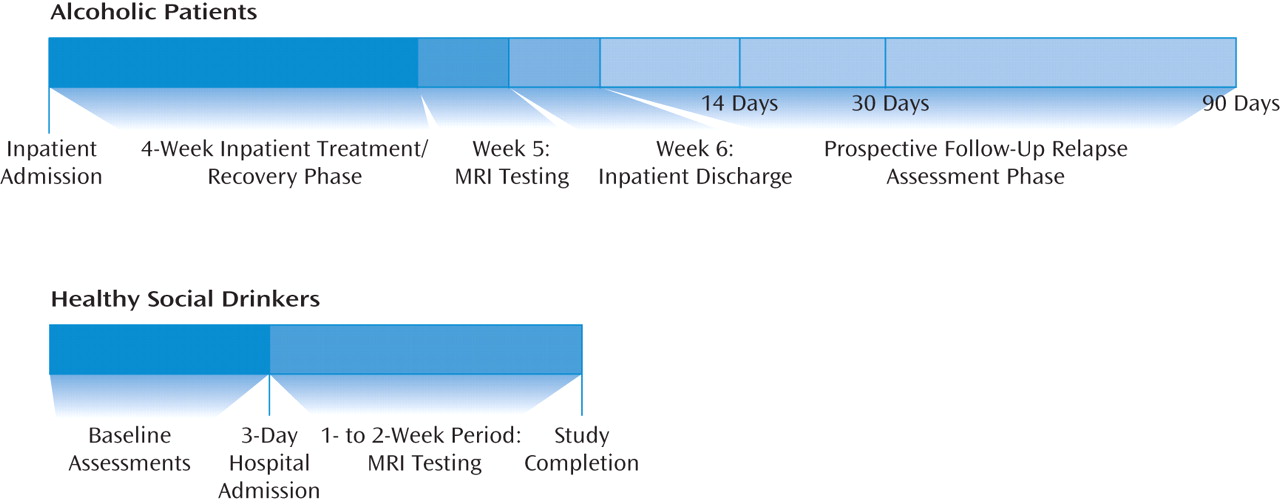
Healthy comparison subjects completed demographic, diagnostic, and alcohol-related assessments in the course of two to three baseline assessment appointments and were then admitted for a 3-day hospital stay at the Yale General Clinical Research Center at Yale-New Haven Hospital for participation in a laboratory study. Within 2 weeks after discharge, they underwent a single structural MRI scan (Figure 1).
FIGURE 1. Schedule of Procedures for Alcohol-Dependent Patients and Healthy Comparison Subjects in a Study of Gray Matter Volume and Time to Alcohol Relapsea
a Alcohol-dependent patients had been in inpatient treatment and abstinent for at least 1 month at the time of MRI scanning. Comparison subjects were assessed within 2 weeks after a research hospital stay. Alcohol-dependent patients were prospectively followed after treatment and participated in face-to-face interviews 14, 30, and 90 days after discharge to assess alcohol relapse outcomes.

MRI Data Acquisition and Preprocessing

Data for each participant consisted of one sagittally acquired high-resolution T1-weighted magnetization-prepared rapid gradient echo scan (voxel size=1 mm3, field of view=256×256, data acquisition matrix=256×256).
Image segmentation and registration were performed using, respectively, the segmentation algorithm (the New Segment procedure) and the DARTEL registration algorithm incorporated in the current release of Statistical Parametric Mapping (SPM8, Wellcome Department of Cognitive Neurology; http://www.fil.ion.ucl.ac.uk/spm). New Segment is an extension of the default Unified Segmentation algorithm retained from SPM5 (20). Unified Segmentation combines tissue classification, bias correction, and image registration steps in an iterative procedure. New Segment improves the registration model and includes additional tissue probability maps to better model CSF and other nonbrain voxels. DARTEL (21) is a high-dimensional, diffeomorphic registration algorithm that has performed well in a comparison of registration algorithms (22). With DARTEL we expected to increase registration accuracy, thereby increasing sensitivity and improving localization in our comparisons of alcohol-dependent and healthy comparison subjects.
As a first processing step, to provide better initial estimates for the segmentation algorithm, the SPM8 Display function was used to manually set the image space origin to the anterior commissure and align each image with the plane of the anterior and posterior commissures. Default settings were used for segmentation. No skull stripping was applied prior to segmentation.
Because manual segmentation is considered the gold standard for evaluating the quality of automated tissue classification (see, for example, references 23 and 24), the resulting segmentations were validated visually (25, 26). Particular attention was given to the thickness of the cortical surface, which was compared visually to each participant's native space image as well as to a published report of cortical thickness (27). Gray matter segmentation demonstrated appropriate face validity in all images.
The segmentation procedure produced rigid-body aligned tissue segments for each image. The gray and white matter segments were input into DARTEL. DARTEL registers the tissue segments to a template generated from their own mean. Because these images have been warped to the space of the mean image, an additional step normalized the warped images to Montreal Neurological Institute template space. The default parameter settings were also used in the DARTEL registration, which include resampling to 1.5-mm3 voxels to reduce memory demands for the large number of parameters estimated by the registration algorithm. Final outputs were modulated gray matter segments (1.5-mm3 voxels) smoothed using an 8-mm Gaussian filter. Gaussian smoothing reduces the effects of residual misregistration on potential group differences and reduces departures from normality that may occur at some voxels (28).

Prospective Follow-Up Interviews

On discharge from the inpatient unit, all alcohol-dependent participants were given appointments for follow-up interviews 14, 30, and 90 days after discharge. Reminders were sent the week before each appointment. Alcohol use was assessed at each appointment using the timeline follow-back method (29, 30) on the Substance Use Calendar (31), an instrument that has been validated in drug-abusing samples (32) and widely used in assessing alcohol use outcomes in treatment research (33). Urine samples (to assess for the alcohol metabolite ethyl glucuronide, detectable for approximately 80 hours after alcohol consumption) and breath samples were also obtained at each follow-up appointment. In addition, patients provided permission to contact three individuals close to them who had knowledge of their alcohol use, to obtain collateral information in the event that the patients were lost to follow-up. Of the 45 patients who completed the MRI scan, one patient was lost to follow-up and not included in the relapse analyses. Two patients attended the 14- and 30-day follow-up interviews but not the 90-day follow-up, and both had relapsed by day 30. Two patients attended only the 14-day follow-up, and collateral information on alcohol use was obtained for the missing follow-up period. Overall, follow-up rates were 98% at day 14, 93% at day 30, and 89% at day 90.
To capture both initial relapse (any use) and relapse to heavy alcohol use (34), relapse was examined using dichotomous variables of any use (no use versus any use) and heavy alcohol use (5 drinks per occasion for men and 4 drinks per occasion for women versus no return to heavy drinking). Data on alcohol use were computed using the follow-up Substance Use Calendar, collateral information, and urine and breath test results. Based on these data, two relapse outcome measures were computed: time to relapse was defined as the first day of any alcohol use after discharge from inpatient treatment; and time to relapse to heavy drinking was defined as the first day of heavy drinking (5 drinks for men and 4 drinks for women).

Data Analysis

Statistical parametric maps were created in SPM8 to perform between-group comparisons using the smoothed, modulated, normalized gray matter tissue segments output by DARTEL.A general linear model was created with diagnostic group (alcohol-dependent patients or comparison subjects) as the factor of interest. Covariates included age, sex, and estimated total intracranial volume, which was calculated by summing voxel-wise the native space gray, white, and CSF segments for each subject. The analysis compared gray matter volume differences adjusted for age effects, as well as the influence of sex and individual differences on global brain size.
The whole-brain statistical analysis was conducted using random field-based cluster-size testing (35) and family-wise error rate correction for multiple comparisons. The cluster-size test increases sensitivity, relative to voxel intensity-based tests, for spatially extended signals (36), and low thresholds increase the power of these tests for signals of large spatial extent (37). The analysis of cluster extent also better characterizes the spatially distributed nature of group differences within spatially smoothed data (38). Clusters were formed from contiguous voxels exceeding an uncorrected one-tailed threshold of p<0.025. The family-wise error-corrected threshold for significant cluster size was set at a one-tailed p<0.025.
To examine whether specific gray matter volume deficits are predictive of alcohol relapse outcomes, significant clusters of gray matter volume differences between the alcohol-dependent and comparison groups identified in SPM analyses were converted to masks to extract gray matter volume measures for each region of interest using MarsBar (http://marsbar.sourceforge.net). The region-of-interest masks and ImCalc (in SPM8) were used to extract the modulated gray matter voxels within each mask. A MATLAB script (MathWorks, Natick, Mass.; G. Ridgway, 2008, http://www.cs.ucl.ac.uk/staff/g.ridgway/vbm/get_totals.m) totaled gray matter volume (in milliliters) for each subject within each significant cluster.
Baseline demographic characteristics, depressive symptoms, and alcohol use measures were examined for significant associations with specific regions with gray matter volume differences observed in the alcohol-dependent relative to the comparison group and whether they were associated with measures of alcohol relapse. If any demographic characteristic, depressive symptom, or alcohol use measure was significantly associated with the relapse outcome measures, it was included as a covariate to examine the independent association between specific regional gray matter volume measures and the specific alcohol relapse measure. Time to relapse (any alcohol use) and time to relapse to heavy drinking were the two dependent measures examined using Cox proportional hazards regression (39).

Results

There were no differences between the alcohol-dependent and healthy comparison groups in sex, race, and lifetime prevalence of mood and anxiety disorders. The patient group was older on average and had a lower mean IQ than the comparison group (Table 1), and these measures were included as covariates in the group difference analyses and in the relapse prediction analyses. As expected, the patient group also had a significantly higher mean number of days of alcohol use, a greater mean number of drinks per day, and a greater mean number of years of alcohol use.
TABLE 1. Demographic and Clinical Characteristics of Alcohol-Dependent Patients and Healthy Comparison Subjects in a Study of Gray Matter Volume and Time to Alcohol Relapse
CharacteristicAlcohol-Dependent Patients (N=45)Healthy Comparison Subjects (N=50)
 N%N%
Male3577.82856.0
Caucasian3066.72856.0
Lifetime prevalence of psychiatric disorders    
    Any mood disorder613.336.0
    Any anxiety disorder48.924.0
 MeanSDMeanSD
Agea (years)38.207.7431.149.04
Estimated IQb108.48.77115.37.13
Prior alcohol use    
    Baseline days of alcohol use per montha19.389.464.125.33
    Number of drinks per montha390.82286.6010.958.34
    Number of drinks per daya15.548.752.221.38
    Years of alcohol usea18.628.658.368.52
a
Alcoholic patients > comparison subjects, p<0.0001.
b
Alcoholic patients < comparison subjects, p<0.0003.

Group Differences in Voxel-Based Morphometric Analyses

As shown in Table 2 and Figure 2, the alcohol-dependent group showed significantly lower gray matter volume than the comparison group in three regional clusters: the lateral prefrontal cortex, including the right dorsolateral and inferolateral prefrontal cortex (family-wise error p<0.004, cluster size=8,060 voxels); a medial frontal cluster including the dorsal anterior cingulate gyrus, the medial and lateral superior frontal gyrus (including the supplementary and presupplementary areas), the middle frontal gyrus, and the posterior cingulate gyrus (family-wise error p<0.013, cluster size=6,513 voxels); and a parietal and occipital cortex cluster including the precuneus and cuneus regions and the posterior cingulate (family-wise error p<0.009, cluster size=6,886 voxels). Montreal Neurological Institute space coordinates from SPM output were converted to Talairach coordinates (40) using the Nonlinear Yale Montreal Neurological Institute to Talairach Conversion Algorithm (41). Anatomical regions for coordinates within significant clusters were identified using the Automated Anatomy Library (42) for Montreal Neurological Institute space and confirmed using the Talairach Daemon (43) for Talairach space within the WFU (Wake Forest University) PickAtlas (44). No clusters were found for which volumes in the alcohol-dependent group were significantly larger than in the comparison group.
TABLE 2. Regions Within Gray Matter Volume Clusters in Which Alcohol-Dependent Patient Volumes < Healthy Comparison Subject Volumesa
ClusterpbVoxels in ClusterCluster Volume (ml)Brodmann's Areas
Lateral prefrontal: inferior frontal gyrus, middle frontal gyrus, superior frontal gyrus0.0048,06027.2010, 11, 45, 46
Parietal-occipital: precuneus, cuneus, posterior cingulate gyrus0.0096,88623.2417, 18, 19, 23/31
Medial frontal: paracentral lobule, superior frontal gyrus, anterior cingulate gyrus0.0136,51321.985, 6, 24/32
a
Using the Nonlinear Yale MNI to Talairach Conversion Algorithm (41).
b
Family-wise error rate cluster p value.
FIGURE 2. Significant Clusters of Gray Matter Volume Deficit in Alcohol-Dependent Patients Relative to Healthy Comparison Subjectsa
a Family-wise error p<0.025. Panel A shows the right lateral prefrontal cortex with crosshairs at Montreal Neurological Institute (MNI) coordinates x=51, y=40, z=19 (Brodmann's area 46; dorsolateral prefrontal cortex). Panel B shows the medial frontal cortex with crosshairs at MNI coordinates x=−5, y=1, z=50 (Brodmann's area 24; anterior cingulate gyrus). Panel C shows the posterior region, including the area surrounding the parietal-occipital sulcus, with the crosshairs at MNI coordinates x=8, y=−58, z=18 (Brodmann's area 31; posterior cingulate). These results were overlaid on the SPM template single_subj_T1 in MNI space. Only statistically significant clusters are displayed.

Correlation of Regional Volume Differences With Baseline Alcohol Use Measures

After controlling for sex, a significant negative correlation was observed between volume in the medial frontal cluster and both the number of years of alcohol use (r=−0.38, df=42, p<0.01) and the number of days of alcohol use during the 90 days preceding treatment (r=−0.32, df=42, p<0.03). Volume in the lateral prefrontal cluster also correlated significantly with both the number of years of alcohol use (r=−0.32, df=42, p<0.03) and the number of days of alcohol use during the 90 days preceding treatment (r=−0.32, df=42, p<0.03). No significant correlations were observed between gray matter volume in the parietal-occipital cluster and baseline alcohol use measures.

Relapse Rates

Alcohol use information pooled from the Substance Use Calendar, urine and breath test results, and collateral information indicated that relapse rates were 25% (11/44) at day 14, 43% (19/44) at day 30, and 68% (30/44) at day 90.

Regional Volume Difference and Prediction of Relapse Outcomes

Age, IQ, gender, and years of education as well as clinical variables such as lifetime mood disorders, lifetime anxiety disorders, depressive symptom scores on the Beck Depression Inventory, lifetime years of alcohol use, and baseline alcohol use measures (total use, number of days of use, and amounts per occasion of use) were assessed independently for prediction of relapse using proportional hazard regression models. Greater number of years of alcohol use (χ2=6.37, df=1, 44, p<0.01; hazard ratio=1.06, 95% CI=1.01-1.11) and greater amount of total alcohol use in the baseline 90-day period (χ2=4.24, df=1, 44, p<0.04; hazard ratio=1.001, 95% CI=1.00-1.001) were each significantly predictive of shorter time to alcohol relapse. Variables that predicted shorter time to relapse but fell short of statistical significance included higher age (χ2=2.66, df=1, 44, p<0.10; hazard ratio=1.04, 95% CI=0.99-1.09) and lower IQ (χ2=2.64, df=1, 44, p<0.10; hazard ratio=0.97, 95% CI=0.93-1.01).
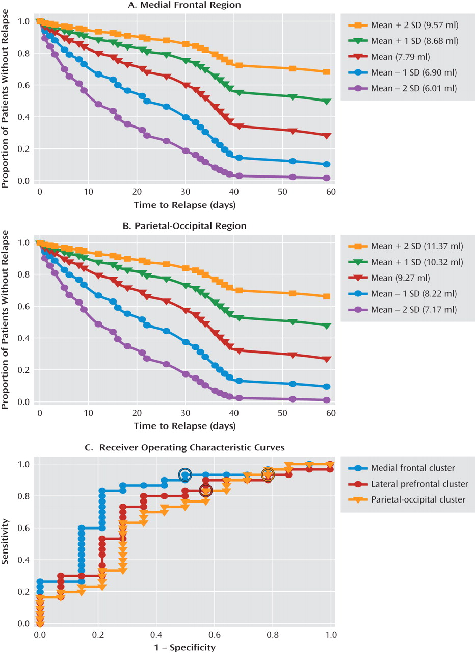
After controlling for age, IQ, years of alcohol use, and total alcohol use in the 90 days preceding inpatient treatment, smaller gray matter volume remained significantly predictive of shorter time to relapse for the medial frontal cluster (χ2=6.7, df=5, 44, p<0.009; hazard ratio=0.52, 95% CI=0.31-0.85) and the parietal-occipital cluster (χ2=9.28, df=5, 44, p<0.002; hazard ratio=0.52, 95% CI=0.34-0.79). Smaller gray matter volume fell short of significance for the lateral frontal cluster (χ2=4.74, df=5, 44, p<0.06; hazard ratio=0.68, 95% CI=0.48-0.96). The hazard ratios indicated that for each 1-ml reduction in gray matter volume in the medial frontal cluster (hazard ratio=0.52) and in the parietal-occipital cluster (hazard ratio=0.52), there was a 48% increase in risk of earlier relapse. Similarly, for relapse to heavy drinking, Cox proportional hazards regression analyses indicated that gray matter volume was significantly associated with time to relapse to heavy drinking in the medial frontal cluster (χ2=4.66, df=5, 44, p<0.04; hazard ratio=0.56, 95% CI=0.33-0.95) and the parietal-occipital cluster (χ2=7.56, df=5, 44, p<0.006; hazard ratio=0.55, 95% CI=0.36-0.84) and fell short of significance in the lateral frontal cluster (χ2=2.91, df=5, 44, p<0.09; hazard ratio=0.74, 95% CI=0.52-1.05). Smaller gray matter volume in the medial frontal and the parietal-occipital cluster predicted shorter time to relapse to heavy drinking (by 44% and 45%, respectively). Figure 3 illustrates the estimated survival functions for the gray matter volume mean and the mean plus one and two standard deviations in either direction for the medial frontal cluster (Figure 3A) and the parietal occipital cluster (Figure 3B) with covariates kept constant.
FIGURE 3. Estimated Survival Risk Functions and Receiver Operating Characteristic Curves for Gray Matter Volumes in Specific Significant Brain Regions in Alcohol-Dependent Patients (N=44)a
a Panels A and B present estimated survival risk functions (with mean age, IQ, and baseline total amount of alcohol consumed held constant) for mean gray matter volumes as well as for volumes one and two standard deviations above and below the mean for the medial frontal cluster (panel A; cluster χ2=6.7, p<0.009; hazard ratio=0.52, 95% CI=0.31-0.85) and the parietal-occipital cluster (panel B; χ2=9.28, p<0.002; hazard ratio=0.52, 95% CI=0.34-0.79). Although the survival function was a 90-day analysis, the graphs are cut off at day 60 because all alcohol-dependent patients with gray matter volumes two standard deviations below the mean for each of the two regions relapsed by day 60. For patients with volumes two standard deviations above the mean in the medial frontal cluster, the estimated survival function at day 60 spans a 0.68 (68%) proportion of surviving relapse, and for those with volumes two standard deviations above the mean in the parietal-occipital cluster, a 0.66 (66%) proportion, whereas for patients with volumes two standard deviations below the mean, the estimated survival function at day 60 for both regions spans only a 0.02% chance of surviving relapse. Panel C shows receiver operating characteristic (ROC) curves for the medial frontal (area under the curve=0.83; 95% CI=0.65-0.96), lateral frontal (area under the curve=0.75, 95% CI=0.55-0.89), and parietal-occipital clusters (area under the curve=0.70, 95% CI=0.49-0.86). The optimal gray matter volume cutoff values are circled in the ROC curves for each of the three regions that best differentiated between those who relapsed and those who did not (medial frontal cluster: χ2=7.04, df=1, 44, p<0.008; odds ratio=0.25, 95% CI=0.09-0.70; correctly classified 80% of relapsers at a 93.3% sensitivity and 50% specificity; optimal cutoff value=8.41; lateral frontal cluster: χ2=4.23, df=1, 44, p<0.04; odds ratio=0.56, 95% CI=0.32-0.97; correctly classified 71% of relapsers at a 83.3% sensitivity and 42.9% specificity; optimal cutoff value=10.6; parietal-occipital cluster: χ2=3.86, df=1, 44, p<0.05; odds ratio=0.47, 95% CI=0.22-0.99; correctly classified 66% of relapsers at a 90% sensitivity and 14.3% specificity; optimal cutoff value=10.4).
In addition, nonparametric receiving operator characteristic (ROC) analysis was applied to assess the sensitivity and specificity with which gray matter volume for each of the three region clusters identified those who relapsed compared with those who did not. The ROC curves for the medial frontal, lateral frontal, and parietal-occipital clusters are shown in Figure 3C. Logistic regression analyses indicated significant probabilities for classifying those who relapsed and those who did not for each of the three regions, along with the optimal cutoff values for gray matter volume for each cluster.

Discussion

Our findings in this study indicate significantly smaller gray matter volume in medial frontal, lateral prefrontal, and posterior parietal-occipital regions of the brain in a sample of alcohol-dependent patients engaged in inpatient treatment after 1 month of abstinence relative to healthy comparison subjects. The observed gray matter volume deficits were located in the medial frontal cortex, including the medial frontal gyrus and the anterior cingulate gyrus, but extending laterally in the superior and middle frontal gyri, the lateral prefrontal regions, primarily including the dorsolateral and inferolateral prefrontal cortex, and lastly a posterior region centered on the parietal-occipital sulcus, overlapping the precuneus, cuneus, and posterior cingulate regions. No areas were found in which the alcohol-dependent sample had larger gray matter volume than the comparison group. These results are consistent with previous studies and provide further evidence of gray matter volume reductions in 1-month abstinent recovering alcoholics relative to comparison subjects. Modest significant correlations were also observed between volume deficit in alcoholic patients and number of years of alcohol use and between volume deficit and baseline number of days of alcohol use prior to treatment. More importantly, using a prospective follow-up study design and after controlling for the influence of demographic variables (age and IQ) and alcohol use history variables (years of alcohol use and total amount of alcohol used for the 90-day period preceding treatment), smaller gray matter volume in the medial frontal cluster and the posterior-occipital cluster were each predictive of shorter time to any alcohol relapse and to heavy drinking relapse. In addition, the medial frontal cluster most accurately classified relapsers versus nonrelapsers at 80%, with a sensitivity of 93.3% and a specificity of 50%.
In support of our hypothesis, our findings indicated that smaller gray matter volume within two regions of medial frontal cortex cluster-the dorsal anterior cingulate (Brodmann's area 24/32) and the presupplementary (Brodmann's area 6) and supplementary motor regions-was predictive of shorter time to relapse and greater relapse risk in the alcohol-dependent sample. The medial frontal cortical regions are involved in cognitive control with top-down processing of sensory inputs, internal states, thoughts, and actions that guide behavior in pursuit of internal goals (45). Cognitive control makes possible flexible, adaptive task-relevant behavior when more automatic responses are inadequate or inappropriate in the interest of long-term goals. Effective cognitive control therefore transcends habitual stimulus-response associations (46). Although this study did not establish a link with functional impairment, the volume deficits in the medial frontal cortical cluster would suggest disruption of cognitive control functions associated with atrophy in these regions. Greater atrophy in these brain regions could therefore weaken a recovering alcohol-dependent patient's ability to override strong, habitual responses to environmental cues, stress, or otherwise cognitively challenging situations and increase his or her susceptibility to relapse, as observed with the association between brain atrophy in this region and relapse risk.
More specifically, the dorsal-caudal region of the anterior cingulate (Brodmann's area 24), which was part of the medial frontal cortex cluster that predicted alcohol relapse, makes up the anterior cingulate cognitive division (47). Evidence suggests a role for the anterior cingulate cognitive division in maintaining attention to goal-relevant stimuli when distracting stimuli conflict with these goals (48), and the inhibition of incorrect actions or facilitation of correct responses (49). Deficits in these executive functions are characteristic of poor impulse control (50) and are known to occur with addiction (51). Altered impulse control has been observed in recently abstinent alcohol-dependent individuals (52). In a recent study using low-resolution brain electromagnetic tomography (53), abnormalities in Brodmann's areas 24 and 32, as measured by reduced visual P3 amplitudes, were observed in alcohol-dependent patients relative to comparison subjects, and in the high-impulsivity participants from the entire sample when compared to the low-impulsivity participants. It is possible that overall gray matter volume loss in Brodmann's area 24/32, as well as the degree of gray matter recovery during abstinence, may have an effect on impulse control and risk of relapse. The presupplementary (Brodmann's area 6) and supplementary motor areas are involved in switching response sets when task rules change (thus altering a stimulus-response association) but also in error and conflict processing. Humans with lesions in these regions demonstrate impaired switching between response sets (54), which could result in difficulties in adaptively responding to alcohol cues in the environment and thus an elevated risk of relapse.
Although not hypothesized, we also found significantly smaller gray matter volume in the alcohol-dependent group in a region surrounding the parietal-occipital sulcus and extending anteriorly into the posterior cingulate gyrus. Gray matter volume deficits relative to comparison subjects have been previously observed in the parietal and occipital lobes of long-term abstinent alcoholics (6 months to 21 years [55]). Our alcohol-dependent sample had been abstinent for 1 month at the time of their imaging sessions, so our finding is consistent with evidence of persistent deficits in posterior gray matter volume. The impact of dysfunction in parietal-occipital areas on alcohol use and relapse is unclear. However, in a functional MRI study (56), abstinent alcohol-dependent patients showed significantly lower activation to visual stimuli in the occipital lobes bilaterally. In a study using multiple regression analyses to examine the relative contributions of tasks assessing visuoperceptual processes, explicit declarative memory, and frontal executive function in the performance of a visuoperceptual learning task (57), the results suggested that male alcoholics invoked executive processes to perform at normal levels, while male control subjects used basic visuoperceptual processes. Alcohol-dependent individuals therefore may be using frontal executive processes to compensate for deficits in visual processing. Interestingly, the authors suggested that the inefficient use of higher executive processes to perform a low-level cognitive task could leave insufficient reserves of higher-level attentional capacity available for additional cognitive demands. To the extent that these executive processes are unavailable to alcohol-dependent individuals because they are being used to compensate for deficits in visual processing, the resources necessary to exert cognitive control may be unavailable, thus increasing vulnerability to alcohol use. Our finding that reduced gray matter volume in visual processing areas of alcohol-dependent subjects predicted shorter time to relapse is consistent with this explanation. The fact that smaller gray matter volume in this region predicted alcohol relapse outcomes suggests a need to examine structural and functional atrophy in this region more closely in both those at risk for alcohol dependence and those at greatest risk of relapse.
There are important clinical implications to our findings. First, MRI volume assessment of medial frontal and posterior parietal-occipital brain regions could be further developed as neural markers for identifying alcoholic patients entering treatment who are at highest risk of relapse and treatment failure. For example, estimated survival functions for gray matter volume values that are one and two standard deviations above the mean (Figure 3A,B) show surviving alcohol relapse and maintaining abstinence at 60 days since discharge, indicating good outcome. By contrast, estimated survival functions for gray matter volume values that are one and two standard deviations below the mean show precipitous decreases in surviving risk of relapse, to levels below 20%. In particular, data from the ROC curves showed that the medial frontal cluster most accurately identified relapsers versus nonrelapsers at an 80% accuracy rate. Second, on further validation in future studies, use of such neural markers in clinical assessment could inform treatment planning by prescribing tailored interventions to improve brain atrophy and associated function early in abstinence in alcoholics who are at greatest risk of relapse. Recent preclinical data with evidence of microglial cell proliferation and neurogenesis during recovery from chronic alcohol exposure in laboratory animals (58) suggest that in alcoholics with significantly smaller gray matter volume, longer-term rehabilitation would be of benefit both for recovery from brain atrophy and in decreasing relapse risk. Finally, our data in this study support the development of pharmacological and other neural therapies that promote neurogenesis and cell proliferation to increase gray matter volume in critical regions in order to decrease relapse vulnerability and promote recovery from alcoholism.
Several limitations of this study are noteworthy. Because of the small number of women included in the sample, we were unable to examine sex-specific effects on gray matter volume deficits. Although the effects of sex were accounted for in the analyses, larger studies that assess sex differences in the association between gray matter volume deficits and relapse risk are recommended, as there is some evidence of greater sensitivity to neurotoxicity in women than in men (59). Furthermore, some recent evidence (60) indicates that MRI findings in treatment-seeking alcoholics may not be generalizable to non-treatment-seeking alcoholics in the community; and while the present study is especially relevant to treatment-seeking clinical alcoholic samples, it would be of benefit to assess gray matter volume deficits in non-treatment-seeking community alcoholic samples. Finally, our relapse assessments used self-reports and urine toxicology assessments that assess alcohol use through levels of alcohol metabolites for a limited time window of up to 80 hours, and hence our study lacked objective long-term assessments of alcohol use. On the other hand, these methods are widely used clinically, and the relapse rates we observed are consistent with those reported in the literature. Despite these limitations, this study is the first to link gray matter volume deficits in a sample of treatment-receiving alcoholics early in abstinence to subsequent time to relapse, and to provide predictive estimates of relapse risk. The findings provide clear evidence that smaller gray matter volumes in specific medial frontal and posterior brain regions play an important role in alcoholism relapse risk and clinical outcome.
Footnotes

Received Feb. 16, 2010; revisions received June 25 and July 26, 2010; accepted Aug. 13, 2010
Dr. Bhagwagar is employed at Bristol-Myers Squibb and has served on the speakers bureau for AstraZeneca and as a consultant for Janssen Pharmaceuticals. Dr. Guarnaccia has served on the speakers bureau and as a consultant for Abbott, Accorda, Bayer, Biogen, Pfizer, Serono, and Teva. Dr. Sinha is on the Scientific Advisory Board for Embera Neurotherapeutics and is a consultant for GlaxoSmithKline.

References

1.
Johnson BA: Update on neuropharmacological treatments for alcoholism: scientific basis and clinical findings. Biochem Pharmacol 2008; 75:34–56
2.
Brandon TH, Vidrine JI, Litvin EB: Relapse and relapse prevention. Annu Rev Clin Psychol 2007; 3:257–284
3.
Sinha R: The role of stress in addiction relapse. Curr Psychiatry Rep 2007; 9:388–395
4.
Fein G, Di Sclafani V, Cardenas VA, Goldmann H, Tolou-Shams M, Meyerhoff DJ: Cortical gray matter loss in treatment-naive alcohol de-pendent individuals. Alcohol Clin Exp Res 2002; 26:558–564
5.
Pfefferbaum A, Lim KO, Zipursky RB, Mathalon DH, Rosenbloom MJ, Lane B, Ha CN, Sullivan EV: Brain gray and white matter volume loss accelerates with aging in chronic alcoholics: a quantitative MRI study. Alcohol Clin Exp Res 1992; 16:1078–1089
6.
Pfefferbaum A, Sullivan EV, Mathalon DH, Lim KO: Frontal lobe volume loss observed with magnetic resonance imaging in older chronic alco-holics. Alcohol Clin Exp Res 1997; 21:521–529
7.
Jernigan TL, Butters N, DiTraglia G, Schafer K, Smith T, Irwin M, Grant I, Schuckit M, Cermak L: Reduced gray matter observed in alcoholics using magnetic resonance imaging. Alcohol Clin Exp Res 1991; 15:418–427
8.
Pfefferbaum A, Sullivan EV, Mathalon DH, Shear PK, Rosenbloom MJ, Lim KO: Longitudinal changes in magnetic resonance imaging brain volumes in abstinent and relapsed alcoholics. Alcohol Clin Exp Res 1995; 19:1177–1191
9.
Sullivan EV, Deshmukh A, Desmond JE, Lim KO, Pfefferbaum A: Cerebellar volume decline in normal aging, alcoholism, and Korsakoff's syn-drome: relation to ataxia. Neuropsychology 2000; 14:341–352
10.
Cardenas VA, Studholme C, Gazdzinski S, Durazzo TC, Meyerhoff DJ: Deformation-based morphometry of brain changes in alcohol depend-ence and abstinence. NeuroImage 2007; 34:879–887
11.
Chanraud S, Martelli C, Delain F, Kostogianni N, Douaud G, Aubin H-J, Reynaud M, Martinot J-L: Brain morphometry and cognitive perform-ance in detoxified alcohol dependents with preserved psycho-social functioning. Neuropsychopharmacology 2007; 32:429–438
12.
Mechtcheriakov S, Brenneis C, Eggar K, Koppelstaetter F, Schocke M, Marksteiner J: A widespread distinct pattern of cerebral atrophy in pa-tients with alcohol addiction revealed by voxel-based morphometry. J Neurol Neurosurg Psychiatry 2007; 78:610–614
13.
Tanabe J, Tregellas JR, Dalwani M, Thompson L, Owens E, Crowley T, Banich M: Medial orbitofrontal cortex gray matter is reduced in absti-nent substance-dependent individuals. Biol Psychiatry 2009; 65:160–164
14.
Pfefferbaum A, Sullivan EV, Rosenbloom MJ, Mathalon DH, Lim KO: A controlled study of cortical gray matter and ventricular changes in al-coholic men over a 5-year interval. Arch Gen Psychiatry 1998; 55:905–912
15.
Gazdzinski S, Durazzo TC, Meyerhoff DJ: Temporal dynamics and determinants of whole brain tissue volume changes during recovery from alcohol dependence. Drug Alcohol Depend 2005; 78:263–273
16.
Wrase J, Makris N, Braus DF, Mann K, Smolka MN, Kennedy DN, Caviness VS, Hodge SM, Tang L, Albaugh M, Ziegler DA, Davis OC, Kissling C, Schumann G, Breiter HC, Heinz A: Amygdala volume associated with alcohol abuse relapse and craving. Am J Psychiatry 2008; 165:1179–1184
17.
Volkow ND, Li TK: Drug addiction: the neurobiology of behaviour gone awry. Nat Rev Neurosci 2004; 5:963–970
18.
Li CS, Sinha R: Inhibitory control and emotional stress regulation: neuroimaging evidence for fronto-limbic dysfunction in psychostimulant ad-diction. Neurosci Biobehav Rev 2008; 32:581–597
19.
First MB, Spitzer RL, Williams JBW, Gibbon M: Structured Clinical Interview for DSM-IV-Patient Edition (SCID-P). Washington, DC, American Psychiatric Press, 1995
20.
Ashburner J, Friston KF: Unified segmentation. Neuroimage 2005; 26:839–851
21.
Ashburner J: A fast diffeomorphic image registration algorithm. Neuroimage 2007; 38:95–113
22.
Klein A, Andersson J, Ardekani BA, Ashburner J, Avants B, Chiang MC, Christensen GE, Collins DL, Gee J, Hellier P, Song JH, Jenkinson M, Lepage C, Rueckert D, Thompson P, Vercauteran T, Woods RP, Mann JJ, Parsey RV: Evaluation of 14 nonlinear deformation algorithms applied to human brain MRI registration. Neuroimage 2009; 46:786–802
23.
Anbeek P, Vincken KL, van Osch MJ, Bisschops RH, van der Grond J: Probabilistic segmentation of white matter lesions in MR imaging. Neuroimage 2004; 21:1037–1044
24.
Bouix S, Martin-Fernandez M, Ungar L, Nakamura M, Koo MS, McCarley RW, Shenton ME: On evaluating brain tissue classifiers without a ground truth. Neuroimage 2007; 36:1207–1224
25.
Cerasa A, Messina D, Nicoletti G, Novellino F, Lanza P, Condino F, Arabia G, Salsone M, Quattrone A: Cerebellar atrophy in essential tremor using an automated segmentation method. AJNR Am J Neuroradiol 2009; 30:1240–1243
26.
Davies RR, Scahill VL, Graham A, Williams GB, Graham KS, Hodges JR: Development of an MRI rating scale for multiple brain regions: com-parison with volumetrics and with voxel-based morphometry. Neuroradiology 2009; 51:491–503
27.
Fischl B, Dale AM: Measuring the thickness of the human cerebral cortex from magnetic resonance images. Proc Natl Acad Sci USA 2000; 97:11050–11055
28.
Ashburner J, Friston KF: Voxel-based morphometry: the methods. Neuroimage 2000; 21(6, pt 1):805–821
29.
Sobell MB, Maisto SA, Sobell LC, Cooper AM, Cooper TC, Sanders B: Developing a prototype for evaluating alcohol treatment effectiveness, in Evaluating Drug and Alcohol Abuse Treatment Effectiveness: Recent Advances. Edited by, Sobell LC, Sobell MB, Ward E. Elmsford, NY, Pergamon Press, 1980
30.
Sobell LC, Sobell MB: Timeline followback: a technique for assessing self-reported alcohol consumption, in Measuring Alcohol Consumption: Pyschosocial and Biochemical Methods. Edited by, Litten RZ, Allen JP. Totowa, NJ, Humana Press, 1992
31.
Miller WR, Del Boca FK: Measurement of drinking behavior using the Form 90 family of instruments. J Stud Alcohol Suppl 1994; 12:112–118
32.
Fals-Stewart W, O'Farrell TJ, Freitas TT, McFarlin SK, Rutiglian P: The timeline followback reports of psychoactive substance use by drug-abusing patients: psychometric properties. J Consult Clin Psychol 2000; 68:134–144
33.
Anton RF, O'Malley SS, Ciraulo DA, Cisler RA, Couper D, Donovan DM, Gastfriend DR, Hosking JD, Johnson BA, LoCastro JS, Longabaugh R, Mason BJ, Mattson ME, Miller WR, Pettinati HM, Randall CL, Swift R, Weiss RD, Williams LD, Zweben A; COMBINE Study Research Group: Combined pharmacotherapies and behavioral interventions for alcohol dependence: the COMBINE study: a randomized controlled trial. JAMA 2006; 295:2003–2017
34.
Rohsenow DJ, Monti PM: Relapse among cocaine abusers: theoretical, methodological, and treatment considerations, in Relapse and Recov-ery in Addictions. Edited by, Tims FM, Leukefeld CG, Platt JJ. New Haven, Conn, Yale University Press, 2001, pp 355–378
35.
Hayasaka S, Nichols TE: Validating cluster size inference: random field and permutation methods. Neuroimage 2003; 20:2343–2356
36.
Poline J-B, Worsley KJ, Evans AC, Friston KJ: Combining spatial extent and peak intensity to test for activations in functional imaging. Neuro-image 1997; 5:83–96
37.
Friston KJ, Holmes A, Poline J-B, Price CJ, Frith CD: Detecting activations in PET and fMRI: levels of inference and power. Neuroimage 1996; 4(3, pt 1):223–235
38.
Chumbley JR, Friston KJ: False discovery rate revisited: FDR and topological inference using Gaussian random fields. Neuroimage 2009; 44:62–70
39.
Cox D: Regression models and life tables (with discussion). J R Stat Soc B 1972; 34:187–220
40.
Talairach J, Tournoux P: Co-Planar Stereotaxic Atlas of the Human Brain. New York, Thieme Medical, 1988
41.
Lacadie CM, Fulbright RK, Rajeevan N, Constable RT, Papademetris X: More accurate Talairach coordinates for neuroimaging using non-linear registration. Neuroimage 2008; 42:717–725
42.
Tzourio-Mazoyer N, Landeau B, Papathanassiou D, Crivello F, Etard O, Delcroix N, Mazoyer B, Joliot M: Automated anatomical labeling of ac-tivations in SPM using a macroscopic anatomical parcellation of the MNI MRI single-subject brain. Neuroimage 2002; 15:273–289
43.
Lancaster JL, Woldorff MG, Parsons LM, Liotti M, Freitas CS, Rainey L, Kochunov PV, Nickerson D, Mikiten SA, Fox PT: Automated Talairach atlas labels for functional brain mapping. Hum Brain Mapp 2000; 10:120–131
44.
Maldjian JA, Laurienti PJ, Burdette JB, Kraft RA: An automated method for neuroanatomic and cytoarchitectonic atlas-based interrogation of fMRI data sets. Neuroimage 2003; 19:1233–1239
45.
Miller EK, Cohen JD: An integrative theory of prefrontal cortex function. Annu Rev Neurosci 2001; 24:167–202
46.
Egner T: Prefrontal cortex and cognitive control: motivating functional hierarchies. Nat Neurosci 2009; 12:821–822
47.
Devinsky O, Morrell MJ, Vogt B: Contributions of the anterior cingulate to behavior. Brain 1995; 118:279–306
48.
Weisman DH, Warner LM, Woldorff MG: The neural mechanisms for minimizing cross-modal distraction. J Neurosci 2004; 24:10941–10949
49.
Bush G, Frazier JA, Rauch SL, Seidman LJ, Whalen PJ, Jenike MA, Rosen BR, Biederman J: Anterior cingulate cortex dysfunction in atten-tion-deficit/hyperactivity disorder revealed by fMRI and the counting Stroop. Biol Psychiatry 1999; 45:1542–1552
50.
Evenden JL: Varieties of impulsivity. Psychopharmacology 1999; 146:348–361
51.
Crews FT, Boettiger CA: Impulsivity, frontal lobes, and risk for addiction. Pharmacol Biochem Behav 2009; 93:237–247
52.
Li C-SR, Luo X, Yan P, Bergquist K, Sinha R: Altered impulse control in alcohol dependence: neural measures of stop-signal performance. Al-cohol Clin Exp Res 2009; 33:740–750
53.
Chen ACH, Porjesz B, Rangaswamy M, Kamarajan C, Tang Y, Jones KA, Chorlian DB, Stimus AT, Begleiter H: Reduced frontal lobe activity in subjects with high impulsivity and alcoholism. Alcohol Clin Exp Res 2007; 31:156–165
54.
Rushworth MFS, Walton ME, Kennerley SW, Bannerman DM: Action sets and decisions in medial frontal cortex. Trends Cogn Sci 2004; 8:410–417
55.
Fein G, Shimotsu R, Chu R, Barakos J: Parietal gray matter volume loss is related to spatial processing deficits in long-term abstinent alcoholic men. Alcohol Clin Exp Res 2009; 33:1–9
56.
Hermann D, Smolka MN, Klein S, Heinz A, Mann K, Braus DF: Reduced fMRI activation of an occipital area in recently detoxified alcohol de-pendent patients in a visual and acoustic paradigm. Addict Biol 2007; 12:117–121
57.
Fama R, Pfefferbaum A, Sullivan EV: Perceptual learning in detoxified alcoholic men: contributions from explicit -memory, executive function, and age. Alcohol Clin Exp Res 2004; 28: 1657–1665
58.
Crews FT, Nixon K: Mechanisms of neurodegeneration and regeneration in alcoholism. Alcohol Alcohol 2009; 44:115–127
59.
Hommer DW, Momenan R, Kaiser E, Rawlings RR: Evidence for a gender-related effect of alcoholism on brain volumes. Am J Psychiatry 2001; 158:198–204
60.
Gazdzinski S, Durazzo TC, Weiner MW, Meyerhoff DJ: Are treated alcoholics representative of the entire population with alcohol use disor-ders? a magnetic resonance study of brain injury. Alcohol 2008; 42:67–76

Information & Authors

Information

Published In

Go to American Journal of Psychiatry
American Journal of Psychiatry
Pages: 183 - 192
PubMed: 21078704

History

Received: 16 February 2010
Revision received: 25 June 2010
Revision received: 26 July 2010
Accepted: 13 August 2010
Published online: 1 February 2011
Published in print: February 2011

Authors

Details

Kenneth Rando, B.S.
From the Departments of Psychiatry and Neurology, Yale University School of Medicine; and the Yale Child Study Center, New Haven.
Kwang-Ik Hong, M.S.
From the Departments of Psychiatry and Neurology, Yale University School of Medicine; and the Yale Child Study Center, New Haven.
Zubin Bhagwagar, M.D., Ph.D.
From the Departments of Psychiatry and Neurology, Yale University School of Medicine; and the Yale Child Study Center, New Haven.
Chiang-Shan Ray Li, M.D., Ph.D.
From the Departments of Psychiatry and Neurology, Yale University School of Medicine; and the Yale Child Study Center, New Haven.
Keri Bergquist, Psy.D.
From the Departments of Psychiatry and Neurology, Yale University School of Medicine; and the Yale Child Study Center, New Haven.
Joseph Guarnaccia, M.D.
From the Departments of Psychiatry and Neurology, Yale University School of Medicine; and the Yale Child Study Center, New Haven.
Rajita Sinha, Ph.D.
From the Departments of Psychiatry and Neurology, Yale University School of Medicine; and the Yale Child Study Center, New Haven.

Notes

Address correspondence and reprint requests to Dr. Sinha, Department of Psychiatry, Yale University School of Medicine, 34 Park St., Rm. S110, New Haven, CT 06519; [email protected] (e-mail).

Funding Information

The other authors report no financial relationships with commercial interests.Supported by NIH grants R01-AA013892 (to Dr. Sinha), K23-MH077914 (to Dr. Bhagwagar), and K02-DA026990 (to Dr. Li); NIH Roadmap Fund for Medical Research grants UL1-DE019586 (to Dr. Sinha) and UL1-RR024139 (Clinical and Translational Science Award to Yale University); and the Connecticut Department of Mental Health and Addiction Services.

Metrics & Citations

Metrics

Citations

Export Citations

If you have the appropriate software installed, you can download article citation data to the citation manager of your choice. Simply select your manager software from the list below and click Download.

For more information or tips please see 'Downloading to a citation manager' in the Help menu.

Format
Citation style
Style
Copy to clipboard

View Options

View options

PDF/EPUB

View PDF/EPUB

Get Access

Login options

Already a subscriber? Access your subscription through your login credentials or your institution for full access to this article.

Personal login Institutional Login Open Athens login
Purchase Options

Purchase this article to access the full text.

PPV Articles - American Journal of Psychiatry

PPV Articles - American Journal of Psychiatry

Not a subscriber?

Subscribe Now / Learn More

PsychiatryOnline subscription options offer access to the DSM-5-TR® library, books, journals, CME, and patient resources. This all-in-one virtual library provides psychiatrists and mental health professionals with key resources for diagnosis, treatment, research, and professional development.

Need more help? PsychiatryOnline Customer Service may be reached by emailing [email protected] or by calling 800-368-5777 (in the U.S.) or 703-907-7322 (outside the U.S.).

Media

Figures

Other

Tables

Share

Share

Share article link

Share