Full access
Perspectives
Published Online: 1 July 2012

Functional Neuroimaging of Major Depressive Disorder: A Meta-Analysis and New Integration of Baseline Activation and Neural Response Data

Abstract

Objective:

Functional neuroimaging investigations of major depressive disorder can advance both the neural theory and treatment of this debilitating illness. Inconsistency of neuroimaging findings and the use of region-of-interest approaches have hindered the development of a comprehensive, empirically informed neural model of major depression. In this context, the authors sought to identify reliable anomalies in baseline neural activity and neural response to affective stimuli in major depressive disorder.

Method:

The authors applied voxel-wise, whole-brain meta-analysis to neuroimaging investigations comparing depressed to healthy comparison groups with respect to baseline neural activity or neural response to positively and/or negatively valenced stimuli.

Results:

Relative to healthy subjects, those with major depression had reliably higher baseline activity, bilaterally, in the pulvinar nucleus. The analysis of neural response studies using negative stimuli showed greater response in the amygdala, insula, and dorsal anterior cingulate cortex and lower response in the dorsal striatum and dorsolateral prefrontal cortex in individuals with major depressive disorder than in healthy subjects.

Conclusions:

The meta-analytic results support an elegant and neuroanatomically viable model of the salience of negative information in major depressive disorder. In this proposed model, high baseline pulvinar activity in depression first potentiates responding of the brain's salience network to negative information; next, and owing potentially to low striatal dopamine levels in depression, this viscerally charged information fails to propagate up the cortical-striatal-pallidal-thalamic circuit to the dorsolateral prefrontal cortex for contextual processing and reappraisal.
Advancing neural theory of major depressive disorder is of great importance given both that this disorder is predicted to be the most burdensome disease in the world in the 21st century (1) and that it is often unresponsive to conventional pharmacologic intervention (2). Over the past two decades, investigations using functional neuroimaging have provided important insights about its neural substrates. Several of these investigations have supported a neural model of depressed mood in which, within a mutually inhibitory limbic and dorsal cortical architecture, limbic activation overcomes dorsal cortical activation, which reciprocally fails to inhibit limbic activation in major depressive disorder (3); researchers examining neural aspects of emotional reactivity in major depression have proposed similar frameworks (4). Investigators examining network-level connectivity in major depressive disorder have reported evidence of limbic inhibition of dorsal cortical activation, partially supporting corticolimbic inhibition models of major depression (5).
While current neural models of major depressive disorder have been important in both understanding and treating this disorder (6), they are equivocal with respect to the primary neural mechanisms of the illness and do not integrate contemporary knowledge of major neuroanatomical pathways and functional subdivisions of the brain. In formulating a more mechanistic and neuroanatomically viable model of major depression, it is important that we first identify and address issues concerning the data that inform neural theories of the disorder. In this context, it is noteworthy that even the neural findings that are considered most foundational in the neuroscience of depression, such as higher than normal baseline activity in the subgenual anterior cingulate cortex (6) and low activation in the dorsolateral prefrontal cortex (7), have not gone unchallenged (810). Further, region-of-interest analytic approaches, which are widely used in clinical neuroimaging (in reference 11, for instance) both to bolster statistical power and to test specific neural models of depression, may nonetheless bias neural models of major depressive disorder by ignoring anomalies in regions that are not of current interest in the study of depression. Thus, we propose that in order to advance neural theory of depression, we must identify, across the whole brain, reliable patterns of anomalous neural response and baseline brain activation in this disorder.
In this context, voxel-wise, whole-brain meta-analysis has the potential to identify robust functional neural findings from the corpus of functional neuroimaging research in major depression; indeed, such an approach has been applied successfully to identifying reliable and interpretable neural responses in anxiety (12) and in emotional responding more generally (13). Further, voxel-wise meta-analysis allows investigators to combine and examine patterns of activation obtained across studies using different types of experimental conditions. For example, findings from studies of neural reactivity to positive stimuli in major depressive disorder can be contrasted with results of investigations of reactivity to negative stimuli, yielding findings that are typically not possible to obtain in a single study. Finally, and particularly important for developing a comprehensive neural theory of major depressive disorder, meta-analysis of whole-brain functional neuroimaging data has the potential to identify anomalous patterns of activation that may not have been accorded adequate attention in previous neural conceptualizations of depression.
While meta-analyses reported by other researchers have produced intriguing results (14), only recently has the number of functional neural investigations of major depression—particularly those using functional MRI (fMRI) to measure neural response—reached a level sufficient to warrant meta-analysis. In the present study we used a whole-brain, voxel-wise meta-analytic technique to identify reliable anomalies in major depressive disorder from both resting-state regional cerebral blood flow (rCBF) and affective-challenge fMRI studies. For affective-challenge fMRI investigations, we examined the results of studies of neural responses to negative relative to positive stimuli in depression. In conducting these analyses, our objective was to develop an empirically informed and rigorous neural model of major depressive disorder.

Method

Overview

We used multilevel kernel density analysis (12, 13) in conducting the present meta-analysis. In this analysis, we first identified functional neuroimaging studies that reported neural functional aberrations among participants diagnosed with major depressive disorder in 1) rCBF as measured with positron emission tomography (PET) or single photon emission computed tomography (SPECT) or 2) neural reactivity (task-based fMRI) to negative and/or positive stimuli. Next we converted results from each comparison of interest in each study into summary binary indicator maps (12). Finally, we merged and conducted voxel-wise comparisons of these maps separately for rCBF and fMRI studies.

Study Selection

We used the ISI Web of Science database to identify functional neuroimaging studies of depression conducted up to Jan. 15, 2011. To identify fMRI studies of major depression, we entered the terms “depress* OR mood” and “functional MRI OR fMRI” in the topic-based search field. To identify PET and SPECT studies of depression, we entered “depress* OR mood” and “positron emission tomography OR PET OR single photon emission OR SPECT OR cerebral-blood flow” in the topic-based search field. Among the studies identified—1,448 fMRI studies and 5,147 rCBF studies—we kept for subsequent meta-analysis only those that were conducted with adults between the ages of 18 and 60 who were diagnosed with a current episode of primary unipolar depression as assessed with the Structured Clinical Interview for DSM (SCID) (15) or with the International Statistical Classification of Diseases and Related Health Problems (ICD) (16). In addition, we required that studies took a whole-brain analysis approach in which the investigators reported coordinates of all suprathreshold clusters in comparing neural activity in subjects with major depressive disorder to that in a comparison group of demographically matched, SCID- or ICD-assessed nondepressed individuals. We also searched the reference lists of studies that met these criteria to ensure that our original web-based search was comprehensive. For PET and SPECT studies to be included, we required that they used a tracer amenable to estimating brain activity: [18F]FDG or [15O]H2O for PET and [99mTc]ECD for SPECT. For fMRI studies to be included, we added the criterion that there was at least one condition in the study in which activation during processing of affectively valenced information was either contrasted to a neutral stimulus condition or parametrically varied. Finally, we required that typical regression analyses, i.e., those using regression to estimate the extent or “height” of the blood-oxygen-level-dependent response, were conducted on fMRI data. Fourteen rCBF studies (6, 1729) and 24 fMRI studies (7, 8, 3051) met our inclusion criteria; 5,133 rCBF and 1,424 fMRI studies identified in our original search were excluded for not meeting one or more of these criteria. While the fMRI studies included in this meta-analysis collectively used a broad range of negatively and positively valenced affective challenges and stimuli, we have combined these studies given that valence is a fundamental component of emotions and affective stimuli in affective circumplex models of emotion (see, for instance, reference 52). Thus, examining differential neural response in major depression to stimulus valence, regardless of its specific form, can inform us from a theoretical perspective about emotional functioning in major depressive disorder.

Computing the Meta-Analytic Statistic

As in other studies employing multilevel kernel density analysis (for instance, 12), the data for the present meta-analysis were coordinates that were reported in three-dimensional standard brain space of peak activation differences between groups. For each study that met the inclusion criteria, we constructed at 1-mm isotropic voxel resolution an indicator map in Talairach space (53) as represented in the AFNI statistical package (54). We converted coordinates reported in the standard space of the Montreal Neurological Institute to Talairach space by using the MATLAB script mni2tal.m (http://imaging.mrc-cbu.cam.ac.uk/imaging/MniTalairach). Each binary indicator map, I, was composed of spheres (value=1) of 10-mm radius—a size chosen to account for limitations in spatial resolution in functional neuroimaging data while maintaining adequate sensitivity to detect neural anomalies in smaller structures—centered at reported locations of peak between-groups difference; in instances of overlap between spheres, we valued the region of intersection at 1.
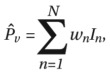
We merged indicator maps into six meta-analytic statistical maps: four from fMRI studies and two from PET/SPECT studies. The fMRI meta-analytic maps showed regions where activation in depressed subjects was greater than in comparison subjects in response to negative stimuli (20 contrasts, 92 coordinates) and positive stimuli (10 contrasts, 40 coordinates) and where activation was greater in comparison subjects than in depressed subjects in response to negative stimuli (19 contrasts, 90 coordinates) and positive stimuli (14 contrasts, 76 coordinates). The PET/SPECT meta-analytic maps indicated where activation was greater in depressed subjects than in comparison subjects (10 contrasts, 37 coordinates) and where it was greater in comparison subjects than in depressed subjects (11 contrasts, 37 coordinates). Indicator maps from N studies per category were merged into meta-analytic statistical maps, where the meta-analytic statistic (^Pv) at each voxel, v, was calculated as
[See PDF for formula]
where wn is the square root of the number of subjects in the full study group (depressed plus comparison subjects) of the nth of N studies. Weighting each map in this fashion meant that studies with larger groups and presumably more reliable results were accorded more weight in the meta-analytic statistic. This approach has several advantages relative to other meta-analytic methods used to examine patterns of brain activation in depression, such as those that use activation likelihood estimation techniques (for instance, reference 14). First, because we treated comparison maps within studies as random effects, no single map could contribute disproportionately to our meta-analytic results. Second, weighting by group size increases accuracy without introducing the complexity of z-score-based weighting for different types of analyses. Finally, relative to methods that incorporate z-score weighting, such as activation likelihood estimation, the approach taken in the present meta-analysis is better suited to examining the replicability of neural responding across studies.

Comparing Meta-Analytic Maps

We subtracted meta-analytic maps showing regions where activation was greater in comparison subjects than in depressed subjects from those showing regions in which activation was greater in depressed subjects for both rCBF studies (21 contrasts, 74 coordinates) and negative- and positive-valence fMRI studies (39 contrasts, 182 coordinates; and 24 contrasts, 106 coordinates, respectively). Further, for the fMRI studies, to test whether significant differences from these comparisons were valence specific, we also subtracted positive from negative meta-analytic maps for comparisons showing where activation was greater in depressed subjects and those showing where activation was greater in comparison subjects (63 contrasts, 288 coordinates). Null-hypothesis distributions for each comparison were calculated from 10,000 Monte Carlo simulations performed in MATLAB (www.mathworks.com) by using relevant variables from the included studies (group size, number of regions reported) but with the assumption of a random distribution of peaks in a standardized brain (AFNI's TT_N27+tlrc). We used the AFNI program AlphaSim to set the significance threshold for each comparison at α=0.05, family-wise-error-rate (FWE) controlled for comparison of multiple voxels across the whole brain. AlphaSim takes as input the parameters used in neuroimaging analysis (e.g., voxel-wise statistical threshold, size of mask over which statistical calculations are performed) and determines, using Monte Carlo simulation, a cluster threshold necessary to assure FWE control. We used a voxel-wise threshold of α=0.001 (resulting in a cluster threshold, k, of 177 voxels) for our fMRI meta-analysis and α=0.005 (k=384 voxels) for our meta-analysis of PET data. We used a more liberal voxel-wise threshold—and, therefore, a more stringent cluster size threshold—for the PET meta-analysis given the lower spatial resolution of PET data and the consequent lower incidence of intersection of PET-derived statistical maps. In addition, to address potential outlier effects and to ensure that significant meta-analytic findings are representative of results from a range of studies, we established that at least three studies had to contribute to a given meta-analytic finding for it to be reported here. Finally, we note that our null-hypothesis distributions were created under the assumption of independence across studies. Several studies that met our inclusion criteria were conducted in the same laboratory (fMRI: Cynthia Fu [7, 35, 36, 43], Thomas Frodl [34, 46], Ian Gotlib [32, 33, 37, 39], Mary Phillips [8, 48]; rCBF: Lewis Baxter [19, 27], Omer Bonne [24, 25]) or included more than one comparison per valence. Therefore, we checked significant meta-analytic findings to ensure that no study was represented more than once. We took the following exclusionary precautions to ensure independence across analyses from the same study: 1) if results from both parametric and neutral stimulus contrasts were reported, only results from the presumably more reliable parametric analyses were included; 2) if a study reported results from contrasting two levels of an emotion variable to a neutral stimulus, then results from only the comparison of the highest-level emotion and the neutral stimulus were included; and 3) if a study reported results from contrasting an experimental condition to more than one control condition, we included results from only one comparison, i.e., that which included the most coordinates. In cases where different studies from the same laboratory contributed to a significant finding, we contacted investigators to ensure that the study groups were independent.

Results

Characteristics of Included Studies

Table 1 and Table 2 present, for the included rCBF and fMRI studies, respectively, the number of subjects, gender composition, and proportion of medicated subjects in each group of depressed participants. Further, for fMRI studies we report the task that was used and the contrasts that were included in the meta-analysis.
TABLE 1. Studies Measuring Differences in Baseline Regional Cerebral Blood Flow Between Persons With Major Depressive Disorder and Healthy Comparison Subjects
 Number of Subjects in GroupCharacteristics of Depressed SubjectsImaging Technique
StudyMajor Depressive DisorderComparisonMedicated (%)Female (%)MethodTracer
Aihara et al. (17)2423063PET[18F]FDG
Bench et al. (18)33235836PET[15O]H2O
Brody et al. (19)2416054PET[18F]FDG
Drevets et al. (20)1333054PET[15O]H2O
Germain et al. (21)1213083PET[18F]FDG
Kennedy et al. (22)132400PET[18F]FDG
Kimbrell et al. (23)3837066PET[18F]FDG
Kohn et al. (24)3325058SPECT[99mTc]ECD
Krausz et al. (25)1010090SPECT[99mTc]ECD
Mayberg et al. (6)6510050PET[15O]H2O
Périco et al. (26)1515080SPECT[99mTc]ECD
Saxena et al. (27)2717052PET[18F]FDG
Skaf et al. (28)912044SPECT[99mTc]ECD
Videbech et al. (29)42479571PET[15O]H2O
TABLE 2. Studies Using Functional Magnetic Resonance Imaging to Estimate Neural Response to Negative and/or Positive Stimuli in Persons With Major Depressive Disorder and Healthy Comparison Subjects
 Number of Subjects in GroupCharacteristics of Depressed Subjects Included Contrasts
StudyMajor Depressive DisorderComparisonMedicated (%)Female (%)Affective ChallengeNegative ValencePositive Valence
Abler et al. (30)1212100100Anticipating and viewing negative, neutral, and positive picturesAnticipating negative versus neutral; viewing negative versus neutralViewing positive versus neutral
Bär et al. (31)13130100Experiencing variable-intensity thermal painParametric thermal pain responseNone
Canli et al. (32)15154780Performing lexical decision task for neutral, happy, sad, and threat-related wordsSad versus neutral; physical threat versus neutral; social threat versus neutralHappy versus neutral
Cooney et al. (33)14144357Thinking about personal traits, abstract ideas, and physical objectsPersonal traits versus abstract ideasNone
Frodl et al. (34)12126758Emotion (explicit) or gender (implicit) matching of negative faces to target; shape-matching controlExplicit versus control; implicit versus controlNone
Fu et al., 2004 (36)1919068Gender identification of variable-intensity sad facesParametric sad face responseNone
Fu et al., 2007 (35)1919068Gender identification of variable-intensity happy facesNoneParametric happy face response
Fu et al., 2008 (7)1616081Gender identification of variable-intensity sad facesParametric sad face responseNone
Gotlib et al. (37)18185072Viewing happy, sad, and neutral facesSad versus neutralHappy versus neutral
Herwig et al. (38)141410057Viewing negative and neutral picturesNegative versus neutralNone
Keedwell et al. (8)12129267Remembering happy, sad, and neutral experiencesSad versus neutralHappy versus neutral
Knutson et al. (39)1412064Anticipating and experiencing monetary gains, losses, and neutral outcomesNoneAnticipating gain versus neutral outcome; experiencing gain versus neutral outcome
Kumari et al. (40)66100100Viewing positive, negative, and neutral picture-caption pairsNegative versus neutralPositive versus neutral
Lawrence et al. (41)91110044Viewing high- and low-intensity happy, sad, and fearful faces; neutral-face controlHigh-intensity fearful versus neutral; high-intensity sad versus neutralHigh-intensity happy versus neutral
Mitterschiffthaler et al., 2003 (42)77100100Viewing positive and neutral imagesNonePositive versus neutral
Mitterschiffthaler et al., 2008 (43)1717082Identifying colors of sad and neutral wordsSad versus neutralNone
Osuch et al. (44)1617669Listening to endorsed favorite and neutral musicNoneFavorite versus neutral
Pizzagalli et al. (45)3031050Anticipating and experiencing monetary gains, losses, and neutral outcomesAnticipating loss versus neutral outcome; experiencing loss versus neutral outcomeAnticipating gain versus neutral outcome; experiencing gain versus neutral outcome
Scheuerecker et al. (46)1315077Emotion (explicit) or gender (implicit) matching of negative faces to target; shape-matching controlExplicit plus implicit versus controlNone
Strigo et al. (47)1515080Anticipating and experiencing painful and nonpainful thermal stimuliAnticipating pain versus nonpain; experiencing pain versus nonpainNone
Surguladze et al. (48)161410038Viewing variable-intensity happy and sad facesParametric sad face responseParametric happy face response
Townsend et al. (49)1515040Matching negative faces to facial emotion or word target; shape-matching controlFace matching versus controlNone
Wang et al. (50)19205863Oddball target detection with sad or neutral picture as backgroundSad versus neutralNone
Yang (51)10100100Viewing erotic and neutral film clipsNoneErotic versus neutral

rCBF Meta-Analysis

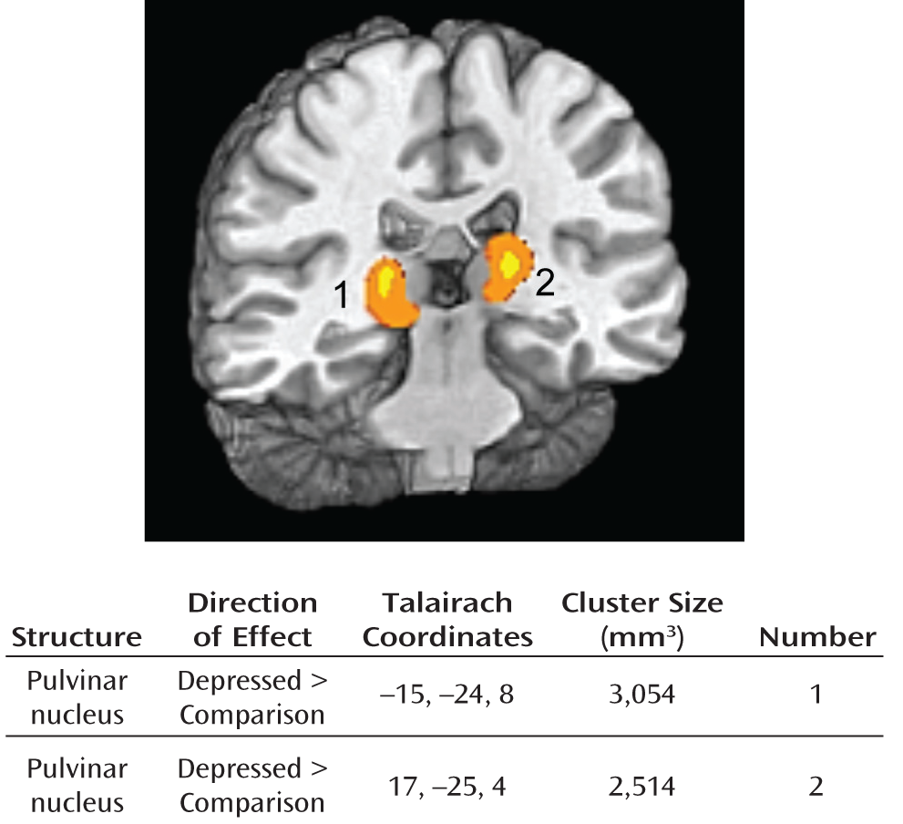
Our meta-analysis of resting-state PET and SPECT studies showed reliably greater activity in the depressed participants, relative to the comparison subjects, in both the right and left pulvinar nuclei of the thalamus (FWE-controlled p<0.05); see Figure 1. The data supplement accompanying the online version of this article contains otherwise significant regional cerebral blood flow anomalies in major depression supported by only two studies.
FIGURE 1. Statistical Map of Reliable Results From Meta-Analytic Synthesis Showing Differences in Baseline Regional Cerebral Blood Flow Between Persons With Major Depressive Disorder and Healthy Comparison Subjectsa
a Presented data were smoothed by using cubic interpolation.

fMRI Meta-Analysis

Negative stimuli.

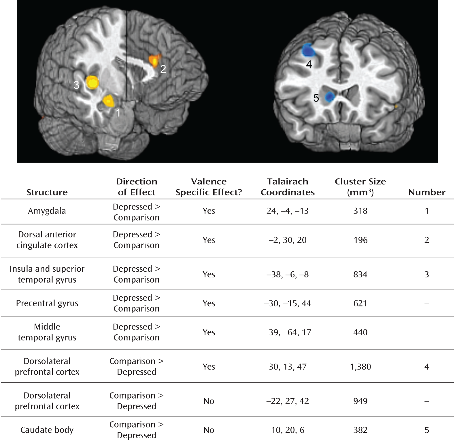
Our analysis showed that individuals diagnosed with major depressive disorder had reliably greater responses to negative stimuli than did the comparison subjects in the amygdala, dorsal anterior cingulate cortex, and insula/superior temporal gyrus; we also found greater activation in response to negative stimuli in depressed individuals in precentral and middle temporal gyri. It is important to note that these effects were also evident in depressed participants when the responses to negative stimuli were contrasted with responses to positive stimuli, indicating that these aberrant responses in depression were specific to negative stimuli. We also found reliably lower activation in response to negative stimuli in depressed participants in the dorsolateral prefrontal cortex bilaterally and in the caudate body (dorsal striatum); however, only the lower response in the right dorsolateral prefrontal cortex in the depressed subjects was found to be specific to negative stimuli. All findings were significant at FWE-controlled p<0.05; see Figure 2 for a graphical rendering of these results.
FIGURE 2. Statistical Map of Reliable Results From Meta-Analytic Synthesis Showing Brain Structures With Different Responsesa to Negative Stimuli Between Persons With Major Depressive Disorder and Healthy Comparison Subjectsb
a As assessed with functional magnetic resonance imaging.
b Presented data were smoothed by using cubic interpolation.

Positive stimuli.

Given that aberrant responses to positive stimuli in major depression do not figure prominently into the framework of this article, we present meta-analytic results relating to positive stimuli in the online supplement to this article.

Discussion

In the present study, we applied whole-brain, voxel-wise meta-analysis to a large corpus of functional neuroimaging studies of baseline activation and neural response in major depressive disorder. Our analysis of rCBF studies showed higher than normal baseline activity, bilaterally, in the pulvinar nucleus in depression. Further, our examination of fMRI studies showed that depressed individuals had greater responses than healthy comparison subjects in the amygdala, dorsal anterior cingulate cortex, and insula and lower responses to negative stimuli in the dorsolateral prefrontal cortex and dorsal striatum.

High Baseline Activity of Pulvinar Nucleus in Depression

In this meta-analysis we found high baseline activation in major depressive disorder in the pulvinar, a large (2,200 mm3) nucleus of the thalamus. In light of evidence of monosynaptic projections from the pulvinar to the amygdala (55), early formulations of pulvinar function proposed that this structure is part of a fast, unconscious processing stream for priming behavioral response in the presence of threat (56). A more recent conceptualization of pulvinar function posits, instead, that this structure plays a key role in emotional attention and awareness (57). This position is supported by the finding that the primary neural drivers of pulvinar activity are cortical, and not subcortical, structures; indeed, both the insula and dorsal anterior cingulate cortex have strong bidirectional connectivity with the pulvinar nucleus (57, 58). Also consistent with the formulation of a central role for the pulvinar in emotional attention is evidence that this structure is necessary for feature binding (59), a key function of attention in integrating the contributions of distinct cell ensembles that code for different perceptual features (60). This model has been confirmed by a recent fMRI study examining pulvinar responding to stimuli that were either affective or neutral and were either detected or undetected. Consistent with the model just described, this study found pulvinar response only to detected affective stimuli (61). Given this role of the pulvinar nucleus in emotional attention and awareness and its connectivity with the amygdala, insula, and dorsal anterior cingulate cortex, we propose that high baseline pulvinar activation in major depressive disorder acts to potentiate response in neural structures that subserve orienting to, awareness of, and responding to affective information.

Overreactive Salience Network and Low Response of Dorsolateral Prefrontal Cortex and Dorsal Striatum to Negative Information in Depression

We found in this meta-analysis that the amygdala, dorsal anterior cingulate cortex, and insula are selectively overreactive to negative stimuli in major depressive disorder; these structures are all prominent nodes in the salience network of the brain (62). In this network, the amygdala is considered essential for sensory tuning to enable adaptive responding (63), whereas the dorsal anterior cingulate cortex is crucial for engaging motor and visceral efferents that constitute the sympathetic division of the autonomic nervous system (64). The insula, which shares extensive bidirectional connectivity with both the amygdala (65) and dorsal anterior cingulate cortex (66), has been postulated to subserve conscious awareness of interoceptive states (67). We also found in this meta-analysis that depressed persons have less response to negative stimuli in the dorsolateral prefrontal cortex than do healthy comparison subjects. The dorsolateral prefrontal cortex is a key component in the brain's executive network (68) and has been implicated in a variety of cognitive control processes, including allocation of attention (69) and interference resolution (70). Complementing its role in subserving cognitive processes is its role in the representation of information (71). Often in tandem with the dorsal striatum—found here to exhibit a lower than normal response to negative information in depression—the dorsolateral prefrontal cortex undergirds representation and maintenance of information (72, 73). The representational capacities of the dorsolateral prefrontal cortex and dorsal striatum have also been observed as components of more complex processes, such as the contextualization and reappraisal of information (71, 74). In the following, we present a model of how low responses of the dorsolateral prefrontal cortex and dorsal striatum to negative stimuli, as well as overreactivity of the salience network to negative information, in major depression might contribute to the pathophysiology of this disorder.

Patterns of Abnormal Neural Responding to Negative Stimuli in Depression: The Ascending Spiral Model of the Cortical-Striatal-Pallidal-Thalamic Circuit

The amygdala and dorsal anterior cingulate cortex, found in this meta-analysis to be overly responsive to negative stimuli in major depressive disorder, are primary input structures of the limbic subdivision of the cortical-striatal-pallidal-thalamic circuit. The basic architecture of this circuit has been identified from a large body of anterograde and retrograde neuronal tracing data from animals, and it provides a general architecture for the flow of information in the brain. In this architecture, information from cortical and allocortical structures travels to the striatum, then to the globus pallidus, then on to the thalamus, and finally, back to the cortex (75). Moreover, investigators have identified segregation within the circuit, in which functionally distinct inputs from limbic and cognitive subdivisions largely maintain segregation through the striatum, globus pallidus, and thalamus (76). Haber and colleagues (77) further refined this model by integrating a large body of anterograde and retrograde tracing data; these researchers posit an ascending spiral configuration of the cortical-striatal-pallidal-thalamic circuit in which information directed to limbic structures follows an upward spiraling trajectory toward dorsal cortical structures, such as the dorsolateral prefrontal cortex. It is important to note here that this upward spiraling trajectory is mediated through dopamine-dependent interactions between the striatum and substantia nigra (77). This ascending trajectory suggests that emotional significance is mapped first to sensory stimuli and that only later are higher-level attributes, such as interpretations and reappraisals, ascribed to these stimuli.

Integrative Neural Model of Heightened Salience of Negative Stimuli in Depression
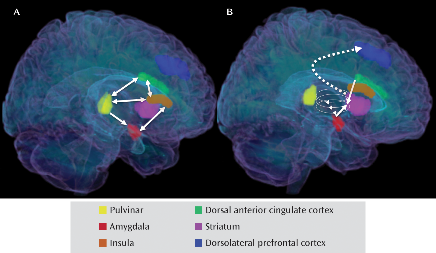
For over three decades, theorists have postulated that negative cognitive biases play a critical role in the onset and maintenance of major depressive disorder (78, 79). The findings of this meta-analysis concerning baseline activity and neural response to affective stimuli cohere into a remarkably clear and neuroanatomically viable model of this greater salience of negative stimuli in depression. Given the conceptualizations we have presented of the pulvinar nucleus, salience network, and dorsal cortical function and connectivity, as well as the dopamine-mediated ascending spiral trajectory of information through the cortical-striatal-pallidal-thalamic circuit, the present meta-analytic findings support a two-step model of enhanced processing of negative information in depression. We propose, first, that through monosynaptic projections to the amygdala, dorsal anterior cingulate cortex, and insula, abnormally high baseline activation in the pulvinar nucleus in depression potentiates responding of these components of the brain's salience network to negative stimuli; this processing may be sustained both by mutually excitatory connections between the insula and amygdala and between the insula and dorsal anterior cingulate cortex and by connections from the insula and dorsal anterior cingulate cortex back to the pulvinar nucleus (Figure 3A). Next, we propose that sensory data fail to propagate to the dorsal caudate and dorsolateral prefrontal cortex (Figure 3B), leading depressed individuals to fail to ascribe emotionally attenuating appraisals and contextual information to viscerally overcharged data from limbic structures. Given that nigrostriatal dopamine projections are instrumental in conducting information up the cortical-striatal-pallidal-thalamic circuit to the dorsolateral prefrontal cortex, both in vivo (80, 81) and postmortem (82) findings of low striatal dopamine activity in major depressive disorder suggest a potential mechanism by which data transfer through the dorsal caudate to the dorsolateral prefrontal cortex is attenuated in major depressive disorder. Recent meta-analytic results from Delaveau and colleagues (83) are important to consider in the present context, as they showed that key aspects of this model—overreaction of the amygdala and insula and underreaction of the dorsolateral prefrontal cortex in depression—resolve with antidepressant treatment, indicating that the present model accounts for state- as opposed to trait-related neural anomalies in major depressive disorder.
FIGURE 3. Neural Model of Biased Responding to Negative Information in Major Depressive Disordera
a Part A: high pulvinar activation at baseline potentiates responding in the amygdala, dorsal anterior cingulate cortex, and insula; mutual excitation among these components of the salience network and connectivity from them back to the pulvinar may sustain their activation. Part B: possibly because of low striatal dopamine levels in major depressive disorder, signals from the dorsal anterior cingulate cortex and amygdala fail to propagate up the ascending cortical-striatal-pallidal-thalamic circuit, resulting in diminished responses to negative stimuli in the dorsal striatum and dorsolateral prefrontal cortex.
This proposed model is distinct in important ways from current cortical-limbic inhibition models of major depressive disorder. First, this model posits a unidirectional, as opposed to a cyclical, pattern of neural influence between limbic and dorsal cortical structures in depression. Consistent with this formulation are the findings in our recent study of depressed participants, providing strong evidence of greater than normal limbic-to-dorsal cortical inhibition but no evidence of lower dorsal cortical-to-limbic inhibition (84) in depression. Second, our model posits a primary neural mechanism, exaggerated tonic pulvinar activation, as critical to initiating a bias toward the processing of negative information in major depressive disorder. Finally, this model accounts for patterns of limbic overresponsiveness and dorsal striatal and dorsal cortical underresponsiveness in depression within a well-validated and integrative neural processing architecture: the upward spiral model of the cortical-striatal-pallidal-thalamic circuit (77). This aspect of the model also accounts well for the lack of dorsal cortical responding—as opposed to dorsal cortical inhibition—that is often observed in major depression (85).
While this proposed model of negative information processing in major depressive disorder offers an integrative account of findings from a large body of neuroimaging research, it nevertheless leaves an important question unaddressed. The finding from our meta-analysis of excess baseline pulvinar activation in depression is primary in our proposed model. However, the reason for this difference is not clear. In this context, it is important to note that genetics may contribute to the anomalies in pulvinar activation reported in major depressive disorder. Indeed, a postmortem brain volumetry study (86) showed higher pulvinar volume in individuals who were homozygous for the short allele in the promoter region of the serotonin transporter gene (5-HTTLPR), a polymorphism that has been associated with greater risk for major depression (87, 88).

Limitations

We should note five limitations of the present study. First, because only a few neuroimaging studies that met the criteria for our meta-analysis involved participants with remitted depression, it was not possible in this meta-analysis to determine whether the anomalies we identified in major depressive disorder are related to the state of depression or are trait-related characteristics of individuals at risk for this disorder. However, the recent meta-analytic finding that amygdala and insula overactivation and dorsolateral prefrontal cortex underreaction normalize after pharmacotherapy for depression (83) indicates that the present meta-analytic results represent state- as opposed to trait-related aspects of neural functioning in depression. Second, while the present meta-analysis was adequately powered to detect reliable functional neural anomalies in major depression, it was not adequately powered to determine the effects of extraexperimental variables, such as psychotropic medication use by the depressed participants. While the meta-analysis of Delaveau and colleagues (83) indicates that key neural anomalies found in the present analysis actually normalize with use of psychotropic medication, our findings cannot speak to the issue of medication effects. Third, we did not include in this meta-analysis data from groups experiencing forms of psychopathology other than major depression. Thus, while we were able to provide a comprehensive neural characterization of major depressive disorder, the issue of the specificity of these findings to major depression remains to be addressed. Fourth, our meta-analysis of resting-state activation studies used data from three different functional neuroimaging modalities; noise introduced by including heterogeneous modalities may have decreased the sensitivity of our analysis. Similarly, noise introduced by the variety of affective challenge techniques used in the fMRI studies that were included in our meta-analysis may have affected the power of this analysis. Finally, because in this meta-analysis we set criteria for finding the strongest and most reliable findings in the literature concerning functional neuroimaging of depression, it is possible that we failed to detect weaker findings that are nevertheless important in understanding neural functioning in major depressive disorder. Indeed, results from previous meta-analytic investigations of major depression (14) indicated a broader array of neural anomalies in depression—both similar to those reported in the present analysis (e.g., greater amygdalar response to negative stimuli in depressed subjects than in healthy subjects) and different from the current findings (e.g., greater bilateral inferior frontal gyrus response to negative stimuli in depression). Certainly, these results should be accorded serious consideration in neural conceptualizations of major depressive disorder.

Summary and Future Directions

From the present meta-analytic synthesis, we formulated what we believe is an elegant and viable model of increased salience of negative information in major depressive disorder. We posit that through monosynaptic projections to the amygdala, dorsal anterior cingulate cortex, and insula, heightened baseline activity in the pulvinar nucleus in depression potentiates responding of these input nodes to the affective subdivision of the cortical-striatal-pallidal-thalamic circuit. We propose further that, potentially due to deficient striatal dopamine activity in major depression, there is a failure at nigrostriatal relays to propagate information to the dorsal striatum and dorsolateral prefrontal cortex; thus, attributions and appraisals that are typically initiated by the dorsolateral prefrontal cortex to reduce the impact of negative stimuli are not applied to viscerally charged sensory data in major depressive disorder.
Theorists have contended that biased processing of negative information plays a critical role in the etiology and maintenance of major depression (89). Moreover, the model supported by the current meta-analytic findings specifies crucial and independent neural anomalies underlying the enhanced processing of negative information in depression—specifically, high baseline pulvinar activation and low striatal dopamine levels. Thus, this model suggests that novel neural-level interventions, including neuroregulatory therapies for down-modulating pulvinar activity and pharmacotherapies for bolstering striatal dopamine availability, may be effective in the treatment of major depressive disorder.

Acknowledgments

The authors thank Ifeanyi Onyeji and Tara Lynn for their contributions to preparation of the manuscript.

Footnote

Received July 23, 2011; revision received Dec. 25, 2011; accepted Feb. 2, 2012.

Supplementary Material

File (appi.ajp.2012.11071105.ds001.pdf)
Supplementary Table and Figure

References

1.
World Health Organization: The Global Burden of Disease: 2004 Update. Geneva, WHO, 2004
2.
Trivedi MH, Rush AJ, Wisniewski SR, Nierenberg AA, Warden D, Ritz L, Norquist G, Howland RH, Lebowitz B, McGrath PJ, Shores-Wilson K, Biggs MM, Balasubramani GK, Fava M STAR*D Study Team: Evaluation of outcomes with citalopram for depression using measurement-based care in STAR*D: implications for clinical practice. Am J Psychiatry 2006; 163:28–40
3.
Mayberg HS: Limbic-cortical dysregulation: a proposed model of depression. J Neuropsychiatry Clin Neurosci 1997; 9:471–481
4.
Siegle GJ, Thompson W, Carter CS, Steinhauer SR, Thase ME: Increased amygdala and decreased dorsolateral prefrontal bold responses in unipolar depression: related and independent features. Biol Psychiatry 2007; 61:198–209
5.
Hamilton JP, Chen G, Thomason ME, Johnson RF, Gotlib IH: Investigating neural primacy in major depressive disorder: multivariate Granger causality analysis of resting-state fMRI time-series data. Mol Psychiatry 2011; 16:763–772
6.
Mayberg HS, Lozano AM, Voon V, McNeely HE, Seminowicz D, Hamani C, Schwalb JM, Kennedy SH: Deep brain stimulation for treatment-resistant depression. Neuron 2005; 45:651–660
7.
Fu CHY, Williams SCR, Cleare AJ, Scott J, Mitterschiffthaler MT, Walsh ND, Donaldson C, Suckling J, Andrew C, Steiner H, Murray RM: Neural responses to sad facial expressions in major depression following cognitive behavioral therapy. Biol Psychiatry 2008; 64:505–512
8.
Keedwell PA, Andrew C, Williams SCR, Brammer MJ, Phillips ML: The neural correlates of anhedonia in major depressive disorder. Biol Psychiatry 2005; 58:843–853
9.
Pizzagalli DA, Oakes TR, Fox AS, Chung MK, Larson CL, Abercrombie HC, Schaefer SM, Benca RM, Davidson RJ: Functional but not structural subgenual prefrontal cortex abnormalities in melancholia. Mol Psychiatry 2004; 9:325, 393–405
10.
Fitzgerald PB, Srithiran A, Benitez J, Daskalakis ZZ, Oxley TJ, Kulkarni J, Egan GF: An fMRI study of prefrontal brain activation during multiple tasks in patients with major depressive disorder. Hum Brain Mapp 2008; 29:490–501
11.
Hamilton JP, Gotlib IH: Neural substrates of increased memory sensitivity for negative stimuli in major depression. Biol Psychiatry 2008; 63:1155–1162
12.
Etkin A, Wager TD: Functional neuroimaging of anxiety: a meta-analysis of emotional processing in PTSD, social anxiety disorder, and specific phobia. Am J Psychiatry 2007; 164:1476–1488
13.
Wager TD, Phan KL, Liberzon I, Taylor SF: Valence, gender, and lateralization of functional brain anatomy in emotion: a meta-analysis of findings from neuroimaging. Neuroimage 2003; 19:513–531
14.
Fitzgerald PB, Laird AR, Maller J, Daskalakis ZJ: A meta-analytic study of changes in brain activation in depression. Hum Brain Mapp 2008; 29:683–695
15.
Spitzer RL, Williams JBW: Structured Clinical Interview for DSM-III-R (SCID). New York, New York State Psychiatric Institute, Biometrics Research, 1985
16.
Sartorius N, Kaelber CT, Cooper JE, Roper MT, Rae DS, Gulbinat W, Ustün TB, Regier DA: Progress toward achieving a common language in psychiatry: results from the field trial of the clinical guidelines accompanying the WHO classification of mental and behavioral disorders in ICD-10. Arch Gen Psychiatry 1993; 50:115–124
17.
Aihara M, Ida I, Yuuki N, Oshima A, Kurnano H, Takahashi K, Fukuda M, Oriuchi N, Endo K, Matsuda H, Mikuni M: HPA axis dysfunction in unmedicated major depressive disorder and its normalization by pharmacotherapy correlates with alteration of neural activity in prefrontal cortex and limbic/paralimbic regions. Psychiatry Res 2007; 155:245–256
18.
Bench CJ, Friston KJ, Brown RG, Scott LC, Frackowiak RSJ, Dolan RJ: The anatomy of melancholia—focal abnormalities of cerebral blood-flow in major depression. Psychol Med 1992; 22:607–615
19.
Brody AL, Saxena S, Mandelkern MA, Fairbanks LA, Ho ML, Baxter LR: Brain metabolic changes associated with symptom factor improvement in major depressive disorder. Biol Psychiatry 2001; 50:171–178
20.
Drevets WC, Videen TO, Price JL, Preskorn SH, Carmichael ST, Raichle ME: A functional anatomical study of unipolar depression. J Neurosci 1992; 12:3628–3641
21.
Germain A, Nofzinger EA, Meltzer CC, Wood A, Kupfer DJ, Moore RY, Buysse DJ: Diurnal variation in regional brain glucose metabolism in depression. Biol Psychiatry 2007; 62:438–445
22.
Kennedy SH, Evans KR, Krüger S, Mayberg HS, Meyer JH, McCann S, Arifuzzman AI, Houle S, Vaccarino FJ: Changes in regional brain glucose metabolism measured with positron emission tomography after paroxetine treatment of major depression. Am J Psychiatry 2001; 158:899–905
23.
Kimbrell TA, Ketter TA, George MS, Little JT, Benson BE, Willis MW, Herscovitch P, Post RM: Regional cerebral glucose utilization in patients with a range of severities of unipolar depression. Biol Psychiatry 2002; 51:237–252
24.
Kohn Y, Freedman N, Lester H, Krausz Y, Chisin R, Lerer B, Bonne O: Tc-99m-HMPAO SPECT study of cerebral perfusion after treatment with medication and electroconvulsive therapy in major depression. J Nuclear Med 2007; 48:1273–1278
25.
Krausz Y, Freedman N, Lester H, Barkai G, Levin T, Bocher M, Chisin R, Lerer B, Bonne O: Brain SPECT study of common ground between hypothyroidism and depression. Int J Neuropsychopharmacology 2007; 10:99–106
26.
Périco CA, Skaf CR, Yamada A, Duran F, Buchpiguel CA, Castro CC, Soares JC, Busatto GF: Relationship between regional cerebral blood flow and separate symptom clusters of major depression: a single photon emission computed tomography study using statistical parametric mapping. Neurosci Lett 2005; 384:265–270
27.
Saxena S, Brody AL, Ho ML, Alborzian S, Ho MK, Maidment KM, Huang SC, Wu HM, Au SC, Baxter LR : Cerebral metabolism in major depression and obsessive-compulsive disorder occurring separately and concurrently. Biol Psychiatry 2001; 50:159–170
28.
Skaf CR, Yamada A, Garrido GEJ, Buchpiguel CA, Akamine S, Castro CC, Busatto GF: Psychotic symptoms in major depressive disorder are associated with reduced regional cerebral blood flow in the subgenual anterior cingulate cortex: a voxel-based single photon emission computed tomography (SPECT) study. J Affect Disord 2002; 68:295–305
29.
Videbech P, Ravnkilde B, Pedersen AR, Egander A, Landbo B, Rasmussen NA, Andersen F, Stødkilde-Jørgensen H, Gjedde A, Rosenberg R: The Danish PET/depression project: PET findings in patients with major depression. Psychol Med 2001; 31:1147–1158
30.
Abler B, Erk S, Herwig U, Walter H: Anticipation of aversive stimuli activates extended amygdala in unipolar depression. J Psychiatr Res 2007; 41:511–522
31.
Bär KJ, Wagner G, Koschke M, Boettger S, Boettger MK, Schlosser R, Sauer H: Increased prefrontal activation during pain perception in major depression. Biol Psychiatry 2007; 62:1281–1287
32.
Canli T, Sivers H, Thomason ME, Whitfield-Gabrieli S, Gabrieli JDE, Gotlib IH: Brain activation to emotional words in depressed vs healthy subjects. Neuroreport 2004; 15:2585–2588
33.
Cooney RE, Joormann J, Eugene F, Dennis EL, Gotlib IH: Neural correlates of rumination in depression. Cogn Affect Behav Neurosci 2010; 10:470–478
34.
Frodl T, Scheuerecker J, Albrecht J, Kleemann AM, Muller-Schunk S, Koutsouleris N, Möller HJ, Brückmann H, Wiesmann M, Meisenzahl E: Neuronal correlates of emotional processing in patients with major depression. World J Biol Psychiatry 2009; 10:202–208
35.
Fu CHY, Williams SCR, Brammer MJ, Suckling J, Kim J, Cleare AJ, Walsh ND, Mitterschiffthaler MT, Andrew CM, Pich EM, Bullmore ET: Neural responses to happy facial expressions in major depression following antidepressant treatment. Am J Psychiatry 2007; 164:599–607
36.
Fu CHY, Williams SCR, Cleare AJ, Brammer MJ, Walsh ND, Kim J, Andrew CM, Pich EM, Williams PM, Reed LJ, Mitterschiffthaler MT, Suckling J, Bullmore ET: Attenuation of the neural response to sad faces in major depression by antidepressant treatment: a prospective, event-related functional magnetic resonance imaging study. Arch Gen Psychiatry 2004; 61:877–889
37.
Gotlib IH, Sivers H, Gabrieli JDE, Whitfield-Gabrieli S, Goldin P, Minor KL, Canli T: Subgenual anterior cingulate activation to valenced emotional stimuli in major depression. Neuroreport 2005; 16:1731–1734
38.
Herwig U, Bruhl AB, Kaffenberger T, Baumgartner T, Boeker H, Jancke L: Neural correlates of 'pessimistic' attitude in depression. Psychol Med 2010; 40:789–800
39.
Knutson B, Bhanji JP, Cooney RE, Atlas LY, Gotlib IH: Neural responses to monetary incentives in major depression. Biol Psychiatry 2008; 63:686–692
40.
Kumari V, Mitterschiffthaler MT, Teasdale JD, Malhi GS, Brown RG, Giampietro V, Brammer MJ, Poon L, Simmons A, Williams SC, Checkley SA, Sharma T: Neural abnormalities during cognitive generation of affect in treatment-resistant depression. Biol Psychiatry 2003; 54:777–791
41.
Lawrence NS, Williams AM, Surguladze S, Giampietro V, Brammer MJ, Andrew C, Frangou S, Ecker C, Phillips ML: Subcortical and ventral prefrontal cortical neural responses to facial expressions distinguish patients with bipolar disorder and major depression. Biol Psychiatry 2004; 55:578–587
42.
Mitterschiffthaler MT, Kumari V, Malhi GS, Brown RG, Giampietro VP, Brammer MJ, Suckling J, Poon L, Simmons A, Andrew C, Sharma T: Neural response to pleasant stimuli in anhedonia: an fMRI study. Neuroreport 2003; 14:177–182
43.
Mitterschiffthaler MT, Williams SCR, Walsh ND, Cleare AJ, Donaldson C, Scott J, Fu CH: Neural basis of the emotional Stroop interference effect in major depression. Psychol Med 2008; 38:247–256
44.
Osuch EA, Bluhm RL, Williamson PC, Theberge J, Densmore M, Neufeld RWJ: Brain activation to favorite music in healthy controls and depressed patients. Neuroreport 2009; 20:1204–1208
45.
Pizzagalli DA, Holmes AJ, Dillon DG, Goetz EL, Birk JL, Bogdan R, Dougherty DD, Iosifescu DV, Rauch SL, Fava M: Reduced caudate and nucleus accumbens response to rewards in unmedicated individuals with major depressive disorder. Am J Psychiatry 2009; 166:702–710
46.
Scheuerecker J, Meisenzahl EM, Koutsouleris N, Roesner M, Schopf V, Linn J, Wiesmann M, Brückmann H, Möller HJ, Frodl T: Orbitofrontal volume reductions during emotion recognition in patients with major depression. J Psychiatry Neurosci 2010; 35:311–320
47.
Strigo IA, Simmons AN, Matthews SC, Craig AD, Paulus MP: Association of major depressive disorder with altered functional brain response during anticipation and processing of heat pain. Arch Gen Psychiatry 2008; 65:1275–1284
48.
Surguladze S, Brammer MJ, Keedwell P, Giampietro V, Young AW, Travis MJ, Williams SC, Phillips ML: A differential pattern of neural response toward sad versus happy facial expressions in major depressive disorder. Biol Psychiatry 2005; 57:201–209
49.
Townsend JD, Eberhart NK, Bookheimer SY, Eisenberger NI, Foland-Ross LC, Cook IA, Sugar CA, Altshuler LL: fMRI activation in the amygdala and the orbitofrontal cortex in unmedicated subjects with major depressive disorder. Psychiatry Res 2010; 183:209–217
50.
Wang LH, Labar KS, Smoski M, Rosenthal MZ, Dolcos F, Lynch TR, Krishnan RR, McCarthy G: Prefrontal mechanisms for executive control over emotional distraction are altered in major depression. Psychiatry Res 2008; 163:143–155
51.
Yang JC: Functional neuroanatomy in depressed patients with sexual dysfunction: blood oxygenation level dependent functional MR imaging. Korean J Radiol 2004; 5:87–95
52.
Green DP, Salovey P: In what sense are positive and negative affect independent? a reply to Tellegen, Watson, and Clark. Psychol Science 1999; 10:304–306
53.
Talairach J, Tournoux P: Co-Planar Stereotaxic Atlas of the Human Brain. Stuttgart, Germany, Thieme, 1988
54.
Cox RW: AFNI: software for analysis and visualization of functional magnetic resonance neuroimages. Comput Biomed Res 1996; 29:162–173
55.
Jones EG, Burton H: Projection from medial pulvinar to amygdala in primates. Brain Res 1976; 104:142–147
56.
LeDoux JE: The Emotional Brain. New York, Simon & Schuster, 1996
57.
Pessoa L, Adolphs R: Emotion processing and the amygdala: from a 'low road' to 'many roads' of evaluating biological significance. Nat Rev Neurosci 2010; 11:773–782
58.
Mufson EJ, Mesulam MM: Thalamic connections of the insula in the rhesus-monkey and comments on the paralimbic connectivity of the medial pulvinar nucleus. J Comp Neurol 1984; 227:109–120
59.
Ward R, Danziger S, Owen V, Rafal R: Deficits in spatial coding and feature binding following damage to spatiotopic maps in the human pulvinar. Nat Neurosci 2002; 5:99–100
60.
Treisman A: Solutions to the binding problem: progress through controversy and convergence. Neuron 1999; 24:105–110
61.
Padmala S, Lim S-L, Pessoa L: Pulvinar and affective significance: responses track moment-to-moment stimulus visibility. Front Hum Neurosci 2010; 4:64
62.
Seeley WW, Menon V, Schatzberg AF, Keller J, Glover GH, Kenna H, Reiss AL, Greicius MD: Dissociable intrinsic connectivity networks for salience processing and executive control. J Neurosci 2007; 27:2349–2356
63.
Davis M, Whalen PJ: The amygdala: vigilance and emotion. Mol Psychiatry 2001; 6:13–34
64.
Critchley HD, Mathias CJ, Josephs O, O'Doherty J, Zanini S, Dewar BK, Cipolotti L, Shallice T, Dolan RJ: Human cingulate cortex and autonomic control: converging neuroimaging and clinical evidence. Brain 2003; 126:2139–2152
65.
Mufson EJ, Mesulam MM, Pandya DN: Insular interconnections with the amygdala in the rhesus-monkey. Neuroscience 1981; 6:1231–1248
66.
Allman JM, Tetreault NA, Hakeem AY, Manaye KF, Semendeferi K, Erwin JM, Park S, Goubert V, Hof PR: The von Economo neurons in frontoinsular and anterior cingulate cortex in great apes and humans. Brain Struct Funct 2010; 214:495–517
67.
Craig AD: How do you feel—now? the anterior insula and human awareness. Nat Rev Neurosci 2009; 10:59–70
68.
Koechlin E, Summerfield C: An information theoretical approach to prefrontal executive function. Trends Cogn Sci 2007; 11:229–235
69.
Kondo H, Osaka N, Osaka M: Cooperation of the anterior cingulate cortex and dorsolateral prefrontal cortex for attention shifting. Neuroimage 2004; 23:670–679
70.
Nee DE, Wager TD, Jonides J: Interference resolution: insights from a meta-analysis of neuroimaging tasks. Cogn Affect Behav Neurosci 2007; 7:1–17
71.
Wood JN, Grafman J: Human prefrontal cortex: processing and representational perspectives. Nat Rev Neurosci 2003; 4:139–147
72.
Braver TS, Cohen JD, Nystrom LE, Jonides J, Smith EE, Noll DC: A parametric study of prefrontal cortex involvement in human working memory. Neuroimage 1997; 5:49–62
73.
Lewis SJG, Dove A, Robbins TW, Barker RA, Owen AM: Striatal contributions to working memory: a functional magnetic resonance imaging study in humans. Eur J Neurosci 2004; 19:755–760
74.
Ochsner KN, Bunge SA, Gross JJ, Gabrieli JDE: Rethinking feelings: an fMRI study of the cognitive regulation of emotion. J Cogn Neurosci 2002; 14:1215–1229
75.
Kemp JM, Powell TPS: Connexions of striatum and globus pallidus—synthesis and speculation. Philos Trans R Soc Lond B Biol Sci 1971; 262:441–457
76.
Alexander GE, Delong MR, Strick PL: Parallel organization of functionally segregated circuits linking basal ganglia and cortex. Annu Rev Neurosci 1986; 9:357–381
77.
Haber SN, Fudge JL, McFarland NR: Striatonigrostriatal pathways in primates form an ascending spiral from the shell to the dorsolateral striatum. J Neurosci 2000; 20:2369–2382
78.
Beck AT: Cognitive Therapy and the Emotional Disorders. New York, International Universities Press, 1976
79.
Gotlib IH, Joormann J: Cognition and depression: current status and future directions. Annu Rev Clin Psychol 2010; 6:285–312
80.
Meyer JH, Kruger S, Wilson AK, Christensen BK, Goulding VS, Schaffer A, Minifie C, Houle S, Hussey D, Kennedy SH: Lower dopamine transporter binding potential in striatum during depression. Neuroreport 2001; 12:4121–4125
81.
Meyer JH, McNeely HE, Sagrati S, Boovariwala A, Martin K, Verhoeff NPLG, Wilson AA, Houle S: Elevated putamen D2 receptor binding potential in major depression with motor retardation: an [11C]raclopride positron emission tomography study. Am J Psychiatry 2006; 163:1594–1602
82.
Bowden C, Cheetham SC, Lowther S, Katona CLE, Crompton MR, Horton RW: Reduced dopamine turnover in the basal ganglia of depressed suicides. Brain Res 1997; 769:135–140
83.
Delaveau P, Jabourian M, Lemogne C, Guionnet S, Bergouignan L, Fossati P: Brain effects of antidepressants in major depression: a meta-analysis of emotional processing studies. J Affect Disord 2011; 130:66–74
84.
Hamilton JP, Chen G, Thomason ME, Schwartz ME, Gotlib IH: Investigating neural primacy in major depressive disorder: multivariate Granger causality analysis of resting-state fMRI time-series data. Mol Psychiatry 2011; 16:763–772
85.
Hooley JM, Gruber SA, Scott LA, Hiller JB, Yurgelun-Todd DA: Activation in dorsolateral prefrontal cortex in response to maternal criticism and praise in recovered depressed and healthy control participants. Biol Psychiatry 2005; 57:809–812
86.
Young KA, Holcomb LA, Bonkale WL, Hicks PB, Yazdani U, German DC: 5HTTLPR polymorphism and enlargement of the pulvinar: unlocking the backdoor to the limbic system. Biol Psychiatry 2007; 61:813–818
87.
Caspi A, Sugden K, Moffitt TE, Taylor A, Craig IW, Harrington H, McClay J, Mill J, Martin J, Braithwaite A, Poulton R: Influence of life stress on depression: moderation by a polymorphism in the 5-HTT gene. Science 2003; 301:386–389
88.
Gotlib IH, Joormann J, Minor KL, Hallmayer J: HPA axis reactivity: a mechanism underlying the associations among 5-HTTLPR, stress, and depression. Biol Psychiatry 2008; 63:847–851
89.
Beck AT, Rush AJ, Shaw BF, Emery G: Cognitive Therapy of Depression. New York, Guilford, 1979

Information & Authors

Information

Published In

Go to American Journal of Psychiatry
American Journal of Psychiatry
Pages: 693 - 703
PubMed: 22535198

History

Received: 23 July 2011
Revision received: 25 December 2011
Accepted: 2 February 2012
Published online: 1 July 2012
Published in print: July 2012

Authors

Details

J. Paul Hamilton, Ph.D.
From the Department of Psychology and the Department of Psychiatry and Behavioral Sciences, Stanford University, Stanford, Calif.; and the Department of Veterans Affairs Palo Alto Health Care System, Palo Alto, Calif.
Amit Etkin, M.D., Ph.D.
From the Department of Psychology and the Department of Psychiatry and Behavioral Sciences, Stanford University, Stanford, Calif.; and the Department of Veterans Affairs Palo Alto Health Care System, Palo Alto, Calif.
Daniella J. Furman, M.A.
From the Department of Psychology and the Department of Psychiatry and Behavioral Sciences, Stanford University, Stanford, Calif.; and the Department of Veterans Affairs Palo Alto Health Care System, Palo Alto, Calif.
Maria G. Lemus, B.A.
From the Department of Psychology and the Department of Psychiatry and Behavioral Sciences, Stanford University, Stanford, Calif.; and the Department of Veterans Affairs Palo Alto Health Care System, Palo Alto, Calif.
Rebecca F. Johnson, B.A.
From the Department of Psychology and the Department of Psychiatry and Behavioral Sciences, Stanford University, Stanford, Calif.; and the Department of Veterans Affairs Palo Alto Health Care System, Palo Alto, Calif.
Ian H. Gotlib, Ph.D.
From the Department of Psychology and the Department of Psychiatry and Behavioral Sciences, Stanford University, Stanford, Calif.; and the Department of Veterans Affairs Palo Alto Health Care System, Palo Alto, Calif.

Notes

Address correspondence to Dr. Hamilton ([email protected]).

Funding Information

All authors report no financial relationships with commercial interests.Preparation of this manuscript was supported by grant MH-59259 from NIMH to Dr. Gotlib and grant MH-079651 from NIMH to Dr. Hamilton.

Metrics & Citations

Metrics

Citations

Export Citations

If you have the appropriate software installed, you can download article citation data to the citation manager of your choice. Simply select your manager software from the list below and click Download.

For more information or tips please see 'Downloading to a citation manager' in the Help menu.

Format
Citation style
Style
Copy to clipboard

View Options

View options

PDF/EPUB

View PDF/EPUB

Login options

Already a subscriber? Access your subscription through your login credentials or your institution for full access to this article.

Personal login Institutional Login Open Athens login
Purchase Options

Purchase this article to access the full text.

PPV Articles - American Journal of Psychiatry

PPV Articles - American Journal of Psychiatry

Not a subscriber?

Subscribe Now / Learn More

PsychiatryOnline subscription options offer access to the DSM-5-TR® library, books, journals, CME, and patient resources. This all-in-one virtual library provides psychiatrists and mental health professionals with key resources for diagnosis, treatment, research, and professional development.

Need more help? PsychiatryOnline Customer Service may be reached by emailing [email protected] or by calling 800-368-5777 (in the U.S.) or 703-907-7322 (outside the U.S.).

Media

Figures

Other

Tables

Share

Share

Share article link

Share