
Abstract

Objective:

Detecting brain abnormalities in clinical high-risk populations before the onset of psychosis is important for tracking pathological pathways and for identifying possible intervention strategies that may impede or prevent the onset of psychotic disorders. Co-occurring cellular and extracellular white matter alterations have previously been implicated after a first psychotic episode. The authors investigated whether or not cellular and extracellular alterations are already present in a predominantly medication-naive cohort of clinical high-risk individuals experiencing attenuated psychotic symptoms.

Methods:

Fifty individuals at clinical high risk, of whom 40 were never medicated, were compared with 50 healthy control subjects, group-matched for age, gender, and parental socioeconomic status. 3-T multishell diffusion MRI data were obtained to estimate free-water imaging white matter measures, including fractional anisotropy of cellular tissue (FAT) and the volume fraction of extracellular free water (FW).

Results:

Significantly lower FAT was observed in the clinical high-risk group compared with the healthy control group, but no statistically significant FW alterations were observed between groups. Lower FAT in the clinical high-risk group was significantly associated with a decline in Global Assessment of Functioning Scale (GAF) score compared with highest GAF score in the previous 12 months.

Conclusions:

Cellular but not extracellular alterations characterized the clinical high-risk group, especially in those who experienced a decline in functioning. These cellular changes suggest an early deficit that possibly reflects a predisposition to develop attenuated psychotic symptoms. In contrast, extracellular alterations were not observed in this clinical high-risk sample, suggesting that previously reported extracellular abnormalities may reflect an acute response to psychosis, which plays a more prominent role closer to or at onset of psychosis.
Schizophrenia is a severe psychiatric disorder that emerges most commonly in late adolescence and early adulthood, affecting approximately 1% of the population (1). Recent studies have focused on detection and treatment intervention in the early stages of psychotic illness (2), especially during the clinical high-risk phase, before a full-blown disorder is present (3, 4). Because psychotic symptoms, cognitive dysfunctions, and disturbances in social functions may all evolve in the clinical high-risk phase (58), investigating brain alterations in clinical high-risk individuals could potentially shed light on their underlying mechanisms.
Studies in the past two decades, and especially those applying diffusion MRI (dMRI) techniques, strongly suggest white matter abnormalities in schizophrenia, where disrupted cortico-cortical connections may play a key role in both clinical symptoms and cognitive dysfunctions (913). Most studies apply diffusion tensor imaging (DTI) (14, 15) to estimate fractional anisotropy (FA), a measure sensitive to the arrangement and microstructure of white matter, often used to assess white matter integrity (9, 12, 13, 16). Lower FA, however, may be caused by different factors, including axon diameter alterations, alterations in myelin sheath, increased extracellular volume fraction, and alterations in the organization of axonal fibers (1723). Consequently, FA provides a nonspecific and less than optimal estimate of white matter integrity (24), making it hard to relate FA changes to a specific underlying pathology. In addition, meta-analysis has shown that FA alterations may be affected by illness stage and by exposure to antipsychotic or antidepressant medications (25). Thus, additional methodological advances and a careful selection of study populations are required to determine the source(s) of white matter alterations and their association with illness progression.
One way to increase FA specificity is to correct for partial volume effects with extracellular water. For example, free-water imaging analysis (19, 23, 26) uses two compartments to separately model diffusion properties of brain tissue and those of surrounding free water, which can be found only in the extracellular space (19). Fractional anisotropy of cellular tissue (FAT), which is more specific to white matter cellular alterations than conventional FA measures, is derived from the tissue compartment after the elimination of the free-water contribution. In addition, this model estimates the fractional volume of the free-water compartment (FW), which is affected by processes that change the extracellular space, such as atrophy and neuroinflammation (18, 27). FW maps can also be estimated using other dMRI techniques (e.g., 17, 28, 29), and across methods, they show high signal in CSF-filled cavities and lower, albeit nonzero, signal throughout the white matter.
Previous free-water imaging studies comparing patients after the first psychotic episode with healthy control subjects have shown widespread higher FW with limited focal lower FAT in frontal lobe regions of patients (18, 30). In chronic patients, the reverse has been noted—that is, a greater extent of significantly lower FAT with less extensive significantly higher FW (27, 30). These results suggest that white matter abnormalities may be explained by co-occurring alterations: extracellular free-water alterations, measured by FW, and microstructural cellular alterations, measured by FAT. The higher FW may thus reflect an acute brain response to psychosis, which is less pronounced in chronic stages, while lower FAT may reflect preexisting neurodevelopmental abnormalities and/or a continuous process of accumulating cellular damage.
An important step toward understanding the source of these different white matter alterations is to study clinical high-risk individuals to determine whether or not extracellular and cellular alterations are present before the onset of psychosis, and if so, to what extent. There have been only a limited number of dMRI studies in clinical high-risk samples, most of which were based on DTI, and most of which had small sample sizes. Two separate white matter fiber tracking studies (31, 32) reported no significant differences between clinical high-risk individuals and healthy control subjects. In contrast, three other studies showed limited frontal regions with lower FA (3335). In two additional tract-based spatial statistics (TBSS) studies (36, 37), extensive regions with lower FA were observed in the corpus callosum, the superior longitudinal fasciculus, the internal capsule, and the corona radiata of clinical high-risk individuals. In one study that applied free-water imaging in a clinical high-risk cohort, group differences were found in FAT in fibers connecting the salience network (38), but FW has not been tested. These studies, taken together, suggest that although white matter alterations are likely present in clinical high-risk individuals, they are inconsistent, may not have a distinct location, and are more subtle than those observed in schizophrenia.
In this study, we went beyond previous studies by performing a dMRI study on a large clinical high-risk sample that was recruited in China, where most individuals were naive to psychotropic medications. Furthermore, we used free-water analysis to separately evaluate the extent and putative roles of extracellular and tissue-related microstructural white matter. Based on our first-episode study, we predicted that both types of alterations (higher FW and lower FAT) would be present in the clinical high-risk stage. In addition, based on the extent of the alterations, we sought to identify which of these processes, extracellular or cellular changes, are more related to attenuated symptoms prior to onset of psychosis.

Methods

Participants

The Shanghai At Risk for Psychosis (SHARP) program of Shanghai Mental Health Center recruited help-seeking clinical high-risk individuals from their first outpatient assessment. A 2-year conversion rate to psychosis of 29.1% was previously reported in this predominantly medication-naive Chinese population (39). All participants met criteria for clinical high risk (Table 1), defined by the Chinese version of the Structured Interview for Prodromal Syndromes (SIPS) and the Scale of Prodromal Symptoms (SOPS) (40), administered by a senior psychiatrist (39, 41). The mean intervals between recruitment and MRI scan was 2.5 days (SD=7.7). The Global Assessment of Functioning Scale (GAF) was administered at the time of recruitment, and retrospectively for the previous 12 months. Functional decline (per SIPS definition) was calculated as the drop in the current GAF score compared with the patient’s highest GAF score in the previous 12 months (39, 42). Conversion to psychosis was assessed at least 1 year after the baseline assessment with the SIPS “presence of psychotic symptoms” criteria (43, 44). The first 50 clinical high-risk individuals who passed imaging quality control evaluation (see below) were included in the study.
TABLE 1. Number of clinical high-risk participants meeting the different criteria for clinical high risk and experiencing symptoms
Criteria or SymptomsN
Attenuated positive symptom syndrome (APSS)45
Brief intermittent psychotic syndrome2
Genetic risk and deterioration syndrome (GRDS)1
APSS + GRDS2
P1, unusual thought content >241
P2, suspiciousness >242
P3, grandiose ideas >20
P4, perceptual abnormalities >222
P5, disorganized communication2
Healthy control subjects were recruited through online advertisements, and 50 healthy subjects who passed imaging quality control and were matched in age and gender to the clinical high-risk sample (Table 2) were included in the study. Exclusion criteria at study entry for all participants included head injury with loss of consciousness of any duration; any history of substance use, neurological disease, severe somatic diseases; IQ below 70; and dementia. Control subjects were additionally excluded if they met criteria for a psychotic disorder or a clinical high-risk syndrome (determined by the SIPS) or any other mental disorder defined by DSM-IV. All study subjects were right-handed by self-report.
TABLE 2. Demographic and clinical characteristics of individuals at clinical high risk for psychosis and healthy control subjectsa
CharacteristicClinical High-Risk Group (N=50)Healthy Control Group (N=50)
 N%N%
Female2040.02040.0
Right-handed50100.050100.0
 MeanSDMeanSD
Ageb (years)19.74.619.23.9
Education (years)10.52.411.22.3
Parental education (years)    
 Father10.23.910.34.2
 Mother9.54.410.33.8
Average in-scanner motion (mm)1.0960.1891.1000.183
Duration of symptoms (months)6.56.2  
Scale of Prodromal Symptoms    
 Positive9.03.2  
 Negative12.76.4  
 Disorganization6.72.9  
 General9.13.2  
Global Assessment of Functioning Scale    
 Highest score in past 12 months76.86.1  
 Current score51.37.0  
 N%  
Antipsychotics or antidepressants1020.0  
a
There were no significant differences between groups on nonclinical variables.
b
Age ranged from 14 to 32 years in the clinical high-risk group and from 14 to 31 years in the healthy control group.
The study protocol and consent form were reviewed and approved by the local ethics committee at the Shanghai Mental Health Center and by the Beth Israel Deaconess Medical Center. Written informed consent was obtained from all participants.

MRI Data Acquisition

All participants underwent MRI scanning at the Shanghai Mental Health Center using a 3-T Verio scanner (Siemens, Munich) with a 32-channel head coil. Multishell dMRI was acquired with 30 gradient directions at b=1000 s/mm2, six at b=500 s/mm2, three at b=200 s/mm2, and five interleaved b0 images. An additional 30 gradient directions at b=3000 s/mm2 were collected for tractography studies, although the latter shell was omitted from calculations in order to circumvent non-Gaussian effects. The sequence had 70 contiguous axial slices, a 256 mm field of view, 2 mm isotropic voxels, a repetition time of 15,800 ms, and an echo time of 109 ms. Acquisition time was 20 minutes 17 seconds, with 6/8 phase partial Fourier, and generalized autocalibrating partial parallel acquisition (GRAPPA) with acceleration factor 2. Scanning parameters and acquisitions at the Shanghai Mental Health Center were supervised by members of the Psychiatry Neuroimaging Laboratory at Brigham and Women’s Hospital, with visits to Shanghai and ongoing communication with the chief technical scientist (Y.T.) at Shanghai.

Image Analysis

All scans included in the study passed quality control, which included visual inspection of the raw data and visual inspection of the output images by trained raters at the Psychiatry Neuroimaging Laboratory. Three clinical high-risk individuals whose scans had incomplete brain coverage and 10 clinical high-risk individuals whose scans had severe image artifacts (e.g., multiple dropped signals, blurring, or ghosting) were excluded from the study. One control subject was excluded because of severe image artifacts. Thus, in total, 63 clinical high-risk individuals and 51 matching healthy control subjects were considered before selecting the 50 clinical high-risk individuals and 50 control subjects who were included in the study. Minor head motion and eddy current distortions were corrected using low-level registration functions (e.g., FLIRT and FNIRT) from FSL, version 5.0.4 (http://fsl.fmrib.ox.ac.uk/fsl) as part of the Psychiatry Neuroimaging Laboratory’s analysis pipeline (https://github.com/pnlbwh/pnlutil). A motion parameter (the between-volume displacement averaged across all volumes) was estimated from these transformations, and gradient orientations were corrected for the rotation component. Brain masks were manually edited in 3D Slicer (www.slicer.org). FA maps were generated by a nonlinear fit estimation of diffusion tensors. We used TBSS to register all subjects to the FSL-provided Montreal Neurological Institute template and to generate a white matter skeleton from the FA maps, which is less susceptible to between-subject misregistration and is focused on the center of fibers, where less partial volume is expected (45, 46).
The free-water analysis has been described in detail elsewhere (18, 19, 27). Briefly, water diffusion within each voxel is modeled using two compartments (19): an isotropic free-water compartment with the diffusion coefficient of free water in body temperature (3×10⁻³ mm2/s), and a second compartment, the tissue compartment, modeled by a diffusion tensor, accounting for water molecules that are not free to diffuse because they are hindered or restricted by tissue components such as membranes. Measures obtained from this model, using a nonlinear and regularized fit (19), are the free-water compartment fraction (FW) and the fractional anisotropy of cellular tissue (FAT), calculated from the tissue compartment’s tensor. For completeness, additional parameters were calculated from the tissue compartment’s tensor, namely, radial diffusivity (RDT) and axial diffusivity (ADT). In contrast to most previously reported studies, here the model was estimated from multishell diffusion imaging data, where the model fit becomes more stable and robust compared with single-shell data, which is typically used in DTI studies (23, 47). FW, FAT, RDT, and ADT maps were calculated for each subject and then projected onto the TBSS skeleton for statistical analysis.

Statistical Analysis

For the first level of analysis, FW and FAT values were averaged across the white matter skeleton in order to identify differences between groups that may not necessarily be location dependent. Linear regression models were constructed to compare clinical high-risk individuals with healthy control subjects, where the average diffusivity measure (FW or FAT) was the dependent variable, and group (healthy control or clinical high-risk), age, gender, and motion were predictor variables. Additional linear regression models were constructed separately for each group. To further interrogate any FAT effects, tests were repeated for the ADT and RDT values.
Bonferroni-corrected Pearson product moment (when normally distributed) or Spearman correlation analyses were used to test for associations between diffusivity measures (FAT and FW) and clinical characteristics (duration of symptoms, functional decline, and the four SOPS scores). In the Results section, p values are reported as significant if they were below the Bonferroni-corrected threshold equivalent to p=0.05. For comparison with previous DTI studies, selected analyses were also performed for the conventional FA measure.
To identify the spatial extent of group differences, in a second level of analysis, permutation tests were used for group comparisons for each voxel on the white matter skeleton, using the randomize tool in FSL (48). Threshold-free cluster enhancement (TFCE) (49) was applied to control for family-wise errors, with a significance threshold (TFCE modified) of p<0.05. Age, gender, and motion were included as covariates. Anatomical locations were identified using the ICBM-DTI-81 atlas (50).

Results

Demographic and Clinical Characteristics

There were no significant differences between the clinical high-risk and healthy control groups in age, gender, education, motion, or handedness (see Table 2). Clinical high-risk individuals experienced attenuated positive symptoms (see Table 1), manifested primarily as elevated symptoms on P1 (unusual thought content/delusional ideas) and P2 (suspiciousness/persecutory ideas) of the SOPS. The mean duration of prodromal symptoms at assessment was 6.5 months (SD=6.2). Forty of the 50 clinical high-risk individuals had never been treated with antipsychotics or antidepressants. Ten clinical high-risk individuals had been treated with antipsychotic or antidepressant medications for less than 3 weeks (aripiprazole, N=3; risperidone, N=2; quetiapine, N=1; sulpiride, N=1; citalopram, N=1; and olanzapine, paroxetine, and lamotrigine, N=1), and one had been treated with an antipsychotic for 3 months (risperidone). The 10 medicated participants (eight of them male; mean age, 19.3 years [SD=3.7]) had significantly more functional decline than the nonmedicated participants (t=2.12, df=48, p=0.04); they also had more negative symptoms, although the difference fell short of significance (t=1.78, df=48, p=0.08). There were no other significant differences in demographic or clinical characteristics between the medicated and nonmedicated clinical high-risk individuals (see Table S1 in the online supplement).
Within the 1-year follow-up evaluation, 11 clinical high-risk individuals (seven of them male; mean age, 18.4 years [SD=2.2]) converted to psychosis and were diagnosed with schizophrenia. The average time between initial assessment and conversion was 6.27 months (SD=5.96, range=1–17). Nine of those who converted to psychosis had never been medicated at baseline. There were no significant differences in age, gender, or clinical scores between clinical high-risk individuals who converted to psychosis and those who did not.

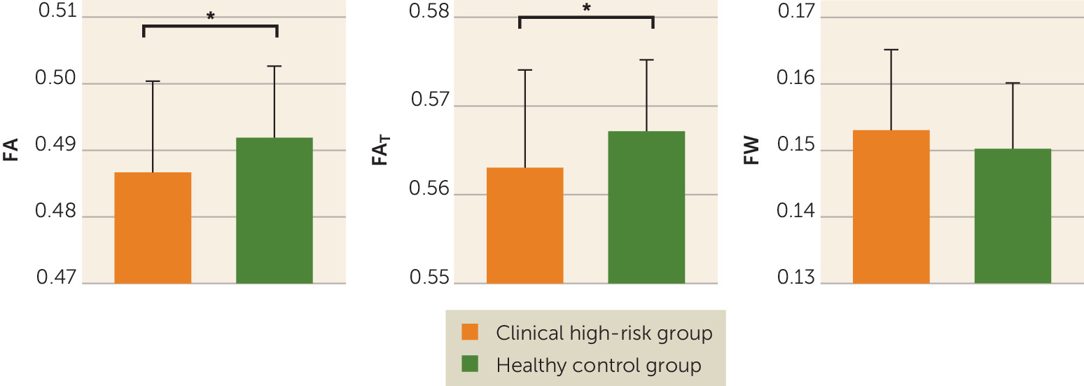
Whole Brain Average Diffusivity Comparison of Clinical High-Risk and Healthy Control Groups

In comparing average diffusivities across the entire white matter skeleton, we found that the clinical high-risk group had significantly lower FA compared with the healthy control group (0.487 [SE=0.002] and 0.492 [SE=0.002], respectively; F=5.672, df=1, 95, p=0.019; Cohen’s d=−0.422) (Figure 1). When applying the free-water model, we found that the clinical high-risk group had significantly lower FAT than the healthy control group (0.563 [SE=0.002] and 0.567 [SE=0.001], respectively; F=5.556, df=1, 95, p=0.020; Cohen’s d=−0.424) but not significantly higher FW (0.153 [SE=0.002] and 0.150 [SE=0.001], respectively; F=2.243, df=1, 95, p=0.137; Cohen’s d=0.034) (Figure 1). The lower FAT coincided with lower ADT (F=6.348, df=1, 95, p=0.013; Cohen’s d=−0.499) but not with higher RD
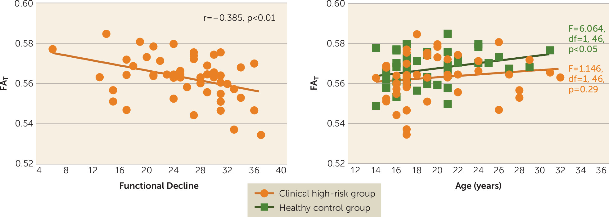
T (F=1.295, df=1, 95, p=0.258; Cohen’s d=0.164). Additionally, FAT was negatively correlated with functional decline in the clinical high-risk group (r=−0.385, df=48, p=0.006) (Figure 2); that is, individuals with lower FAT tended to have more functional decline. Significant correlations with functional decline were also found for the ADT measure (r=−0.299, df=48, p=0.035) and the RDT measure (r=0.300, df=48, p=0.034). The uncorrected FA measure was also correlated with functional decline, although to a lesser degree than FAT (r=−0.357, df=48, p=0.011). There was no significant correlation between FW and functional decline (r=0.161, df=48, p=0.265). There were also no significant correlations between the diffusion measures (FA, FAT, ADT, RDT, and FW) and any other clinical score.
FIGURE 1. Fractional anisotropy, fractional anisotropy of cellular tissue, and extracellular free water in individuals at clinical high risk for psychosis and healthy control subjectsa
a The clinical high-risk group had statistically significantly lower average fractional anisotropy (FA) over the whole white matter skeleton compared with the healthy control group. Free-water imaging measures showed that the clinical high-risk group had significant lower fractional anisotropy of cellular tissue (FAT) but no group difference in the volume fraction of extracellular free water (FW), suggesting that the lower FA may be explained by cellular changes rather than by extracellular changes.
*p<0.05.
FIGURE 2. Association of functional decline and age with fractional anisotropy of cellular tissue in individuals at clinical high risk for psychosis and healthy control subjectsa
a Fractional anisotropy of cellular tissue (FAT) was statistically significantly negatively correlated with functional decline (calculated as the drop in the current Global Assessment of Functioning Scale score compared with the individual's highest GAF score in the previous 12 months) in the clinical high-risk group. FAT was also statistically significantly positively correlated with age (right) in the healthy control group but not in the clinical high-risk group.
Gender (F=4.337, df=1, 95, p=0.040) and age (F=5.951, df=1, 95, p=0.017) were significant predictors of FAT across the entire sample. There was no significant interaction of group and gender (F=1.706, df=2, 97, p=0.187). However, for FAT there was a significant interaction of group and age (F=4.789, df=2, 97, p=0.010), suggesting age-related differences between groups. Follow-up linear models within the healthy control group showed that FAT was positively correlated with age (F=6.064, df=1, 46, p=0.018). However, in the clinical high-risk group, FAT was not correlated with age (F=1.146, df=1, 46, p=0.290).
There were no significant differences between nonmedicated clinical high-risk individuals and those who received antipsychotics or antidepressants on either of the free-water measures (for FAT, F=2.003, df=1, 45, p=0.164; Cohen’s d=0.425; and for FW, F=0.051, df=1, 45, p=0.822; Cohen’s d=0.018). For the nonmedicated clinical high-risk group, correlation with functional decline remained significant (r=−0.397, df=38, p=0.011), although the decrease in FAT compared with the healthy control group fell short of significance (F=2.931, df=1, 85, p=0.091; Cohen’s d=−0.278). There were no significant group differences in FAT (F=2.078, df=1, 45, p=0.156; Cohen’s d=0.423) or FW (F=0.006, df=1, 45, p=0.939; Cohen’s d=0.128) between clinical high-risk individuals who converted to psychosis and those who did not.
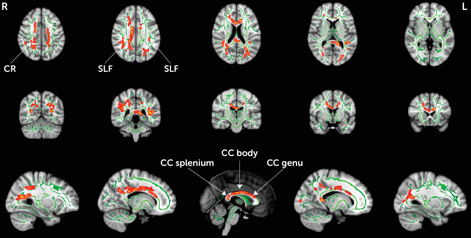
Voxel-Wise Analyses

To better understand the extent of microstructural alterations, FAT and FW were compared between the clinical high-risk and healthy control groups using a voxel-wise TBSS analysis. Similar to the whole brain analysis above, there were no group differences in FW. The statistical analysis identified lower FAT in the clinical high-risk group in 4% of the skeleton (5,363 voxels), and in the following locations: the genu, body, and splenium of the corpus callosum; the right anterior, superior, and posterior corona radiata; and the left and right superior longitudinal fasciculus (Figure 3). These locations largely overlapped with the group differences in FA (see Figure S1 in the online supplement), which covered 10.6% of the skeleton (14,267 voxels). There were no voxel-wise correlations between FAT and clinical parameters. However, there was a significant correlation (r=−0.405, df=48, p=0.004) between functional decline and FAT averaged across all voxels that showed significant FAT group differences. There were no significant voxel-wise group differences in ADT or RDT. Also, we did not identify any significant group differences between clinical high-risk individuals who converted to psychosis and those who did not.
FIGURE 3. Voxel-wise tract-based spatial statistics analysis of fractional anisotropy of cellular tissue in individuals at clinical high risk for psychosis and healthy control subjectsa
a Tract-based spatial statistics analysis showed significantly lower values for fractional anisotropy of cellular tissue (FAT) (p<0.05, corrected for family-wise error) in the clinical high-risk group compared with the healthy control group. Significant lower FAT values (red to yellow) are drawn on top of the white matter skeleton (green) within the clusters including the genu, body, and splenium of the corpus callosum (CC); the right anterior, superior, and posterior corona radiata (CR); and the left and right superior longitudinal fasciculus (SLF).

Discussion

We report differences in the white matter of a large and predominantly medication-naive cohort of clinical high-risk individuals compared with healthy control subjects. We demonstrated that the abnormalities observed in the clinical high-risk group originate from cellular (FAT) rather than from extracellular differences (FW) in the brain. This is in contrast to previous findings in first-psychotic-episode patients who presented with a large extent of extracellular differences in addition to cellular differences (18, 30). We further note that the cellular differences are manifested as an abnormal age-related effect and are associated with functional decline. We note, however, that our analyses assumed a linear relationship of the diffusion parameters with age and were based on cross-sectional data. More accurate longitudinal models that are based on larger samples are needed to better characterize the relationship with age, as well as group differences in age-related effects. Our findings thus suggest that early cellular alterations appear prior to psychosis onset in clinical high-risk individuals experiencing attenuated psychotic symptoms.
The finding of lower FAT is in agreement with previous studies that have observed lower FA and FAT in clinical high-risk individuals (3638). FAT is meant to improve the specificity of the measurement to processes that occur within or around neuronal tissue. FAT is expected to show less difference between groups than FA when much of the group differences originates from extracellular processes (e.g., 18, 30, 51). On the other hand, in some instances, extracellular processes may dominate the signal but may not show differences between the groups, in which case FAT would show more findings than FA (e.g., 52). Finally, if there is not a large contribution to the signal from extracellular processes, FAT is expected to be similar to FA (19). Our study goes further, therefore, in that it helps establish that microstructural abnormalities, which are already apparent in the clinical high-risk phase, likely originate from the tissue itself and not from extracellular free water. Of further note, in this study, the exact underlying pathology related to cellular alterations is difficult to determine. Nevertheless, the cellular alterations were correlated with more functional decline over time, rather than with current symptom levels, which may suggest an ongoing deterioration. This correlation remained significant after medicated clinical high-risk individuals were excluded, which provides reassurance that this is not a medication effect. In addition, the positive correlation between FAT and age in the healthy control group, which was not observed in the clinical high-risk group, suggests an abnormal age-related effect in the clinical high-risk group.
Within the age range of our sample, many of the white matter bundles are still at a stage of development before reaching a peak in anisotropy (53). The lack of a positive correlation between FAT and age in the clinical high-risk group may therefore suggest that white matter fiber development in the clinical high-risk individuals was prematurely disrupted, which possibly points to a neurodevelopmental anomaly. Also supporting this hypothesis is that FAT changes were associated with ADT changes, which are thought to be more related to axonal organization, and not with RDT changes, which are thought to be more related to neurodegeneration or demyelination (54). Taken together, the FAT group differences raise the possibility that initial preexisting cellular anomalies confer a predisposition for the development of symptoms and that any subsequent cellular deterioration determines the severity of the functional decline experienced by the clinical high-risk individual. This possibility could be tested by obtaining longitudinal imaging in clinical high-risk individuals prior to onset of psychosis.
The location of FAT group differences included the genu, body, and splenium of the corpus callosum; the right anterior, superior, and posterior corona radiata; and the left and right superior longitudinal fasciculus. These differences found in a Chinese sample are in line with previous whole-brain studies of clinical high-risk cohorts (33, 34, 36, 37). In particular, white matter abnormalities within the superior longitudinal fasciculus, which connects the fronto-parieto-temporal regions, are the most frequently reported abnormalities in clinical high-risk individuals (33, 34, 36). Interestingly, we report more locations with FAT group differences than in our previous studies of first-episode patients with psychosis (18, 30). Since it is not likely that cellular changes diminish with the progression of the disorder, we believe that several differences in the design of the present study likely led to higher sensitivity for identifying FAT group differences. These design differences include a larger sample size, which increases the study’s power to identify group differences, and an improved multishell acquisition method, which provides better signal-to-noise ratio and a more robust model fit. The higher sensitivity of the multishell sequence can also be appreciated by noting the higher spatial extent of FAT group differences in this study compared with a previous single-shell study with a similar population (38).
Of particular note, based on our previous studies of patients after their first psychotic episode (18, 30), we predicted that FW would be higher in the clinical high-risk group. However, we did not find extracellular FW group differences in the present clinical high-risk sample. Since this study is better powered and uses a more sophisticated diffusion MRI sequence than previous studies, the lack of FW findings suggests that FW changes are either absent or much more subtle than those observed in first-episode patients. Thus, this study suggests that substantial FW alterations may appear closer to psychosis onset, as opposed to the present clinical high-risk sample, in which psychosis was not present. The absence of higher FW in clinical high-risk individuals substantiates a hypothesis that an increase in FW likely represents an acute response of the brain to psychosis and that the increase reaches a peak after the first psychotic episode and then declines in the chronic phases (27). Such a hypothesis could be tested in future studies, especially by following clinical high-risk individuals longitudinally, where an increase in FW is predicted in those individuals who convert to psychosis compared with those who do not. Relating increased FW with acute response to psychosis may lead to treatment approaches that may prevent such an increase at the clinical high-risk stage (e.g., anti-inflammatory treatments), which may also prevent the outbreak of psychosis.
Limitations of this study include the cross-sectional nature of the analysis and the fact that the sample was not large enough for us to identify differences between participants who developed psychosis and those who did not. Clinical high risk is an early, potentially recoverable stage of psychotic disorder (55), and thus it could be argued that the clinical high-risk state may not be representative of psychosis. However, all clinical high-risk individuals in our sample experienced attenuated psychotic symptoms, and it may be that conversion to psychosis is determined by the interaction of neuropathology with external factors and life events, quality of the support system, environmental factors, treatment strategies and response to treatment, or supervening pathophysiological processes, such as neuroinflammation, that add to an already existing biological vulnerability (56). Future assessments in a large, longitudinal sample of clinical high-risk individuals who converted to psychosis will be important in validating our findings in this study. While our cohort in this study was largely drug naive, our results suggest that group differences may be attributed in part to medication status or may be a proxy of symptom severity. Studies with a sufficient number of medicated and nonmedicated clinical high-risk individuals are needed to establish whether or not medication has an effect on the diffusion MRI measures. We note that while this cohort was recruited in China, we used the same procedures and protocols as in our other studies with cohorts in the United States. Therefore, we do not expect technical differences to affect our results, although genetic differences between Chinese and Western cohorts may contribute to some of the differences between this and previous studies. The specific multishell sequence that we used here was designed to support the model-based quantitative analysis in addition to tractography studies. Free water is visible in the low b-value range, and here we added two lower shells. In low b-values, the signal profile is isotropic and therefore requires fewer gradient orientations, although the acquisition could be further optimized in future work. We note that the high b-value shell was not used in this study because it is not appropriate to include in a Gaussian diffusion model such as the free-water imaging. The information in the high b-value shell awaits further work with non-Gaussian models or tractography studies.
In conclusion, our data indicate that microstructural white matter alterations are present in clinical high-risk individuals who are experiencing attenuated psychotic symptoms, prior to the onset of psychosis, and that these alterations are associated with functional decline, as well as possibly reflecting a predisposition to develop attenuated psychotic symptoms. In contrast, higher extracellular FW was not observed in this clinical high-risk sample, suggesting that previously reported extracellular processes, such as neuroinflammation, may play a more prominent role closer to or at onset of psychosis. As we follow this sample longitudinally, we will be able to provide important new information bearing on the relationship between white matter changes and the emergence of psychosis.

Footnote

Dr. McCarley died in May 2017. Dr. Seidman died in September 2017.

Supplementary Material

File (appi.ajp.2019.18091044.ds001.pdf)

References

1.
Tandon R, Keshavan MS, Nasrallah HA: Schizophrenia, “just the facts” what we know in 2008: 2. Epidemiology and etiology. Schizophr Res 2008; 102:1–18
2.
Yung AR, Yuen HP, McGorry PD, et al: Mapping the onset of psychosis: the comprehensive assessment of at-risk mental states. Aust N Z J Psychiatry 2005; 39:964–971
3.
Carpenter WT, Schiffman J: Diagnostic concepts in the context of clinical high risk/attenuated psychosis syndrome. Schizophr Bull 2015; 41:1001–1002
4.
Fusar-Poli P, Borgwardt S, Bechdolf A, et al: The psychosis high-risk state: a comprehensive state-of-the-art review. JAMA Psychiatry 2013; 70:107–120
5.
Cannon TD, Cadenhead K, Cornblatt B, et al: Prediction of psychosis in youth at high clinical risk: a multisite longitudinal study in North America. Arch Gen Psychiatry 2008; 65:28–37
6.
Cannon TD, Chung Y, He G, et al: Progressive reduction in cortical thickness as psychosis develops: a multisite longitudinal neuroimaging study of youth at elevated clinical risk. Biol Psychiatry 2015; 77:147–157
7.
Fusar-Poli P, Bonoldi I, Yung AR, et al: Predicting psychosis: meta-analysis of transition outcomes in individuals at high clinical risk. Arch Gen Psychiatry 2012; 69:220–229
8.
Mechelli A, Riecher-Rössler A, Meisenzahl EM, et al: Neuroanatomical abnormalities that predate the onset of psychosis: a multicenter study. Arch Gen Psychiatry 2011; 68:489–495
9.
Fitzsimmons J, Kubicki M, Shenton ME: Review of functional and anatomical brain connectivity findings in schizophrenia. Curr Opin Psychiatry 2013; 26:172–187
10.
Friston KJ, Frith CD: Schizophrenia: a disconnection syndrome? Clin Neurosci 1995; 3:89–97
11.
Heinze K, Reniers RLEP, Nelson B, et al: Discrete alterations of brain network structural covariance in individuals at ultra-high risk for psychosis. Biol Psychiatry 2015; 77:989–996
12.
Samartzis L, Dima D, Fusar-Poli P, et al: White matter alterations in early stages of schizophrenia: a systematic review of diffusion tensor imaging studies. J Neuroimaging 2014; 24:101–110
13.
Wheeler AL, Voineskos AN: A review of structural neuroimaging in schizophrenia: from connectivity to connectomics. Front Hum Neurosci 2014; 8:653
14.
Basser PJ, Mattiello J, LeBihan D: MR diffusion tensor spectroscopy and imaging. Biophys J 1994; 66:259–267
15.
Assaf Y, Pasternak O: Diffusion tensor imaging (DTI)-based white matter mapping in brain research: a review. J Mol Neurosci 2008; 34:51–61
16.
Kubicki M, McCarley R, Westin C-F, et al: A review of diffusion tensor imaging studies in schizophrenia. J Psychiatr Res 2007; 41:15–30
17.
Zhang H, Schneider T, Wheeler-Kingshott CA, et al: NODDI: practical in vivo neurite orientation dispersion and density imaging of the human brain. Neuroimage 2012; 61:1000–1016
18.
Pasternak O, Westin C-F, Bouix S, et al: Excessive extracellular volume reveals a neurodegenerative pattern in schizophrenia onset. J Neurosci 2012; 32:17365–17372
19.
Pasternak O, Sochen N, Gur Y, et al: Free water elimination and mapping from diffusion MRI. Magn Reson Med 2009; 62:717–730
20.
O’Donnell LJ, Pasternak O: Does diffusion MRI tell us anything about the white matter? An overview of methods and pitfalls. Schizophr Res 2015; 161:133–141
21.
Kochunov P, Chiappelli J, Wright SN, et al: Multimodal white matter imaging to investigate reduced fractional anisotropy and its age-related decline in schizophrenia. Psychiatry Res 2014; 223:148–156
22.
Kochunov P, Chiappelli J, Hong LE: Permeability-diffusivity modeling vs fractional anisotropy on white matter integrity assessment and application in schizophrenia. Neuroimage Clin 2013; 3:18–26
23.
Pasternak O, Shenton ME, Westin CF: Estimation of extracellular volume from regularized multi-shell diffusion MRI. Med Image Comput Comput Assist Interv 2012; 15(Pt 2):305–312
24.
Jones DK, Knösche TR, Turner R: White matter integrity, fiber count, and other fallacies: the do’s and don’ts of diffusion MRI. Neuroimage 2013; 73:239–254
25.
Melonakos ED, Shenton ME, Rathi Y, et al: Voxel-based morphometry (VBM) studies in schizophrenia: can white matter changes be reliably detected with VBM? Psychiatry Res 2011; 193:65–70
26.
Pasternak O, Kubicki M, Shenton ME: In vivo imaging of neuroinflammation in schizophrenia. Schizophr Res 2016; 173:200–212.
27.
Pasternak O, Westin C-F, Dahlben B, et al: The extent of diffusion MRI markers of neuroinflammation and white matter deterioration in chronic schizophrenia. Schizophr Res 2015; 161:113–118
28.
Wang X, Cusick MF, Wang Y, et al: Diffusion basis spectrum imaging detects and distinguishes coexisting subclinical inflammation, demyelination, and axonal injury in experimental autoimmune encephalomyelitis mice. NMR Biomed 2014; 27:843–852
29.
Scherrer B, Warfield SK: Parametric representation of multiple white matter fascicles from cube and sphere diffusion MRI. PLoS One 2012; 7:e48232
30.
Lyall AE, Pasternak O, Robinson DG, et al: Greater extracellular free-water in first-episode psychosis predicts better neurocognitive functioning. Mol Psychiatry 2018; 23:701–707
31.
Peters BD, de Haan L, Dekker N, et al: White matter fibertracking in first-episode schizophrenia, schizoaffective patients, and subjects at ultra-high risk of psychosis. Neuropsychobiology 2008; 58:19–28
32.
Peters BD, Dingemans PM, Dekker N, et al: White matter connectivity and psychosis in ultra-high-risk subjects: a diffusion tensor fiber tracking study. Psychiatry Res 2010; 181:44–50
33.
Bloemen OJN, de Koning MB, Schmitz N, et al: White-matter markers for psychosis in a prospective ultra-high-risk cohort. Psychol Med 2010; 40:1297–1304
34.
Karlsgodt KH, Niendam TA, Bearden CE, et al: White matter integrity and prediction of social and role functioning in subjects at ultra-high risk for psychosis. Biol Psychiatry 2009; 66:562–569
35.
Peters BD, Schmitz N, Dingemans PM, et al: Preliminary evidence for reduced frontal white matter integrity in subjects at ultra-high-risk for psychosis. Schizophr Res 2009; 111:192–193
36.
Carletti F, Woolley JB, Bhattacharyya S, et al: Alterations in white matter evident before the onset of psychosis. Schizophr Bull 2012; 38:1170–1179
37.
von Hohenberg CC, Pasternak O, Kubicki M, et al: White matter microstructure in individuals at clinical high risk of psychosis: a whole-brain diffusion tensor imaging study. Schizophr Bull 2014; 40:895–903
38.
Wang C, Ji F, Hong Z, et al: Disrupted salience network functional connectivity and white-matter microstructure in persons at risk for psychosis: findings from the LYRIKS study. Psychol Med 2016; 46:2771–2783
39.
Zhang T, Li H, Woodberry KA, et al: Prodromal psychosis detection in a counseling center population in China: an epidemiological and clinical study. Schizophr Res 2014; 152:391–399
40.
Zheng L, Wang J, Zhang T, et al: The Chinese version of the SIPS/SOPS: a pilot study of reliability and validity. Chin Ment Health J 2012; 26:571–576
41.
Zhang T, Li H, Tang Y, et al: Screening schizotypal personality disorder for detection of clinical high risk of psychosis in Chinese mental health services. Psychiatry Res 2015; 228:664–670
42.
Chon MW, Lee TY, Kim SN, et al: Factors contributing to the duration of untreated prodromal positive symptoms in individuals at ultra-high risk for psychosis. Schizophr Res 2015; 162:64–66
43.
McGlashan TH, Walsh BC, Woods SW: The Psychosis-Risk Syndrome: Handbook for Diagnosis and Follow-Up. New York, Oxford University Press, 2010
44.
Zhang TH, Li HJ, Woodberry KA, et al: Two-year follow-up of a Chinese sample at clinical high risk for psychosis: timeline of symptoms, help-seeking, and conversion. Epidemiol Psychiatr Sci 2017; 26:287–298
45.
Smith SM, Jenkinson M, Johansen-Berg H, et al: Tract-based spatial statistics: voxelwise analysis of multi-subject diffusion data. Neuroimage 2006; 31:1487–1505
46.
Smith SM, Jenkinson M, Woolrich MW, et al: Advances in functional and structural MR image analysis and implementation as FSL. Neuroimage 2004; 23(suppl 1):S208–S219
47.
Taquet M, Scherrer B, Boumal N, et al: Improved fidelity of brain microstructure mapping from single-shell diffusion MRI. Med Image Anal 2015; 26:268–286
48.
Winkler AM, Ridgway GR, Webster MA, et al: Permutation inference for the general linear model. Neuroimage 2014; 92:381–397
49.
Smith SM, Nichols TE: Threshold-free cluster enhancement: addressing problems of smoothing, threshold dependence, and localisation in cluster inference. Neuroimage 2009; 44:83–98
50.
Mori S, Wakana S, Van Zijl PCM, et al: MRI Atlas of Human White Matter. Amsterdam, Elsevier, 2005
51.
Tuozzo C, Lyall AE, Pasternak O, et al: Patients with chronic bipolar disorder exhibit widespread increases in extracellular free water. Bipolar Disord 2018; 20:523–530
52.
Maier-Hein KH, Westin CF, Shenton ME, et al: Widespread white matter degeneration preceding the onset of dementia. Alzheimers Dement 2015; 11:485–493.e2
53.
Kochunov P, Hong LE: Neurodevelopmental and neurodegenerative models of schizophrenia: white matter at the center stage. Schizophr Bull 2014; 40:721–728
54.
Song SK, Sun SW, Ramsbottom MJ, et al: Dysmyelination revealed through MRI as increased radial (but unchanged axial) diffusion of water. Neuroimage 2002; 17:1429–1436
55.
McGorry PD: Issues for DSM-V: clinical staging: a heuristic pathway to valid nosology and safer, more effective treatment in psychiatry. Am J Psychiatry 2007; 164:859–860
56.
McGorry P, Keshavan M, Goldstone S, et al: Biomarkers and clinical staging in psychiatry. World Psychiatry 2014; 13:211–223

Information & Authors

Information

Published In

Go to American Journal of Psychiatry
American Journal of Psychiatry
Pages: 820 - 828
PubMed: 31230461

History

Received: 9 September 2018
Revision received: 9 January 2019
Revision received: 5 March 2019
Accepted: 1 April 2019
Published online: 24 June 2019
Published in print: October 01, 2019

Keywords

  1. Psychosis
  2. Clinical High-Risk
  3. Diffusion Tensor Imaging
  4. Free Water
  5. White Matter
  6. Neurodevelopment

Authors

Affiliations

Yingying Tang, Ph.D.
Shanghai Key Laboratory of Psychotic Disorders, Shanghai Mental Health Center, Shanghai Jiao Tong University School of Medicine, Shanghai, China (Tang, Zhang, Junjie Wang, Xu, Qian, Jijun Wang); the Psychiatry Neuroimaging Laboratory, Department of Psychiatry, Brigham and Women’s Hospital, Harvard Medical School, Boston (Tang, Pasternak, Kubicki, Rathi, Zhang, Zhu, Shenton); the Department of Radiology, Brigham and Women’s Hospital, Harvard Medical School, Boston (Pasternak, Kubicki, Rathi, Shenton); the Institute of Mental Health, Suzhou Psychiatric Hospital, Affiliated Guangji Hospital of Soochow University, Suzhou, Jiangsu, China (Junjie Wang); the Department of Psychology, Florida A&M University, Tallahassee (Li); the Massachusetts Mental Health Center, Public Psychiatry Division, Beth Israel Deaconess Medical Center, Harvard Medical School, Boston (Woodberry, Keshavan, Stone, Seidman); the McGovern Institute for Brain Research and the Poitras Center for Affective Disorders Research, Massachusetts Institute of Technology, Cambridge (Whitfield-Gabrieli); the Department of Psychiatry (Niznikiewicz, McCarley) and Research and Development (Shenton), Veterans Affairs Boston Healthcare System, Brockton Division, Brockton, Mass.; the Institute of Psychology and Behavioral Science, Shanghai Jiao Tong University, Shanghai, China (Jijun Wang); the CAS Center for Excellence in Brain Science and Intelligence Technology (CEBSIT), Chinese Academy of Science, China (Jijun Wang); the Department of Psychiatry, Massachusetts General Hospital, Harvard Medical School, Boston (Seidman).
Ofer Pasternak, Ph.D. [email protected]
Shanghai Key Laboratory of Psychotic Disorders, Shanghai Mental Health Center, Shanghai Jiao Tong University School of Medicine, Shanghai, China (Tang, Zhang, Junjie Wang, Xu, Qian, Jijun Wang); the Psychiatry Neuroimaging Laboratory, Department of Psychiatry, Brigham and Women’s Hospital, Harvard Medical School, Boston (Tang, Pasternak, Kubicki, Rathi, Zhang, Zhu, Shenton); the Department of Radiology, Brigham and Women’s Hospital, Harvard Medical School, Boston (Pasternak, Kubicki, Rathi, Shenton); the Institute of Mental Health, Suzhou Psychiatric Hospital, Affiliated Guangji Hospital of Soochow University, Suzhou, Jiangsu, China (Junjie Wang); the Department of Psychology, Florida A&M University, Tallahassee (Li); the Massachusetts Mental Health Center, Public Psychiatry Division, Beth Israel Deaconess Medical Center, Harvard Medical School, Boston (Woodberry, Keshavan, Stone, Seidman); the McGovern Institute for Brain Research and the Poitras Center for Affective Disorders Research, Massachusetts Institute of Technology, Cambridge (Whitfield-Gabrieli); the Department of Psychiatry (Niznikiewicz, McCarley) and Research and Development (Shenton), Veterans Affairs Boston Healthcare System, Brockton Division, Brockton, Mass.; the Institute of Psychology and Behavioral Science, Shanghai Jiao Tong University, Shanghai, China (Jijun Wang); the CAS Center for Excellence in Brain Science and Intelligence Technology (CEBSIT), Chinese Academy of Science, China (Jijun Wang); the Department of Psychiatry, Massachusetts General Hospital, Harvard Medical School, Boston (Seidman).
Marek Kubicki, M.D., Ph.D.
Shanghai Key Laboratory of Psychotic Disorders, Shanghai Mental Health Center, Shanghai Jiao Tong University School of Medicine, Shanghai, China (Tang, Zhang, Junjie Wang, Xu, Qian, Jijun Wang); the Psychiatry Neuroimaging Laboratory, Department of Psychiatry, Brigham and Women’s Hospital, Harvard Medical School, Boston (Tang, Pasternak, Kubicki, Rathi, Zhang, Zhu, Shenton); the Department of Radiology, Brigham and Women’s Hospital, Harvard Medical School, Boston (Pasternak, Kubicki, Rathi, Shenton); the Institute of Mental Health, Suzhou Psychiatric Hospital, Affiliated Guangji Hospital of Soochow University, Suzhou, Jiangsu, China (Junjie Wang); the Department of Psychology, Florida A&M University, Tallahassee (Li); the Massachusetts Mental Health Center, Public Psychiatry Division, Beth Israel Deaconess Medical Center, Harvard Medical School, Boston (Woodberry, Keshavan, Stone, Seidman); the McGovern Institute for Brain Research and the Poitras Center for Affective Disorders Research, Massachusetts Institute of Technology, Cambridge (Whitfield-Gabrieli); the Department of Psychiatry (Niznikiewicz, McCarley) and Research and Development (Shenton), Veterans Affairs Boston Healthcare System, Brockton Division, Brockton, Mass.; the Institute of Psychology and Behavioral Science, Shanghai Jiao Tong University, Shanghai, China (Jijun Wang); the CAS Center for Excellence in Brain Science and Intelligence Technology (CEBSIT), Chinese Academy of Science, China (Jijun Wang); the Department of Psychiatry, Massachusetts General Hospital, Harvard Medical School, Boston (Seidman).
Yogesh Rathi, Ph.D.
Shanghai Key Laboratory of Psychotic Disorders, Shanghai Mental Health Center, Shanghai Jiao Tong University School of Medicine, Shanghai, China (Tang, Zhang, Junjie Wang, Xu, Qian, Jijun Wang); the Psychiatry Neuroimaging Laboratory, Department of Psychiatry, Brigham and Women’s Hospital, Harvard Medical School, Boston (Tang, Pasternak, Kubicki, Rathi, Zhang, Zhu, Shenton); the Department of Radiology, Brigham and Women’s Hospital, Harvard Medical School, Boston (Pasternak, Kubicki, Rathi, Shenton); the Institute of Mental Health, Suzhou Psychiatric Hospital, Affiliated Guangji Hospital of Soochow University, Suzhou, Jiangsu, China (Junjie Wang); the Department of Psychology, Florida A&M University, Tallahassee (Li); the Massachusetts Mental Health Center, Public Psychiatry Division, Beth Israel Deaconess Medical Center, Harvard Medical School, Boston (Woodberry, Keshavan, Stone, Seidman); the McGovern Institute for Brain Research and the Poitras Center for Affective Disorders Research, Massachusetts Institute of Technology, Cambridge (Whitfield-Gabrieli); the Department of Psychiatry (Niznikiewicz, McCarley) and Research and Development (Shenton), Veterans Affairs Boston Healthcare System, Brockton Division, Brockton, Mass.; the Institute of Psychology and Behavioral Science, Shanghai Jiao Tong University, Shanghai, China (Jijun Wang); the CAS Center for Excellence in Brain Science and Intelligence Technology (CEBSIT), Chinese Academy of Science, China (Jijun Wang); the Department of Psychiatry, Massachusetts General Hospital, Harvard Medical School, Boston (Seidman).
Tianhong Zhang, M.D., Ph.D.
Shanghai Key Laboratory of Psychotic Disorders, Shanghai Mental Health Center, Shanghai Jiao Tong University School of Medicine, Shanghai, China (Tang, Zhang, Junjie Wang, Xu, Qian, Jijun Wang); the Psychiatry Neuroimaging Laboratory, Department of Psychiatry, Brigham and Women’s Hospital, Harvard Medical School, Boston (Tang, Pasternak, Kubicki, Rathi, Zhang, Zhu, Shenton); the Department of Radiology, Brigham and Women’s Hospital, Harvard Medical School, Boston (Pasternak, Kubicki, Rathi, Shenton); the Institute of Mental Health, Suzhou Psychiatric Hospital, Affiliated Guangji Hospital of Soochow University, Suzhou, Jiangsu, China (Junjie Wang); the Department of Psychology, Florida A&M University, Tallahassee (Li); the Massachusetts Mental Health Center, Public Psychiatry Division, Beth Israel Deaconess Medical Center, Harvard Medical School, Boston (Woodberry, Keshavan, Stone, Seidman); the McGovern Institute for Brain Research and the Poitras Center for Affective Disorders Research, Massachusetts Institute of Technology, Cambridge (Whitfield-Gabrieli); the Department of Psychiatry (Niznikiewicz, McCarley) and Research and Development (Shenton), Veterans Affairs Boston Healthcare System, Brockton Division, Brockton, Mass.; the Institute of Psychology and Behavioral Science, Shanghai Jiao Tong University, Shanghai, China (Jijun Wang); the CAS Center for Excellence in Brain Science and Intelligence Technology (CEBSIT), Chinese Academy of Science, China (Jijun Wang); the Department of Psychiatry, Massachusetts General Hospital, Harvard Medical School, Boston (Seidman).
Junjie Wang, Ph.D.
Shanghai Key Laboratory of Psychotic Disorders, Shanghai Mental Health Center, Shanghai Jiao Tong University School of Medicine, Shanghai, China (Tang, Zhang, Junjie Wang, Xu, Qian, Jijun Wang); the Psychiatry Neuroimaging Laboratory, Department of Psychiatry, Brigham and Women’s Hospital, Harvard Medical School, Boston (Tang, Pasternak, Kubicki, Rathi, Zhang, Zhu, Shenton); the Department of Radiology, Brigham and Women’s Hospital, Harvard Medical School, Boston (Pasternak, Kubicki, Rathi, Shenton); the Institute of Mental Health, Suzhou Psychiatric Hospital, Affiliated Guangji Hospital of Soochow University, Suzhou, Jiangsu, China (Junjie Wang); the Department of Psychology, Florida A&M University, Tallahassee (Li); the Massachusetts Mental Health Center, Public Psychiatry Division, Beth Israel Deaconess Medical Center, Harvard Medical School, Boston (Woodberry, Keshavan, Stone, Seidman); the McGovern Institute for Brain Research and the Poitras Center for Affective Disorders Research, Massachusetts Institute of Technology, Cambridge (Whitfield-Gabrieli); the Department of Psychiatry (Niznikiewicz, McCarley) and Research and Development (Shenton), Veterans Affairs Boston Healthcare System, Brockton Division, Brockton, Mass.; the Institute of Psychology and Behavioral Science, Shanghai Jiao Tong University, Shanghai, China (Jijun Wang); the CAS Center for Excellence in Brain Science and Intelligence Technology (CEBSIT), Chinese Academy of Science, China (Jijun Wang); the Department of Psychiatry, Massachusetts General Hospital, Harvard Medical School, Boston (Seidman).
Huijun Li, Ph.D.
Shanghai Key Laboratory of Psychotic Disorders, Shanghai Mental Health Center, Shanghai Jiao Tong University School of Medicine, Shanghai, China (Tang, Zhang, Junjie Wang, Xu, Qian, Jijun Wang); the Psychiatry Neuroimaging Laboratory, Department of Psychiatry, Brigham and Women’s Hospital, Harvard Medical School, Boston (Tang, Pasternak, Kubicki, Rathi, Zhang, Zhu, Shenton); the Department of Radiology, Brigham and Women’s Hospital, Harvard Medical School, Boston (Pasternak, Kubicki, Rathi, Shenton); the Institute of Mental Health, Suzhou Psychiatric Hospital, Affiliated Guangji Hospital of Soochow University, Suzhou, Jiangsu, China (Junjie Wang); the Department of Psychology, Florida A&M University, Tallahassee (Li); the Massachusetts Mental Health Center, Public Psychiatry Division, Beth Israel Deaconess Medical Center, Harvard Medical School, Boston (Woodberry, Keshavan, Stone, Seidman); the McGovern Institute for Brain Research and the Poitras Center for Affective Disorders Research, Massachusetts Institute of Technology, Cambridge (Whitfield-Gabrieli); the Department of Psychiatry (Niznikiewicz, McCarley) and Research and Development (Shenton), Veterans Affairs Boston Healthcare System, Brockton Division, Brockton, Mass.; the Institute of Psychology and Behavioral Science, Shanghai Jiao Tong University, Shanghai, China (Jijun Wang); the CAS Center for Excellence in Brain Science and Intelligence Technology (CEBSIT), Chinese Academy of Science, China (Jijun Wang); the Department of Psychiatry, Massachusetts General Hospital, Harvard Medical School, Boston (Seidman).
Kristen A. Woodberry, M.S.W., Ph.D.
Shanghai Key Laboratory of Psychotic Disorders, Shanghai Mental Health Center, Shanghai Jiao Tong University School of Medicine, Shanghai, China (Tang, Zhang, Junjie Wang, Xu, Qian, Jijun Wang); the Psychiatry Neuroimaging Laboratory, Department of Psychiatry, Brigham and Women’s Hospital, Harvard Medical School, Boston (Tang, Pasternak, Kubicki, Rathi, Zhang, Zhu, Shenton); the Department of Radiology, Brigham and Women’s Hospital, Harvard Medical School, Boston (Pasternak, Kubicki, Rathi, Shenton); the Institute of Mental Health, Suzhou Psychiatric Hospital, Affiliated Guangji Hospital of Soochow University, Suzhou, Jiangsu, China (Junjie Wang); the Department of Psychology, Florida A&M University, Tallahassee (Li); the Massachusetts Mental Health Center, Public Psychiatry Division, Beth Israel Deaconess Medical Center, Harvard Medical School, Boston (Woodberry, Keshavan, Stone, Seidman); the McGovern Institute for Brain Research and the Poitras Center for Affective Disorders Research, Massachusetts Institute of Technology, Cambridge (Whitfield-Gabrieli); the Department of Psychiatry (Niznikiewicz, McCarley) and Research and Development (Shenton), Veterans Affairs Boston Healthcare System, Brockton Division, Brockton, Mass.; the Institute of Psychology and Behavioral Science, Shanghai Jiao Tong University, Shanghai, China (Jijun Wang); the CAS Center for Excellence in Brain Science and Intelligence Technology (CEBSIT), Chinese Academy of Science, China (Jijun Wang); the Department of Psychiatry, Massachusetts General Hospital, Harvard Medical School, Boston (Seidman).
Lihua Xu, Ph.D.
Shanghai Key Laboratory of Psychotic Disorders, Shanghai Mental Health Center, Shanghai Jiao Tong University School of Medicine, Shanghai, China (Tang, Zhang, Junjie Wang, Xu, Qian, Jijun Wang); the Psychiatry Neuroimaging Laboratory, Department of Psychiatry, Brigham and Women’s Hospital, Harvard Medical School, Boston (Tang, Pasternak, Kubicki, Rathi, Zhang, Zhu, Shenton); the Department of Radiology, Brigham and Women’s Hospital, Harvard Medical School, Boston (Pasternak, Kubicki, Rathi, Shenton); the Institute of Mental Health, Suzhou Psychiatric Hospital, Affiliated Guangji Hospital of Soochow University, Suzhou, Jiangsu, China (Junjie Wang); the Department of Psychology, Florida A&M University, Tallahassee (Li); the Massachusetts Mental Health Center, Public Psychiatry Division, Beth Israel Deaconess Medical Center, Harvard Medical School, Boston (Woodberry, Keshavan, Stone, Seidman); the McGovern Institute for Brain Research and the Poitras Center for Affective Disorders Research, Massachusetts Institute of Technology, Cambridge (Whitfield-Gabrieli); the Department of Psychiatry (Niznikiewicz, McCarley) and Research and Development (Shenton), Veterans Affairs Boston Healthcare System, Brockton Division, Brockton, Mass.; the Institute of Psychology and Behavioral Science, Shanghai Jiao Tong University, Shanghai, China (Jijun Wang); the CAS Center for Excellence in Brain Science and Intelligence Technology (CEBSIT), Chinese Academy of Science, China (Jijun Wang); the Department of Psychiatry, Massachusetts General Hospital, Harvard Medical School, Boston (Seidman).
Zhenying Qian, B.A.
Shanghai Key Laboratory of Psychotic Disorders, Shanghai Mental Health Center, Shanghai Jiao Tong University School of Medicine, Shanghai, China (Tang, Zhang, Junjie Wang, Xu, Qian, Jijun Wang); the Psychiatry Neuroimaging Laboratory, Department of Psychiatry, Brigham and Women’s Hospital, Harvard Medical School, Boston (Tang, Pasternak, Kubicki, Rathi, Zhang, Zhu, Shenton); the Department of Radiology, Brigham and Women’s Hospital, Harvard Medical School, Boston (Pasternak, Kubicki, Rathi, Shenton); the Institute of Mental Health, Suzhou Psychiatric Hospital, Affiliated Guangji Hospital of Soochow University, Suzhou, Jiangsu, China (Junjie Wang); the Department of Psychology, Florida A&M University, Tallahassee (Li); the Massachusetts Mental Health Center, Public Psychiatry Division, Beth Israel Deaconess Medical Center, Harvard Medical School, Boston (Woodberry, Keshavan, Stone, Seidman); the McGovern Institute for Brain Research and the Poitras Center for Affective Disorders Research, Massachusetts Institute of Technology, Cambridge (Whitfield-Gabrieli); the Department of Psychiatry (Niznikiewicz, McCarley) and Research and Development (Shenton), Veterans Affairs Boston Healthcare System, Brockton Division, Brockton, Mass.; the Institute of Psychology and Behavioral Science, Shanghai Jiao Tong University, Shanghai, China (Jijun Wang); the CAS Center for Excellence in Brain Science and Intelligence Technology (CEBSIT), Chinese Academy of Science, China (Jijun Wang); the Department of Psychiatry, Massachusetts General Hospital, Harvard Medical School, Boston (Seidman).
Anni Zhu, B.A.
Shanghai Key Laboratory of Psychotic Disorders, Shanghai Mental Health Center, Shanghai Jiao Tong University School of Medicine, Shanghai, China (Tang, Zhang, Junjie Wang, Xu, Qian, Jijun Wang); the Psychiatry Neuroimaging Laboratory, Department of Psychiatry, Brigham and Women’s Hospital, Harvard Medical School, Boston (Tang, Pasternak, Kubicki, Rathi, Zhang, Zhu, Shenton); the Department of Radiology, Brigham and Women’s Hospital, Harvard Medical School, Boston (Pasternak, Kubicki, Rathi, Shenton); the Institute of Mental Health, Suzhou Psychiatric Hospital, Affiliated Guangji Hospital of Soochow University, Suzhou, Jiangsu, China (Junjie Wang); the Department of Psychology, Florida A&M University, Tallahassee (Li); the Massachusetts Mental Health Center, Public Psychiatry Division, Beth Israel Deaconess Medical Center, Harvard Medical School, Boston (Woodberry, Keshavan, Stone, Seidman); the McGovern Institute for Brain Research and the Poitras Center for Affective Disorders Research, Massachusetts Institute of Technology, Cambridge (Whitfield-Gabrieli); the Department of Psychiatry (Niznikiewicz, McCarley) and Research and Development (Shenton), Veterans Affairs Boston Healthcare System, Brockton Division, Brockton, Mass.; the Institute of Psychology and Behavioral Science, Shanghai Jiao Tong University, Shanghai, China (Jijun Wang); the CAS Center for Excellence in Brain Science and Intelligence Technology (CEBSIT), Chinese Academy of Science, China (Jijun Wang); the Department of Psychiatry, Massachusetts General Hospital, Harvard Medical School, Boston (Seidman).
Susan Whitfield-Gabrieli, Ph.D.
Shanghai Key Laboratory of Psychotic Disorders, Shanghai Mental Health Center, Shanghai Jiao Tong University School of Medicine, Shanghai, China (Tang, Zhang, Junjie Wang, Xu, Qian, Jijun Wang); the Psychiatry Neuroimaging Laboratory, Department of Psychiatry, Brigham and Women’s Hospital, Harvard Medical School, Boston (Tang, Pasternak, Kubicki, Rathi, Zhang, Zhu, Shenton); the Department of Radiology, Brigham and Women’s Hospital, Harvard Medical School, Boston (Pasternak, Kubicki, Rathi, Shenton); the Institute of Mental Health, Suzhou Psychiatric Hospital, Affiliated Guangji Hospital of Soochow University, Suzhou, Jiangsu, China (Junjie Wang); the Department of Psychology, Florida A&M University, Tallahassee (Li); the Massachusetts Mental Health Center, Public Psychiatry Division, Beth Israel Deaconess Medical Center, Harvard Medical School, Boston (Woodberry, Keshavan, Stone, Seidman); the McGovern Institute for Brain Research and the Poitras Center for Affective Disorders Research, Massachusetts Institute of Technology, Cambridge (Whitfield-Gabrieli); the Department of Psychiatry (Niznikiewicz, McCarley) and Research and Development (Shenton), Veterans Affairs Boston Healthcare System, Brockton Division, Brockton, Mass.; the Institute of Psychology and Behavioral Science, Shanghai Jiao Tong University, Shanghai, China (Jijun Wang); the CAS Center for Excellence in Brain Science and Intelligence Technology (CEBSIT), Chinese Academy of Science, China (Jijun Wang); the Department of Psychiatry, Massachusetts General Hospital, Harvard Medical School, Boston (Seidman).
Matcheri S. Keshavan, M.D.
Shanghai Key Laboratory of Psychotic Disorders, Shanghai Mental Health Center, Shanghai Jiao Tong University School of Medicine, Shanghai, China (Tang, Zhang, Junjie Wang, Xu, Qian, Jijun Wang); the Psychiatry Neuroimaging Laboratory, Department of Psychiatry, Brigham and Women’s Hospital, Harvard Medical School, Boston (Tang, Pasternak, Kubicki, Rathi, Zhang, Zhu, Shenton); the Department of Radiology, Brigham and Women’s Hospital, Harvard Medical School, Boston (Pasternak, Kubicki, Rathi, Shenton); the Institute of Mental Health, Suzhou Psychiatric Hospital, Affiliated Guangji Hospital of Soochow University, Suzhou, Jiangsu, China (Junjie Wang); the Department of Psychology, Florida A&M University, Tallahassee (Li); the Massachusetts Mental Health Center, Public Psychiatry Division, Beth Israel Deaconess Medical Center, Harvard Medical School, Boston (Woodberry, Keshavan, Stone, Seidman); the McGovern Institute for Brain Research and the Poitras Center for Affective Disorders Research, Massachusetts Institute of Technology, Cambridge (Whitfield-Gabrieli); the Department of Psychiatry (Niznikiewicz, McCarley) and Research and Development (Shenton), Veterans Affairs Boston Healthcare System, Brockton Division, Brockton, Mass.; the Institute of Psychology and Behavioral Science, Shanghai Jiao Tong University, Shanghai, China (Jijun Wang); the CAS Center for Excellence in Brain Science and Intelligence Technology (CEBSIT), Chinese Academy of Science, China (Jijun Wang); the Department of Psychiatry, Massachusetts General Hospital, Harvard Medical School, Boston (Seidman).
Margaret Niznikiewicz, Ph.D.
Shanghai Key Laboratory of Psychotic Disorders, Shanghai Mental Health Center, Shanghai Jiao Tong University School of Medicine, Shanghai, China (Tang, Zhang, Junjie Wang, Xu, Qian, Jijun Wang); the Psychiatry Neuroimaging Laboratory, Department of Psychiatry, Brigham and Women’s Hospital, Harvard Medical School, Boston (Tang, Pasternak, Kubicki, Rathi, Zhang, Zhu, Shenton); the Department of Radiology, Brigham and Women’s Hospital, Harvard Medical School, Boston (Pasternak, Kubicki, Rathi, Shenton); the Institute of Mental Health, Suzhou Psychiatric Hospital, Affiliated Guangji Hospital of Soochow University, Suzhou, Jiangsu, China (Junjie Wang); the Department of Psychology, Florida A&M University, Tallahassee (Li); the Massachusetts Mental Health Center, Public Psychiatry Division, Beth Israel Deaconess Medical Center, Harvard Medical School, Boston (Woodberry, Keshavan, Stone, Seidman); the McGovern Institute for Brain Research and the Poitras Center for Affective Disorders Research, Massachusetts Institute of Technology, Cambridge (Whitfield-Gabrieli); the Department of Psychiatry (Niznikiewicz, McCarley) and Research and Development (Shenton), Veterans Affairs Boston Healthcare System, Brockton Division, Brockton, Mass.; the Institute of Psychology and Behavioral Science, Shanghai Jiao Tong University, Shanghai, China (Jijun Wang); the CAS Center for Excellence in Brain Science and Intelligence Technology (CEBSIT), Chinese Academy of Science, China (Jijun Wang); the Department of Psychiatry, Massachusetts General Hospital, Harvard Medical School, Boston (Seidman).
William S. Stone, Ph.D.
Shanghai Key Laboratory of Psychotic Disorders, Shanghai Mental Health Center, Shanghai Jiao Tong University School of Medicine, Shanghai, China (Tang, Zhang, Junjie Wang, Xu, Qian, Jijun Wang); the Psychiatry Neuroimaging Laboratory, Department of Psychiatry, Brigham and Women’s Hospital, Harvard Medical School, Boston (Tang, Pasternak, Kubicki, Rathi, Zhang, Zhu, Shenton); the Department of Radiology, Brigham and Women’s Hospital, Harvard Medical School, Boston (Pasternak, Kubicki, Rathi, Shenton); the Institute of Mental Health, Suzhou Psychiatric Hospital, Affiliated Guangji Hospital of Soochow University, Suzhou, Jiangsu, China (Junjie Wang); the Department of Psychology, Florida A&M University, Tallahassee (Li); the Massachusetts Mental Health Center, Public Psychiatry Division, Beth Israel Deaconess Medical Center, Harvard Medical School, Boston (Woodberry, Keshavan, Stone, Seidman); the McGovern Institute for Brain Research and the Poitras Center for Affective Disorders Research, Massachusetts Institute of Technology, Cambridge (Whitfield-Gabrieli); the Department of Psychiatry (Niznikiewicz, McCarley) and Research and Development (Shenton), Veterans Affairs Boston Healthcare System, Brockton Division, Brockton, Mass.; the Institute of Psychology and Behavioral Science, Shanghai Jiao Tong University, Shanghai, China (Jijun Wang); the CAS Center for Excellence in Brain Science and Intelligence Technology (CEBSIT), Chinese Academy of Science, China (Jijun Wang); the Department of Psychiatry, Massachusetts General Hospital, Harvard Medical School, Boston (Seidman).
Robert W. McCarley, M.D.
Shanghai Key Laboratory of Psychotic Disorders, Shanghai Mental Health Center, Shanghai Jiao Tong University School of Medicine, Shanghai, China (Tang, Zhang, Junjie Wang, Xu, Qian, Jijun Wang); the Psychiatry Neuroimaging Laboratory, Department of Psychiatry, Brigham and Women’s Hospital, Harvard Medical School, Boston (Tang, Pasternak, Kubicki, Rathi, Zhang, Zhu, Shenton); the Department of Radiology, Brigham and Women’s Hospital, Harvard Medical School, Boston (Pasternak, Kubicki, Rathi, Shenton); the Institute of Mental Health, Suzhou Psychiatric Hospital, Affiliated Guangji Hospital of Soochow University, Suzhou, Jiangsu, China (Junjie Wang); the Department of Psychology, Florida A&M University, Tallahassee (Li); the Massachusetts Mental Health Center, Public Psychiatry Division, Beth Israel Deaconess Medical Center, Harvard Medical School, Boston (Woodberry, Keshavan, Stone, Seidman); the McGovern Institute for Brain Research and the Poitras Center for Affective Disorders Research, Massachusetts Institute of Technology, Cambridge (Whitfield-Gabrieli); the Department of Psychiatry (Niznikiewicz, McCarley) and Research and Development (Shenton), Veterans Affairs Boston Healthcare System, Brockton Division, Brockton, Mass.; the Institute of Psychology and Behavioral Science, Shanghai Jiao Tong University, Shanghai, China (Jijun Wang); the CAS Center for Excellence in Brain Science and Intelligence Technology (CEBSIT), Chinese Academy of Science, China (Jijun Wang); the Department of Psychiatry, Massachusetts General Hospital, Harvard Medical School, Boston (Seidman).
Martha E. Shenton, Ph.D.
Shanghai Key Laboratory of Psychotic Disorders, Shanghai Mental Health Center, Shanghai Jiao Tong University School of Medicine, Shanghai, China (Tang, Zhang, Junjie Wang, Xu, Qian, Jijun Wang); the Psychiatry Neuroimaging Laboratory, Department of Psychiatry, Brigham and Women’s Hospital, Harvard Medical School, Boston (Tang, Pasternak, Kubicki, Rathi, Zhang, Zhu, Shenton); the Department of Radiology, Brigham and Women’s Hospital, Harvard Medical School, Boston (Pasternak, Kubicki, Rathi, Shenton); the Institute of Mental Health, Suzhou Psychiatric Hospital, Affiliated Guangji Hospital of Soochow University, Suzhou, Jiangsu, China (Junjie Wang); the Department of Psychology, Florida A&M University, Tallahassee (Li); the Massachusetts Mental Health Center, Public Psychiatry Division, Beth Israel Deaconess Medical Center, Harvard Medical School, Boston (Woodberry, Keshavan, Stone, Seidman); the McGovern Institute for Brain Research and the Poitras Center for Affective Disorders Research, Massachusetts Institute of Technology, Cambridge (Whitfield-Gabrieli); the Department of Psychiatry (Niznikiewicz, McCarley) and Research and Development (Shenton), Veterans Affairs Boston Healthcare System, Brockton Division, Brockton, Mass.; the Institute of Psychology and Behavioral Science, Shanghai Jiao Tong University, Shanghai, China (Jijun Wang); the CAS Center for Excellence in Brain Science and Intelligence Technology (CEBSIT), Chinese Academy of Science, China (Jijun Wang); the Department of Psychiatry, Massachusetts General Hospital, Harvard Medical School, Boston (Seidman).
Jijun Wang, M.D., Ph.D. [email protected]
Shanghai Key Laboratory of Psychotic Disorders, Shanghai Mental Health Center, Shanghai Jiao Tong University School of Medicine, Shanghai, China (Tang, Zhang, Junjie Wang, Xu, Qian, Jijun Wang); the Psychiatry Neuroimaging Laboratory, Department of Psychiatry, Brigham and Women’s Hospital, Harvard Medical School, Boston (Tang, Pasternak, Kubicki, Rathi, Zhang, Zhu, Shenton); the Department of Radiology, Brigham and Women’s Hospital, Harvard Medical School, Boston (Pasternak, Kubicki, Rathi, Shenton); the Institute of Mental Health, Suzhou Psychiatric Hospital, Affiliated Guangji Hospital of Soochow University, Suzhou, Jiangsu, China (Junjie Wang); the Department of Psychology, Florida A&M University, Tallahassee (Li); the Massachusetts Mental Health Center, Public Psychiatry Division, Beth Israel Deaconess Medical Center, Harvard Medical School, Boston (Woodberry, Keshavan, Stone, Seidman); the McGovern Institute for Brain Research and the Poitras Center for Affective Disorders Research, Massachusetts Institute of Technology, Cambridge (Whitfield-Gabrieli); the Department of Psychiatry (Niznikiewicz, McCarley) and Research and Development (Shenton), Veterans Affairs Boston Healthcare System, Brockton Division, Brockton, Mass.; the Institute of Psychology and Behavioral Science, Shanghai Jiao Tong University, Shanghai, China (Jijun Wang); the CAS Center for Excellence in Brain Science and Intelligence Technology (CEBSIT), Chinese Academy of Science, China (Jijun Wang); the Department of Psychiatry, Massachusetts General Hospital, Harvard Medical School, Boston (Seidman).
Larry J. Seidman, Ph.D.
Shanghai Key Laboratory of Psychotic Disorders, Shanghai Mental Health Center, Shanghai Jiao Tong University School of Medicine, Shanghai, China (Tang, Zhang, Junjie Wang, Xu, Qian, Jijun Wang); the Psychiatry Neuroimaging Laboratory, Department of Psychiatry, Brigham and Women’s Hospital, Harvard Medical School, Boston (Tang, Pasternak, Kubicki, Rathi, Zhang, Zhu, Shenton); the Department of Radiology, Brigham and Women’s Hospital, Harvard Medical School, Boston (Pasternak, Kubicki, Rathi, Shenton); the Institute of Mental Health, Suzhou Psychiatric Hospital, Affiliated Guangji Hospital of Soochow University, Suzhou, Jiangsu, China (Junjie Wang); the Department of Psychology, Florida A&M University, Tallahassee (Li); the Massachusetts Mental Health Center, Public Psychiatry Division, Beth Israel Deaconess Medical Center, Harvard Medical School, Boston (Woodberry, Keshavan, Stone, Seidman); the McGovern Institute for Brain Research and the Poitras Center for Affective Disorders Research, Massachusetts Institute of Technology, Cambridge (Whitfield-Gabrieli); the Department of Psychiatry (Niznikiewicz, McCarley) and Research and Development (Shenton), Veterans Affairs Boston Healthcare System, Brockton Division, Brockton, Mass.; the Institute of Psychology and Behavioral Science, Shanghai Jiao Tong University, Shanghai, China (Jijun Wang); the CAS Center for Excellence in Brain Science and Intelligence Technology (CEBSIT), Chinese Academy of Science, China (Jijun Wang); the Department of Psychiatry, Massachusetts General Hospital, Harvard Medical School, Boston (Seidman).

Notes

Send correspondence to Dr. Jijun Wang ([email protected]) and Dr. Pasternak ([email protected]).

Author Contributions

Drs. Tang and Pasternak contributed equally and share first authorship. Drs. Shenton, Jijun Wang, and Seidman contributed equally and share senior authorship.

Competing Interests

The authors report no financial relationships with commercial interests.

Funding Information

Science and Technology Commission of Shanghai Municipalityhttp://dx.doi.org/10.13039/501100003399: 16JC1420200, 17411953100
Shanghai Municipal Human Resources and Social Security Bureauhttp://dx.doi.org/10.13039/501100009018: 2017YQ069
National Institute of Mental Healthhttp://dx.doi.org/10.13039/100000025: K23MH102358, R01MH074794, R01MH101052, R01MH102377, R01MH108574, R01MH111448
National Nature Science Foundation of China: 81361120403, 81671329, 81671332
Ministry of Science and Technology of China, National Key R&D Program of China: 2016YFC1306800
U.S. Department of Veterans Affairshttp://dx.doi.org/10.13039/100000738: CX000157, CX000176
Supported by Ministry of Science and Technology of China, National Key R&D Program of China grant 2016YFC1306800; NIMH grants R01MH101052, R01MH111448, R01MH074794, R01MH102377, R01MH108574, and K23MH102358; National Nature Science Foundation of China grants 81361120403, 81671332, 81671329, and 81871050; VA grants 2I01 CX000176 and CX000157; Shanghai Science and Technology Committee grants 16JC1420200 and 17411953100; and Clinical Research Center at Shanghai Mental Health Center grants CRC2018ZD01, CRC2018ZD04, and CRC2018YB01. Dr. Tang was funded by a Municipal Human Resources Development Program for Outstanding Young Talents in Medical and Health Sciences in Shanghai (2017YQ069).

Metrics & Citations

Metrics

Citations

Export Citations

If you have the appropriate software installed, you can download article citation data to the citation manager of your choice. Simply select your manager software from the list below and click Download.

For more information or tips please see 'Downloading to a citation manager' in the Help menu.

Format
Citation style
Style
Copy to clipboard

There are no citations for this item

View Options

View options

PDF/ePub

View PDF/ePub

Get Access

Login options

Already a subscriber? Access your subscription through your login credentials or your institution for full access to this article.

Personal login Institutional Login Open Athens login
Purchase Options

Purchase this article to access the full text.

PPV Articles - American Journal of Psychiatry

PPV Articles - American Journal of Psychiatry

Not a subscriber?

Subscribe Now / Learn More

PsychiatryOnline subscription options offer access to the DSM-5-TR® library, books, journals, CME, and patient resources. This all-in-one virtual library provides psychiatrists and mental health professionals with key resources for diagnosis, treatment, research, and professional development.

Need more help? PsychiatryOnline Customer Service may be reached by emailing [email protected] or by calling 800-368-5777 (in the U.S.) or 703-907-7322 (outside the U.S.).

Media

Figures

Other

Tables

Share

Share

Share article link

Share