Full access
Reviews and Overviews
Published Online: 1 January 2021

Harmonizing the Neurobiology and Treatment of Obsessive-Compulsive Disorder

Abstract

Obsessive-compulsive disorder (OCD) is a common, chronic, and oftentimes disabling disorder. The only established first-line treatments for OCD are exposure and response prevention, and serotonin reuptake inhibitor medications (SRIs). However, a subset of patients fails to respond to either modality, and few experience complete remission. Beyond SRI monotherapy, antipsychotic augmentation is the only medication approach for OCD with substantial empirical support. Our incomplete understanding of the neurobiology of OCD has hampered efforts to develop new treatments or enhance extant interventions. This review focuses on several promising areas of research that may help elucidate the pathophysiology of OCD and advance treatment. Multiple studies support a significant genetic contribution to OCD, but pinpointing the specific genetic determinants requires additional investigation. The preferential efficacy of SRIs in OCD has neither led to discovery of serotonergic abnormalities in OCD nor to development of new serotonergic medications for OCD. Several lines of preclinical and clinical evidence suggest dysfunction of the glutamatergic system in OCD, prompting testing of several promising glutamate modulating agents. Functional imaging studies in OCD show consistent evidence for increased activity in brain regions that form a cortico-striato-thalamo-cortical (CSTC) loop. Neuromodulation treatments with either noninvasive devices (e.g., transcranial magnetic stimulation) or invasive procedures (e.g., deep brain stimulation) provide further support for the CSTC model of OCD. A common substrate for various interventions (whether drug, behavioral, or device) may be modulation (at different nodes or connections) of the CSTC circuit that mediates the symptoms of OCD.
Obsessive-compulsive disorder (OCD) is a common, chronic, and oftentimes disabling disorder characterized by unwanted and distressing thoughts (obsessions) and repetitive behaviors that the individual feels driven to perform (compulsions) (1, 2). Compulsions can be either overt acts or mental rituals that typically serve to reduce distress engendered by the obsessions. A cardinal feature of OCD is that patients retain insight (to variable degrees) into the irrationality and excessiveness of their obsessive-compulsive (OC) behaviors. OCD affects 2%−3% of the U.S. population (3) and is responsible for substantial functional impairment (4, 5) and increased risk of early mortality (6). The only established first-line treatments for OCD are cognitive-behavioral therapy with exposure/response prevention (ERP) (7, 8) and serotonin reuptake inhibitor medications (SRIs) (812). Approximately 25%−40% of patients fail to respond to either modality (13, 14), and few patients experience complete symptom resolution (15). Beyond SRI monotherapy, the only medication approach for OCD with substantial empirical support is antipsychotic augmentation, particularly in patients with a comorbid chronic tic disorder (1618).
Our incomplete understanding of the neurobiology of OCD has hampered efforts to develop new treatments or enhance extant interventions. As there are numerous clues to the biological underpinnings of OCD symptoms, armed with new technologies and tools in the lab and clinic, we may be on the threshold of testing hypotheses that will lead to optimization of existing treatments and discovery of new medications and devices that will alter the outcomes for patients with refractory OCD. In this review, we will not attempt to survey the whole field, rather we concentrate on several promising areas of research that may help elucidate the pathophysiology of OCD and advance treatment. We begin with what is currently known about the heritability and genetics of OCD.

OCD as Familial And Heritable

Twin and family aggregation studies support a significant genetic contribution to OCD and related disorders (19). For example, based on a study of 5409 twin pairs (20), higher concordance rates in monozygotic versus dizygotic twins resulted in an OCD heritability estimate of 48% (19). Population-based studies have confirmed substantial heritability in OCD (21). A multigenerational family clustering study of nearly 25,000 individuals with OCD—identified through the Swedish national registers—found that the risk for OCD among relatives increased according to the degree of genetic relatedness to the proband (21). Shared environmental factors did not contribute additional risk for OCD (21). Familial risk of OCD was previously reported as higher for probands with childhood-onset OCD compared with adult-onset probands (22); however, the aforementioned population-based study did not find significant differences between these groups (21). Based on their family data, Mataix-Cols et al. estimated the genetic contribution to OCD as approximately 50% (21). A recent large study of individuals from the Swedish National Patient Register showed that 44%−48% of the total variance in risk for OCD is due to direct genetic effects, suggesting that the inflation of heritability from twin studies was modest (23). However, after accounting for maternal effects, the estimate of heritability was 35% (95% CI: 32.3%−36.9%).
Maternal effects are influences on the offspring phenotype that result from maternal phenotypes and as such can be partitioned into genetic maternal effects and environmental maternal effects. Mahjani et al. (23) showed that genetic maternal effects accounted for 7.6% of the total variance in risk for OCD, while shared environmental maternal effects had little or no effect on risk. Assortative mating among individuals with OCD has been reported in the literature (24, 25), meaning that individuals with OCD choose a partner with OCD more frequently than expected under a random mating pattern. Assortative mating is estimated to inflate direct genetic effects by 4%−5% (23).

Genetic Studies

Numerous candidate gene association studies in OCD have failed to deliver reproducible results (19). The majority of these studies have examined genes associated with serotonin, dopamine, and glutamate containing pathways, representing the neurotransmitters most often implicated in the pathophysiology and treatment of OCD (26, 27). Several candidate genes have also been identified through studies of animal models of OCD-like behaviors (e.g., excessive self-grooming alleviated by fluoxetine), including SAPAP3 and SLITRK5 (28). The strongest OCD candidate gene to date is SLC1A1, which encodes the neuronal glutamate transporter EAAT3 (29). Several gene variants influencing SLC1A1 expression have been associated with OCD (19). Thus far, SLC1A1 has not acquired the stringent level of statistical evidence to be considered a definitive risk gene for OCD.
Two genome-wide association studies (GWAS) have been conducted to date, each involving about 1,400 cases (30, 31). Neither study identified single-nucleotide polymorphisms (SNPs) associated with OCD at genome-wide significance level, nor did a meta-analyses of the two studies (32). However, the Psychiatric Genomics Consortium (PGC) is currently working on a substantially larger meta-analysis that will include at least 14,000 individuals with OCD and over 560,000 controls.
Complex and heterogeneous psychiatric disorders like OCD may involve multiple common variants of small effects (33); accordingly, existing GWAS in OCD may lack the statistical power to detect loci that reach genome-wide significance. Another strategy is to search for rare structural variants with large effects. Several copy number variation (CNV) studies have been conducted in patients with OCD (19). The IOCDF Genetics Collaborative conducted a cross-disorder study of CNV in OCD (N=1,613), Tourette’s syndrome (N=1,086), and control subjects (N=1,789) (34). Although no global CNV burden was detected in OCD (or Tourette’s), they found a 3.3-fold increased burden of large deletions on chromosome 16p13.1, a region previously associated with other neurodevelopmental disorders (34).
A substantial portion of the genetic contribution to OCD is still unknown (35). Larger sample sizes are needed to identify both common and rare variants involved in risk for OCD. A large-scale, population-based, prospective study of the genetics of OCD and chronic tic disorders is currently in progress that addresses several limitations of prior studies (36).

Accidents of Nature and Structural Brain Abnormalities

Although widely believed to result from the interplay of genetic and environmental factors, some cases of OCD may be traced to specific neurological etiologies (19). Evidence for a role of the basal ganglia in the pathophysiology of OCD comes from case reports of “accidents of nature” such as Sydenham’s chorea (37), von Economo’s encephalitis (38), and ischemic events (39, 40) in which insults to the basal ganglia, particularly the globus pallidus and caudate, produced OC behaviors.
The OCD Working Group within the Enhancing Neuro Imaging Genetics through Meta Analysis (ENIGMA) Consortium has been leading the effort to identify structural brain abnormalities in OCD. Analysis of 16 pediatric and 30 adult datasets found asymmetry of subcortical structures in pediatric, but not adult, cases with OCD (41). In another study from ENIGMA, no detectable brain structural differences were identified in a comparison of MRIs obtained from 2,304 OCD patients and 2,068 healthy controls (42). The two groups (patients versus controls) could not be separated on the basis of their brain structural scans until medication status was factored into the analysis (42).
Few postmortem studies of patients with OCD have been published. However, two recent studies found evidence for abnormalities in the orbitofrontal cortex of patients with OCD compared with controls. One study found layer-specific reduced neuronal density in the orbitofrontal cortex of seven subjects with OCD (43). This study is limited by small sample size and that diagnoses were made postmortem. Another study investigated expression of synaptic genes from several brain regions (e.g., orbitofrontal cortex and striatum) implicated in OCD (44). The authors found evidence for lower excitatory synaptic gene expression in the orbitofrontal cortex of the subjects (N=8) compared with unaffected controls (44).

Pediatric Acute-Onset Neuropsychiatric Syndrome

In 1998, Swedo et al. (45) proposed that some cases of childhood onset OCD might be caused by development of an autoimmune response to infection with group A beta-hemolytic Streptococcus (GAS), analogous to Sydenham’s chorea. This syndrome was named pediatric autoimmune neuropsychiatric disorders associated with streptococcal infections or PANDAS. Revisions to this theory allow for other pathogens besides GAS to serve as triggers. A broader term in use today is Pediatric Acute-onset Neuropsychiatric Syndrome (PANS). The clinical presentation of PANS/PANDAS is differentiated from classic OCD by its abrupt, dramatic onset and associated symptoms (e.g., deteriorated handwriting) and episodic course (46). Treatment strategies for presumed cases of PANDAS have included antibiotics and immune therapies such as intravenous immunoglobulin therapy (IVIG) (47) as well as standard therapies (i.e., ERP, SRIs).
PANDAS and their treatment have been controversial (48). One of the challenges in validating PANDAS has been the absence of reliable biomarkers for the illness. Serological evidence of a recent GAS infection is insufficient proof of a bona fide case of PANS because of the high background rate of these markers of infection. Recently, new evidence has been uncovered by Xu et al. that antibodies from serum of children with PANDAS bind specifically to striatal cholinergic interneurons (CINs) (49). Serum from 27 children with PANDAS showed binding of IgG to CINs in the striatum of mice but not in other brain regions (49). This study provides strong evidence that striatal CINs may be the cellular target in rapid-onset OCD in children and breathes new life into PANDAS.

Serotonin Hypothesis and Efficacy Of SRIs

Like medication treatment of other psychiatric disorders (e.g., chlorpromazine for schizophrenia [50] and imipramine for depression [51]), the initial finding that SRIs are effective in OCD was a result of serendipity coupled with keen clinical observation. The observation in 1975 that clomipramine was beneficial in OCD (52) sparked interest in the role of serotonin in this disorder. In contrast to other tricyclic antidepressants (TCAs), clomipramine is a potent inhibitor of the serotonin transporter. Clomipramine was shown to be more effective in OCD than the TCA desipramine, which is predominantly a norepinephrine reuptake inhibitor (53). Later, selective SRIs (SSRIs) such as fluvoxamine and sertraline were also shown to be superior to desipramine (9, 54). A large series of randomized clinical trials (RCTs) confirmed that clomipramine and SSRIs (e.g., fluoxetine, fluvoxamine, sertraline, etc.) were superior to placebo (9, 10, 5557), establishing these medications as the mainstay for pharmacological management of OCD.
Since SRIs have a broad spectrum of action in psychiatric conditions, including depression and anxiety disorders, their efficacy in OCD is not a distinguishing feature. What stands out is that SRIs are effective in OCD, whereas other antidepressants that do not bind with high affinity to the serotonin transporter are generally ineffective in OCD (9). These drug response data led researchers to hypothesize that dysfunction in brain serotonergic systems might underlie the pathophysiology of OCD (26). A number of studies into the role of the serotonin (5-hydroxytryptamine [5-HT]) system in OCD were launched in the 1980s and 1990s. Many involved pharmacological challenge paradigms with probes of serotonin function (58, 59); however, the findings were either equivocal or conflicting (5860). One surprising finding was that acute tryptophan depletion, which temporarily reduces brain 5-HT levels, did not induce elevations in OC symptoms in a cohort of OCD patients who were SRI responders (61). In contrast, depression ratings were increased during this tryptophan depletion challenge (61). Positron emission tomography studies with radioligands that bind to the 5-HT transporter or the 5-HT2a receptor have thus far not revealed consistent group differences between OCD subjects and matched healthy subjects (62). To date, direct evidence for serotonergic abnormalities in OCD remains elusive.
Multiple drug trials of serotonin modulating agents (e.g., 5-HT1a partial agonist buspirone [63], 5-HT precursor tryptophan [64], and 5-HT3 receptor antagonist ondansetron [65]) were conducted, mostly as adjuncts to SSRIs in partial or nonresponders, some with encouraging initial results. Unfortunately, subsequent RCTs failed to confirm efficacy of augmentation with serotonergic agents (63, 64, 66, 67). Further testing of high-dose ondansetron may be warranted (67). The preferential efficacy of SRIs in OCD remains a tantalizing clue about a role for serotonin in treatment OCD, but so far it has not yielded insights into pathophysiology nor led to new treatment approaches (26). There is a lesson here about the pitfalls of inferring pathophysiology from treatment response data and the presumed mechanism of action of the intervention.
Conceivably, potent SRIs may act through an intact serotonergic system to compensate for a disturbance in a functionally coupled system that is more directly tied to the underlying neurobiology of OCD (26). Several research groups have hypothesized that the therapeutic action of SRIs in OCD may involve dampening of activity in hyperactive orbitofrontal-subcortical circuits that drive OC behavior, possibly restoring balance between direct and indirect frontal-cortical pathways (see subsequent discussion of neurocircuitry) (68). Serotonergic neurons originating in the midbrain include projections to the orbitofrontal cortex and ventral striatum (69), two brain regions implicated in the pathophysiology of OCD (68). Preclinical studies have shown that 8 weeks of chronic SSRI administration, but not other antidepressants tested, leads to desensitization of 5-HT autoreceptors on axons terminating in the orbitofrontal cortex (70).

Learning Theory and Efficacy of CBT

Learning theory models of OCD gained influence as a result of the success of cognitive behavioral therapy (CBT) (71) and the growth of cognitive neuroscience (72). Exposure and response prevention therapy is the first-line psychotherapy for OCD, having demonstrated large treatment effects (14, 73). This approach originated on a conceptual framework emphasizing the salience of intrusive thoughts that motivate distress reducing, albeit problematic, behavioral responses (74). Although nearly all individuals experience thoughts that share content with obsessions (75), obsessions and associated distress result from cognitive misattributions of intrusive thoughts (e.g., equating thoughts with intent or action) that lead to a benign thought being interpreted as highly salient and dangerous. Such obsessional distress motivates compulsions or avoidance to reduce distress and prevent the undesirable outcome from occurring. Compulsions/avoidance are negatively reinforcing and prevent the person from realizing that the feared outcome was unlikely to occur and, if it had, the person could cope effectively. ERP directly targets this cycle and involves gradual and systematic exposure to distress-evoking stimuli while refraining from distress-reducing rituals or avoidance. However, the extent to which ERP informs the underlying pathophysiology of OCD remains unclear.
Mechanistically, the cognitive-behavioral model of OCD to understand symptom development, maintenance, and treatment is based on fear acquisition and extinction (76). Conditioned fear occurs when an affectively neutral stimulus (conditioned stimulus [CS]) is associated with an aversive unconditioned stimulus (US), e.g., a person with unwanted intrusive beliefs that an otherwise affectively benign steak knife (originally the US) could be used to harm a loved one on impulse, thereby eliciting distress. After development of the conditioned relationship, the CS (i.e., knife) elicits a conditioned response (CR) of fear/distress. Many OCD patients further generalize these learned associations to other stimuli (e.g., sharp objects) (77). Extinction involves the process of new learning in which repeated exposure to the CS in the absence of the feared outcome (e.g., illness/death) and/or engagement in safety behaviors (e.g., avoidance, compulsive rituals) results in an attenuated CR. Importantly, this process forms new learning of a CS/no US association that inhibits expression of the existing CS/US association and becomes more robust with repeated pairings (78). A growing literature suggests that individuals with OCD differ from controls in the manner in which CS/US associations are developed, maintained, and extinguished (7983), which may have implications for understanding treatment response.
Studies examining neural correlates of ERP response are another method of understanding the neurobiology of OCD. Yet, the literature has yielded inconsistent findings likely due to differing imaging protocols, ERP protocols, small sample sizes, and patient characteristics (e.g., medication status, treatment history). Overall, the cingulo-opercular and orbito-striato-thalamic networks have been implicated, supporting contemporary circuits models of OCD (72). The cingulo-opercular network, which includes the dorsal anterior cingulate cortex (ACC) and anterior insula/frontal operculum, mediates attentional control (84). A meta-analysis of fMRI studies found cingulo-opercular hyperactivity during error processing tasks in OCD (84). With respect to orbito-striato-thalamic networks, multiple functioning imaging studies have found orbito-striatal hyperconnectivity in OCD at baseline that normalizes during treatment (85). For example, post-ERP reductions in OCD symptom severity have been associated with decreased metabolism of the caudate nucleus (86, 87) and thalamus (88); however, effects of ERP on ACC have been mixed (88, 89). Functional magnetic resonance imaging (fMRI) studies comparing pre- and post-ERP found decreased resting-state functional connectivity between left dorsolateral prefrontal cortex (dlPFC) and superior temporal gyrus, cuneus, and right precuneus (core component of the default-mode network [90]) that correlated with reduced OC symptoms (91). Another fMRI study in children with OCD found decreased recruitment of the dlPFC and left parietal cortex during an executive functioning task that normalized after ERP and correlated with symptom improvement (92). Recently in a sample of 12–45-year-old patients with OCD, greater pretreatment activation in two networks—orbito-striato-thalamic during reward processing and cingulo-opercular during cognitive control—was associated with better treatment response to ERP (93). Norman et al. (93) advances the field by demonstrating the specificity of activated neural regions to ERP, suggesting potential biomarkers that may facilitate more personalized intervention.
Attempts to translate findings to improve treatment outcomes have yielded variable results. One approach involves targeting the N-methyl-d-aspartate (NMDA) receptor in the amygdala, which is critically involved in fear extinction, with the NMDA partial agonist d-cycloserine (DCS). While individual studies have mixed findings, mega-analytic results of studies of DCS augmentation of exposure therapy in OCD and anxiety disorders demonstrated a small effect relative to placebo augmentation (−0.25) (94) that could be further enhanced with optimized dosing and timing parameters (95). While the potential of DCS augmentation has not been fully actualized, it represents an attempt to link ERP and the neurobiology of OCD by considering basic science research on the molecular mechanisms of fear extinction.

Glutamate: From Neurobiology to Treatment

A role for the glutamatergic system in the neurobiology of OCD has been gaining traction as a result of emerging imaging data (96), genomic studies (97), biochemical studies of cerebrospinal fluid (CSF) (98, 99), and animal models of aberrant grooming behavior (100) (for a detailed review of the glutamatergic hypothesis of OCD, see Pittenger et al. [27]). These findings have stimulated interest in examining the efficacy of medications that modulate glutamate function (10). Most of the medications tested act to reduce glutamatergic activity. Involvement of glutamate in the pathophysiology of OCD is highly compatible with circuit-based theories of OCD that will be subsequently discussed.
The ubiquitous amino acid glutamate serves as the primary excitatory neurotransmitter in the adult brain (27). Along with the inhibitory neurotransmitter gamma-aminobutyric acid (GABA), glutamatergic projections participate in the connections forming the cortico-striato-thalamo-cortical (CSTC) circuit that has been implicated in the pathophysiology of OCD. Regulation of glutamatergic activity is complex. Glutamate released from the presynaptic membrane diffuses and binds to ionotropic receptors (NMDA, α-amino-3-hydroxy-5–4-isoxazoleproprionic acid [AMPA], and kainite) and metabotropic glutamate receptors (mGluR3 and mGluR5) located postsynaptically and presynaptically and to transporters (excitatory amino acid transporter [EAAT] 1 and EAAT2) on glial cells, which remove glutamate from the extracellular space.
One of the first glutamate-modulating agents to be tested in the treatment of OCD is riluzole (27), a medication with Food and Drug Administration (FDA) approval for amyotrophic lateral sclerosis (ALS). It exerts a number of actions on glutamatergic activity, including inhibiting presynaptic release of glutamate, direct effects on ionotropic receptors, and increased expression and function of EAAT2, which may increase clearance of glutamate (101). Several positive open-label studies of riluzole augmentation were reported among adults and adolescents with treatment-resistant OCD (27). These were followed by two RCTs of adjunctive riluzole versus placebo in adults (102) and children (103) who had inadequate responses to SRIs alone. Neither study found significant group differences on the primary endpoints, but a secondary analysis showed a trend favoring riluzole in adult outpatients (102). Although generally well tolerated, riluzole is associated with elevated transaminase levels, and pancreatitis occurred in two children with OCD (103). Troriluzole (formerly BHV-4157) is a prodrug conjugate of riluzole that bypasses first-pass metabolism, a property that confers improved bioavailability and, potentially, lower risk for hepatoxicity (104). Troriluzole is currently undergoing evaluation as an adjunctive medication in adults with OCD (NCT03299166).
Memantine is a low-affinity noncompetitive NMDA blocker with an FDA indication for treatment of Alzheimer’s disease. A series of open-label studies showed benefit of adjunctive memantine in patients with OCD (10). In a case control study of 44 patients treated at McLean Hospital, 22 who received memantine showed greater improvement (105). A pair of RCTs from a different center claimed that memantine was superior to placebo either as an adjunct or monotherapy; response rates were higher than expected: drug 81% versus placebo 19% (106, 107). The validity of these findings, and a subsequent meta-analysis of memantine in OCD (108), has been questioned (109). Of the four studies included in the meta-analysis, only one enrolled SRI-refractory patients (109), dropout rates were high, and completer analyses (instead of more rigorous intent-to-treat analyses) were performed in each RCT (109). Additional controlled trials of memantine by independent research groups are warranted.
Ketamine, a noncompetitive antagonist at NMDA receptors, is a rapid-acting antidepressant (110). Esketamine, the s-enantiomer of ketamine, is FDA-approved for treatment-resistant depression as a nasal spray. An open-label study of intravenous ketamine was conducted in 10 patients with treatment-resistant OCD (111). Both OC and depressive symptoms improved significantly at 3 days postinfusion compared with baseline, but none of the subjects met criteria as an OCD responder, defined as a 35% reduction on the Y-BOCS (112). Another research group performed an RCT of intravenous ketamine versus placebo in 15 medication-free subjects with OCD (113). At one-week postinfusion, 50% of the ketamine group were OCD responders compared with 0% of the placebo-administered group. These positive findings reinforce the need for larger confirmatory RCTs.
N-Acetylcysteine (NAC) is a prodrug of the amino acid cysteine that is available over the counter and generally well-tolerated (114). NAC modulates glutamate via glial cystine/glutamate exchange (115) and increases cellular production of the antioxidant glutathione. NAC is used as an antidote for acetaminophen poisoning. A placebo-controlled RCT of adjunctive NAC (3,000 mg daily) was carried out in 40 adults with treatment-resistant OCD (116). No significant between-group differences were found at the conclusion of this 16-week trial (116) nor in a small parallel study in youth (117). A secondary analysis suggested that NAC may reduce anxiety (116).
The nonessential amino acid glycine functions as an allosteric agonist at NMDA receptors (118). Several compounds that influence glycine availability, including glycine itself, or act on glycine receptors or transporters, have been investigated in OCD. Efficacy of adding glycine to patients with OCD (N=24) stabilized on SRIs was evaluated in a double-blind, placebo-controlled trial (119). The high dropout rate because of gastrointestinal side effects from glycine confounded interpretation of the findings (119). Sarcosine is an endogenous inhibitor of the glycine transporter type 1 (GlyT-1) and is thought to increase levels of glycine (120). In an open-label trial of adjunctive sarcosine, eight (32%) of 25 subjects were responders (120). A multicenter RCT (N=99) of the specific GlyT-1 inhibitor bitopertin was performed in patients with SSRI-refractory OCD (121). Bitopertin failed to separate from placebo in augmenting response to ongoing SSRIs at week 12 of the double-blind phase (121). DCS is a partial agonist at the glycine/d-serine site on NMDA receptors. Its potential to augment ERP by enhancement of extinction learning was discussed earlier.

Neurocircuitry of OCD

A confluence of evidence—from studies of brain imaging, cognitive-affective neuroscience, neuromodulation, and animal models—suggests that OCD is a prime example of a disorder borne of dysfunction not within a single region in the brain but rather within networks of brain regions (122). One productive way of conceptualizing these networks and studying their dysfunction in OCD is based on the motif of CSTC loops and related regions (123). Using this framework, functional brain imaging studies in OCD have generally demonstrated consistent results (123). Both positron emission tomography (PET) (124) and fMRI (125) have shown increased activation in regions of the orbitofrontal cortex (OFC), anterior cingulate cortex (ACC), and portions of the basal ganglia (particularly caudate nucleus) in the symptomatic state compared with healthy controls (85). These areas of abnormal activation tend to normalize with successful treatment upon repeated testing, whether with medications (↓ACC, ↓caudate, ↓OFC) or ERP (↓caudate) (85, 87, 126). Other effective treatment modalities are also associated with reductions in brain activity compared with baseline in OCD: deep brain stimulation (DBS) (↓ACC, ↓PFC) (127, 128), neurosurgical lesions (↓ACC, ↓caudate, ↓PFC, ↓thalamus) (129, 130), and transcranial magnetic stimulation (TMS) (↓ACC, ↓OFC, ↓PFC) (131, 132).
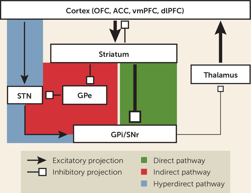
One of the most important technical and conceptual advances in circuit-based theories of OCD has been the change in focus from static regions of interest to interrogation of functional networks subserving different cognitive or behavioral functions germane to the symptomatology of OCD (133140). The seminal work by Alexander et al. (141) influenced OCD researchers to consider certain parallel segregated CSTC loops, subserving different motor or cognitive functions, as the neuroanatomical substrate for OC behavior (85). The canonical model of a CSTC loop consists of projections from a specific region of the frontal cortex that terminate in the striatum, and then travel by either a “direct” (net excitatory) or “indirect” (net inhibitory) pathway through the basal ganglia to the thalamus, and finally returning via recurrent projections to the same cortical region to close the loop (see Figure 1). The striatum is the largest structure of the basal ganglia and serves as its main input region. It is composed of the caudate, putamen, and ventral striatum (VS), which contains the nucleus accumbens (NAc). The main output structure of the basal ganglia is the globus pallidus interna/substantia nigra pars reticulata (GPi/SNr) complex, which largely projects to the thalamus. Other important relay structures within the basal ganglia are the globus pallidus externa (GPe) and subthalamic nucleus (STN).
FIGURE 1. Schematic of cortico-striato-thalamo-cortical (CSTC) circuit model for obsessive-compulsive disorder (OCD)a
a CSTC loops engage functionally related regions of the cortex, striatum, and thalamus relevant to a particular behavior. Cortical regions relevant for OCD pathophysiology span regions of the prefrontal cortex (PFC), including the orbitofrontal cortex (OFC), anterior cingulate cortex (ACC), ventromedial PFC (vmPFC), and dorsolateral PFC (dlPFC). The direct pathway is net excitatory and tends to facilitate behavior, whereas the indirect pathway is net inhibitory and tends to restrain behavior. Overactivity of the direct pathway is a hypothesized feature of the pathogenesis of OCD. This hyperactivity is denoted by a thick line of excitatory input from the cortex to striatum. The striatum in turn has increased inhibitory tone (thick line) on the globus pallidus interna/substantia nigra pars reticulata complex (GPi/SNr), which causes decreased inhibition (thin line) of the thalamus. The thalamus is thereby disinhibited and increases its excitatory input (thick line) to the cortex. The net effect is therefore increased symptomatic behavior. A more recently highlighted “hyperdirect” pathway bypasses the striatum and synapses directly on the subthalamic nucleus (STN). Modulating this pathway has gained in prominence as a mechanism for therapeutic intervention. GPe=globus pallidus externa.
Most projections within the basal ganglia, with the main exception of the STN, are inhibitory. Thus, the overall balance of inhibition and excitation is determined by relative contributions of direct and indirect pathway components. The direct pathway projects from the striatum to the GPi/SNr. Striatal inhibition of the GPi/SNr disinhibits the thalamus, producing net excitatory tone. In contrast, the indirect pathway projects from the striatum to the GPe, and then to the STN, before returning to the GPi/SNr and rejoining the common pathway (85). These additional relays add one node of inhibition, resulting in a net inhibitory tone within the circuit. Revisions of this model paint a more complex picture of the organization of CSTC loops (123), showing more overlap and functional integration between loops than previously appreciated and the importance of other interconnected structures such as the amygdala and hippocampus (142).
Several research groups have hypothesized that excessive tone in the direct pathway (relative to the indirect pathway) would explain some of the neuroimaging findings and phenomenological features of OCD (85, 143). According to this model, such an imbalance in activity between these pathways would bias the individual to selection of, and failure to inhibit, abnormal and repetitive behavioral sequences (85, 143, 144). Hyperactivity of orbitofrontal-subcortical pathways has been found in both human imaging studies in OCD and mouse models of OCD-like behaviors is (144). An influential study by Ahmari et al. (145) used optogenetics in mice to test the effects of CSTC hyperactivation on behavior. Repeated stimulation (over multiple days) of OFC-ventral striatum projections induced increased grooming behaviors, which persisted after stimulation cessation. Chronic administration of fluoxetine reversed the aberrant grooming (145). These data provide strong support for a role of OFC hyperactivity in the genesis of abnormal repetitive behaviors. Together, converging lines of clinical and preclinical evidence suggest that OCD involves dysfunction of limbic CTSC loops that include the OFC, vmPFC, ACC, and ventral striatum (146).

Transcranial Magnetic Stimulation

Recently, the FDA cleared a form of repetitive transcranial magnetic stimulation (TMS), referred to as deep TMS (dTMS), for the treatment of OCD based on the results of a pivotal trial (147). In this double-blind RCT of adults (N=99) who had limited response to previous treatments, 38% responded to the device compared with 11% who received sham treatment (147). This device uses an H-shaped coil that was designed to reach deeper structures, 3 cm to 5 cm penetration, compared with electromagnetic stimulation depth of about 2 cm with conventional figure-8 coils (148). The brain regions targeted with dTMS were midline structures, medial PFC (mPFC) and ACC, two brain areas thought to be hyperactive in OCD (147).
Other studies of rTMS in OCD have targeted different cortical brain regions, including the dlPFC and OFC, with mixed results (147). Dunlop et al. (131) used rTMS targeting the dorsomedial PFC (dmPFC) to treat 20 adults with treatment-resistant OCD. Resting state fMRI (rsfMRI) was performed pre- and posttreatment. Ten subjects met stringent response criteria. Responders had higher dmPFC-ventral striatal connectivity at baseline (131). Furthermore, dmPFC-rTMS treatment was associated with reductions in hyperconnectivity that correlated with improvement on the Y-BOCS (131). These findings with noninvasive neuromodulation provide additional evidence for cortico-striatal hyperactivity underlying the symptoms of OCD.
Neurosurgery for OCD

Beyond the aforementioned modalities (i.e., ERP, SRIs, antipsychotic augmentation of SRIs), there are few proven treatment alternatives. TMS is an emerging option, but additional studies are needed to determine its efficacy in highly treatment-resistant cases of OCD. For the most severe and refractory cases, neurosurgical interventions—either ablative approaches (149152) or DBS (146, 153, 154)—merit consideration in adults with OCD. Another contribution to circuit-based hypotheses has been the therapeutic benefit of neurosurgery in intractable OCD (153). Current conceptualization of the therapeutic mechanism of these interventions is that although they are performed in a specific region, their influence spreads to include the larger dysfunctional network. These surgical interventions are thus inherently network-minded, and their results have the capacity to inform us about the neurobiology of the disorder (146).

Stereotactic Ablation

The introduction of the stereotaxic frame in the middle of the last century facilitated the development of more precise and focal lesion procedures for refractory psychiatric disorders (155, 156). Of the four major ablation procedures (157), the two that are most commonly used in current practice are dorsal anterior cingulotomy (150) and anterior capsulotomy (158). In cingulotomy, a lesion is placed in the cingulum bundle, interrupting communication between the dorsal ACC and OFC, ventral striatum (VS), and other limbic structures. In anterior capsulotomy, a lesion in the anterior limb of the internal capsule (ALIC) is thought to disconnect the OFC and dACC from the VS and thalamus. Both procedures interrupt pathways in CSTC circuits hypothesized to be hyperactive in network models of OCD (159). A 2016 systematic review of the literature among 193 patients with OCD found a 41% responder rate to cingulotomy versus a 54% response rate to capsulotomy (160).
Beyond the therapeutic effects of neurosurgical procedures, their focality also provides the opportunity to understand the neurobiology of the circuits where they intervene. For example, a recent study using resting state fMRI demonstrated decreased functional connectivity between VS and dACC proportional to YBOCs improvement in patients undergoing capsulotomy (130). Studies such as these can help test causal hypotheses regarding network effects of targeted interventions.

Deep Brain Stimulation

Deep brain stimulation (DBS) is used worldwide to treat patients with movement disorders such as Parkinson’s disease and essential tremor. In contrast to ablative procedures, DBS has the advantages of being reversible (explantable) and adjustable (154, 161). In 1999, DBS targeting the ALIC was found beneficial in three of four cases of intractable OCD (162). Since then, DBS of the ALIC (163) or neighboring targets (i.e., the ventral striatum [VS] or nucleus accumbens [NAc], a subregion of the VS, and the bed nucleus of the stria terminalis [BNST] [164]) have shown response rates in the range of 40%−70% (153, 161, 165169). In 2009, the FDA approved a Humanitarian Device Exemption (HDE) for Ventral Capsule/Ventral Striatum (VC/VS) DBS in intractable OCD based upon the device’s safety record and evidence of benefit for cases of severe and treatment refractory OCD (161). In the same year, the European Union granted a “Conformité Européenne” (CE) mark for the procedure, signifying that it meets the EU’s health and safety standards. Due to several factors including cost, differences in health insurance coverage, challenges in access and awareness, and expertise required to manage this therapy, the number of DBS for OCD procedures since these approvals still only numbers in the hundreds worldwide. Unlike in the field of surgery for movement disorders, in which DBS has largely supplanted lesion surgery, both ablative procedures and DBS coexist today for treating severe, intractable OCD.
The exact “target” for the DBS electrode has been a matter of substantial discussion. Some studies have focused on deep gray matter structures such as the VS/NAc or BNST (170) as critical mediators of response. Support for this argument includes the central role of the VS/NAc in approach behavior (142) and role of the BNST in anxiety and context conditioning (171). On the other hand, others have suggested that these nuclei are useful guideposts but that the white matter fibers connecting the PFC and thalamus that course through the ventral ALIC superjacent to these nuclei are critical, as they convey the influence of the injected neuromodulation to the wider symptomatic network (172174). The fact that DBS targeting similar white matter pathways in disparate brain regions (e.g., VC/VS and STN) achieves comparable results (175) provides support for the white matter hypothesis.
The original hypothesis that high frequency DBS (e.g., 130 Hz) would act as “functional ablation” has been challenged by emerging basic neuroscience research showing that the therapeutic mechanisms of DBS are far more complex (176, 177). Effects of VC/VS DBS may be mediated in part by antidromic activation of descending cortical-striatal fibers (159, 178). The end result appears to be modulation of OFC, ACC, and thalamic activity (160), components of the CTSC circuit linked to OCD. In a seminal study of NAc DBS in OCD, repeated resting-state fMRI scans showed that DBS normalized (increased) NAc activity and reduced excessive connectivity between NAc and PFC (178). Degree of reduction in frontostriatal connectivity following DBS treatment was correlated with improvement on the Y-BOCS (178).
DBS targeting the limbic region of the subthalamic nucleus (STN) seems to have grossly similar efficacy to VC/VS DBS in the treatment of OCD (167), but one recent head-to-head comparison between these targets may be able to teach us a useful neurobiological lesson. Tyagi et al. (175) confirmed similar Y-BOCS reductions for the two targets but found greater improvement in comorbid depression with VC/VS DBS versus greater improvement in cognitive flexibility with STN DBS. A white matter tractography analysis has revealed overlapping but distinct regions influenced by these targets (173), suggesting that differential responses to DBS may be predictable based on the connectivity of the stimulating contacts. This study and another recent review of both monkey tract-tracing and human tractography data (174) also highlight the importance of the “hyperdirect” pathway, a monosynaptic connection between cortex and STN that bypasses the striatum entirely (see Figure 1). Just as the hyperdirect motor pathway (motor cortex to STN) may be the therapeutic target in STN DBS for Parkinson’s disease (179, 180), the hyperdirect limbic pathway (OFC and/or ACC to STN) may be an important target in DBS for OCD, whether accessed near the STN or in the ventral ALIC. These types of connectomic studies suggest that future efforts may allow individualization of DBS targeting based on clinical or neurobiological measures.
While VC/VS DBS is an important option for intractable OCD, there is room for improvement in clinical outcomes and mitigation of DBS-induced side effects, including induction of hypomania (161). NIH-funded studies are underway using next generation DBS devices that can record local field potentials (LFPs) as well as deliver neurostimulation (NCT03457675, NCT03244852) (181). These studies may yield insights into the neural signatures of behavioral states associated with changes in OCD symptom severity (181). The ability to record neural data from patients in their natural environment, time locked with behavior and physiology, offers a unique research opportunity to test hypotheses about the neurocircuitry of OCD.

Conclusions

Understanding the neurobiology of OCD is critical to the development of more effective treatments, especially for those patients resistant to conventional therapies. Multiple studies support a significant genetic contribution to OCD, but pinpointing the specific genetic determinants requires additional investigation. The finding that serotonin reuptake inhibitors (SRIs) are preferentially effective in OCD is one of the distinguishing features of this disorder. However, direct support for a role of serotonin in the pathophysiology of OCD is lacking. Several lines of preclinical and clinical evidence suggest dysfunction of the glutamatergic system in OCD, prompting testing of several promising glutamate modulating agents. Functional imaging studies in OCD show consistent evidence for increased activity in brain regions that form a CSTC loop. Neuromodulation treatments with either noninvasive devices (e.g., transcranial magnetic stimulation) or invasive procedures (e.g., deep brain stimulation) provide further support for the CSTC model of OCD. A common substrate for various interventions (whether drug, behavioral, or device) may be modulation (at different nodes or connections) of the CSTC circuit that mediates the symptoms of OCD. Hypotheses that integrate knowledge over multiple levels of analysis (e.g., genetics, neurochemical, and circuit networks) stand the best chance of advancing our understanding of OCD (182). Research using DBS devices that allow real-time recordings of neural activity, time-locked with behavioral state, may generate new insights into the neurocircuitry of OCD (181).

References

1.
American Psychiatric Association: Diagnostic and Statistical Manual of Mental Disorders, 5th ed. Washington, DC, American Psychiatric Association, 2013.
2.
Rasmussen SA, Eisen JL: The epidemiology and clinical features of obsessive compulsive disorder. Psychiatr Clin North Am 1992; 15:743–758
3.
Ruscio AM, Stein DJ, Chiu WT, et al: The epidemiology of obsessive-compulsive disorder in the National Comorbidity Survey Replication. Mol Psychiatry 2010; 15:53–63
4.
Norberg MM, Calamari JE, Cohen RJ, et al: Quality of life in obsessive-compulsive disorder: an evaluation of impairment and a preliminary analysis of the ameliorating effects of treatment. Depress Anxiety 2008; 25:248–259
5.
Adam Y, Meinlschmidt G, Gloster AT, et al: Obsessive-compulsive disorder in the community: 12-month prevalence, comorbidity and impairment. Soc Psychiatry Psychiatr Epidemiol 2012; 47:339–349
6.
Meier SM, Mattheisen M, Mors O, et al: Mortality among persons with obsessive-compulsive disorder in Denmark. JAMA Psychiatry 2016; 73:268–274
7.
Deacon BJ, Abramowitz JS: Cognitive and behavioral treatments for anxiety disorders: a review of meta-analytic findings. J Clin Psychol 2004; 60:429–441
8.
Koran LM, Hanna GL, Hollander E, et al: Practice guideline for the treatment of patients with obsessive-compulsive disorder. Am J Psychiatry 2007; 164(Suppl):5–53
9.
Goodman WK, Price LH, Delgado PL, et al: Specificity of serotonin reuptake inhibitors in the treatment of obsessive-compulsive disorder. Comparison of fluvoxamine and desipramine. Arch Gen Psychiatry 1990; 47:577–585
10.
Pittenger C, Bloch MH: Pharmacological treatment of obsessive-compulsive disorder. Psychiatr Clin North Am 2014; 37:375–391
11.
Pigott TA, Seay SM: A review of the efficacy of selective serotonin reuptake inhibitors in obsessive-compulsive disorder. J Clin Psychiatry 1999; 60:101–106
12.
Ackerman DL, Greenland S: Multivariate meta-analysis of controlled drug studies for obsessive-compulsive disorder. J Clin Psychopharmacol 2002; 22:309–317
13.
Romanelli RJ, Wu FM, Gamba R, et al: Behavioral therapy and serotonin reuptake inhibitor pharmacotherapy in the treatment of obsessive-compulsive disorder: a systematic review and meta-analysis of head-to-head randomized controlled trials. Depress Anxiety 2014; 31:641–652
14.
Öst LG, Havnen A, Hansen B, et al: Cognitive behavioral treatments of obsessive-compulsive disorder. A systematic review and meta-analysis of studies published 1993-2014. Clin Psychol Rev 2015; 40:156–169
15.
Simpson HB, Huppert JD, Petkova E, et al: Response versus remission in obsessive-compulsive disorder. J Clin Psychiatry 2006; 67:269–276
16.
McDougle CJ, Goodman WK, Leckman JF, et al: Haloperidol addition in fluvoxamine-refractory obsessive-compulsive disorder. A double-blind, placebo-controlled study in patients with and without tics. Arch Gen Psychiatry 1994; 51:302–308
17.
Bloch MH, Landeros-Weisenberger A, Kelmendi B, et al: A systematic review: antipsychotic augmentation with treatment refractory obsessive-compulsive disorder. Mol Psychiatry 2006; 11:622–632
18.
Dold M, Aigner M, Lanzenberger R, et al: Antipsychotic augmentation of serotonin reuptake inhibitors in treatment-resistant obsessive-compulsive disorder: a meta-analysis of double-blind, randomized, placebo-controlled trials. Int J Neuropsychopharmacol 2013; 16:557–574
19.
Fernandez TV, Leckman JF, Pittenger C: Genetic susceptibility in obsessive-compulsive disorder. Handb Clin Neurol 2018; 148:767–781
20.
Monzani B, Rijsdijk F, Harris J, et al: The structure of genetic and environmental risk factors for dimensional representations of DSM-5 obsessive-compulsive spectrum disorders. JAMA Psychiatry 2014; 71:182–189
21.
Mataix-Cols D, Boman M, Monzani B, et al: Population-based, multigenerational family clustering study of obsessive-compulsive disorder. JAMA Psychiatry 2013; 70:709–717
22.
Taylor S: Early versus late onset obsessive-compulsive disorder: evidence for distinct subtypes. Clin Psychol Rev 2011; 31:1083–1100
23.
Mahjani B, Klei L, Hultman CM, et al: Maternal effects as causes of risk for obsessive-compulsive disorder. Biol Psychiatry 2020; 87:1045–1051
24.
Nordsletten AE, Larsson H, Crowley JJ, et al: Patterns of nonrandom mating within and across 11 major psychiatric disorders. JAMA Psychiatry 2016; 73:354–361
25.
Peyrot WJ, Robinson MR, Penninx BW, et al: Exploring boundaries for the genetic consequences of assortative mating for psychiatric traits. JAMA Psychiatry 2016; 73:1189–1195
26.
Goodman WK, McDougle CJ, Price LH, et al: Beyond the serotonin hypothesis: a role for dopamine in some forms of obsessive compulsive disorder? J Clin Psychiatry 1990; 51(Suppl):36–43, discussion 55–58
27.
Pittenger C, Bloch MH, Williams K: Glutamate abnormalities in obsessive compulsive disorder: neurobiology, pathophysiology, and treatment. Pharmacol Ther 2011; 132:314–332
28.
Zai G, Barta C, Cath D, et al: New insights and perspectives on the genetics of obsessive-compulsive disorder. Psychiatr Genet 2019; 29:142–151
29.
Escobar AP, Wendland JR, Chávez AE, et al: The neuronal glutamate transporter EAAT3 in obsessive-compulsive disorder. Front Pharmacol 2019; 10:1362
30.
Stewart SE, Yu D, Scharf JM, et al: Genome-wide association study of obsessive-compulsive disorder. Mol Psychiatry 2013; 18:788–798
31.
Mattheisen M, Samuels JF, Wang Y, et al: Genome-wide association study in obsessive-compulsive disorder: results from the OCGAS. Mol Psychiatry 2015; 20:337–344
32.
International Obsessive Compulsive Disorder Foundation Genetics Collaborative (IOCDF-GC) and OCD Collaborative Genetics Association Studies (OCGAS): Revealing the complex genetic architecture of obsessive-compulsive disorder using meta-analysis. Mol Psychiatry 2018; 23:1181–1188
33.
Sullivan PF, Daly MJ, O’Donovan M: Genetic architectures of psychiatric disorders: the emerging picture and its implications. Nat Rev Genet 2012; 13:537–551
34.
McGrath LM, Yu D, Marshall C, et al: Copy number variation in obsessive-compulsive disorder and tourette syndrome: a cross-disorder study. J Am Acad Child Adolesc Psychiatry 2014; 53:910–919
35.
Grice DE: Don’t worry, the genetics of obsessive-compulsive disorder is finally catching up. Biol Psychiatry 2020; 87:1017–1018
36.
Mahjani B, Dellenvall K, Grahnat AS, et al: Cohort profile: Epidemiology and Genetics of Obsessive-compulsive disorder and chronic tic disorders in Sweden (EGOS). Soc Psychiatry Psychiatr Epidemiol 2020; 55:1383–1393
37.
Asbahr FR, Garvey MA, Snider LA, et al: Obsessive-compulsive symptoms among patients with Sydenham chorea. Biol Psychiatry 2005; 57:1073–1076
38.
Cheyette SR, Cummings JL: Encephalitis lethargica: lessons for contemporary neuropsychiatry. J Neuropsychiatry Clin Neurosci 1995; 7:125–134
39.
Thobois S, Jouanneau E, Bouvard M, et al: Obsessive-compulsive disorder after unilateral caudate nucleus bleeding. Acta Neurochir (Wien) 2004; 146:1027–1031, discussion 1031
40.
Grados MA: Obsessive-compulsive disorder after traumatic brain injury. Int Rev Psychiatry 2003; 15:350–358
41.
Kong XZ, Boedhoe PSW, Abe Y, et al: Mapping cortical and subcortical asymmetry in obsessive-compulsive disorder: findings from the ENIGMA consortium. Biol Psychiatry 2020; 87:1022–1034
42.
Bruin WB, Taylor L, Thomas RM, et al: Structural neuroimaging biomarkers for obsessive-compulsive disorder in the ENIGMA-OCD consortium: medication matters. Transl Psychiatry 2020; 10:342
43.
de Oliveira KC, Grinberg LT, Hoexter MQ, et al: Layer-specific reduced neuronal density in the orbitofrontal cortex of older adults with obsessive-compulsive disorder. Brain Struct Funct 2019; 224:191–203
44.
Piantadosi SC, Chamberlain BL, Glausier JR, et al: Lower excitatory synaptic gene expression in orbitofrontal cortex and striatum in an initial study of subjects with obsessive compulsive disorder. Mol Psychiatry 2019
45.
Swedo SE, Leonard HL, Garvey M, et al: Pediatric autoimmune neuropsychiatric disorders associated with streptococcal infections: clinical description of the first 50 cases. Am J Psychiatry 1998; 155:264–271
46.
Murphy TK, Patel PD, McGuire JF, et al: Characterization of the pediatric acute-onset neuropsychiatric syndrome phenotype. J Child Adolesc Psychopharmacol 2015; 25:14–25
47.
Williams KA, Swedo SE, Farmer CA, et al: Randomized, controlled trial of intravenous immunoglobulin for pediatric autoimmune neuropsychiatric disorders associated with streptococcal infections. J Am Acad Child Adolesc Psychiatry 2016; 55:860–867.e2, e862
48.
Kurlan R, Kaplan EL: The pediatric autoimmune neuropsychiatric disorders associated with streptococcal infection (PANDAS) etiology for tics and obsessive-compulsive symptoms: hypothesis or entity? Practical considerations for the clinician. Pediatrics 2004; 113:883–886
49.
Xu J, Liu RJ, Fahey S, et al: Antibodies from children with PANDAS bind specifically to striatal cholinergic interneurons and alter their activity. Am J Psychiatry 2021; 178:48–64
50.
Carpenter WT Jr, Davis JM: Another view of the history of antipsychotic drug discovery and development. Mol Psychiatry 2012; 17:1168–1173
51.
Brown WA, Rosdolsky M: The clinical discovery of imipramine. Am J Psychiatry 2015; 172:426–429
52.
Yaryura-Tobias JA, Neziroglu F: The action of chlorimipramine in obsessive-compulsive neurosis: a pilot study. Curr Ther Res Clin Exp 1975; 17:111–116
53.
Leonard HL, Swedo SE, Rapoport JL, et al: Treatment of obsessive-compulsive disorder with clomipramine and desipramine in children and adolescents. A double-blind crossover comparison. Arch Gen Psychiatry 1989; 46:1088–1092
54.
Hoehn-Saric R, Ninan P, Black DW, et al: Multicenter double-blind comparison of sertraline and desipramine for concurrent obsessive-compulsive and major depressive disorders. Arch Gen Psychiatry 2000; 57:76–82
55.
Katz RJ, DeVeaugh-Geiss J, Landau P: Clomipramine in obsessive-compulsive disorder. Biol Psychiatry 1990; 28:401–414
56.
Goodman WK, Price LH, Rasmussen SA, et al: Efficacy of fluvoxamine in obsessive-compulsive disorder. A double-blind comparison with placebo. Arch Gen Psychiatry 1989; 46:36–44
57.
Perse TL, Greist JH, Jefferson JW, et al: Fluvoxamine treatment of obsessive-compulsive disorder. Am J Psychiatry 1987; 144:1543–1548
58.
Zohar J, Mueller EA, Insel TR, et al: Serotonergic responsivity in obsessive-compulsive disorder. Comparison of patients and healthy controls. Arch Gen Psychiatry 1987; 44:946–951
59.
Goodman WK, McDougle CJ, Price LH, et al: m-Chlorophenylpiperazine in patients with obsessive-compulsive disorder: absence of symptom exacerbation. Biol Psychiatry 1995; 38:138–149
60.
Charney DS, Goodman WK, Price LH, et al: Serotonin function in obsessive-compulsive disorder. A comparison of the effects of tryptophan and m-chlorophenylpiperazine in patients and healthy subjects. Arch Gen Psychiatry 1988; 45:177–185
61.
Barr LC, Goodman WK, McDougle CJ, et al: Tryptophan depletion in patients with obsessive-compulsive disorder who respond to serotonin reuptake inhibitors. Arch Gen Psychiatry 1994; 51:309–317
62.
Simpson HB, Slifstein M, Bender J Jr, et al: Serotonin 2A receptors in obsessive-compulsive disorder: a positron emission tomography study with [11C]MDL 100907. Biol Psychiatry 2011; 70:897–904
63.
McDougle CJ, Goodman WK, Leckman JF, et al: Limited therapeutic effect of addition of buspirone in fluvoxamine-refractory obsessive-compulsive disorder. Am J Psychiatry 1993; 150:647–649
64.
Goodman WK, McDougle CJ, Barr LC, et al: Biological approaches to treatment-resistant obsessive compulsive disorder. J Clin Psychiatry 1993; 54(Suppl):16–26
65.
Pallanti S, Bernardi S, Antonini S, et al: Ondansetron augmentation in patients with obsessive-compulsive disorder who are inadequate responders to serotonin reuptake inhibitors: improvement with treatment and worsening following discontinuation. Eur Neuropsychopharmacol 2014; 24:375–380
66.
McDougle CJ, Price LH, Goodman WK, et al: A controlled trial of lithium augmentation in fluvoxamine-refractory obsessive-compulsive disorder: lack of efficacy. J Clin Psychopharmacol 1991; 11:175–184
67.
Stern ER, Shahab R, Grimaldi SJ, et al: High-dose ondansetron reduces activation of interoceptive and sensorimotor brain regions. Neuropsychopharmacology 2019; 44:390–398
68.
Saxena S, Brody AL, Maidment KM, et al: Localized orbitofrontal and subcortical metabolic changes and predictors of response to paroxetine treatment in obsessive-compulsive disorder. Neuropsychopharmacology 1999; 21:683–693
69.
Michelsen KA, Prickaerts J, Steinbusch HW: The dorsal raphe nucleus and serotonin: implications for neuroplasticity linked to major depression and Alzheimer’s disease. Prog Brain Res 2008; 172:233–264
70.
Bergqvist PB, Bouchard C, Blier P: Effect of long-term administration of antidepressant treatments on serotonin release in brain regions involved in obsessive-compulsive disorder. Biol Psychiatry 1999; 45:164–174
71.
Rachman SJ: Obsessions and compulsions. Englewood Cliffs, N.J., Prentice-Hall, 1980
72.
Stern ER, Taylor SF: Cognitive neuroscience of obsessive-compulsive disorder. Psychiatr Clin North Am 2014; 37:337–352
73.
McGuire JF, Piacentini J, Lewin AB, et al: A meta-analysis of cognitive behavior therapy and medication for child obsessive-compulsive disorder: moderators of treatment efficacy, response, and remission. Depress Anxiety 2015; 32:580–593
74.
Abramowitz JS, Taylor S, McKay D: Obsessive-compulsive disorder. Lancet 2009; 374:491–499
75.
Berry L, Laskey B: A review of obsessive intrusive thoughts in the general population. J Obsessive Compuls Relat Disord 2012; 1:125–132
76.
McGuire JF, Orr SP, Essoe JK, et al: Extinction learning in childhood anxiety disorders, obsessive compulsive disorder and post-traumatic stress disorder: implications for treatment. Expert Rev Neurother 2016; 16:1155–1174
77.
Tolin DF, Worhunsky P, Maltby N: Sympathetic magic in contamination-related OCD. J Behav Ther Exp Psychiatry 2004; 35:193–205
78.
Myers KM, Davis M: Mechanisms of fear extinction. Mol Psychiatry 2007; 12:120–150
79.
McGuire JF, Orr SP, Wu MS, et al: Fear conditioning and extinction in youth with obsessive-compulsive disorder. Depress Anxiety 2016; 33:229–237
80.
Milad MR, Furtak SC, Greenberg JL, et al: Deficits in conditioned fear extinction in obsessive-compulsive disorder and neurobiological changes in the fear circuit. JAMA Psychiatry 2013; 70:608–618, quiz 554
81.
Fyer AJ, Schneier FR, Simpson HB, et al: Heterogeneity in fear processing across and within anxiety, eating, and compulsive disorders. J Affect Disord 2020; 275:329–338
82.
Geller DA, McGuire JF, Orr SP, et al: Fear conditioning and extinction in pediatric obsessive-compulsive disorder. Ann Clin Psychiatry 2017; 29:17–26
83.
Geller DA, McGuire JF, Orr SP, et al: Fear extinction learning as a predictor of response to cognitive behavioral therapy for pediatric obsessive compulsive disorder. J Anxiety Disord 2019; 64:1–8
84.
Norman LJ, Taylor SF, Liu Y, et al: Error processing and inhibitory control in obsessive-compulsive disorder: a meta-analysis using statistical parametric maps. Biol Psychiatry 2019; 85:713–725
85.
Saxena S, Rauch SL: Functional neuroimaging and the neuroanatomy of obsessive-compulsive disorder. Psychiatr Clin North Am 2000; 23:563–586
86.
Baxter LR Jr, Schwartz JM, Bergman KS, et al: Caudate glucose metabolic rate changes with both drug and behavior therapy for obsessive-compulsive disorder. Arch Gen Psychiatry 1992; 49:681–689
87.
Schwartz JM, Stoessel PW, Baxter LR Jr, et al: Systematic changes in cerebral glucose metabolic rate after successful behavior modification treatment of obsessive-compulsive disorder. Arch Gen Psychiatry 1996; 53:109–113
88.
Saxena S, Gorbis E, O’Neill J, et al: Rapid effects of brief intensive cognitive-behavioral therapy on brain glucose metabolism in obsessive-compulsive disorder. Mol Psychiatry 2009; 14:197–205
89.
Yamanishi T, Nakaaki S, Omori IM, et al: Changes after behavior therapy among responsive and nonresponsive patients with obsessive-compulsive disorder. Psychiatry Res 2009; 172:242–250
90.
Utevsky AV, Smith DV, Huettel SA: Precuneus is a functional core of the default-mode network. J Neurosci 2014; 34:932–940
91.
Li P, Yang X, Greenshaw AJ, et al: The effects of cognitive behavioral therapy on resting-state functional brain network in drug-naive patients with obsessive-compulsive disorder. Brain Behav 2018; 8:e00963
92.
Huyser C, Veltman DJ, Wolters LH, et al: Functional magnetic resonance imaging during planning before and after cognitive-behavioral therapy in pediatric obsessive-compulsive disorder. J Am Acad Child Adolesc Psychiatry 2010; 49:1238–1248, 1248.e1–1248.e5
93.
Norman LJ, Mannella KA, Yang H, et al: Treatment-specific associations between brain activation and symptom reduction in OCD following CBT: a randomized fMRI trial. Am J Psychiatry 2020; p202019080886
94.
Mataix-Cols D, Fernández de la Cruz L, Monzani B, et al: D-Cycloserine augmentation of exposure-based cognitive behavior therapy for anxiety, obsessive-compulsive, and posttraumatic stress disorders: a systematic review and meta-analysis of individual participant data. JAMA Psychiatry 2017; 74:501–510
95.
Rosenfield D, Smits JAJ, Hofmann SG, et al: Changes in dosing and dose timing of D-cycloserine explain its apparent declining efficacy for augmenting exposure therapy for anxiety-related disorders: an individual participant-data meta-analysis. J Anxiety Disord 2019; 68:102149
96.
Brennan BP, Rauch SL, Jensen JE, et al: A critical review of magnetic resonance spectroscopy studies of obsessive-compulsive disorder. Biol Psychiatry 2013; 73:24–31
97.
Stewart SE, Mayerfeld C, Arnold PD, et al: Meta-analysis of association between obsessive-compulsive disorder and the 3¢ region of neuronal glutamate transporter gene SLC1A1. Am J Med Genet B Neuropsychiatr Genet 2013; 162B:367–379
98.
Bhattacharyya S, Khanna S, Chakrabarty K, et al: Anti-brain autoantibodies and altered excitatory neurotransmitters in obsessive-compulsive disorder. Neuropsychopharmacology 2009; 34:2489–2496
99.
Chakrabarty K, Bhattacharyya S, Christopher R, et al: Glutamatergic dysfunction in OCD. Neuropsychopharmacology 2005; 30:1735–1740
100.
Bienvenu OJ, Wang Y, Shugart YY, et al: Sapap3 and pathological grooming in humans: Results from the OCD collaborative genetics study. Am J Med Genet B Neuropsychiatr Genet 2009; 150B:710–720
101.
Sakurai H, Dording C, Yeung A, et al: Longer-term open-label study of adjunctive riluzole in treatment-resistant depression. J Affect Disord 2019; 258:102–108
102.
Pittenger C, Bloch MH, Wasylink S, et al: Riluzole augmentation in treatment-refractory obsessive-compulsive disorder: a pilot randomized placebo-controlled trial. J Clin Psychiatry 2015; 76:1075–1084
103.
Grant PJ, Joseph LA, Farmer CA, et al: 12-week, placebo-controlled trial of add-on riluzole in the treatment of childhood-onset obsessive-compulsive disorder. Neuropsychopharmacology 2014; 39:1453–1459
104.
BHV-4157 in Adult Subjects with Obsessive Compulsive Disorder. ClinicalTrials.gov.
105.
Stewart SE, Jenike EA, Hezel DM, et al: A single-blinded case-control study of memantine in severe obsessive-compulsive disorder. J Clin Psychopharmacol 2010; 30:34–39
106.
Ghaleiha A, Entezari N, Modabbernia A, et al: Memantine add-on in moderate to severe obsessive-compulsive disorder: randomized double-blind placebo-controlled study. J Psychiatr Res 2013; 47:175–180
107.
Haghighi M, Jahangard L, Mohammad-Beigi H, et al: In a double-blind, randomized and placebo-controlled trial, adjuvant memantine improved symptoms in inpatients suffering from refractory obsessive-compulsive disorders (OCD). Psychopharmacology (Berl) 2013; 228:633–640
108.
Modarresi A, Chaibakhsh S, Koulaeinejad N, et al: A systematic review and meta-analysis: Memantine augmentation in moderate to severe obsessive-compulsive disorder. Psychiatry Res 2019; 282:112602
109.
Andrade C: Augmentation with memantine in obsessive-compulsive disorder. J Clin Psychiatry 2019; 80: 19f13163
110.
Zarate CA Jr, Singh JB, Carlson PJ, et al: A randomized trial of an N-methyl-D-aspartate antagonist in treatment-resistant major depression. Arch Gen Psychiatry 2006; 63:856–864
111.
Bloch MH, Wasylink S, Landeros-Weisenberger A, et al: Effects of ketamine in treatment-refractory obsessive-compulsive disorder. Biol Psychiatry 2012; 72:964–970
112.
Goodman WK, Price LH, Rasmussen SA, et al: The Yale-Brown Obsessive Compulsive Scale. I. Development, use, and reliability. Arch Gen Psychiatry 1989; 46:1006–1011
113.
Rodriguez CI, Kegeles LS, Levinson A, et al: Randomized controlled crossover trial of ketamine in obsessive-compulsive disorder: proof-of-concept. Neuropsychopharmacology 2013; 38:2475–2483
114.
Deepmala SJ, Slattery J, Kumar N, et al: Clinical trials of N-acetylcysteine in psychiatry and neurology: A systematic review. Neurosci Biobehav Rev 2015; 55:294–321
115.
Kupchik YM, Moussawi K, Tang XC, et al: The effect of N-acetylcysteine in the nucleus accumbens on neurotransmission and relapse to cocaine. Biol Psychiatry 2012; 71:978–986
116.
Costa DLC, Diniz JB, Requena G, et al: Randomized, double-blind, placebo-controlled trial of N-acetylcysteine augmentation for treatment-resistant obsessive-compulsive disorder. J Clin Psychiatry 2017; 78:e766–e773
117.
Li F, Welling MC, Johnson JA, et al: N-Acetylcysteine for pediatric obsessive-compulsive disorder: a small pilot study. J Child Adolesc Psychopharmacol 2020; 30:32–37
118.
Pittenger C: Glutamatergic agents for OCD and related disorders. Curr Treat Options Psychiatry 2015; 2:271–283
119.
Greenberg WM, Benedict MM, Doerfer J, et al: Adjunctive glycine in the treatment of obsessive-compulsive disorder in adults. J Psychiatr Res 2009; 43:664–670
120.
Wu PL, Tang HS, Lane HY, et al: Sarcosine therapy for obsessive compulsive disorder: a prospective, open-label study. J Clin Psychopharmacol 2011; 31:369–374
121.
Singer P, Yee BK: Pharmacotherapy through the inhibition of glycine transporters: an update on and beyond schizophrenia, in Psychiatry and Neuroscience Update, vol 2. Edited by Gargiulo P, Mesones-Arroyo H. Cham, Switzerland, Springer, 2017
122.
Yuste R: From the neuron doctrine to neural networks. Nat Rev Neurosci 2015; 16:487–497
123.
Milad MR, Rauch SL: Obsessive-compulsive disorder: beyond segregated cortico-striatal pathways. Trends Cogn Sci 2012; 16:43–51
124.
Baxter LR Jr, Schwartz JM, Mazziotta JC, et al: Cerebral glucose metabolic rates in nondepressed patients with obsessive-compulsive disorder. Am J Psychiatry 1988; 145:1560–1563
125.
Breiter HC, Rauch SL, Kwong KK, et al: Functional magnetic resonance imaging of symptom provocation in obsessive-compulsive disorder. Arch Gen Psychiatry 1996; 53:595–606
126.
Benkelfat C, Nordahl TE, Semple WE, et al: Local cerebral glucose metabolic rates in obsessive-compulsive disorder. Patients treated with clomipramine. Arch Gen Psychiatry 1990; 47:840–848
127.
Park HR, Kim IH, Kang H, et al: Electrophysiological and imaging evidence of sustained inhibition in limbic and frontal networks following deep brain stimulation for treatment refractory obsessive compulsive disorder. PLoS One 2019; 14:e0219578
128.
Le Jeune F, Vérin M, N’Diaye K, et al: Decrease of prefrontal metabolism after subthalamic stimulation in obsessive-compulsive disorder: a positron emission tomography study. Biol Psychiatry 2010; 68:1016–1022
129.
Zuo C, Ma Y, Sun B, et al: Metabolic imaging of bilateral anterior capsulotomy in refractory obsessive compulsive disorder: an FDG PET study. J Cereb Blood Flow Metab 2013; 33:880–887
130.
Yin D, Zhang C, Lv Q, et al: Dissociable frontostriatal connectivity: mechanism and predictor of the clinical efficacy of capsulotomy in obsessive-compulsive disorder. Biol Psychiatry 2018; 84:926–936
131.
Dunlop K, Woodside B, Olmsted M, et al: Reductions in cortico-striatal hyperconnectivity accompany successful treatment of obsessive-compulsive disorder with dorsomedial prefrontal rTMS. Neuropsychopharmacology 2016; 41:1395–1403
132.
Nauczyciel C, Le Jeune F, Naudet F, et al: Repetitive transcranial magnetic stimulation over the orbitofrontal cortex for obsessive-compulsive disorder: a double-blind, crossover study. Transl Psychiatry 2014; 4:e436
133.
Stern ER, Welsh RC, Fitzgerald KD, et al: Hyperactive error responses and altered connectivity in ventromedial and frontoinsular cortices in obsessive-compulsive disorder. Biol Psychiatry 2011; 69:583–591
134.
Anticevic A, Hu S, Zhang S, et al: Global resting-state functional magnetic resonance imaging analysis identifies frontal cortex, striatal, and cerebellar dysconnectivity in obsessive-compulsive disorder. Biol Psychiatry 2014; 75:595–605
135.
Beucke JC, Sepulcre J, Talukdar T, et al: Abnormally high degree connectivity of the orbitofrontal cortex in obsessive-compulsive disorder. JAMA Psychiatry 2013; 70:619–629
136.
Cocchi L, Harrison BJ, Pujol J, et al: Functional alterations of large-scale brain networks related to cognitive control in obsessive-compulsive disorder. Hum Brain Mapp 2012; 33:1089–1106
137.
Fitzgerald KD, Welsh RC, Stern ER, et al: Developmental alterations of frontal-striatal-thalamic connectivity in obsessive-compulsive disorder. J Am Acad Child Adolesc Psychiatry 2011; 50:938–948.e3, e933
138.
Harrison BJ, Soriano-Mas C, Pujol J, et al: Altered corticostriatal functional connectivity in obsessive-compulsive disorder. Arch Gen Psychiatry 2009; 66:1189–1200
139.
Jang JH, Kim JH, Jung WH, et al: Functional connectivity in fronto-subcortical circuitry during the resting state in obsessive-compulsive disorder. Neurosci Lett 2010; 474:158–162
140.
Stern ER, Fitzgerald KD, Welsh RC, et al: Resting-state functional connectivity between fronto-parietal and default mode networks in obsessive-compulsive disorder. PLoS One 2012; 7:e36356
141.
Alexander GE, DeLong MR, Strick PL: Parallel organization of functionally segregated circuits linking basal ganglia and cortex. Annu Rev Neurosci 1986; 9:357–381
142.
Haber SN, Knutson B: The reward circuit: linking primate anatomy and human imaging. Neuropsychopharmacology 2010; 35:4–26
143.
Ahmari SE, Dougherty DD: Dissecting OCD circuits: from animal models to targeted treatments. Depress Anxiety 2015; 32:550–562
144.
Ting JT, Feng G: Neurobiology of obsessive-compulsive disorder: insights into neural circuitry dysfunction through mouse genetics. Curr Opin Neurobiol 2011; 21:842–848
145.
Ahmari SE, Spellman T, Douglass NL, et al: Repeated cortico-striatal stimulation generates persistent OCD-like behavior. Science 2013; 340:1234–1239
146.
Greenberg BD, Rauch SL, Haber SN: Invasive circuitry-based neurotherapeutics: stereotactic ablation and deep brain stimulation for OCD. Neuropsychopharmacology 2010; 35:317–336
147.
Carmi L, Tendler A, Bystritsky A, et al: Efficacy and safety of deep transcranial magnetic stimulation for obsessive-compulsive disorder: a prospective multicenter randomized double-blind placebo-controlled trial. Am J Psychiatry 2019; 176:931–938
148.
McCathern AG, Mathai DS, Cho RY, et al: Deep transcranial magnetic stimulation for obsessive compulsive disorder. Expert Rev Neurother 2020; 20:1029–1036
149.
Dougherty DD, Baer L, Cosgrove GR, et al: Prospective long-term follow-up of 44 patients who received cingulotomy for treatment-refractory obsessive-compulsive disorder. Am J Psychiatry 2002; 159:269–275
150.
Sheth SA, Neal J, Tangherlini F, et al: Limbic system surgery for treatment-refractory obsessive-compulsive disorder: a prospective long-term follow-up of 64 patients. J Neurosurg 2013; 118:491–497
151.
Lopes AC, Greenberg BD, Canteras MM, et al: Gamma ventral capsulotomy for obsessive-compulsive disorder: a randomized clinical trial. JAMA Psychiatry 2014; 71:1066–1076
152.
Lopes AC, Greenberg BD, Pereira CA, et al: Notice of Retraction and Replacement. Lopes et al. Gamma ventral capsulotomy for obsessive-compulsive disorder: a randomized clinical trial. JAMA Psychiatry. 2014;71(9):1066-1076. JAMA Psychiatry 2015; 72:1258
153.
Greenberg BD, Gabriels LA, Malone DA Jr, et al: Deep brain stimulation of the ventral internal capsule/ventral striatum for obsessive-compulsive disorder: worldwide experience. Mol Psychiatry 2010; 15:64–79
154.
Goodman WK, Insel TR: Deep brain stimulation in psychiatry: concentrating on the road ahead. Biol Psychiatry 2009; 65:263–266
155.
Ballantine HT Jr, Cassidy WL, Flanagan NB, et al: Stereotaxic anterior cingulotomy for neuropsychiatric illness and intractable pain. J Neurosurg 1967; 26:488–495
156.
Leksell L: A stereotaxic apparatus for intracerebral surgery. Acta Chir Scand 1949; 229–233
157.
Patel SR, Aronson JP, Sheth SA, et al: Lesion procedures in psychiatric neurosurgery. World Neurosurg 2013; 80:31.e9–31.e16, e39–e16
158.
Miguel EC, Lopes AC, McLaughlin NCR, et al: Evolution of gamma knife capsulotomy for intractable obsessive-compulsive disorder. Mol Psychiatry 2019; 24:218–240
159.
Bourne SK, Eckhardt CA, Sheth SA, et al: Mechanisms of deep brain stimulation for obsessive compulsive disorder: effects upon cells and circuits. Front Integr Nuerosci 2012; 6:29
160.
Brown LT, Mikell CB, Youngerman BE, et al: Dorsal anterior cingulotomy and anterior capsulotomy for severe, refractory obsessive-compulsive disorder: a systematic review of observational studies. J Neurosurg 2016; 124:77–89
161.
Goodman WK, Alterman RL: Deep brain stimulation for intractable psychiatric disorders. Annu Rev Med 2012; 63:511–524
162.
Nuttin B, Cosyns P, Demeulemeester H, et al: Electrical stimulation in anterior limbs of internal capsules in patients with obsessive-compulsive disorder. Lancet 1999; 354:1526
163.
Abelson JL, Curtis GC, Sagher O, et al: Deep brain stimulation for refractory obsessive-compulsive disorder. Biol Psychiatry 2005; 57:510–516
164.
Nuttin B, Gielen F, van Kuyck K, et al: Targeting bed nucleus of the stria terminalis for severe obsessive-compulsive disorder: more unexpected lead placement in obsessive-compulsive disorder than in surgery for movement disorders. World Neurosurg 2013; 80:30.e11–30.e16, e11–e36
165.
Goodman WK, Foote KD, Greenberg BD, et al: Deep brain stimulation for intractable obsessive compulsive disorder: pilot study using a blinded, staggered-onset design. Biol Psychiatry 2010; 67:535–542
166.
de Koning PP, Figee M, van den Munckhof P, et al: Current status of deep brain stimulation for obsessive-compulsive disorder: a clinical review of different targets. Curr Psychiatry Rep 2011; 13:274–282
167.
Alonso P, Cuadras D, Gabriëls L, et al: Deep brain stimulation for obsessive-compulsive disorder: a meta-analysis of treatment outcome and predictors of response. PLoS One 2015; 10:e0133591
168.
Denys D, Mantione M, Figee M, et al: Deep brain stimulation of the nucleus accumbens for treatment-refractory obsessive-compulsive disorder. Arch Gen Psychiatry 2010; 67:1061–1068
169.
Denys D, Graat I, Mocking R, et al: Efficacy of deep brain stimulation of the ventral anterior limb of the internal capsule for refractory obsessive-compulsive disorder: a clinical cohort of 70 patients. Am J Psychiatry 2020; 177:265–271
170.
Luyten L, Hendrickx S, Raymaekers S, et al: Electrical stimulation in the bed nucleus of the stria terminalis alleviates severe obsessive-compulsive disorder. Mol Psychiatry 2016; 21:1272–1280
171.
Luyten L, van Kuyck K, Vansteenwegen D, et al: Electrolytic lesions of the bed nucleus of the stria terminalis disrupt freezing and startle potentiation in a conditioned context. Behav Brain Res 2011; 222:357–362
172.
van den Munckhof P, Bosch DA, Mantione MH, et al: Active stimulation site of nucleus accumbens deep brain stimulation in obsessive-compulsive disorder is localized in the ventral internal capsule. Acta Neurochir Suppl (Wien) 2013; 117:53–59
173.
Li N, Baldermann JC, Kibleur A, et al: A unified connectomic target for deep brain stimulation in obsessive-compulsive disorder. Nat Commun 2020; 11:3364
174.
Haber SN, Yendiki A, Jbabdi S: Four deep brain stimulation targets for obsessive-compulsive disorder: are they different? Biol Psychiatry 2020; S0006-3223(20)31773-X
175.
Tyagi H, Apergis-Schoute AM, Akram H, et al: A randomized trial directly comparing ventral capsule and anteromedial subthalamic nucleus stimulation in obsessive-compulsive disorder: clinical and imaging evidence for dissociable effects. Biol Psychiatry 2019; 85:726–734
176.
McCracken CB, Grace AA: High-frequency deep brain stimulation of the nucleus accumbens region suppresses neuronal activity and selectively modulates afferent drive in rat orbitofrontal cortex in vivo. J Neurosci 2007; 27:12601–12610
177.
Gradinaru V, Mogri M, Thompson KR, et al: Optical deconstruction of parkinsonian neural circuitry. Science 2009; 324:354–359
178.
Figee M, Luigjes J, Smolders R, et al: Deep brain stimulation restores frontostriatal network activity in obsessive-compulsive disorder. Nat Neurosci 2013; 16:386–387
179.
Miocinovic S, de Hemptinne C, Chen W, et al: Cortical potentials evoked by subthalamic stimulation demonstrate a short latency hyperdirect pathway in humans. J Neurosci 2018; 38:9129–9141
180.
Johnson LA, Wang J, Nebeck SD, et al: Direct activation of primary motor cortex during subthalamic but not pallidal deep brain stimulation. J Neurosci 2020; 40:2166–2177
181.
Provenza NR, Matteson ER, Allawala AB, et al: The case for adaptive neuromodulation to treat severe intractable mental disorders. Front Neurosci 2019; 13:152
182.
Pauls DL, Abramovitch A, Rauch SL, et al: Obsessive-compulsive disorder: an integrative genetic and neurobiological perspective. Nat Rev Neurosci 2014; 15:410–424

Information & Authors

Information

Published In

Go to American Journal of Psychiatry
American Journal of Psychiatry
Pages: 17 - 29
PubMed: 33384007

History

Accepted: 13 November 2020
Published online: 1 January 2021
Published in print: January 01, 2021

Keywords

  1. OCD
  2. PANDAS
  3. Genetics
  4. Genomics
  5. Neurostimulation
  6. Neurobiology
  7. Exposure Response Prevention
  8. Serotonin
  9. Glutamate
  10. Deep Brain Stimulation
  11. Transcranial Magnetic Stimulation

Authors

Details

Wayne K. Goodman, M.D. [email protected]
Menninger Department of Psychiatry and Behavioral Sciences (all authors) and Department of Neurosurgery (Sheth), Baylor College of Medicine, Houston.
Eric A. Storch, Ph.D.
Menninger Department of Psychiatry and Behavioral Sciences (all authors) and Department of Neurosurgery (Sheth), Baylor College of Medicine, Houston.
Sameer A. Sheth, M.D., Ph.D.
Menninger Department of Psychiatry and Behavioral Sciences (all authors) and Department of Neurosurgery (Sheth), Baylor College of Medicine, Houston.

Notes

Send correspondence to Dr. Goodman ([email protected]).

Funding Information

Dr. Goodman receives research funding from Biohaven Pharmaceutics, the International OCD Foundation, McNair Medical Foundation, and NIH; he has received honoraria from Biohaven and Neurocrine Biosciences; and he has received donated devices from Medtronic. Dr. Storch receives research funding from the McNair Foundation, NIH, ReBuild Texas, the Red Cross, and Texas Higher Education Coordinating Board; he serves as a consultant to Levo Therapeutics; he receives speaker’s and travel fees from the International OCD Foundation to provide training in behavioral therapy; and he receives royalties from Elsevier, Jessica Kingsley, Lawrence Erlbaum, Oxford University Press, Springer, and Wiley. Dr. Sheth serves as a consultant to Abbott, Zimmer Biomet, Boston Scientific, and Koh Young.

Metrics & Citations

Metrics

Citations

Export Citations

If you have the appropriate software installed, you can download article citation data to the citation manager of your choice. Simply select your manager software from the list below and click Download.

For more information or tips please see 'Downloading to a citation manager' in the Help menu.

Format
Citation style
Style
Copy to clipboard

View Options

View options

PDF/EPUB

View PDF/EPUB

Login options

Already a subscriber? Access your subscription through your login credentials or your institution for full access to this article.

Personal login Institutional Login Open Athens login
Purchase Options

Purchase this article to access the full text.

PPV Articles - American Journal of Psychiatry

PPV Articles - American Journal of Psychiatry

Not a subscriber?

Subscribe Now / Learn More

PsychiatryOnline subscription options offer access to the DSM-5-TR® library, books, journals, CME, and patient resources. This all-in-one virtual library provides psychiatrists and mental health professionals with key resources for diagnosis, treatment, research, and professional development.

Need more help? PsychiatryOnline Customer Service may be reached by emailing [email protected] or by calling 800-368-5777 (in the U.S.) or 703-907-7322 (outside the U.S.).

Media

Figures

Other

Tables

Share

Share

Share article link

Share