Site maintenance Wednesday, November 13th, 2024. Please note that access to some content and account information will be unavailable on this date.
Skip to main content
Full access
Clinical and Research Reports
Published Online: 30 May 2018

The Neural Correlates of Self-Regulatory Fatigability During Inhibitory Control of Eye Blinking

Publication: The Journal of Neuropsychiatry and Clinical Neurosciences

Abstract

The capacity to regulate urges is an important human characteristic associated with a range of social and health outcomes. Self-regulatory capacity has been postulated to have a limited reserve, which when depleted leads to failure. The authors aimed to investigate the neural correlates of self-regulatory fatigability. Functional MRI was used to detect brain activations in 19 right-handed healthy subjects during inhibition of eye blinking, in a block design. The increase in number of blinks during blink inhibition from the first to the last block was used as covariate of interest. There was an increase in the number of eye blinks escaping inhibitory control across blink inhibition blocks, whereas there was no change in the number of eye blinks occurring during rest blocks. Inhibition of blinking activated a wide network bilaterally, including the inferior frontal gyrus, dorsolateral prefrontal cortex, dorsal anterior cingulate cortex, supplementary motor area, and caudate. Deteriorating performance was associated with activity in orbitofrontal cortex, ventromedial prefrontal cortex, rostroventral anterior cingulate cortex, precuneus, somatosensory, and parietal areas. As anticipated, effortful eye-blink control resulted in activation of prefrontal control areas and regions involved in urge and interoceptive processing. Worsening performance was associated with activations in brain areas involved in urge, as well as regions involved in motivational evaluation. These findings suggest that self-regulatory fatigability is associated with relatively less recruitment of prefrontal cortical regions involved in executive control.
The capacity to regulate urges is an important human characteristic associated with a range of academic, financial, legal, physical, and mental health outcomes.17 Early experiments that started in the 1960s by Walter Mischel and colleagues found that preschoolers’ ability to delay gratification was predictive of better performance based on various social, psychological, and health measures even 40 years later.1,2 A series of follow-up studies showed that the number of seconds preschoolers were able to wait for a delayed reward correlated with higher sense of self-worth and self-esteem, better distress tolerance, fewer interpersonal difficulties, and less substance abuse.2 In another long-term follow-up investigation, limited ability to delay gratification at age 4 was found to be associated with increased likelihood of being overweight at age 11,5 with similar findings obtained in yet another such study.6 Similar results have been obtained from the Dunedin Multidisciplinary Health and Development Study, in which measures of self-control in childhood predicted better health and functional outcomes in adulthood.3,7 These findings underscore the important role of self-regulatory capacity in mental health, physical health, and psychosocial functioning.
Self-regulatory capacity has been postulated to involve limited reserve, which, when depleted, leads to failure.8,9 An alternative explanation to this resource depletion model is that self-regulatory failure after initial effort is mediated by a shift in motivation, emotion, and attention toward more inherently rewarding action.10,11 These models may not be mutually exclusive and may in fact be complementary. Fatigue can be seen as a cost/benefit calculation and may be caused by increased cost of continued effort, decreased value of reward, and depleted internal resources.12 Thus, continued effort in an activity without a commensurate reward can result in an unfavorable cost/benefit evaluation and a shift in motivation toward activity that is more rewarding. Self-regulatory fatigability has been postulated as a process through which efforts to resist cravings in addiction can fail.13 Indeed, several studies have implicated self-regulatory fatigability in substance abuse and addiction.1418 In addition, deficits in sustained attention, recognized as a feature of attention deficit hyperactivity disorder (ADHD),1921 can also be conceptualized as a form of self-regulatory fatigability.22 Interestingly, subjective perception of mental effort during attention tasks correlates with extent of reported ADHD symptoms.23,24 Self-regulatory fatigability may also be involved in other psychiatric conditions that are known to entail deficits in self-regulation, including Tourette’s syndrome, obsessive-compulsive disorder (OCD), behavioral addictions (pathological gambling, video game addiction, sex/pornography addiction), bulimia nervosa, and sleep deprivation. Of note, frontal control neural systems have been found to be implicated in these conditions.
The neural basis of self-regulation is believed to involve prefrontal cortical executive control, particularly the dorsal, lateral, and medial regions of the prefrontal cortex (PFC).25,26 There have been few studies that investigated the neural basis of the phenomenon of self-regulatory fatigability. These have used a variety of different self-regulatory tasks, including emotion suppression, cognitive control, and impulse control. An early study found that white volunteers performed more poorly on the Stroop task following interaction with black individuals; the degree of worsening in Stroop performance correlated highly with racial bias scores on the implicit association test, an unobtrusive behavioral measure of racial bias.27 The rationale is that white volunteers with higher racial bias scores required more self-regulatory effort during interaction with black individuals, which resulted in more self-regulatory fatigability during their Stroop performance. In a separate session, functional magnetic resonance imaging (fMRI) showed that activity in the right dorsolateral prefrontal cortex (DLPFC) in response to viewing black faces predicted the Stroop interference that followed the interracial interaction.27 A later study by Inzlicht and Gutsell27 found that after a self-control activity in which subjects were required to suppress their emotions while watching an emotional movie, subsequent performance on the Stroop task was worse compared with that of participants who watched the movie normally; moreover, electroencephalographic recordings during the Stroop task showed that the worsened performance was mediated by a weaker dorsal anterior cingulate (dACC)-associated error-related negativity signal.28 This has been replicated more recently by another group.29 Of interest, in the more recent replication, when subjects were instructed to regulate their emotions by reappraisal rather than suppression, there was no worsening of performance on the subsequent Stroop task and no weakening of the error-related negativity signal.29 In an fMRI study, following an attention control task that involved having to pay attention to a film while ignoring distracting words appearing on screen, participants showed increased activity in the amygdala when shown negative emotional scenes, as well as decreased functional connectivity between the left amygdala and the ventromedial PFC.30 The same attention control task was used by the same group with chronic dieter participants, who subsequently had greater food-cue-related activity in the orbitofrontal cortex (OFC), and decreased functional connectivity between the OFC and the inferior frontal gyrus (IFG) in comparison to chronic dieter participants not exposed to the attention control task.31 In another fMRI study, participants who suppressed their emotions while viewing images that included negatively valenced pictures subsequently had worse performance on the Stroop task, with decreased activity in the right DLPFC compared with control participants.32 A particularly interesting fMRI study recruited individuals who had taken part in the original preschooler gratification delay studies 40 years earlier to perform a go/no-go task; those individuals who had been identified as “high delayers” (i.e., those that were better able to delay gratification) showed significantly greater right IFG activation in the no-go versus the go trials, in comparison to the participants identified as “low delayers”33. Finally, another, more recent fMRI study with participants from the original preschooler gratification delay studies suggested that the “high delayers” recruited a network of brain areas including IFG, ACC/superior frontal gyrus more efficiently in comparison to the “low delayers” during a working memory task.34 Overall, studies investigating the neural basis of self-regulatory fatigability have implicated the PFC—in particular, the dorsal, lateral, and medial regions.
Here, we contribute to this emerging literature with an fMRI investigation of the neural mechanisms of the phenomenon of self-regulatory fatigability via an eye-blink inhibitory control paradigm. We hypothesized that repeated effortful eye-blink inhibition would show evidence of self-regulatory fatigability manifesting as an increase in the number of eye blinks escaping inhibitory control; that inhibition of blinking would invoke known cortical frontal control areas, as well as regions involved in the experience of urge and interoceptive processing; and that self-regulatory fatigability would involve altered activity in PFC subregions involved in self-regulatory control, including the DLPFC, IFG, and ACC, and interoceptive processing areas such as the ventral PFC and parietal cortex.

Methods

Participants

A total of 20 right-handed adult subjects participated in the study. Participants were recruited through postings at local university hospitals and the university campus. Subjects underwent psychiatric assessments by a study psychiatrist (EA-J) to rule out any psychiatric or other health condition. One of the subjects was found to have had a history of cerebral palsy and was excluded. The group mean age was 32 years (range: 20–49). The male:female ratio was 14:5. The subject assessments, neuroimaging scans, and data analysis were carried out at the Toronto Western Hospital–University Health Network, Toronto, Canada. The study was approved by the relevant institutional research ethics board. The study was conducted in compliance with the Code of Ethics of the World Medical Association (Declaration of Helsinki) and the standards established by the relevant institutional review board and granting agencies. All participants received financial reimbursement. After complete description of the study to the subjects, written informed consent was obtained from all study participants prior to any procedures.

Behavioral Task

The task, carried out in a block design, consisted of 1-minute “no blink” blocks of effortful inhibition of eye blinking, alternating with 1-minute “blink” blocks without such inhibition. There were two runs of 6 minutes each. Participants were given visual prompts for each block type, delivered via goggles: a horizontal oval to indicate the blocks for keeping the eyes open; a horizontal line to indicate that participants could blink. Escape blinks (i.e., blinks that occurred during “no blink” blocks) were monitored via an infrared camera attached to the participants’ goggles and were manually recorded. There was a 5-minute break between runs, during which the participants viewed images of water (for a separate study). Participants were told how many runs to expect.

Image Acquisition

Images were acquired with a 3.0-T GE clinical scanner (GE Medical Systems, Milwaukee, Wisc.), using an 8-channel head coil. T1-weighted image parameters were as follows: repetition time=12.0 seconds, echo time=5.1–15.0 ms, flip angle=20°, slice thickness=1.5 mm, number of slices=106, field of view=20×20 cm2, and matrix size=256×256. T2*-weighted image parameters were as follows: repetition time=2.0 seconds, echo time=30 ms, flip angle=85°, slice thickness=4.5 mm, number of slices=30, field of view=24×24 cm2, and matrix size=64×64.

Image Analysis

Image analysis was carried out in SPM8 (Wellcome Department of Imaging Neuroscience, London [http://www.fil.ion.ucl.ac.uk/spm]). Images underwent standard preprocessing that included slice timing correction, motion correction, and normalization to the Montreal Neurological Institute EPI template, followed by spatial smoothing with a Gaussian function at 8 mm full width at half-maximum. Subsequently, voxelwise statistical parametric analysis was carried out to obtain general linear model contrasts between the “no blink” and “blink” blocks. The last 40 seconds of each 1-minute block was used for analyses in order to maximize the capture of effort and fatigue. Following this, the increase in the number of escape blinks was used as a covariate of interest. Finally, the top performers (i.e., those who had the least increase in escape blinks) were contrasted with participants who had the poorest performance (i.e., those who had the most increase in escape blinks). For the behavioral measures, statistical significance was defined at a p value threshold of 0.05. For the imaging data, the main effects analyses (“no blink” and “blink” block contrasts) significance was set at 0.05, family-wise-error-corrected. Due to decreased power, in the subsequent analyses p value correction was done via extent thresholding. For the analyses involving escape blinks as a covariate, significance was set at 0.01, with extent thresholding k=25 voxels. For the comparison of the participants based on performance, significance was set at 0.05, with extent thresholding k=50 voxels. Voxelwise contrast images were visualized using the xjView toolbox (http://www.alivelearn.net/xjview).

Results

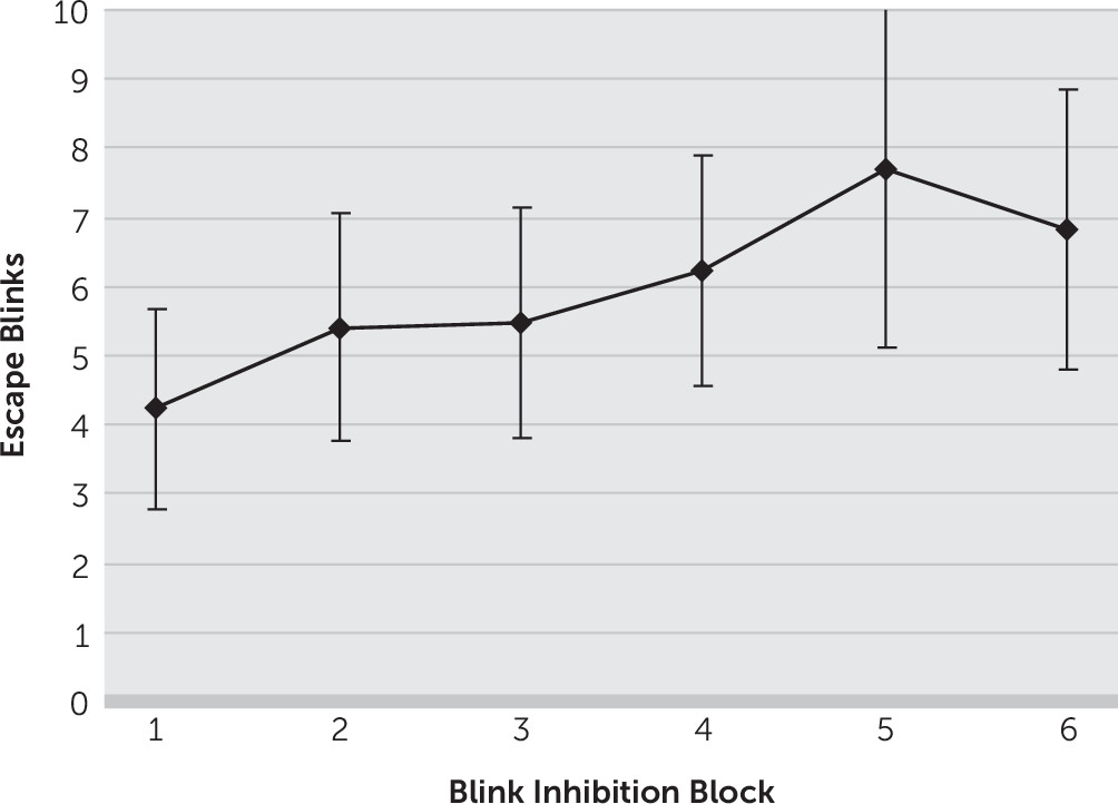
There was an increase in the number of eye blinks escaping inhibitory control across blink inhibition blocks from an average (± standard deviation) of 4.2 (±5.9) escape blinks in the first “no blink” block to 6.8 (±8.4) in the sixth “no blink” block (Figure 1). Thus, there was an average increase of 2.6 (±3.8) escape blinks from the first to the last “no blink” blocks (p=0.01). In contrast, there was no significant change in rest blink rates from baseline, with 27.8 (±13.6) and 28.5 (±19.0) blinks in the first and last “blink” blocks, respectively (p=0.82).
FIGURE 1. Increase in Escape Blinks Across Eye-Blink Inhibition Blocks
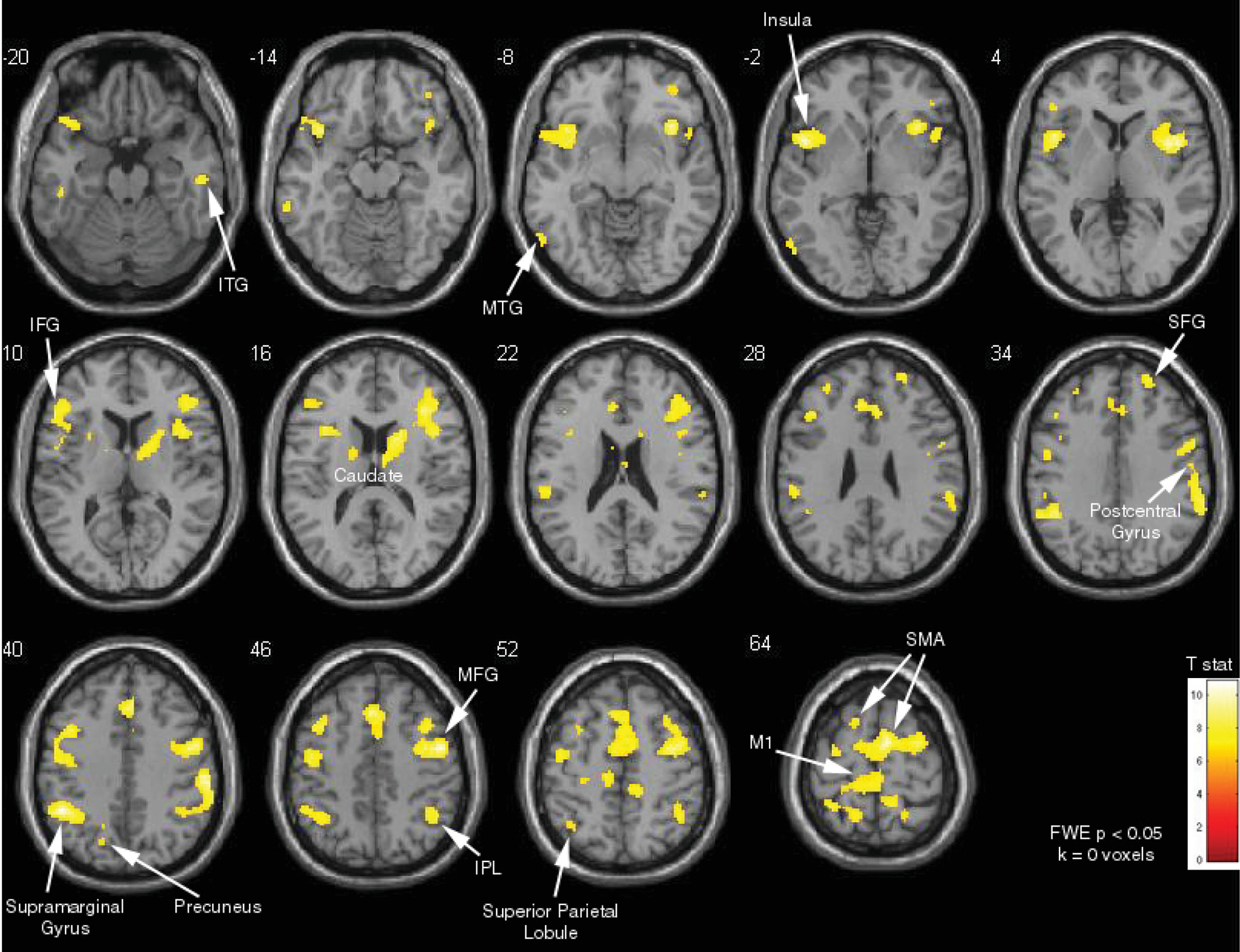
Contrasting the “no blink” and “blink” blocks revealed that inhibition of blinking activated a wide network bilaterally, including the IFG, DLPFC, dACC, frontal eye fields, supplementary motor area, and caudate (family-wise-error-corrected, p<0.05) (Figure 2). There were also bilateral activations in the inferior parietal, anterior insula, precuneus, and sensory association areas (p<0.05, family-wise-error-corrected).
FIGURE 2. Prefrontal Cortical Control Areas and Regions Involved in Urges and Interoceptive Processing Activated by Effortful Eye-Blink Controla
a FWE=family-wise-error corrected.
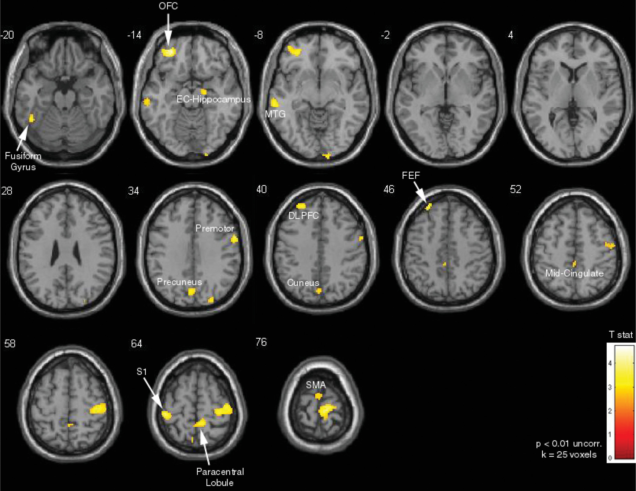
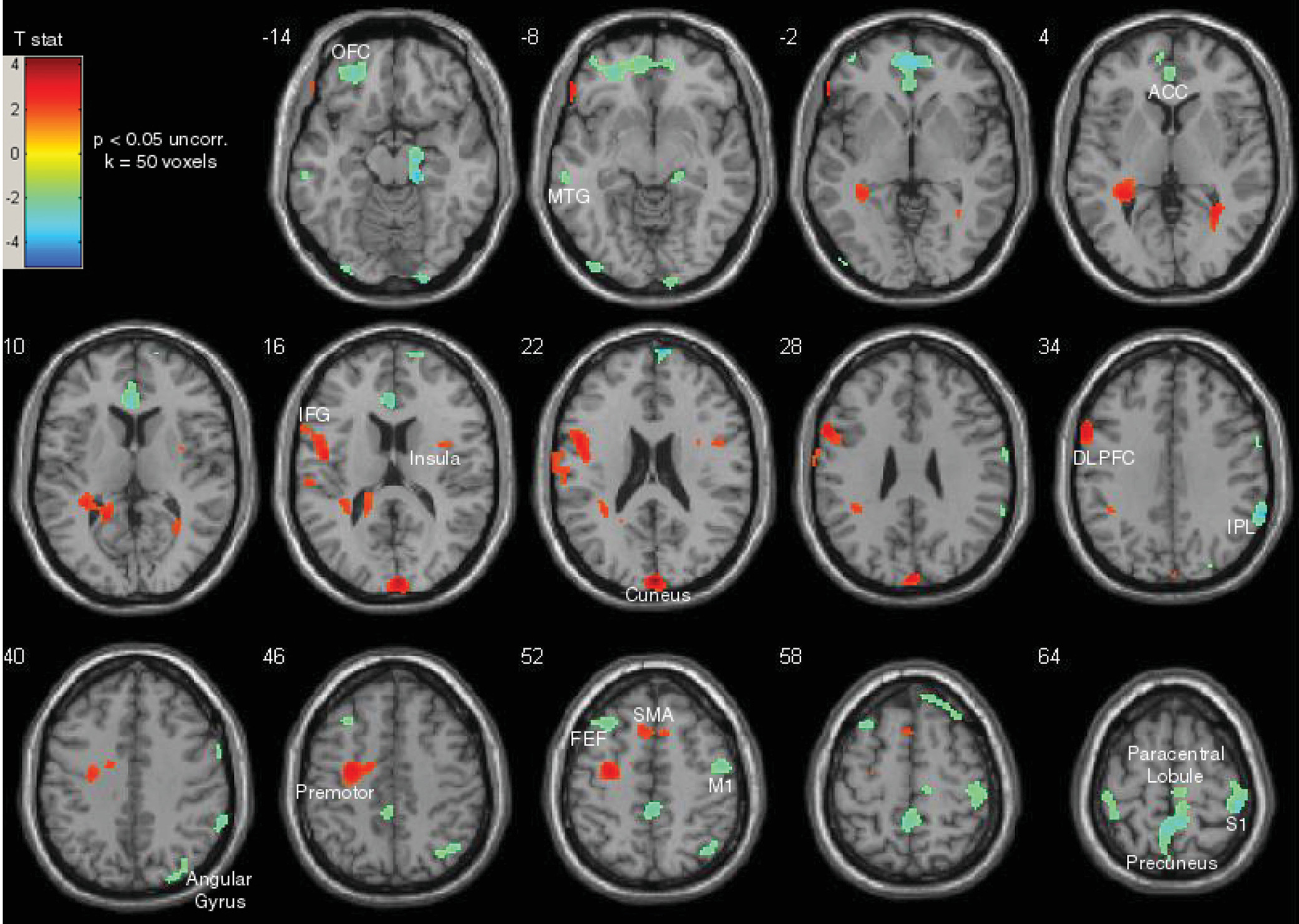
Areas positively correlated with increase in escape blinks, identified by using the increase in number of escape blinks as a covariate of interest in the “no blink” versus “blink” block contrast analysis, included the left OFC, left DLPFC, bilateral supplementary motor area, cuneus, precuneus, midcingulate, and bilateral somatosensory areas (p<0.01 uncorrected, extent thresholding k=25 voxels) (Figure 3). Finally, the top performers (i.e., those who had the least increase in escape blinks) (N=7) were contrasted with participants who had the poorest performance (i.e., those who had the most increase in escape blinks) (N=7). The top performers showed relatively higher task-related activity in frontal control areas, including the left DLPFC, left IFG, and bilateral supplementary motor areas (p<0.05 uncorrected, extent thresholding k=50 voxels) (Figure 4). In contrast, participants who had the poorest performance had relatively higher task-related activity in the left OFC, ventral ACC, right inferior parietal lobule, and bilateral somatosensory cortex.
FIGURE 3. Increase in Escape Blinks Associated With Activity in the Left Orbitofrontal Cortex (OFC) and the Left Dorsolateral Prefrontal Cortex (DLPFC)
FIGURE 4. Activity in the Frontal Control Areas Among Top Performers Compared With Activity in the Interoceptive and Likely Compensatory Frontal Areas Among Poor Performersa
a The top performers (N=7) showed activity in frontal control areas (red) compared with activity in the interoceptive and likely compensatory frontal areas (blue-green) among poor performers (N=7).

Discussion

In this study, we demonstrated behavioral evidence of self-regulatory fatigability in an eye-blink inhibition task. There was a progressive, significant increase in escape eye blinks across blink inhibition blocks (Figure 1). In contrast, there was no significant change in blink rates during rest blocks. As anticipated, inhibition of blinking activated PFC control areas, including the DLPFC, IFG, dACC, supplementary motor areas, and areas involved in interoceptive processing, which included the anterior insula, inferior parietal lobule, and sensory association areas (Figure 2). This is consistent with previous studies of self-control using suppression of bodily urges, including eye-blink inhibition,35,36 cough suppression,37 breath holding,38 urinary bladder control,39,40 and control over the urge to scratch an itch.41 Worsening performance was associated with activity in overlapping areas, including the cuneus, precuneus, DLPFC, and SMA, as well as the OFC, midcingulate, and somatosensory areas (Figure 3). The cuneus has been implicated in inhibitory control in persons with bipolar depression42 and, along with the precuneus, in differential activation between pathological gamblers and control subjects in response to visual gambling cues.43 Furthermore, given its functional connectivity to the motor cortex, dorsomedial prefrontal cortex, and DLPFC, the precuneus is believed to be involved in sensorimotor and cognitive functions related to these regions.44 The midcingulate cortex has been involved in itch relief41,45 and other sensory processing functions.46,47 The OFC has been associated with subjective sensations of fatigue with prolonged performance on the Trail-Making Test48 and craving in response to visual cues among individuals who abuse cocaine.49 More generally, the OFC is known to have an evaluative role regarding the rewarding or aversive consequences of actions.12 The positive correlation between escape blinks and the DLPFC, SMA and premotor areas may be a compensatory response to fatigue.50 Contrasting the top performers with those that most demonstrated the phenomenon of self-regulatory fatigability showed a particularly interesting pattern: while the former had relatively higher activation in known PFC control areas—the IFG, DLPFC, SMA—the latter had relatively higher activation in the ventromedial PFC, rostroventral ACC, and OFC (Figure 4), areas more related to response to hedonic impulses.25 Self-regulatory fatigability was also associated with relatively higher activation of sensory areas and the inferior parietal lobule.
Our findings are consistent with a model of response inhibition and salience attribution that has been used to explain emotional, cognitive, and behavioral changes seen in addiction.25 According to this model, higher-order control or “cold” processes are subserved by dorsal and lateral subregions of the PFC. On the other hand, automatic, emotion-laden, or “hot” processes, are associated with ventral subregions of the PFC. Thus, the activations in the high performers suggest that the urge to blink remains controlled by the control subregions of the PFC. In contrast, self-regulatory fatigue was associated with activations in ventral subregions of the PFC, suggesting reduced self-control, with emotion and attentional resources directed to craving. Interestingly, this group also showed relatively higher activation of somatosensory and interoceptive processing areas, including the inferior parietal lobule, which has been found to be involved in the experience of urges. In a 2-deoxy-2[18F]fluoro-d-glucose positron emission tomography study conducted by Volkow and colleagues,49 instructions for individuals who abuse cocaine to suppress their craving when watching a cocaine-cue video resulted in decreased activity in the rostroventral ACC, insula, and OFC, with associated activation in right inferior frontal areas. In an fMRI study, tobacco-dependent smokers who were showed cigarette cues with instructions to resist their craving demonstrated increased activity in the dorsal ACC and superior frontal gyrus and decreased activity in sensory areas.51 A similar study by another group showed that control over craving among smokers is associated with activity in the dorsomedial PFC, DLPFC, and ventrolateral PFC and decreased activity in the OFC and subgenual cingulate.52 Additionally, in a particularly interesting fMRI and ecological study, increased right IFG and SMA activation during a go/no-go task was associated with weakening of the link between cravings and subsequent smoking.53 These findings overlap with results from another fMRI and ecological study that found that the IFG mediated the effect of resistance to giving in to the temptation to eat.54
On the basis of this model, when persons who are addicted to alcohol,15 persons who abuse cocaine,16 or persons who are smokers18 are exposed to the relevant cues from the relevant substance, subsequent performance on self-control tasks deteriorates as “hot processes,” subserved by ventral PFC regions, become relatively more active. Of more general clinical significance, resources used for symptom suppression may result in self-regulatory fatigability and difficulty in maintaining executive control. For example, in Tourette’s syndrome, a condition known to involve multiple comorbid conditions (particularly OCD and ADHD), sustained efforts to achieve tic suppression may exacerbate difficulties with attention and affect school performance; in addition, tics and comorbid symptoms are known to be worse with fatigue, which may be a reason why tics tend to generally become worse at the end of the day.55 In ADHD, deficits in sustained self-control1921 may help to explain the high comorbidity with substance abuse.13,56 In fact, deficits in delayed gratification in ADHD57 can be conceptualized as a form of executive fatigability. Interestingly, subjective perception of mental effort during attention tasks correlates with the extent of reported ADHD symptoms.23,24 It therefore makes sense that environmental interventions that decrease executive demand, such as a predictable structure, planned frequent breaks, and reduction in the amount of undue stress, can be helpful in managing ADHD.
Although our study cannot settle the debate regarding the processes leading to the phenomenon of self-regulatory fatigability (i.e., the resource depletion model versus the more recent non-resource-based competing model), our findings do show distinct dorsal and lateral PFC subregions associated with consistent self-control, in contrast to the ventral PFC and other somatosensory urge-related brain areas that are associated with self-regulatory fatigability. A possible explanation, which would encompass both models as complementary, is that depletion of resources required to maintain a certain level of activity in dorsal and lateral PFC control regions results in a relative shift toward ventral PFC regions, with concomitant deterioration of performance. Accordingly, resource depletion would be accompanied by a shift in motivation, emotion, and attention toward giving in to the urge to blink. It is noteworthy that in the original studies of the delay of gratification among preschoolers, attentional control away from the immediate reward was found to play an important role in the preschoolers’ ability to maintain self-control.1 This appears to fit with our findings, as well as with the competing model to explain the phenomenon of self-regulatory fatigability.10 Also of interest, in the original delay-of-gratification studies, cognitive reappraisal was another successful self-control strategy.2 This is consistent with findings from more recent neuroimaging research demonstrating that while suppression of emotions impairs subsequent executive function,28,29,32 task performance was not adversely affected when reappraisal was used as the emotional regulation strategy.29 In fact, distraction and environmental modification are recognized strategies for self-control in substance abuse, dieting, and daily life situations.58 Such strategies involve activation of dorsal and lateral PFC self-control regions59 and may function by helping to ensure that attention, motivation, and emotion resources are not directed toward hedonic, immediate-reward cues.
There are several limitations to consider here. First, although our total sample size is comparable to that of other fMRI studies, the analysis comparing the subjects on the basis of performance entailed a smaller number of subjects. It is for this reason that p-value correction was conducted via extent thresholding, as opposed to consistently via family-wise error. Another limitation was that we did not assess for motivation, nor did we have an online rating for urge and discomfort during the task. All of these are factors that could influence performance. Furthermore, we did not control for time of day, smoking, caffeine intake, or medication, all of which could affect the results, for example, through effects on arousal. Finally, our task design did not entail a longitudinal analysis that could presumably show a shift of brain area activations with self-regulatory fatigability. As such, we are unable to uncover potential compensatory changes in response to increasing fatigue.
In summary, we demonstrated behavioral evidence of self-regulatory fatigability in an eye-blink inhibition task. As anticipated, effortful eye-blink control resulted in activation of prefrontal control areas and regions involved in urge and interoceptive processing. Worsening performance was associated with activation in regions involved in affective salience attribution and craving. In contrast, high performers showed relatively more activation in prefrontal cortical control areas, including those involved in attentional control. Our findings suggest that the phenomenon of self-regulatory fatigability is associated with relatively less recruitment of prefrontal cortical control regions.
Future larger studies that include analyses of brain activation changes over time during task performance are warranted. Furthermore, investigation of changes in effective connectivity in relation to task performance could help elucidate the neural dynamics that mediate self-regulatory fatigability. Additionally, there are many opportunities for more research of self-regulatory fatigability in clinical conditions, such as substance abuse, ADHD, and Tourette’s syndrome, among others. Finally, improved knowledge in this field promises to help in the development of interventions to improve executive functions in young children,60,61 which has the potential to have a significant positive impact on long-term outcomes.
Acknowledgments

The authors thank Drs. David Kideckel, Tomas Paus, Mary Pat McAndrews, Aaron Kucyi, Karen Davis, Cornelia Mccormick, Rostom Mabrouk, and Jane Lawrence for helpful discussions on this topic; Dr. David Mikulis for clinical input regarding the scans; Keith Ta and Eugen Hlasny for technical assistance with scanning; and all of the participants who took part in this study.

Footnote

Dr. Sandor has received unrestricted grants in support of conferences from Purdue and Shire; he has received CME speaker’s fees from Purdue; he has received clinical trial support from Otsuka; and he has served on the data safety monitoring committee for Psyadon. All other authors report no financial relationships with commercial interests.

References

1.
Mischel W, Shoda Y, Rodriguez MI: Delay of gratification in children. Science 1989; 244:933–938
2.
Mischel W, Ayduk O, Berman MG, et al: ‘Willpower’ over the life span: decomposing self-regulation. Soc Cogn Affect Neurosci 2011; 6:252–256
3.
Moffitt TE, Arseneault L, Belsky D, et al: A gradient of childhood self-control predicts health, wealth, and public safety. Proc Natl Acad Sci USA 2011; 108:2693–2698
4.
Kubzansky LD, Martin LT, Buka SL: Early manifestations of personality and adult health: a life course perspective. Health Psychol 2009; 28:364–372
5.
Seeyave DM, Coleman S, Appugliese D, et al: Ability to delay gratification at age 4 years and risk of overweight at age 11 years. Arch Pediatr Adolesc Med 2009; 163:303–308
6.
Francis LA, Susman EJ: Self-regulation and rapid weight gain in children from age 3 to 12 years. Arch Pediatr Adolesc Med 2009; 163:297–302
7.
Poulton R, Moffitt TE, Silva PA: The Dunedin Multidisciplinary Health and Development Study: overview of the first 40 years, with an eye to the future. Soc Psychiatry Psychiatr Epidemiol 2015; 50:679–693
8.
Baumeister RF, Heatherton TF: Self-regulation failure: an overview. Psychol Inq 1996; 7:1–15
9.
Hagger MS, Wood C, Stiff C, et al: Ego depletion and the strength model of self-control: a meta-analysis. Psychol Bull 2010; 136:495–525
10.
Inzlicht M, Schmeichel BJ, Macrae CN: Why self-control seems (but may not be) limited. Trends Cogn Sci 2014; 18:127–133
11.
Dang J, Björklund F, Bäckström M: Self-control depletion impairs goal maintenance: A meta-analysis. Scand J Psychol 2017; 58:284–293
12.
Boksem MAS, Tops M: Mental fatigue: costs and benefits. Brain Res Brain Res Rev 2008; 59:125–139
13.
Levy N: Addiction, autonomy and ego-depletion: a response to Bennett Foddy and Julian Savulescu. Bioethics 2006; 20:16–20
14.
Dvorak RD, Simons JS: Moderation of resource depletion in the self-control strength model: differing effects of two modes of self-control. Pers Soc Psychol Bull 2009; 35:572–583
15.
Gauggel S, Heusinger A, Forkmann T, et al: Effects of alcohol cue exposure on response inhibition in detoxified alcohol-dependent patients. Alcohol Clin Exp Res 2010; 34:1584–1589
16.
Moeller SJ, Tomasi D, Honorio J, et al: Dopaminergic involvement during mental fatigue in health and cocaine addiction. Transl Psychiatry 2012; 2:e176
17.
Hofmann W, Vohs KD, Baumeister RF: What people desire, feel conflicted about, and try to resist in everyday life. Psychol Sci 2012; 23:582–588
18.
Hagger MS, Leaver E, Esser K, et al: Cue-induced smoking urges deplete cigarette smokers’ self-control resources. Ann Behav Med 2013; 46:394–400
19.
Barkley RA: Behavioral inhibition, sustained attention, and executive functions: constructing a unifying theory of ADHD. Psychol Bull 1997; 121:65–94
20.
Gmehlin D, Fuermaier ABM, Walther S, et al: Attentional lapses of adults with attention deficit hyperactivity disorder in tasks of sustained attention. Arch Clin Neuropsychol 2016; 31:343–357
21.
Tucha L, Fuermaier ABM, Koerts J, et al.: Sustained attention in adult ADHD: time-on-task effects of various measures of attention. J Neural Transm Vienna Austria 2017;124(Supp. 1):39–53.
22.
Thomson DR, Besner D, Smilek D: A resource-control account of sustained attention: evidence from mind-wandering and vigilance paradigms. Perspect Psychol Sci 2015; 10:82–96
23.
Panetta L, Phinnemore R, Eastwood J, et al: The experience of mental effort during a sustained attention task in individuals with self-reported attention problems. Pers Individ Dif 2016; 101:503
24.
Hsu C-F, Eastwood JD, Toplak ME: Differences in perceived mental effort required and discomfort during a working memory task between individuals at-risk and not at-risk for ADHD. Front Psychol 2017; 8:407
25.
Goldstein RZ, Volkow ND: Dysfunction of the prefrontal cortex in addiction: neuroimaging findings and clinical implications. Nat Rev Neurosci 2011; 12:652–669
26.
Kelley WM, Wagner DD, Heatherton TF: In search of a human self-regulation system. Annu Rev Neurosci 2015; 38:389–411
27.
Richeson JA, Baird AA, Gordon HL, et al: An fMRI investigation of the impact of interracial contact on executive function. Nat Neurosci 2003; 6:1323–1328
28.
Inzlicht M, Gutsell JN: Running on empty: neural signals for self-control failure. Psychol Sci 2007; 18:933–937
29.
Wang Y, Yang L, Wang Y: Suppression (but not reappraisal) impairs subsequent error detection: an ERP study of emotion regulation’s resource-depleting effect. PLoS One 2014; 9:e96339
30.
Wagner DD, Heatherton TF: Self-regulatory depletion increases emotional reactivity in the amygdala. Soc Cogn Affect Neurosci 2013; 8:410–417
31.
Wagner DD, Altman M, Boswell RG, et al: Self-regulatory depletion enhances neural responses to rewards and impairs top-down control. Psychol Sci 2013; 24:2262–2271
32.
Friese M, Binder J, Luechinger R, et al: Suppressing emotions impairs subsequent Stroop performance and reduces prefrontal brain activation. PLoS One 2013; 8:e60385
33.
Casey BJ, Somerville LH, Gotlib IH, et al: Behavioral and neural correlates of delay of gratification 40 years later. Proc Natl Acad Sci USA 2011; 108:14998–15003
34.
Berman MG, Yourganov G, Askren MK, et al: Dimensionality of brain networks linked to life-long individual differences in self-control. Nat Commun 2013; 4:1373
35.
Berman BD, Horovitz SG, Morel B, et al: Neural correlates of blink suppression and the buildup of a natural bodily urge. Neuroimage 2012; 59:1441–1450
36.
Lerner A, Bagic A, Hanakawa T, et al.: Involvement of insula and cingulate cortices in control and suppression of natural urges. Cereb Cortex 2009; 19:218–223
37.
Mazzone SB, Cole LJ, Ando A, et al: Investigation of the neural control of cough and cough suppression in humans using functional brain imaging. J Neurosci 2011; 31:2948–2958
38.
McKay LC, Adams L, Frackowiak RSJ, et al: A bilateral cortico-bulbar network associated with breath holding in humans, determined by functional magnetic resonance imaging. Neuroimage 2008; 40:1824–1832
39.
Kuhtz-Buschbeck JP, Gilster R, van der Horst C, et al: Control of bladder sensations: an fMRI study of brain activity and effective connectivity. Neuroimage 2009; 47:18–27
40.
Seseke S, Baudewig J, Kallenberg K, et al: Voluntary pelvic floor muscle control: an fMRI study. Neuroimage 2006; 31:1399–1407
41.
Mochizuki H, Papoiu ADP, Yosipovitch G: Brain processing of itch and scratching, in Itch: Mechanisms and Treatment. Edited by Carstens E, Akiyama T. Boca Raton, Fla, CRC Press/Taylor and Francis, 2014
42.
Haldane M, Cunningham G, Androutsos C, et al: Structural brain correlates of response inhibition in bipolar disorder I. J Psychopharmacol Oxf Engl 2008; 22:138–143
43.
Crockford DN, Goodyear B, Edwards J, et al: Cue-induced brain activity in pathological gamblers. Biol Psychiatry 2005; 58:787–795
44.
Margulies DS, Vincent JL, Kelly C, et al: Precuneus shares intrinsic functional architecture in humans and monkeys. Proc Natl Acad Sci USA 2009; 106:20069–20074
45.
Papoiu ADP, Nattkemper LA, Sanders KM, et al: Brain’s reward circuits mediate itch relief: a functional MRI study of active scratching. PLoS One 2013; 8:e82389
46.
Vogt BA: Midcingulate cortex: Structure, connections, homologies, functions and diseases. J Chem Neuroanat 2016; 74:28–46
47.
Kleckner IR, Zhang J, Touroutoglou A, et al: Evidence for a large-scale brain system supporting allostasis and interoception in humans. Nat Hum Behav 2017; 1:69
48.
Tajima S, Yamamoto S, Tanaka M, et al: Medial orbitofrontal cortex is associated with fatigue sensation. Neurol Res Int 2010; 2010:e671421
49.
Volkow ND, Fowler JS, Wang G-J, et al: Cognitive control of drug craving inhibits brain reward regions in cocaine abusers. Neuroimage 2010; 49:2536–2543
50.
Wang C, Trongnetrpunya A, Samuel IBH, et al: Compensatory neural activity in response to cognitive fatigue. J Neurosci 2016; 36:3919–3924
51.
Brody AL, Mandelkern MA, Olmstead RE, et al: Neural substrates of resisting craving during cigarette cue exposure. Biol Psychiatry 2007; 62:642–651
52.
Kober H, Mende-Siedlecki P, Kross EF, et al: Prefrontal-striatal pathway underlies cognitive regulation of craving. Proc Natl Acad Sci USA 2010; 107:14811–14816
53.
Berkman ET, Falk EB, Lieberman MD: In the trenches of real-world self-control: neural correlates of breaking the link between craving and smoking. Psychol Sci 2011; 22:498–506
54.
Lopez RB, Hofmann W, Wagner DD, et al: Neural predictors of giving in to temptation in daily life. Psychol Sci 2014; 25:1337–1344
55.
Cohen SC, Leckman JF, Bloch MH: Clinical assessment of Tourette syndrome and tic disorders. Neurosci Biobehav Rev 2013; 37:997–1007
56.
Zulauf CA, Sprich SE, Safren SA, et al: The complicated relationship between attention deficit/hyperactivity disorder and substance use disorders. Curr Psychiatry Rep 2014; 16:436
57.
Solanto MV, Abikoff H, Sonuga-Barke E, et al: The ecological validity of delay aversion and response inhibition as measures of impulsivity in AD/HD: a supplement to the NIMH multimodal treatment study of AD/HD. J Abnorm Child Psychol 2001; 29:215–228
58.
Duckworth AL, Gendler TS, Gross JJ: Situational strategies for self-control. Perspect Psychol Sci 2016; 11:35–55
59.
Jasinska AJ, Stein EA, Kaiser J, et al: Factors modulating neural reactivity to drug cues in addiction: a survey of human neuroimaging studies. Neurosci Biobehav Rev 2014; 38:1–16
60.
Tang Y-Y, Posner MI: Attention training and attention state training. Trends Cogn Sci 2009; 13:222–227
61.
Diamond A, Ling DS: Conclusions about interventions, programs, and approaches for improving executive functions that appear justified and those that, despite much hype, do not. Dev Cogn Neurosci 2016; 18:34–48

Information & Authors

Information

Published In

Go to The Journal of Neuropsychiatry and Clinical Neurosciences
The Journal of Neuropsychiatry and Clinical Neurosciences
Pages: 325 - 333
PubMed: 29843584

History

Received: 19 July 2017
Revision received: 30 October 2017
Revision received: 19 January 2018
Accepted: 14 February 2018
Published online: 30 May 2018
Published in print: Fall 2018

Keywords

  1. Neurology
  2. Headache and Facial Pain
  3. Self-Regulatory Control
  4. Self-Regulatory Failure
  5. Self-Regulatory Fatigue
  6. Self-Regulatory Fatigability
  7. Executive Fatigue
  8. Executive Fatigability
  9. Resource Depletion
  10. Eye Blinking
  11. fMRI
  12. Orbitofrontal Cortex
  13. Dorsolateral Prefrontal Cortex
  14. Inferior Frontal Gyrus
  15. Anterior Cingulate Cortex

Authors

Details

Elia Abi-Jaoude, M.Sc., M.D., F.R.C.P.C. [email protected]
From the Department of Psychiatry, The Hospital for Sick Children, University of Toronto, Ontario, Canada (EA-J); the Department of Psychiatry, University Health Network, University of Toronto, Ontario, Canada (EA-J, PS); the Department of Medicine, Faculty of Medicine and Health Science, Institute of Neuroscience, University of Barcelona, Catalonia, Spain (BS); the Research Imaging Centre, Centre for Addiction and Mental Health, Toronto, Ontario, Canada (SC); the Department of Medical Imaging, University Health Network, University of Toronto, Ontario, Canada (AC); and the Youthdale Treatment Centers, Toronto, Ontario, Canada (PS).
Barbara Segura, Ph.D.
From the Department of Psychiatry, The Hospital for Sick Children, University of Toronto, Ontario, Canada (EA-J); the Department of Psychiatry, University Health Network, University of Toronto, Ontario, Canada (EA-J, PS); the Department of Medicine, Faculty of Medicine and Health Science, Institute of Neuroscience, University of Barcelona, Catalonia, Spain (BS); the Research Imaging Centre, Centre for Addiction and Mental Health, Toronto, Ontario, Canada (SC); the Department of Medical Imaging, University Health Network, University of Toronto, Ontario, Canada (AC); and the Youthdale Treatment Centers, Toronto, Ontario, Canada (PS).
Sang Soo Cho, Ph.D.
From the Department of Psychiatry, The Hospital for Sick Children, University of Toronto, Ontario, Canada (EA-J); the Department of Psychiatry, University Health Network, University of Toronto, Ontario, Canada (EA-J, PS); the Department of Medicine, Faculty of Medicine and Health Science, Institute of Neuroscience, University of Barcelona, Catalonia, Spain (BS); the Research Imaging Centre, Centre for Addiction and Mental Health, Toronto, Ontario, Canada (SC); the Department of Medical Imaging, University Health Network, University of Toronto, Ontario, Canada (AC); and the Youthdale Treatment Centers, Toronto, Ontario, Canada (PS).
Adrian Crawley, Ph.D.
From the Department of Psychiatry, The Hospital for Sick Children, University of Toronto, Ontario, Canada (EA-J); the Department of Psychiatry, University Health Network, University of Toronto, Ontario, Canada (EA-J, PS); the Department of Medicine, Faculty of Medicine and Health Science, Institute of Neuroscience, University of Barcelona, Catalonia, Spain (BS); the Research Imaging Centre, Centre for Addiction and Mental Health, Toronto, Ontario, Canada (SC); the Department of Medical Imaging, University Health Network, University of Toronto, Ontario, Canada (AC); and the Youthdale Treatment Centers, Toronto, Ontario, Canada (PS).
Paul Sandor, M.D., F.R.C.P.C.
From the Department of Psychiatry, The Hospital for Sick Children, University of Toronto, Ontario, Canada (EA-J); the Department of Psychiatry, University Health Network, University of Toronto, Ontario, Canada (EA-J, PS); the Department of Medicine, Faculty of Medicine and Health Science, Institute of Neuroscience, University of Barcelona, Catalonia, Spain (BS); the Research Imaging Centre, Centre for Addiction and Mental Health, Toronto, Ontario, Canada (SC); the Department of Medical Imaging, University Health Network, University of Toronto, Ontario, Canada (AC); and the Youthdale Treatment Centers, Toronto, Ontario, Canada (PS).

Notes

Send correspondence to Dr. Abi-Jaoude; e-mail: [email protected]
Previously presented at the 24th Annual Meeting of the American Neuropsychiatric Association, Boston, April 3–6, 2013, and the 19th Annual Meeting of the Organization for Human Brain Mapping, Seattle, June 17, 2013.

Funding Information

Onatrio Mental Health Foundation:
Supported in part by the Toronto General and Western Hospital Foundation and an Ontario Mental Health Foundation studentship (to Dr. Abi-Jaoude).

Metrics & Citations

Metrics

Citations

Export Citations

If you have the appropriate software installed, you can download article citation data to the citation manager of your choice. Simply select your manager software from the list below and click Download.

For more information or tips please see 'Downloading to a citation manager' in the Help menu.

Format
Citation style
Style
Copy to clipboard

View Options

View options

PDF/EPUB

View PDF/EPUB

Get Access

Login options

Already a subscriber? Access your subscription through your login credentials or your institution for full access to this article.

Personal login Institutional Login Open Athens login
Purchase Options

Purchase this article to access the full text.

PPV Articles - Journal of Neuropsychiatry and Clinical Neurosciences

PPV Articles - Journal of Neuropsychiatry and Clinical Neurosciences

Not a subscriber?

Subscribe Now / Learn More

PsychiatryOnline subscription options offer access to the DSM-5-TR® library, books, journals, CME, and patient resources. This all-in-one virtual library provides psychiatrists and mental health professionals with key resources for diagnosis, treatment, research, and professional development.

Need more help? PsychiatryOnline Customer Service may be reached by emailing [email protected] or by calling 800-368-5777 (in the U.S.) or 703-907-7322 (outside the U.S.).

Media

Figures

Other

Tables

Share

Share

Share article link

Share